Embed Size (px)
Citation preview

1The Gonda Brain Sciences Center,Bar-Ilan University, Ramat Gan, Israel2Yale University Child Study Center,New Haven, CT, USA
*Correspondence:[email protected] (R. Feldman).
Feature ReviewThe Neurobiology of HumanAttachmentsRuth Feldman1,2,*
Attachment bonds are a defining feature of mammals. A conceptual frameworkon human attachments is presented, integrating insights from animal researchwith neuroimaging studies. Four mammalian bonds are described, includingparent–infant, pair–bonds, peers, and conspecifics, all built upon systemsshaped by maternal provisions during sensitive periods, and evolution fromrodents to humans is detailed. Bonding is underpinned by crosstalk of oxytocinand dopamine in striatum, combining motivation and vigor with social focus,and their time sensitivity/pulsatility enables reorganization of neural networks.Humans’ representation-based attachments are characterized by biobehavioralsynchrony and integrate subcortical with cortical networks implicated inreward/motivation, embodied simulation, and mentalization. The neurobiologyof love may open perspectives on the ‘situated’ brain and initiate dialog betweenscience and humanities, arts, and clinical wisdom.
The measure of the intensity of love
Is measure, also, of the verve of earth.
For me, the firefly's quick, electric stroke
Ticks tediously the time of one more year.
And you? Remember how the crickets came
Out of their mother grass, like little kin,
In the pale nights, when your first imagery
Found inklings of your bond to all that dust.
Wallace Stevens, Harmonium
Human Attachments throughout Life Share Underlying NeurobiologySince the dawn of humanity, ‘the intensity of love’ has been depicted by modalities vastlydifferent from scientific ‘measurement’: cave paintings, clay figures, story-telling, dance, music,and poetry. Over the last decades, studies in animal models, particularly rats and monogamousprairie voles, began to uncover the cellular, neural, and endocrine mechanisms implicated inmaternal care and pair bonding. Those gave rise to a new field of inquiry – the neurobiology ofhuman attachments – which integrates insights from other mammals with new tools available forhuman research – brain imaging, neuroendocrinology, genetics and epigenetics, and peptideadministration – to test the biological basis of human attachments. This emerging field isgenerating a growing body of knowledge which requires a new conceptual framework, onethat integrates cross-species comparability with the distinct features of human love [1,2].
The goal of this paper is to provide such a conceptual frame for the neurobiology of humanattachments and address its tenets, parameters, and neural basis. While this conceptual
80 Trends in Cognitive Sciences, February 2017, Vol. 21, No. 2 http://dx.doi.org/10.1016/j.tics.2016.11.007
© 2016 Elsevier Ltd. All rights reserved.

GlossaryActivity-dependent facilitation:occurs when presynaptic spikeactivity is paired with activity offacilitatory interneurons, leading toenhanced neuronal activation [198].Alloparenting: the care of infants byadults other than the biologicalmother. Alloparenting is commonacross primate species andenhances infant survival [84].Biobehavioral synchrony: thecoordination of biological andbehavioral processes betweenattachment partners during socialcontact. It is a key feature of humanattachments.Heteromers: G protein-coupledreceptors (GPCRs) consist of sevenmembrane-spanning alpha-helicalsegments that combine into a singlereceptor. However, GPCRs can formheteromers by combining two ormore GPCR subunits. The oxytocinreceptor belongs to the G protein-coupled receptor family. NA shell
framework is built upon empirical data in animals and humans, some of its details are speculativeand may guide future research. I argue that the study of mammalian bonding must be conductedfrom a developmental perspective, both in relation to the life of an individual and in the context ofanimal evolution, and that this is especially the case in humans whose large associative cortex iswired, to a large extent, by early experiences within child-rearing contexts [3–5]. Later attach-ments, with romantic partners, close friends, mentors, or in-group members from sports teamsto nations, repurpose the basic machinery established by the mother–offspring bond duringearly ‘sensitive periods’ (see Glossary). I further suggest that the neurobiology of attachmentrides on systems that maintain brain plasticity through time-sensitive pulsatility – dopamine (DA)and oxytocin (OT) – which, by forming tighter crosstalk during periods of bond formation [6],integrates reward salience with social focus to reorganize neural networks around the newattachment [7–9]. Studies in rodents have shown that the integration of OT and DA in striatumsupports the formation of maternal–infant and pair bonds [10]. Yet, while similar mechanisms arethought to underpin bond formation in humans, attachment bonds acquired substantial com-plexity, duration, and flexibility across mammalian evolution, reaching their apex in humans’ long-term exclusive attachments that integrate subcortical reward with higher-order representationalcomponents (Figure 1, Key Figure). Such flexibility affords not only the immense variability ofhuman attachments across cultural contexts but also enables later reparation, and while earlybonds shape the social brain and its underlying neurobiology, humans can repair, via top–downprocessing, commitment, and discipline, the effects of early maladaptive relationships by laterbenevolent ones. Key propositions of the model are summarized in Box 1.
contains oxytocin receptors that mayform heteromers with D2 receptorson medium spiny neuron. The ligand-binding properties and neuralpathways of heteromers integrateaspects of both parent receptors.Long-term depression: reduction inthe efficacy of neuronal synapsesfollowing a long repeated stimulus.Medium spiny neurons (MSNs):projection neurons that areGABAergic (inhibitory) and compriseover 90% of neurons in the humanstriatum. MSN come in two formats.The D1-type (D1 dopamine receptor-expressing neurons) functions in the‘direct pathway’, that is, convey theirinformation directly to the outputnuclei of basal ganglia. The D2-type(D2 dopamine receptor-expressingneurons) functions in the ‘indirectpathway’, conveying information tobasal ganglia indirectly via pallidalneurons. Some MSNs express forboth D1-type and D2-type receptors.Myoactivity: the stimulation ofrhythmic tissue contraction.Myoactivity is the most conservedfunction of the oxytocin system,which has been integrated inmammals into the oxytocin-controlleduterine contractions and milk ejection[43,62].Sensitive periods: specific timewindows in early life when the brainmust experience certainenvironmental inputs for propermaturation [2]. In the context of
Box 1. Key Propositions of the Neurobiology of Human Attachments Model� Research on human attachments implicates a developmental perspective: Mammalian bonding is supported by
neurobiological systems shaped by the mother–offspring relationship during early sensitive periods [36].� Continuity in neurobiological systems underpins human bonds: Human attachments repurpose the basic machinery
established by the parent–offspring bond in the formation of other attachments throughout life, such as romanticattachment or close friendships [1].
� Human bonds are selective and enduring: Bonds are specific to attachment target and last for extended periods, oftena lifetime. Gradients of the selective and enduring components define the various human bonds (see Figure 1 in maintext).
� Bonding is behavior based triggered by the expression of species-specific, person-specific, and culture-specificbehavioral patterns: Bonding implicates bottom–up processes. Bonding-related brain and neuroendocrine systemsare activated by attachment-related behavior [5,37].
� Biobehavioral synchrony is a key feature of human attachments: Human attachments are characterized by thecoupling of coordinated nonverbal behavior with coordinated physiological response among partners during socialcontact [101] (see Figure 2 in main text).
� Central role of the oxytocin system and oxytocin–dopamine connectivity: OT is implicated in human mothering,fathering, coparenting, romantic attachment, and close friendship. Integration of OT and DA in striatum ignitesbonding, imbuing attachments with motivation and vigor [9].
� Bond formation involves increased activity and tighter crosstalk among relevant systems: Activation and closer linksamong systems underpinning affiliation, reward, and stress management are observed during periods of attachmentformation [6].
� Human attachments promote homeostasis, health, and well-being throughout life: Social attachments enhance healthand happiness while social isolation increases stress, impaired health, and death [176].
� Patterns of attachment are transferred across generations: Behavioral patterns experienced in early life organize OTavailability and receptor localization in the infant's brain, shaping the capacity to parent the next generation [66,129].
� The human brain is a situated organ, shaped by the mother–infant attachment and proximity to mother's body tofunction within the social ecology: The young mammal's immature brain at birth and need for close proximity to anursing mother shape the brain as a ‘situated’ organ, constantly responding online to the social world [71]. Humans’protracted maturity sculpts the dialogical nature of the human brain and its constant need for social affiliations.
� Human bonds experienced throughout life are transformative and have the potential to repair early negative relation-ships by later benevolent ones: The great plasticity of the human social brain and its behavior-based nature enablelater attachments to reorganize neural networks and repair, at least partly, negative early experiences. This highlightsthe translational potential of research on the neurobiology of human attachments [177].
Trends in Cognitive Sciences, February 2017, Vol. 21, No. 2 81

bonding, these involve the species-typical parenting behaviors.Striatum: a subcortical structureserving key role in the rewardsystem. The striatum receivesdopaminergic inputs from multiplebrain areas and is the central input tobasal ganglia. The ventral striatumcontains the NA and the dorsalstriatum includes the putamen andcaudate. The ‘corpus striatum’
comprises the striatum and globuspallidus.Trophallaxis: the exchange ofsensory signals among members of asocial group [199]. The term wasextended to include social stimuli[103] and to denote the reciprocalmultisensory stimulation of lowintensity that elicits approachresponse. Parenting marks aprototypical form of trophallaxis [105].
The Neurobiology of Mammalian Affiliation: Crosstalk of DA and OTOur proposed model suggests that the ‘intensity of love’, the quality of attachment, is measuredin terms of the vigor [11] of DA action in striatum, particularly in the nucleus accumbens (NA). DAacts in the NA to organize goal-directed reward-related behavior characterized by initiation andvigor by inhibiting the inhibitory output of NA GABAergic (inhibitory) medium spiny neurons,which then release ventral pallidum (VP) neurons in the basal ganglia to enable motor action [12–15]. Disinhibition of the inhibitory control in the accumbens–pallidal indirect pathway, pathwayfrom striatum to basal ganglia which is not direct but acts via pallidal neurons, renders the VPaccessible to glutamate (excitatory neurons) inputs, leading to energetic behavior and markingthe striatum as a limbic–motor interface where reward translates into action marked by vigor andgoal directedness [16,17].
Yet, while DA neurons function as general-purpose stimulators to reward-related targets, it istheir close links with OT receptors in striatum, where OT receptors abound [18,19], that imbueattachment bonds with incentive value and direct action toward affiliative goals, such asmaternal care [9,20,21]. Actions of DA D1-type and D2-type neurons on OT receptors in NAshell establish maternal memory and form repetitive patterns of caregiving, as shown in rats[22]. OT stimulates the accumbens–pallidal indirect pathway, strengthening synaptic activityin VP and forming memories of the attachment context. NA shell contains OT receptors thatmay form heteromers, neurons expressing for multiple receptors, with DA D2-type receptorson medium spiny neurons, and OT binding to OT receptors increases affinity of DA toassociated D2-type receptors [23]. Since D2 receptors depress activity of medium spinyneurons, OT can potentiate these inhibitory effects. The combination of DA D1-type andD2-type receptors and OT receptors in NA shell functions to depress the striatal inhibitoryinput to VP and enables supernormal excitation of basolateral amygdala projections to VP,which, through activity-dependent facilitation, creates the effects of mesolimbic DA onattachment-focused action [24,25]. The coactivation of D2-expressing neurons in the NA byOT enables neurons specifically suited to identify sensory–motor reward patterns to employreward computations of D2 neurons to encode the temporal patterns of social reward, asshown in rhesus macaques [26,27]. This allows the brain to internalize the social partner andits preferences, encode relationship-specific patterns of social exchange, and draw rewardfrom the matching of self and partner's actions (i.e., social synchrony), which lead toconsolidation of the bond [13,26].
Overall, the tighter OT–DA crosstalk in the NA during bond formation enables plasticity of thebrain reward system and its flexible adaptation to incorporate the new bond into the self[18,28,29]. Studies in prairie voles show that the NA receives OT receptors containing inputsfrom multiple cortical regions, including sensorimotor and associative cortices and theseenable the formation of sensory and motor memories of attachment experiences [30].Furthermore, OT receptor density in the NA in infancy was found to predict the time spenthuddling with partner in female prairie voles [31], and bereavement-like behavior and activa-tion of stress-related neurohormonal systems following partner loss in male prairie voles wereassociated with suppression of OT signaling in the NA [32]. These findings highlight the role ofaccumbens OT in forming continuity from parental to pair bonds and in buttressing theprotective function of long-term attachments. Finally, research in rats describes the role of OTin long-term depression in amygdala, attenuating amygdalar response to aversive socialstimuli [33,34]. Such long-term depression reduces fear and facilitates the approach orienta-tion required for bonding [35]. Thus, while DA affords vigor and motivation, OT provides thesoothing and tranquility necessary for bond formation via its regulatory effects on hypotha-lamic–pituitary–adrenal axis activity [36,37] and anxiolytic properties [38]. This creates aunique neurobiological state of ‘immobility without fear’ [39], a state specifically suited forthe formation of new attachments.
82 Trends in Cognitive Sciences, February 2017, Vol. 21, No. 2

Key Figure
Attachment Bonds across Mammalian Evolution
Model systemsC. elegans
OxytocinAncient func�ons
Dopamine
Lamprey
Evolu�onary origins
• Sensory–neurocircuitry coupling • Striato–pallidal pathway• Regula�on of motor ac�on• Regula�on of basic life func�ons
Parental(maternal)
Roman�c(pair)
Integra�on of OT and DA in striatum ignites mammalian bonding
Friendship(peer/clan)
Fellow humans(conspecifics)
Olfactory-based,
Group-living,expecta�ons-based,cor�cal-monitoredaffilia�ons
Representa�on-based, long-term,culture-informeda�achments
nest-bound,hormone-dependentbonds Rodents
Primates
Humans
• Non-exclusive
• Short-lived• Subcor�cal
• Evidence of response to distress of familiar conspecific
• Mostly none• Prarie voles:
ma�ng-basedrecogni�on
• No evidence
• Mostly none• Monogamous primates: proximity-based partner preferenc e
• Exclusive, alloparental care• Hormone primed, not dependent
• Empathy/social behavior to group members• Hormonal
the context ofcoordina�on in
childrearing
• Time together needed
• Exclusive, • Exclusive,poten�ally
culture-defined,
independent, long-term
associa�on-based,
hormone-
a�achments a�achments
long-term ,
independent , memory-base d
ma�ng-
• Personal, • Affilia�ve behavior long-term, memory-based,
neurobiology ofand ac�vates the
affilia�on
and empathy thatac�vate the
affilia�on
term a�achment
neurobiology of
• Capacity for long-
to other species
Figure 1. The figure describes the four bonds observed across mammalian species – parent–infant, pair bonds (romantic attachment), peers (close friendships), andconspecifics (fellow humans) – and charts their expression in rodents, primates, and humans. Attachment bonds in mammals are underpinned by functioning of twoancient systems, OT and DA, which maintained basic organization across vertebrate evolution and supported group living in harsh ecologies (OT) and motivational goal-directed action (DA) throughout animal evolution. Caenorhabditis elegans and lamprey are presented here to illustrate model systems used by research to study theancient functions of OT and DA. The model proposes, on the basis of studies in rodents and primates, that the integration of OT and DA in striatum ignites mammalianbonding. In humans, attachment bonds are marked by two main features: selective (specific to attachment target) and enduring (long lasting). The figure describes howthese two features undergo substantial reorganization across mammalian evolution. The decrease in color intensity from parental to pair to peer to conspecific indexes thegradual weakening of intensity in these selective and enduring features from the parent–infant attachment (most intense color) to humans interactions with strangers (leastintense color). Abbreviations: DA, dopamine; OT, oxytocin.
Trends in Cognitive Sciences, February 2017, Vol. 21, No. 2 83

Humans are wired for social affiliation via activity of this limbic circuit, comprising the OT-producing hypothalamus, extended amygdala network, and striatum [including the ventraltegmental area (VTA), which projects to striatum, and VP, which receives projections fromit]. This limbic network regulates critical survival and motivation functions and redirects them inthe service of social life [40]. Yet, while DA–OT links chart a mammalian-general mechanism ofbonding, as observed in rat mothers, prairie voles, and primates, connectivity of this limbicsystem with cortical sites via multiple ascending and descending projections supports humans’long-term exclusive attachments [5,41,42]. Notably, striatal activity has been detected in nearlyall imaging studies of human attachments; however, striatal/VTA activations are typically coupledwith both subcortical (amygdala and hypothalamus) and cortical structures, particularly theanterior cingulate cortex (ACC), medial prefrontal cortex (mPFC), and orbitofrontal cortex (OFC)of the reward system, in addition to structures supporting mentalizing, including the superiortemporal sulcus (STS) and temporoparietal junction (TPJ), and those underpinning embodiedsimulation functions, such as anterior insula (AI), inferior parietal lobule (IPL), inferior frontal gyrus(IFG), and supplementary motor area (SMA). Aspects of OT system functionality have similarlybeen implicated in human maternal, paternal, romantic, and friendship attachments in researchemploying OT administration, peripheral measures, or allelic variability and methylation of the OTreceptor gene (Box 2) [6,43–54]. Longitudinal studies following humans from infancy to adult-hood describe OT involvement in the transfer of attachment from parents to friends and romanticpartners [55,56]. Human studies indicate tighter connections between OT and DA in response tobonding-related cues as mediated by social synchrony; for instance, coactivation of OT- andDA-rich brain areas in response to infant stimuli [47,53], heightened VTA response to romanticpartner following OT administration [7], or increased coupling of peripheral biomarkers duringparental and romantic bonding [6]. This enables the subcortical system of motor ‘vigor’ to extract
Box 2. Time Sensitivity/Pulsatility of the Dopamine and Oxytocin Systems Enables Plasticity of NeuralNetworks to Incorporate the New Attachment
DopaminePhasic DA striatal neurons process the timing of reward and encode reward anticipation, which builds the internal senseof time in the brain [13]. Since DA neurons are sensitive to reward stemming from social interaction [26] and can linkreward to attachment experiences, they ground reward in cycles of caregiving actions. DA is implicated in circadiantiming and encoding the temporal component of experiences [60], which imbues attachments with sense of continuityover time. Computations in striatal DA neurons, while not uniquely social, can integrate social components into theirtemporal predictions, partly through OT's role in augmenting the salience of social stimuli [61]. This permits social signalsto act upon a pre-established synaptic tract and associate it with specific reward outcome [24]. It also enables an initialbrief, unselective, and broad increase in DA activity to become subjective and social, and function with accuracy, energy,and specificity [13].
OxytocinPulsatility is a defining feature of OT functionality across evolution [43,62]. OT is released from both OT-producinghypothalamic neurons and dendrites, the branched projections of neurons, and this enables OT to operate at farlocations from OT-producing sites [178]. Dendritic release primes vesicle (small fluid-consisting structure within a cell)stores for activity-dependent release and this enables a diffuse signal to maintain long half-life in the central nervoussystem and extracellular fluid to execute far-reaching behavioral goals [179]. OT signals cause dendrite release withoutincreasing electrical activity, which, via peptidergic feedback, can become self-sustaining by creating autoregulatedrelease primed by salient experiences [180–183]. OT functions at both presynapsis and postsynapsis to attenuateGABAergic inhibitory neurons, enlarging extracellular interactions among OT cells while reducing interactions with otherstimuli, which leads to synchronization across multiple brain areas [184–187]. Once activated, release is repeated in time-sensitive bursts. Special primed signals, such as attachment-specific cues, can trigger dendrite release, relocating OT invesicles from reserve to releasable stores [188,189] and future release is then shaped by the primed cue [190,191]. Inaddition, OT participates in modifying the excitation-to-inhibition balance by causing GABAergic signaling to change fromexcitatory to inhibitory around birth [192,193]. This opens a time window of increased neural sensitivity to specificattachment cues (i.e., sensitive periods). Such time-sensitive mechanisms chart the way by which attachment experi-ences impact the infant's OT release, the stimuli that will trigger it in future attachments, and its ultimate organization inspecific sites in the brain [2]. This is also how OT transmission, which is not between neurons but between populations ofneurons, can act across great distances and cause coherent, long-lasting, self-sustaining effects on behavior[113,129,194].
84 Trends in Cognitive Sciences, February 2017, Vol. 21, No. 2

from repeated attachment experiences the representation of love and transfer it to other bondsthroughout life, extending the neurobiology of maternal–infant bonding across human attach-ments and beyond.
Time-Keeping PulsatilityAnother important feature of the neurobiology of attachment is the time sensitivity of both DA andOT, which is critical for their role in neural plasticity. OT and (phasic) DA are characterized bypulsatile release that supports time-keeping mechanisms implicated in patterned action andseasonal rhythmicity. OT and DA are evolutionary-ancient systems involved in the regulation ofbasic life functions across vertebrate evolution [57–59] (Figure 1). The pulsatility/time sensitivity ofOT and DA enabled their involvement in neural plasticity, which is required for selectiverecognition and long-term memory, the two key features of human attachments.
DopamineWhile phasic DA striatal neurons encode for general reward and reward anticipation, theseneurons can incorporate social reward into their computations [13,26]. This lends support to thehypothesis that attachment experiences, which are repeated and predictable in nature, maybecome particularly salient targets for the reward computations of striatal DA neurons. The roleof DA in circadian rhythmicity [60] facilitates the consolidation of attachments, imbuing them witha sense of regularity and stability. Studies in primates have shown that striatal DA neurons candifferentiate reward stemming from self and partner, anticipate predictable social interactions,and draw reward from the matching of self and partner's actions [26]. It is thus assumed that thetemporal sensitivity of DA neurons enables humans to draw reward from the experience ofbiobehavioral synchrony, which is built on familiarity with the partner's repeated socialpatterns. Since OT increases the salience of social stimuli [61], the integration of OT and DAin striatum enhances the experience of social synchrony, leading to cycles of coordinatedmoments within attachment bonds that become rewarding and therefore repetitive, receivingmotivation and vigor from DA and social focus and tranquility from OT.
OxytocinPulsatility is a defining feature of OT functionality across evolution, and myoactivity is OT's mostconserved feature [43,62]. Pulsatility is critical for the unique dendritic release that supports OT'srole in bonding (Box 2). This mode of functioning leads to autoregulated, feed-forward releasethat is triggered by primed attachment experiences, which, once activated, release repeatedrhythmic bursts. Such time-sensitive mechanisms chart the way by which early attachmentexperiences shape the ultimate organization of OT in specific sites in the infant's brain, the stimulithat will trigger it in future attachments, and its cross-generational transfer via the expression ofparenting behavior in the next generation [2].
The molecular underpinnings of the time sensitive mechanisms for the two systems aredescribed in Box 2.
Mammalian Affiliative Bonds: Parents, Partners, and PeersAttachment bonds are a defining feature of mammals. Pregnancy, birth, and lactation and theirunderlying neurobiology define class mammalia [1,2,58,63,64]. Life for a young mammal beginswith two constraints; an immature brain at birth and the need for close proximity to a nursingmother. Consequently, provisions embedded in the mother's body and the species-typicalmaternal behaviors organize the immature brain, orienting it to social life via relationship with themother [65]. Networks supporting mammalian sociality develop in the context of the mother–offspring bond and are shaped by variations in maternal care [4,66]. In the 3–5% of mammalianspecies that are biparental, the father's presence and parenting behaviors also play a role ininfant brain development both directly and through their effect on reducing maternal stress [67].
Trends in Cognitive Sciences, February 2017, Vol. 21, No. 2 85

The mother's body provides the first environment for the developing mammal. Maternal heartrhythms, smell, touch, movement patterns, arousal dynamics, social cues, and stressresponse mark the first environmental signals the brain encounters, programming it to livein close proximity with others, signaling to the developing central nervous system the amountof stress the environment contains [68,69], and tuning the brain to function as a ‘situated’organ [70], constantly receiving information, updating predictions, and responding online tothe social world [71–73]. It has been recently argued [74] that the brain's modus operandi isnot solipsistic but situated and research on the ‘situated’ brain should become the focus ofsocial neuroscience. This position resonates with the central hypothesis proposed here, thatis, the social embeddedness of the immature mammalian brain at birth shapes it as afundamentally dialectic organ and that elucidating the mechanisms by which early attach-ments program the brain may provide new understanding into the brain's basic mode ofaction.
Attachment bonds are marked by two key features; they are selective (specific to attachmenttarget) and enduring (long-lasting) [37]. These two components underwent substantial reorga-nization across mammalian evolution and specific combinations of their gradients define thevarious human bonds (Figure 1). The human parent–offspring bond is selective and enduring.Romantic bonds are also selective – at least in most cultures and recent human history – andenduring, but more precarious; while humans rarely abandon their children, romantic attach-ments can terminate under normative conditions and this may account for the tighter crosstalk ofthe OT and reward systems during romantic bonding [6,75]. Close friendships are selective andenduring, but these appear in a weaker form, with humans nurturing numerous friendshipssimultaneously and long friendships dwindling with no overt breakup. Humans’ relationship toconspecifics is neither selective nor enduring; yet humans are unique in their ability to activate thebehavioral and neurobiological systems of affiliation toward unfamiliar fellow humans. Forinstance, humans express empathy or synchronize gaze with strangers and both activatethe OT system [76,77].
The ancient OT and DA systems foreshadow the selective and enduring features of mammalianbonds. Across vertebrate evolution, the OT-family molecule has repurposed the basic lifefunctions controlled by the ancient vasotocin molecule in the service of social life, adapting itto the social hierarchies, seasonality, and social organization of each species [59,78]. Thesensory–neurocircuitry coupling among group members in lower species, modulated by OT'spulsatile release, transformed from group collaboration into the biobehavioral synchrony ofnursing mother and young in mammals [43]. The ancient DA system integrated repetitive motoraction with goal-directed reward via organization of the striatal–pallidal indirect network and theevolution of GABAergic (inhibitory) control over motor neurons. Motor inhibition, a criticalcomponent of any higher-order motor program [79], enabled the quiescence required forthe formation of attachment bonds.
Yet, as seen in Figure 1, humans’ selective and enduring bonds mark a long progress from theirancient origins. The mother–offspring bond in rodents is nonselective and short lived; rodentmothers care for any infant in their surrounding (bond to a generic infant) and bonds are bound tothe nest, primed by hormones of pregnancy, and depend on olfactory cues [1,10,80]. Withinthese constraints, however, it is research in rodent mothers and monogamous prairie voles thatuncovered the cellular and molecular basis of the selective and enduring components ofattachment and charted commonalities between parental and pair bonds [10,81].
Primates’ enlarged neocortex enables the formation of selective attachments that rely oncomplex social signals which are necessary for life in large groups [82,83]. The widespreadpractice of alloparenting highlights the primates’ parental brain as an adaptive template that
86 Trends in Cognitive Sciences, February 2017, Vol. 21, No. 2

can flexibly activate via bottom–up caregiving behavior [84,85]. Primates’ bonding is hormoneprimed but not hormone dependent and olfactory cues are integrated into visually guided socialbonds [86]. Most primates, like rodents, are not monogamous and across both primates androdents, extended paternal care is observed only in monogamous species, indicating that male–female mating and physical proximity are required to trigger the neurobiology of fathering [87,88].This is in contrast to humans, for whom the paternal and pair bonds are independent [48]. As to‘peers’, rodents [89] and primates [90] show behavioral and hormonal contingencies to distressof a conspecific (member of the same species), such as elevation in cortisol or behavioralmimicking, and cooperative breeding marmosets also display OT synchrony among childrearingadults [91]. Peer-reared rhesus macaques, while exhibiting lifetime aberrations in social behav-ior, express bonding to peers [92,93], indicating that friendshiplike behavior is common acrossprimate species.
Humans’ cortical complexity enables integration of the subcortical limbic network and ancientOT and DA systems into love that is built on representations and memory, translates multisen-sory experiences into higher-order associations, adapts to cultural norms to carry bonds acrossgenerations and ground them in meaning systems, conceives both the overlapping andautonomy of self and other [94], incorporates sociocognitive abilities of empathy and trust tomaintain long-term affiliations, and extends the here-and-now so that love can be felt in itsabsence (e.g., deceased parents) and transcend to abstract ideas (God, homeland), human-kind, and other species (pets). Notably, all these forms of love activate the neurobiology ofaffiliation [95–97] and are built on early attachment experiences [2,98,99]. Humans, as Wilsonrecently noted [100], can create a feeling that life is meaningful by activating their affiliative biologytoward continuum of experience, from Earth's biosphere of flora and fauna to highest levels ofabstraction and the arts.
Biobehavioral SynchronyBiobehavioral synchrony, the coordination of biological and behavioral processes betweenattachment partners during social contact, is a critical component of human attachments[1,2,25,101]. Biobehavioral synchrony evolved from the coordinated group activity of lowerspecies where joint motor action (involving DA) is locked with coupled physiology to achievesurvival-related collaborative goals (involving OT); for instance, ants carrying a grain of wheat toshelter, fish swimming to ward off a shark, or birds flocking toward warmer climates[1,2,37,102–104].
Life within social groups requires constant exchange of social signals among members and theterm ‘trophallaxis’ [95,105] has been coined by entomologists to denote the exchange ofsensory and social signals among members of a social group. Three aspects of trophallaxis havebeen integrated into mammalian bonds: the low intensity and arousal-modulatory nature ofattachments, the social reciprocity and online construction embedded in them, and the troph-allaxic process as charting a line from parent–offspring bond to life within social groups [1]. Mostimportantly, humans’ biobehavioral synchrony is straightforwardly built on trophallaxis in highlysocial invertebrate species, such as ants.
Biobehavioral synchrony is observed across human attachments, in parental, romantic, friend-ship, and fellow–human interactions, and employs great flexibility so that human synchrony is notmetronome precise but unfolds a stochastic process, follows dynamic systems’ principles, andintegrates patterned order and local variability [106,107]. The basic characteristics of biobehav-ioral synchrony, however, are maintained across evolution; it involves the coordination ofbiological processes and species-typical behaviors expressed during social contact, it initiatesyoung to life in social groups, it assembles online from the inputs of multiple parties, and it relieson the ancient OT system [37,101].
Trends in Cognitive Sciences, February 2017, Vol. 21, No. 2 87

During or immediately following social contact, human synchrony is evident in four systems:behavior, autonomic, hormones, and brain, and, to varying degrees, this coupling is foundacross the four human attachment constellations (Figure 2). Synchrony between partners’nonverbal behaviors in the gaze, affect, vocal, and touch modalities has been observed inmother–infant interactions since the 1950s and thought to entrain the neonate's physiologicalperiodicities of sucking, crying, and circadian rhythmicity [108,109]. Fathers similarly engage inbehavioral synchrony, but utilize a quick-paced, high-arousal temporal pattern [110]. Synchro-nous parent–infant interactions are accompanied by physiological coordination [2,5,101,111];parent and infant's heart rhythms are coupled during episodes of behavioral synchrony, but notduring nonsynchronous moments [112]; and following synchronous interactions, OT release iscoordinated between parent and child [113]. Synchronous moments induce brain-to-braincoupling between mother and child in key nodes of the social brain.
Synchronous interactions experienced during early sensitive periods are expressed in laterattachments throughout life. Matched interactions are observed between romantic partners andshow similar second-by-second coordination of gaze and affect [6,114]. Heart-rate coordination[115], OT coupling [116], and brain-to-brain synchrony have been described among couples.Mother and father show a coordinated brain response in structures of the embodied simulationand mentalizing networks (STS, IPL, and AI) when viewing a video of their own infant [117].Interactions among close friends show behavioral reciprocity; however, interactions amongfriends are not as tightly coupled as those observed in parental or romantic attachment [55,118].Evidence suggests that OT increases following contact with friends, albeit the increase is notcoupled [55]. Finally, teams trained for coordinated action and group cohesion, such as militaryunits, exhibit wide response across the social brain in the alpha band to vignettes depictingsynchronous group activities, particularly coordinated unit in battle [119].
Unlike other mammals which require familiarity with conspecific for biobehavioral coordination,humans display behavioral synchrony toward strangers; humans coordinate gaze and vocalturn-taking during conversations with strangers while touch synchrony is preserved for intimatebonds [1]. When strangers sit in close proximity and execute joint tasks, they also display heart-rate coupling, brain-to-brain synchrony of alpha rhythms, and coordinated brain response intemporoparietal structures, such as STS and IPL [120–122]. Empathy to strangers in distress isimpacted by OT administration and observing groups in collaborative action elicits OT response[119]. Furthermore, the brain responds to ‘similar to me’ synchronous action; mothers observingsynchronous interactions of unfamiliar mothers and their infants activate areas of the rewardsystem (NA, dorsal ACC), and the degree of activation parallels their own behavioral synchrony[123]. Finally, evidence highlights humans’ preference for synchrony in large crowds in walking,marching, or rowing [124]. Such group-maintaining mechanisms are rooted in earliest mam-malian experiences – the matching of physiology and behavior between mother and younggeneralized by humans’ large associative cortex across time, place, and person.
Longitudinal studies show long-term associations between the degree of parent–infant syn-chrony and the quality of later attachments with close friends and romantic partners and withabilities that support participation in human social life, including empathy, moral orientation,theory-of-mind, and culture-specific modes of self-regulation [125–127]. Early synchronousinteractions also shape children's social brain. For instance, parent–infant synchrony longitudi-nally predicts intactness of the brain basis of empathy in adolescence [128]. Similarly, parents’brain response to infant cues is combined with behavioral synchrony to predict preschoolers’emotion regulation and socialization [129], and parental OT integrates with parent–infantsynchrony to shape children's social reciprocity toward best friends [55]. These studies dem-onstrate that the parent's affiliative neurobiology and synchronous behavior shape humanchildren's attachments to nonparental figures and buttress human-specific social
88 Trends in Cognitive Sciences, February 2017, Vol. 21, No. 2

Biobehavioral synchrony in human a�achments
Behavioralsynchrony
• Synchronized behavior in gaze, affect, vocal, and touch• Mother-specific father-specific
• Coordina�on of brain response in mentalizing network in parents• Coordina�on of gamma oscilla�ons in temporal cortex in lovers
• Synchronized HR during synchronized interac�ons
• HR coordina�on during or following interac�on
• Coordina�on of OT and cor�sol among parents
• Coordina�on of OT among lovers
• Teams coordinate heart rythms during joint ac�on
• OT is released during interac�ons with friends
• Alpha response to behavioral synchrony among teams in social brain
• Coordina�on among teams in mirror network
• Evidence for coordinated ac�va�on in mentalizing areas during interac�on
• No evidence for coupling
• OT is implicated in acts of empathy
• No evidence for coupling
• Evidence for some coordina�on during joint ac�on in close proximity
• Synchronized nonverbal pa�erns
• Coordinated self-disclosure + empathy
• Pa�erns of social reciprocity
• Coordina�on of culture-spcific display rules (e.g., eye gaze)
• Coordinated OT response following contact
• Coordinated brain oscilla�ons in alpha and gamma rythms
• Coordinated cor�sol response to stress
Parents
Roman�cpartners
Friends
Strangers
Heart ratecoupling
Endocrine fit Brain-to-brainsynchrony
Figure 2. Biobehavioral Synchrony in Human Attachments. Human attachments are characterized by the coupling of the partners’ physiological and behavioralprocesses during moments of social contact. Such coupling is observed across four systems: matching of nonverbal behavior, coupling of heart rhythms and autonomicfunctioning, coordination of hormonal release, and brain-to-brain synchrony. In humans, biobehavioral synchrony is observed in the four affiliative bond constellations:parental, romantic, friendship, and strangers. Abbreviations: HR, heart rate; OT, oxytocin.
Trends in Cognitive Sciences, February 2017, Vol. 21, No. 2 89

competencies, highlighting the lifetime effects of the neurobiology of human attachment and itscross-generational nature.
Notably, our focus on the OT system stems from its critical role in the formation and maintenanceof attachment bonds in humans and other mammals. Other hormones, including cortisol,testosterone, prolactin, progesterone, vasopressin, beta-endorphin, and estradiol, have beenstudied in relation to human attachments. Our hypothesis, supported by studies from ourlaboratory [1], suggests that OT serves a key integrative function by providing a neuroendocrinemilieu for the effects of multiple hormones on the development of human attachments.
The various measures used in research on human attachments are described in Box 3.
Human Attachments and the BrainHumans’ rootedness in mammalian affiliative biology renders the integration of OT and DA instriatum an important foundation for human attachment, albeit the role of other neurohormonaland neurotransmitter systems has been described [9,10,81]. Recently, fMRI studies began totest the neural basis of humans’ multiple attachments by assessing brain responses to auditory,
Box 3. The ‘Measurement of Love’
Neuroscience research has utilized the following tools to measure human attachments:
Micro/macroanalysis of social behavior and behavioral synchrony: Observation of interactions between mothers andinfants and among couples has been conducted for nearly a century. Studies use either global rating scales addressingthe quality of the partners’ behaviors (e.g., ‘responsive’, ‘intrusive’) or focus on micro-level detection of bonding-relatedbehaviors (e.g., ‘motherese’ vocalizations, social gazing) and their coordination among partners [98]. Studies employedsimilar micro- and macro-level analysis in other attachments, including romantic/marital relationships and friendships.
Autonomic response: Parenting and couple studies employed measures of heart period and respiratory sinus arrhythmiaas indices of parasympathetic functioning during social interactions, evaluating changes from baseline and during variousdyadic stressors (e.g., parent–infant ‘still-face’, couples ‘conflict dialog’). Skin conductance has been used to indexsympathetic activity and multiple autonomic measures have been integrated into a single index of autonomic arousal[195,196].
Hormones: Peripheral measures of hormones from plasma, saliva, urine, and, less commonly, from cerebrospinal fluid,have been tested as correlates of human attachment bonds. Hormones studied in attachment contexts mainly includecortisol, testosterone, oxytocin, vasopressin, prolactin, progesterone, estradiol, salivary alpha amylase, and betaendorphin.
Peptide administration: Nasal administration of OT and, to a lesser extent, vasopressin has been used to address theeffects of neuropeptides on affiliative response and the brain networks supporting human social abilities. Studies typicallyemploy a double-blind design where peptide administration is compared with placebo in a within-subject or between-subject design [197].
Brain oscillations: Electroencephalogram and emerging studies in MEG have been used to examine questions related tobond formation and social affiliation, in addition to understanding the role of brain oscillations in human social functions[119,128,174]. Research on brain oscillations and their cortical generators afforded by MEG can open new vistas on theneurobiology of attachment.
Brain imaging: fMRI has been recently used to address various aspects of human attachments and these studies aresummarized in the Supplemental Information online. Studies often use auditory, visual, or multimodal stimuli of theattachment partner (infant, romantic partner) as fMRI stimuli.
Genetics/Epigenetics: Individual differences related to allelic variability on genes related to the oxytocin–vasopressinpathway (OXTR, AVPR1a), dopamine (DRD4, DRD2, DAT1, COMT), or serotonin (5-HTT) have been used to examinegenetic markers associated with individual differences in attachment behavior or the effects of various early attachmentexperiences on later functioning. Recent studies have also tested methylation on the OXTR gene in relation toattachment-related outcomes [43].
90 Trends in Cognitive Sciences, February 2017, Vol. 21, No. 2
(A) Straitum detailed view
NAcc
PFC
IPLSTGpre-SMAdACC
CA
MFG
FPNAcc
IFG
AI AMG
CA
PC
SMA
IFG
OFC AI TP
IPLTPJSTS
mPFC ACC
PCCSTRVTA
vmPFC OFC AMG
RewardKey:
Embodied simula�on
Mentalizing
PCC
TPJ
THAL
VTA
dmPFC
ACCmPFCNAcc
vmPFCHT
OFC rACC AMGPHG
ParentalRoman�cFriendship
ParentalPair bonds
GP STS
GP CA VP AMG Pu HPC
Human a�achment networksSagi�al plane
(B)
(C)
(See figure legend on the bottom of the next page.)
Trends in Cognitive Sciences, February 2017, Vol. 21, No. 2 91

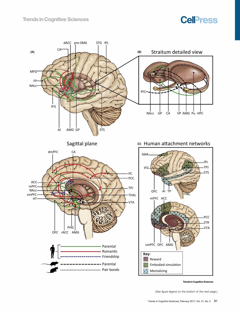
visual, or multimodal stimuli of the attachment target; for example, infants to their parents,romantic partners, or friends. Overall, various findings indicate that while the VTA/ventral striatumignites the brain's experience of attachment and imbues it with reward and vigor, humanattachments integrate subcortical with multiple cortical reward- and sociocognitive-relatednetworks (Figure 3).
A survey of published studies using fMRI to investigate humans’ various social attachments (seethe Supplemental Information online for more details) provides evidence for three main inter-connected neural systems that integrate to establish, maintain, and enhance our affiliative bondswith others. Some brain areas participate in more than one system.
The first is the ‘reward-motivation’ system, including the striatum (NA, caudate, putamen),amygdala, VTA, OFC, ventromedial prefrontal cortex (vmPFC), and ACC, employing DA- andOT-rich pathways [12,130,131], and supporting multiple attachment-related motivationalbehaviors, such as social orienting, social seeking, and maintaining contact across extendedperiods [132,133]. Attachments have intrinsic motivational value that combines immediatehedonic response with approach motivation, goal-directed behavior, and learning [134]. Thestriatum and its massive projections from both frontal cortex and amygdala [130,135,136] areimplicated in detecting attachment-relevant cues, appraising their valence, and guiding action bycoding the affective properties of stimuli [137–139]. Notably, even within the striatum there is ashift from ventral (NA) to dorsal striatum (caudate) in corticostriatal connectivity with thestabilization of the bond [140,141], reflecting a shift from reward-related drive and noveltyseeking to familiarity processing and predictability [142,143]. Caudate–cortical connectivity isassociated less with passion, sexual desire, or parental response to vulnerable infants but withlong-term relationships, habit formation, and companionship among couples [141], and socialcooperation and trust among friends [144]. The caudate is involved in reinforcement learning,goal-directed action, and weighing the relative values of outcomes [135,145] and authorssuggest that the shift from ventral to dorsal striatal functioning accompanies attachments asthey settle into joint goals, mutual habitat, and reciprocity, and is mediated by OT [140]. Theexistence of convergent projections from the cortex to striatum, along with hippocampal andamygdala-striatal projections, places the striatum as a central entry port for processing emo-tional/motivational information supporting human attachments.
The amygdala and its associated network play a critical role in human attachment, particularlymothering [5,10,146] and romantic attachment [88]. Maternal bonding requires vigilance forinfant safety and romantic attachment involves heightened emotionality and interoceptivesensitivity to signs of safety and danger. Amygdala activations have been detected in ‘all’imaging studies of mothers [5] and in most studies of fathers, romantic partners, and close
Figure 3. The Brain Basis of Human Attachments. (A) Connections among subcortical and cortical structures supporting attachment in the human brain areindicated by solid green lines for parent–infant bonds, red lines for romantic bonds, and broken blue lines for suggested connections in friendship bonds (taken from 86fMRI papers on humans’ various social attachments, see the Supplemental Information online for details of each study). Neural models for parental care in rats areindicated by broken black lines and for pair bonding formation in prairie voles by dotted black lines [10,19,80]. (B) The basal ganglia intraconnections (striatum: NA, Pu,CA; VP; and GP) and interconnections with cortical structures (PFC) and subcortical structures (AMG and HPC). (C) The human affiliation networks. Three maininterconnected neural systems underpinning human attachments (some areas participate in more than one system). The reward network (broken lines/pink colored),supporting approach motivation, social orienting and seeking, goal-directed behavior, social learning, and the incentive value of attachment cues [134], includes thestriatum, OFC, ACC, vmPFC, VTA, and AMG. The embodied simulation network (broken lines/green colored) enables individuals to resonate with other's mental stateand emotion via embodiment mechanisms and grounds experience in the present moment [94] and includes the AI, ACC, IFG, IPL, and SMA. The mentalizing network(dotted/bright blue colored), supporting social cognition, mental-state understanding, and social goal interpretation [164], comprises frontotemporal–parietal regionsincluding the STS, PCC, TPJ, TP, and mPFC. Abbreviations: ACC, anterior cingulated cortex; AI, anterior insula; AMG, amygdala; CA, caudate; CT, cortisol; dACC, dorsalanterior cingulated cortex; GP, globus pallidus; HPC, hippocampus; HT, hypothalamus; IFG, inferior frontal gyrus; IPL, inferior parietal lobule; MFG, middle frontal gyrus;NA, nucleus accumbens; OFC, orbitofrontal cortex; PC, precuneus; PCC, posterior cingulated cortex; PFC, prefrontal cortex; PHG, parahippocampal gyrus; Pu,putamen; rACC, rostral anterior cingulated cortex; SMA, supplementary motor area; STG, superior temporal gyrus; STS, superior temporal sulcus; TP, temporal pole;TPJ, temporoparietal junction; THAL, thalamus; vmPFC, ventromedial PFC; VP, ventral pallidum; VTA, ventral tegmental area.
92 Trends in Cognitive Sciences, February 2017, Vol. 21, No. 2

friends. The amygdala guides attention to biologically relevant stimuli, adjusts social orienting,codes the intensity of reward, and computes the salience of social information [147,148].Interestingly, amygdala activation in human attachments is typically coupled with other areasof the subcortical limbic circuit (hypothalamus, VTA, VP), insular–cingulate cortices, and tem-poral–frontal areas [STS, superior temporal gyrus, prefrontal cortex (PFC)] [47,129,149].
Human attachments require complex higher-order processes that involve learning, memory,planning, and predictions and depend on frontostriatal connections of the reward circuit,particularly vmPFC, OFC, and ACC. These connections enable the encoding of reward-relatedexpectations, associations, and representations; evaluation of the affective valence of attach-ment stimuli; and maintenance of flexible representations to guide action [150,151]. Suchcorticostriatal connections provide the foundation for the human capacity to combine reward,passion, proximity seeking, ‘vigor’, and unconscious motivation with higher-order abilities thatmark the top–down control, trust, empathy, and commitment of human attachments and enablehumans to tend and maintain them. The OFC, the latest evolving structure of the human brainand the end point of the reward pathway [152], implicates in the representation of ‘pleasantness’and in effortful, goal-directed actions to tend long-term relationships [153]. The OFC selectsamong rewards, enables the resistance of immediate rewards toward long-term attachmentgoals, and shapes affiliations in the color of culture, ritual, and personal preferences with stabilityand far-sightedness [154]. The vmPFC enables representation of love and its entire ‘ripples’ ofassociations, sense of ‘yearning’ that is critical for human love (maintaining love in its absence),the appraisal of safety, and the sense of self as both overlapping and separate from attachmentpartner, an overlap that defines the experience of intersubjectivity [155,156]. The vmPFC exertsinhibitory control over limbic regions, reducing anxiety/avoidance in safe environments and long-term attachments [157].
The second system underpinning human attachment is the ‘embodied simulation/empathynetwork’, including the insula, ACC, IFG, IPL, and SMA. Embodied simulation is an evolutionary-ancient mechanism, which, via automatic interoception and internal representations, recreatesother's state in one's brain. Embodied simulation is critical for grounding a ‘shared world’ in thebrain and underpins the human capacity to build and maintain attachments [94,158]. This formof interpersonal ‘matching’ relies on neural pathways that involve both the experience of internalbody formats and the perception of similar states in others via perceptual–motor coupling[120,159]. This network enables the parent/partner to integrate interoceptive and affectiveinformation, resonate with mental states and emotions, and ground experience in the presentmoment thus giving it color, immediacy, and ‘situatedness’ [160,161]. von Economo neurons,projection neurons located in layer V of the anterior cingulate and frontoinsular cortices, areimplicated in the conscious perception of bodily states and afford the integrated representationsof social moments as they are lived [162,163].
Finally, interoceptive mechanisms are insufficient to support representation-based humanattachments. Human bonds rely on ‘mentalizing’ processes – higher-order cognitive processesinvolving complex top–down inferences of others’ mental states by attributing beliefs, thoughts,and intentions to others to create a full sense of ‘togetherness’ [164,165]. Mentalizing processesunderpin attachment formation by building on the individual's ability to appreciate multipleperspectives, understand partner's goals and motives, and keep in mind his/her values andconcerns [74,166]. Frontotemporal–parietal structures, particularly the STS, posterior cingu-lated cortex, TPJ, temporal pole, and mPFC, are components of the ‘mentalizing system’, thethird network of the ‘global human attachment network’ (Figure 3). The STS and TPJ are centralregions of this network [167] and play a vital role in social cognition [164], evaluation of others’state, social goal interpretation, and prediction making and their online updating [168–170]. TheSTS combines embodied simulation (mirror) and mentalizing properties [171], integrates fast
Trends in Cognitive Sciences, February 2017, Vol. 21, No. 2 93

Outstanding QuestionsCan the neurobiology of attachmentprovide insights into the mechanismsthat support brain-to-brain coordina-tion among humans during social con-tact and foster the transition from a‘solipsistic’ to a ‘situated’ model ofthe human brain?Can knowledge on the neurobiology ofattachment help understand the ‘neu-robiology of reparation’: what pro-cesses enable humans to thrive afterdisrupted early attachments? Whichindividuals are better disposed toreparation following deficits in earlybonding? And what components muststill exist in the early environment tomake reparation possible?Can the ‘measurement of love’ inte-grate as a valid area of scientificinquiry? Can we develop novel toolsand formulate new models?How do cultural ecologies and mean-ing systems shape the neurobiology ofattachment? Can conditions such asnuclear versus extended family living,traditional versus modern mate selec-tion, or monogamous versus openpartner relationships impact brain andendocrine systems implicated inbonding?What are the lifetime effects of humanattachments on health, well-being, andhappiness? What are the processes bywhich attachment bonds exert theirimpact on health and longevity?Can the neurobiology of attachmentprovide a unique entry point for theintegration of science with the huma-nities, arts, ethics, and clinical wisdom?
bottom–up simulations (biological motion) with slower top–down understanding (theory-of-mind), and provides critical support for the process of attachment formation [172]. Interestingly,the STS has been shown as particularly relevant to the development of fathering, a bond builtmore on top–down understanding of infant signals than on the ancient limbic structures thatsupport mothering [48].
Finally, research is beginning to use magnetoencephalography (MEG) to study the participationof neural oscillations and their cortical generators in human attachments. Overall, it has beensuggested [70,74] that brain oscillations underpin brain-to-brain synchrony among attachmentpartners or between humans during social interactions and participate in binding members intoa social group. In several recent studies, my colleagues and I detected the involvement of alpha(8–14 Hz) and gamma (30–60 Hz) rhythms in human bonding, the two frequencies defining top–down control mechanisms and bottom–up rapid processing, respectively, with gamma rhythmsoften integrating into alpha to create the interplay of automaticity and control needed for bondformation [173]. We found that empathy to others’ pain is supported by alpha rhythms inmentalizing structures [174]; that adolescents exposed to maternal depression across the firstyears of life terminate alpha response in posterior STS at a late time window (900–1100 spoststimulus), suggesting aborted top–down processing of empathic resonance [128]; and thatbrain-to-brain synchrony of alpha rhythms in SMA (embodied simulation) binds members of agroup and differentiates them from the outgroup [167]. Importantly, MEG studies of humanattachment are rare and may provide valuable information on the temporal course of the brainresponse to attachment-related cues.
Concluding Remarks and Future DirectionsCan the scientific measurement of love and research on the ‘affiliative brain’ (Box 3; seeOutstanding Questions) open new vistas not only for understanding human attachments butalso for neuroscience in general? I submit that the answer is affirmative for three reasons.
First, when humans are asked about the most important aspect of their life, they often describetheir affiliations. The capacity to give and receive love and maintain long-term bonds is increas-ingly recognized as key to human thriving, impacting well-being, positive outlook in the face ofadversity, physical health, and better aging [175]. Yet, we still know relatively little about themechanisms by which social bonds impact our immune system, express throughout the lifespan, predict better aging, or transmit from parent to offspring. Knowledge is also limited as tohow two humans coordinate their brain response online during social interactions and how earlyexperiences longitudinally tune the brain to social life. A better understanding of the neurobiologyof affiliation can shed light on how to live a personally meaningful life, as well as how we mightrepair developmental and affiliative disruptions, as can occur in cases of maternal postpartumdepression, premature birth, or impoverished or dangerous environments where the socialenvelop provides no safe haven for proper bonding.
Second, the flip side of the neurobiology of affiliation is the neurobiology of intergroup conflict,racial bias, and tribal hatred, both activating the same ancient systems, which evolved to helporganisms rapidly distinguish friend from foe [77]. It is critical we understand how the brain shutsdown its empathic response when the inflicted is a member of the ‘outgroup’, particularlyoutgroup perceived as potentially threatening. We recently found that shutting down the brain'sempathy centers is accomplished by tightening brain-to-brain synchrony among ingroupmembers, increasing OT production, and imposing top–down attenuating processes on bot-tom–up automatic response to the distress of outgroup [77,167].
Finally, the neurobiology of affiliation stands at the crossroad between science and humanities,with the potential to provide deeper integration of the two after centuries of separation into
94 Trends in Cognitive Sciences, February 2017, Vol. 21, No. 2
distinct branches of knowledge. While Aristotle and Leonardo De Vinci were well-versed in boththe sciences and arts of their period, such integration is no longer available or encouraged. Theneurobiology of human affiliation requires that a biologically based evolutionary perspective,which provides mechanistic understanding but pays little attention to the individual, is supple-mented by perspectives that focus precisely on the individual with his or her experiences,expressions, and aspirations, and are committed to the individual's well-being, health, andthriving. To study the neurobiology of human attachment, one must season the objectivity ofscience with the wisdom of the clinician, foresight of the philosopher, and creativity of the artistinto a unified endeavor that can shed new light on the loftiest – and oldest – of humanexperiences: ‘love’
AcknowledgmentsSupported by the Simms-Mann Foundation and the Irving B. Harris Foundation. I wish to express my deepest gratitude to
Eyal Abraham for his hard work, insights, and wisdom, and to Maayan Harel for her exceptional talent and dedication in
creating the graphic work.
Supplemental InformationSupplemental information related to this article can be found, in the online version, at http://dx.doi.org/10.1016/j.tics.2016.
11.007.
References
1. Feldman, R. (2016) The neurobiology of mammalian parentingand the biosocial context of human caregiving. Horm. Behav. 77,3–17
2. Feldman, R. (2015) Sensitive periods in human social develop-ment: new insights from research on oxytocin, synchrony, andhigh-risk parenting. Dev. Psychopathol. 27, 369–395
3. Rilling, J.K. (2014) Comparative primate neuroimaging: insightsinto human brain evolution. Trends Cogn. Sci. 18, 46–55
4. Kundakovic, M. and Champagne, F.A. (2015) Early-life experi-ence, epigenetics, and the developing brain. Neuropsychophar-macology 40, 141–153
5. Feldman, R. (2015) The adaptive human parental brain: implica-tions for children's social development. Trends Neurosci. 38,387–399
6. Ulmer-Yaniv, A. et al. (2016) Affiliation, reward, and immunebiomarkers coalesce to support social synchrony during periodsof bond formation in humans. Brain Behav. Immun. 56, 130–139
7. Scheele, D. et al. (2013) Oxytocin enhances brain reward systemresponses in men viewing the face of their female partner. Proc.Natl. Acad. Sci. U.S.A. 110, 20308–20313
8. Groppe, S.E. et al. (2013) Oxytocin influences processing ofsocially relevant cues in the ventral tegmental area of the humanbrain. Biol. Psychiatry 74, 172–179
9. Love, T.M. (2014) Oxytocin, motivation and the role of dopamine.Pharmacol. Biochem. Behav. 119, 49–60
10. Numan, M. and Young, L.J. (2016) Neural mechanisms ofmother–infant bonding and pair bonding: similarities, differences,and broader implications. Horm. Behav. 77, 98–112
11. Ko, D. and Wanat, M.J. (2016) Phasic dopamine transmissionreflects initiation vigor and exerted effort in an action-and region-specific manner. J. Neurosci. 36, 2202–2211
12. Schultz, W. (2000) Multiple reward signals in the brain. Nat. Rev.Neurosci. 1, 199–207
13. Schultz, W. (2016) Reward functions of the basal ganglia.J. Neural Transm. (Vienna). 123, 679–693
14. Aggarwal, M. et al. (2012) Neural control of dopamine neuro-transmission: implications for reinforcement learning. Eur. J.Neurosci. 35, 1115–1123
15. Floresco, S.B. (2015) The nucleus accumbens: an interfacebetween cognition, emotion, and action. Annu. Rev. Psychol.66, 25–52
16. Maldonado-Irizarry, C.S. and Kelley, A.E. (1994) Differentialbehavioral effects following microinjection of an NMDA
antagonist into nucleus accumbens subregions. Psychopharma-cology (Berl) 116, 65–72
17. Grillner, S. et al. (2005) Mechanisms for selection of basic motorprograms – roles for the striatum and pallidum. Trends Neurosci.28, 364–370
18. Dölen, G. and Malenka, R.C. (2014) The emerging role ofnucleus accumbens oxytocin in social cognition. Biol. Psychia-try 76, 354–355
19. Young, L.J. and Wang, Z. (2004) The neurobiology of pair bond-ing. Nat. Neurosci. 7, 1048–1054
20. Shahrokh, D.K. et al. (2010) Oxytocin-dopamine interactionsmediate variations in maternal behavior in the rat. Endocrinology151, 2276–2286
21. Strathearn, L. (2011) Maternal neglect: oxytocin, dopamineand the neurobiology of attachment. J. Neuroendocrinol.23, 1054–1065
22. Parada, M. et al. (2008) The roles of accumbal dopamine D1 andD2 receptors in maternal memory in rats. Behav. Neurosci. 122,368–376
23. Olazabal, D.E. and Young, L.J. (2006) Species and individualdifferences in juvenile female alloparental care are associated withoxytocin receptor density in the striatum and the lateral septum.Horm. Behav. 49, 681–687
24. Brzosko, Z. et al. (2015) Retroactive modulation of spike timing-dependent plasticity by dopamine. Elife 4, e09685
25. Romero-Fernandez, W. et al. (2013) Evidence for the existence ofdopamine D2-oxytocin receptor heteromers in the ventral anddorsal striatum with facilitatory receptor–receptor interactions.Mol. Psychiatry 18, 849–850
26. Báez-Mendoza, R. and Schultz, W. (2013) The role of the stria-tum in social behavior. Front. Neurosci. 2013 7, 233
27. Ross, H.E. and Young, L.J. (2009) Oxytocin and the neuralmechanisms regulating social cognition and affiliative behavior.Front. Neurosci. 30, 534–547
28. Dölen, G. et al. (2013) Social reward requires coordinated activity ofnucleus accumbens oxytocin and serotonin. Nature 501, 179–184
29. Johns, J.M. et al. (2005) The effects of dopaminergic/serotoner-gic reuptake inhibition on maternal behavior, maternal aggres-sion, and oxytocin in the rat. Pharmacol. Biochem. Behav. 81,769–785
30. Ross, H.E. et al. (2009) Characterization of the oxytocin systemregulating affiliative behavior in female prairie voles. Neuroscience162, 892–903
Trends in Cognitive Sciences, February 2017, Vol. 21, No. 2 95

31. Barrett, C.E. et al. (2015) The oxytocin system promotes resil-ience to the effects of neonatal isolation on adult social attach-ment in female prairie voles. Transl. Psychiatry 5, e606
32. Bosch, O.J. et al. (2016) Oxytocin in the nucleus accumbens shellreverses CRFR2-evoked passive stress-coping after partner lossin monogamous male prairie voles. Psychoneuroendocrinology64, 66–78
33. Gamer, M. et al. (2010) Different amygdala subregions mediatevalence-related and attentional effects of oxytocin in humans.Proc. Natl. Acad. Sci. U.S.A. 107, 9400–9405
34. De Dreu, C.K. (2012) Oxytocin modulates the link between adultattachment and cooperation through reduced betrayal aversion.Psychoneuroendocrinology 37, 871–880
35. Maroun, M. and Wagner, S. (2016) Oxytocin and memory ofemotional stimuli: some dance to remember, some dance toforget. Biol. Psychiatry 79, 203–212
36. Carter, C.S. (2014) Oxytocin pathways and the evolution ofhuman behavior. Annu. Rev. Psychol. 65, 17–39
37. Feldman, R. (2012) Oxytocin and social affiliation in humans.Horm. Behav. 61, 380–391
38. Neumann, I.D. (2008) Brain oxytocin: a key regulator of emotionaland social behaviours in both females and males. J. Neuro-endocrinol. 20, 858–865
39. Carter, C.S. and Porges, S.W. (2013) The biochemistry of love:an oxytocin hypothesis. EMBO Rep. 14, 12–16
40. Sokolowski, K. and Corbin, J.G. (2012) Wired for behaviors: fromdevelopment to function of innate limbic system circuitry. Front.Mol. Neurosci. 5, 55
41. Kanat, M. et al. (2014) Oxytocin and the social brain: neuralmechanisms and perspectives in human research. Brain Res.1580, 160–171
42. Valk, S.L. et al. (2016) Socio-cognitive phenotypes differentiallymodulate large-scale structural covariance networks. Cereb.Cortex. http://dx.doi.org/10.1093/cercor/bhv319
43. Feldman, R. et al. (2016) Oxytocin pathway genes: evolutionaryancient system impacting on human affiliation, sociality, andpsychopathology. Biol. Psychiatry 79, 174–184
44. Bakermans-Kranenburg, M.J. and van IJzendoorn, M.H. (2008)Oxytocin receptor (OXTR) and serotonin transporter (5-HTT)genes associated with observed parenting. Soc. Cogn. Affect.Neurosci. 3, 128–134
45. Bakermans-Kranenburg, M.J. and van IJzendoorn, M.H. (2014)A sociability gene? Meta-analysis of oxytocin receptor genotypeeffects in humans. Psychiatr. Genet. 24, 45–51
46. Riem, M.M. et al. (2011) Oxytocin receptor gene and depressivesymptoms associated with physiological reactivity to infant cry-ing. Soc. Cogn. Affect. Neurosci. 6, 294–300
47. Atzil, S. et al. (2011) Specifying the neurobiological basis ofhuman attachment: brain, hormones, and behavior in synchro-nous and intrusive mothers. Neuropsychopharmacology 36,2603–2615
48. Abraham, E. et al. (2014) Father's brain is sensitive to child-care experiences. Proc. Natl. Acad. Sci. U.S.A. 111, 9792–9797
49. Walum, H. et al. (2012) Variation in the oxytocin receptor gene isassociated with pair-bonding and social behavior. Biol. Psychia-try 71, 419–426
50. Kogan, A. et al. (2011) Thin-slicing study of the oxytocin recep-tor (OXTR) gene and the evaluation and expression of theprosocial disposition. Proc. Natl. Acad. Sci. U.S.A. 108,19189–19192
51. Kusui, C. et al. (2001) DNA methylation of the human oxytocinreceptor gene promoter regulates tissue-specific gene suppres-sion. Biochem. Biophys. Res. Commun. 289, 681–686
52. Szyf, M. et al. (2008) The social environment and the epigenome.Environ. Mol. Mutagen. 49, 46–60
53. Strathearn, L. et al. (2009) Adult attachment predicts maternalbrain and oxytocin response to infant cues. Neuropsychophar-macology 34, 2655–2666
54. Weisman, O. et al. (2012) Oxytocin administration to parentenhances infant physiological and behavioral readiness for socialengagement. Biol. Psychiatry 72, 982–989
96 Trends in Cognitive Sciences, February 2017, Vol. 21, No. 2
55. Feldman, R. et al. (2013) Parental oxytocin and early caregivingjointly shape children's oxytocin response and social reciprocity.Neuropsychopharmacology 38, 1154–1162
56. Lee Raby, K. et al. (2013) Genetic contributions to continuity andchange in attachment security: a prospective, longitudinal inves-tigation from infancy to young adulthood. J. Child Psychol.Psychiatry 54, 1223–1230
57. Grillner, S. et al. (2013) The evolutionary origin of the vertebratebasal ganglia and its role in action selection. J. Physiol. 591,5425–5431
58. Donaldson, Z.R. and Young, L.J. (2008) Oxytocin, vasopressin,and the neurogenetics of sociality. Science 322, 900–904
59. Grimmelikhuijzen, C.J. and Hauser, F. (2012) Mini-review: theevolution of neuropeptide signaling. Regul. Pept. 177, S6–S9
60. Agostino, P.V. and Cheng, R.K. (2016) Contributions of dopa-minergic signaling to timing accuracy and precision. Curr. Opin.Behav. Sci. 8, 153–160
61. Bartz, J.A. et al. (2010) Oxytocin selectively improves empathicaccuracy. Psychol. Sci. 21, 1426–1428
62. Kenkel, W.M. et al. (2014) Is oxytocin a maternal–foetal signallingmolecule at birth? Implications for development. J. Neuroendoc-rinol. 26, 739–749
63. Neumann, I.D. et al. (2013) Increased brain and plasma oxytocinafter nasal and peripheral administration in rats and mice. Psy-choneuroendocrinology 38, 1985–1993
64. Olazábal, D.E. et al. (2013) Flexibility and adaptation of the neuralsubstrate that supports maternal behavior in mammals. Neuro-sci. Biobehav. Rev. 37, 1875–1892
65. Hofer, M.A. (1987) Early social relationships: a psychobiologist'sview. Child Dev. 58, 633–647
66. Weaver, I.C. et al. (2004) Epigenetic programming by maternalbehavior. Nat. Neurosci. 7, 847–854
67. Braun, K. and Champagne, F.A. (2014) Paternal influences onoffspring development: behavioural and epigenetic pathways. J.Neuroendocrinol. 26, 697–706
68. Lyons, D.M. et al. (2010) Animal models of early life stress:implications for understanding resilience. Dev. Psychobiol. 52,616–624
69. Hofer, M.A. (1996) Multiple regulators of ultrasonic vocalization inthe infant rat. Psychoneuroendocrinology 21, 203–217
70. Hari, R. and Kujala, M.V. (2009) Brain basis of human socialinteraction: from concepts to brain imaging. Physiol. Rev. 89,453–479
71. Akers, K.G. et al. (2008) Social competitiveness and plasticity ofneuroendocrine function in old age: influence of neonatal noveltyexposure and maternal care reliability. PLoS One 3, e2840
72. Meaney, M.J. and Szyf, M. (2005) Maternal care as a model forexperience-dependent chromatin plasticity? Trends Neurosci.28, 456–463
73. Parker, K.J. et al. (2006) Maternal mediation, stress inoculation,and the development of neuroendocrine stress resistance inprimates. Proc. Natl. Acad. Sci. U.S.A. 103, 3000–3005
74. Hari, R. et al. (2015) Centrality of social interaction in human brainfunction. Neuron 88, 181–193
75. Gavrilets, S. (2012) Human origins and the transition from pro-miscuity to pair-bonding. Proc. Natl. Acad. Sci. U.S.A. 109,9923–9928
76. Guastella, A.J. et al. (2008) Oxytocin increases gaze to the eyeregion of human faces. Biol. Psychiatry 63, 3–5
77. De Dreu, C.K. et al. (2010) The neuropeptide oxytocin regulatesparochial altruism in intergroup conflict among humans. Science328, 1408–1411
78. Goodson, J.L. et al. (2012) Evolving nonapeptide mechanisms ofgregariousness and social diversity in birds. Horm. Behav. 61,239–250
79. Grillner, S. and Robertson, B. (2015) The basal ganglia down-stream control of brainstem motor centres – an evolutionarilyconserved strategy. Curr. Opin. Neurobiol. 33, 47–52
80. Rilling, J.K. and Young, L.J. (2014) The biology of mammalianparenting and its effect on offspring social development. Science345, 771–776

81. Lieberwirth, C. and Wang, Z. (2016) The neurobiology of pairbond formation, bond disruption, and social buffering. Curr.Opin. Neurobiol. 40, 8–13
82. Dunbar, R.I. (1992) Neocortex size as a constraint on group sizein primates. J. Hum. Evol. 22, 469–493
83. Dunbar, R.I.M. (2003) The social brain: mind, language, andsociety in evolutionary perspective. Ann. Rev. Anthropol. 32,163–181
84. Hrdy, S.B. (1999) Mother Nature: Natural Selection and theFemale of the Species, Chatto & Windus
85. Royle, N.J. et al. (2014) The evolution of flexible parenting.Science 345, 776–781
86. Keverne, E.B. (2014) Significance of epigenetics for understand-ing brain development, brain evolution and behaviour. Neurosci-ence 264, 207–217
87. Fernandez-Duque, E. et al. (2009) The biology of paternal care inhuman and nonhuman primates. Annu. Rev. Anthropol. 38, 115–130
88. Johnson, Z.V. and Young, L.J. (2015) Neurobiological mecha-nisms of social attachment and pair bonding. Curr. Opin. Behav.Sci. 3, 38–44
89. Burkett, J.P. et al. (2016) Oxytocin-dependent consolationbehavior in rodents. Science 351, 375–378
90. de Waal, F.B. (2012) The antiquity of empathy. Science 336,874–876
91. Finkenwirth, C. et al. (2016) Oxytocin is associated with infant-care behavior and motivation in cooperatively breeding marmo-set monkeys. Horm. Behav. 80, 10–18
92. Suomi, S.J. (1999) Attachment in rhesus monkeys. In Handbookof Attachment: Theory, Research, and Clinical Applications, pp.181–197, Guilford Press
93. Suomi, S.J. (2005) Mother-infant attachment, peer relationships,and the development of social networks in rhesus monkeys.Hum. Dev. 48, 67–79
94. Gallese, V. (2015) Bodily selves in relation: embodied simulationas second-person perspective on intersubjectivity. Philos. Trans.R. Soc. Lond. B Biol. Sci. 369, 20130177
95. Nagasawa, M. et al. (2015) Oxytocin-gaze positive loop and thecoevolution of human-dog bonds. Science 348, 333–336
96. Urgesi, C. et al. (2010) The spiritual brain: selective cortical lesionsmodulate human self-transcendence. Neuron 65, 309–319
97. Tankersley, D. et al. (2007) Altruism is associated with anincreased neural response to agency. Nat. Neurosci. 10, 150–152
98. Feldman, R. (2007) Parent–infant synchrony and the constructionof shared timing; physiological precursors, developmental out-comes, and risk conditions. J. Child Psychol. Psychiatry 48, 329–354
99. Sroufe, L.A. et al. (2009) The Development of the Person: TheMinnesota Study of Risk and Adaptation from Birth to Adulthood,Guilford Press
100. Wilson, E.O. (2014) The Meaning of Human Existence, WWNorton and Company
101. Feldman, R. (2012) Bio-behavioral synchrony: a model for inte-grating biological and microsocial behavioral processes in thestudy of parenting. Parenting 12, 154–164
102. Rosenblatt, J.S. (1965) Effects of experience on sexual behaviorin male cats. In Sex & Behavior (Beach, F.A., ed.), pp. 416–439,John Wiley & Sons, Inc
103. Schneirla, T.C. (1946) Problems in the bio-psychology of socialorganization. J. Abnorm. Soc. Psychol. 41, 385
104. Wilson, E.O. (2012) The Social Conquest of Earth, WW Nortonand Company
105. Schneirla, T.C. and Rosenblatt, J.S. (1963) “Critical periods” inthe development of behavior. Science 139, 1110–1115
106. Fogel, A. (1993) Developing through Relationships: Origins ofCommunication, Self, and Culture, University of Chicago Press
107. Thelen, E. and Smith, L.B. (1996) A Dynamic Systems Approachto the Development of Cognition and Action, MIT Press
108. Feldman, R. (2006) From biological rhythms to social rhythms:physiological precursors of mother-infant synchrony. Dev. Psy-chol. 42, 175
109. Stern, D.N. et al. (1977) The infant's stimulus world during socialinteraction: a study of caregiver behaviors with particular refer-ence to repetition and timing. In Studies in Mother–Infant Inter-action (Schaffer, H.R., ed.), pp. 177–202, Academic Press
110. Feldman, R. (2003) Infant–mother and infant–father synchrony: thecoregulation of positive arousal. Infant Ment. Health J. 24, 1–23
111. Feldman, R. and Eidelman, A.I. (2007) Maternal postpartumbehavior and the emergence of infant–mother and infant–fathersynchrony in preterm and full-term infants: the role of neonatalvagal tone. Dev. Psychobiol. 49, 290–302
112. Feldman, R. et al. (2011) Mother and infant coordinate heartrhythms through episodes of interaction synchrony. Infant Behav.Dev. 34, 569–577
113. Feldman, R. et al. (2010) The cross-generation transmission ofoxytocin in humans. Horm. Behav. 58, 669–676
114. Schneiderman, I. et al. (2012) Oxytocin during the initial stages ofromantic attachment: relations to couples’ interactive reciprocity.Psychoneuroendocrinology 37, 1277–1285
115. Helm, J.L. et al. (2012) Assessing cross-partner associations inphysiological responses via coupled oscillator models. Emotion12, 748–762
116. Schneiderman, I. et al. (2014) Mutual influences between part-ners’ hormones shape conflict dialog and relationship duration atthe initiation of romantic love. Soc. Neurosci. 9, 337–351
117. Atzil, S. et al. (2012) Synchrony and specificity in the maternal andthe paternal brain: relations to oxytocin and vasopressin. J. Am.Acad. Child Adolesc. Psychiatry 51, 798–811
118. Feldman, R. et al. (2013) Parent-specific reciprocity from infancyto adolescence shapes children's social competence and dia-logical skills. Attach. Hum. Dev. 15, 407–423
119. Levy, J. et al. (2016) Oxytocin selectively modulates brainresponse to stimuli probing social synchrony. Neuroimage124, 923–930
120. Hasson, U. and Frith, C.D. (2016) Mirroring and beyond: coupleddynamics as a generalized framework for modelling social inter-actions. Philos. Trans. R Soc. Lond. B Biol. Sci. 371, 20150366
121. Dumas, G. et al. (2010) Inter-brain synchronization during socialinteraction. PLoS One 5, e12166
122. Golland, Y. and Levit-Binnun, N. (2013) The mere co-presence:studying the physiological effects of the presence of others. J.Mol. Neurosci. 51, S48
123. Atzil, S. et al. (2014) The brain basis of social synchrony. Soc.Cogn. Affect. Neurosci. 9, 1193–1202
124. Fridman, N. and Kaminka, G.A. (2007) Towards a cognitivemodel of crowd behavior based on social comparison theory.In Proceedings of the National Conference on Artificial Intelli-gence. AAAI Press
125. Feldman, R. et al. (2006) Microregulatory patterns of familyinteractions: cultural pathways to toddlers’ self-regulation. J.Fam. Psychol. 20, 614–623
126. Feldman, R. (2007) Mother-infant synchrony and the develop-ment of moral orientation in childhood and adolescence: directand indirect mechanisms of developmental continuity. Am. J.Orthopsychiatry 77, 582–597
127. Feldman, R. et al. (2010) Conflict resolution in the parent–child,marital, and peer contexts and children's aggression in the peergroup: a process-oriented cultural perspective. Dev. Psychol. 46,310
128. Pratt, M. et al. (2016) Maternal depression across the first yearsof life impacts the neural basis of empathy in preadolescence. J.Am. Acad. Child Adolesc. Psychiatry. http://dx.doi.org/10.1016/j.jaac.2016.10.012
129. Abraham, E. et al. (2016) Network integrity of the parental brain ininfancy supports the development of children's social compe-tencies. Soc. Cogn. Affect. Neurosci. 11, 1707–1718
130. Haber, S.N. and Knutson, B. (2010) The reward circuit: linkingprimate anatomy and human imaging. Neuropsychopharmacol-ogy 35, 4–26
131. Berridge, K.C. et al. (2009) Dissecting components of reward:‘liking’, ‘wanting’, and learning. Curr. Opin. Pharmacol. 9, 65–73
132. Chevallier, C. et al. (2012) The social motivation theory of autism.Trends Cogn. Sci. 16, 231–239
Trends in Cognitive Sciences, February 2017, Vol. 21, No. 2 97

133. Acevedo, B.P. et al. (2012) Neural correlates of long-term intenseromantic love. Soc. Cogn. Affect. Neurosci. 7, 145–159
134. Berridge, K.C. and Robinson, T.E. (1998) What is the role ofdopamine in reward: hedonic impact, reward learning, or incen-tive salience? Brain Res. Rev. 28, 309–369
135. Pauli, W.M. et al. (2016) Regional specialization within the humanstriatum for diverse psychological functions. Proc. Natl. Acad.Sci. U.S.A. 113, 1907–1912
136. Robinson, J.L. et al. (2012) The functional connectivity of thehuman caudate: an application of meta-analytic connectivitymodeling with behavioral filtering. Neuroimage 60, 117–129
137. Grahn, J.A. et al. (2008) The cognitive functions of the caudatenucleus. Prog. Neurobiol. 86, 141–155
138. Wise, R.A. (2004) Dopamine, learning and motivation. Nat. Rev.Neurosci. 5, 483–494
139. Bromberg-Martin, E.S. et al. (2010) Dopamine in motivationalcontrol: rewarding, aversive, and alerting. Neuron 68, 815–834
140. Tops, M. et al. (2014) Why social attachment and oxytocinprotect against addiction and stress: insights from the dynamicsbetween ventral and dorsal corticostriatal systems. Pharmacol.Biochem. Behav. 119, 39–48
141. Diamond, L.M. and Dickenson, J.A. (2012) The neuroimaging oflove and desire: review and future directions. Clin. Neuropsychi-atry 9, 39–46
142. Alcaro, A. and Panksepp, J. (2011) The seeking mind: primalneuro-affective substrates for appetitive incentive states and theirpathological dynamics in addictions and depression. Neurosci.Biobehav. Rev. 35, 1805–1820
143. Tops, M. and Boksem, M.A. (2012) “What's that?” “What wentwrong?” Positive and negative surprise and the rostral–ventral tocaudal–dorsal functional gradient in the brain. Front. Psychol. 3, 21
144. Declerck, C.H. et al. (2013) When do people cooperate? Theneuroeconomics of prosocial decision making. Brain Cogn. 81,95–117
145. Aron, A. et al. (2005) Reward, motivation, and emotion systemsassociated with early-stage intense romantic love. J. Neurophy-siol. 94, 327–337
146. Kim, P. et al. (2016) The maternal brain and its plasticity inhumans. Horm. Behav. 77, 113–123
147. Adolphs, R. and Spezio, M. (2006) Role of the amygdala inprocessing visual social stimuli. Prog. Brain Res. 156, 363–378
148. Bickart, K.C. et al. (2014) The amygdala as a hub in brain net-works that support social life. Neuropsychologia 63, 235–248
149. Bickart, K.C. et al. (2012) Intrinsic amygdala–cortical functionalconnectivity predicts social network size in humans. J. Neurosci.32, 14729–14741
150. Chaumon, M. et al. (2014) Visual predictions in the orbitofrontalcortex rely on associative content. Cereb. Cortex 24, 2899–2907
151. Adolphs, R. (2003) Cognitive neuroscience of human socialbehaviour. Nat. Rev. Neurosci. 4, 165–178
152. Stalnaker, T.A. et al. (2015) What the orbitofrontal cortex doesnot do. Nat. Neurosci. 18, 620–627
153. Kringelbach, M.L. (2005) The human orbitofrontal cortex: linkingreward to hedonic experience. Nat. Rev. Neurosci. 6, 691–702
154. Parsons, C.E. et al. (2013) Understanding the human parentalbrain: a critical role of the orbitofrontal cortex. Soc. Neurosci. 8,525–543
155. Moore, W.E. et al. (2014) ‘Like me?’: ventromedial prefrontalcortex is sensitive to both personal relevance and self-similarityduring social comparisons. Soc. Cogn. Affect. Neurosci. 9,421–426
156. Winecoff, A. et al. (2013) Ventromedial prefrontal cortex encodesemotional value. J. Neurosci. 33, 11032–11039
157. Etkin, A. et al. (2015) The neural bases of emotion regulation. Nat.Rev. Neurosci. 16, 693–700
158. Craig, A.D. (2009) How do you feel – now? the anterior insula andhuman awareness. Nat. Rev. Neurosci. 10, 59–70
159. Keysers, C. and Gazzola, V. (2007) Integrating simulation andtheory of mind: from self to social cognition. Space 8, 108–114
160. Keysers, C. et al. (2013) Mirror neuron system and social cogni-tion. In In Understanding Other Minds: Perspectives from
98 Trends in Cognitive Sciences, February 2017, Vol. 21, No. 2
Developmental Social Neuroscience, pp. 233–263, Oxford Uni-versity Press
161. Decety, J. (2015) The neural pathways, development and func-tions of empathy. Curr. Opin. Behav. Sci. 3, 1–6
162. Allman, J.M. et al. (2005) Intuition and autism: a possible role forVon Economo neurons. Trends Cogn. Sci. 9, 367–373
163. Butti, C. et al. (2013) Von Economo neurons: clinical and evolu-tionary perspectives. Cortex 49, 312–326
164. Frith, C.D. and Frith, U. (2006) The neural basis of mentalizing.Neuron 50, 531–534
165. Van Overwalle, F. (2009) Social cognition and the brain: a meta-analysis. Hum. Brain Mapp. 30, 829–858
166. Ciaramidaro, A. et al. (2013) Do you mean me? Communicativeintentions recruit the mirror and the mentalizing system. Soc.Cogn. Affect. Neurosci. 9, 909–916
167. Levy, J. et al. (2016) Adolescents growing up amidst intractableconflict attenuate brain response to pain of outgroup. Proc. Natl.Acad. Sci. U.S.A. 113, 13696–13701
168. Hein, G. and Knight, R.T. (2008) Superior temporal sulcus – it'smy area: or is it? J. Cogn. Neurosci. 20, 2125–2136
169. Allison, T. et al. (2000) Social perception from visual cues: role ofthe STS region. Trends Cogn. Sci. 4, 267–278
170. Rice, K. et al. (2016) Perceived live interaction modulates thedeveloping social brain. Soc. Cogn. Affect. Neurosci. 11, 1354–1362
171. Yang, D.Y.J. et al. (2015) An integrative neural model of socialperception, action observation, and theory of mind. Neurosci.Biobehav. Rev. 51, 263–275
172. Nolte, T. et al. (2013) Brain mechanisms underlying the impact ofattachment-related stress on social cognition. Front. Hum. Neu-rosci. 7, 816
173. Fries, P. (2015) Rhythms for cognition: communication throughcoherence. Neuron 88, 220–235
174. Eidelman-Rothman, M. et al. (2016) Prior exposure to extremepain alters neural response to pain in others. Cogn. Affect. Behav.Neurosci. 16, 662–671
175. Valliant, G.E. (2012) The Triumphs of Experience: The Men of theHarvard Grant Study, Belknap Press
176. Cacioppo, J.T. et al. (2015) The neuroendocrinology of socialisolation. Annu. Rev. Psychol. 66, 733–767
177. Schore, A.N. (2013) Relational trauma, brain development, anddissociation. In Treating Complex Traumatic Stress Disorders inChildren and Adolescents, Scientific Foundations and Therapeu-tic Models (Ford, J.D. and Courtois, C.A., eds), pp. 3–23, TheGuilford Press
178. Theodosis, D.T. et al. (1986) Oxytocin induces morphologicalplasticity in the adult hypothalamo-neurohypophysial system.Nature 322, 738–740
179. Ludwig, M. and Leng, G. (2006) Dendritic peptide release andpeptide-dependent behaviours. Nat. Rev. Neurosci. 7, 126–136
180. Oettl, L.L. et al. (2016) Oxytocin enhances social recognition bymodulating cortical control of early olfactory processing. Neuron90, 609–621
181. Hammock, E.A. (2015) Developmental perspectives on oxytocinand vasopressin. Neuropsychopharmacology 40, 24–42
182. Dayanithi, G. et al. (2000) Intracellular calcium signalling in mag-nocellular neurones of the rat supraoptic nucleus: understandingthe autoregulatory mechanisms. Exp. Physiol. 85, 75S–84S
183. Lopatina, O. et al. (2010) Oxytocin-induced elevation of ADP-ribosyl cyclase activity, cyclic ADP-riboseor Ca2+ concentrationsis involved in autoregulation of oxytocin secretionin the hypothal-amus and posterior pituitary in male mice. Neuropharmacology58, 50–55
184. Tyzio, R. et al. (2014) Oxytocin-mediated GABA inhibition duringdelivery attenuates autism pathogenesis in rodent offspring. Sci-ence 343, 675–679
185. Brussaard, A.B. et al. (1997) Plasticity in fast synaptic inhibition ofadult oxytocin neurons caused by switch in GABA A receptorsubunit expression. Neuron 19, 1103–1114
186. Johnson, Z.V. et al. (2016) Central oxytocin receptors mediatemating-induced partner preferences and enhance correlated

activation across forebrain nuclei in male prairie voles. Horm.Behav. 79, 8–17
187. Owen, S.F. et al. (2013) Oxytocin enhances hippocampal spiketransmission by modulating fast-spiking interneurons. Nature500, 458–462
188. Ludwig, M. et al. (2002) Intracellular calcium stores regulateactivity-dependent neuropeptide release from dendrites. Nature418, 85–89
189. Sabatier, N. et al. (2003) /-Melanocyte-stimulating hormonestimulates oxytocin release from the dendrites of hypothalamicneurons while inhibiting oxytocin release from their terminals inthe neurohypophysis. J. Neurosci. 23, 10351–10358
190. Winslow, J.T. et al. (2003) Rearing effects on cerebrospinal fluidoxytocin concentration and social buffering in rhesus monkeys.Neuropsychopharmacology 28, 910–918
191. Kojima, S. et al. (2012) Maternal contact differentially modulatescentral and peripheral oxytocin in rat pups during a brief regime ofmother–pup interaction that induces a filial huddling preference.J. Neuroendocrinol. 24, 831–840
192. Bali, B. and Kovacs, K.J. (2003) GABAergic control of neuropep-tide gene expression in parvocellular neurons of the hypothalamicparaventricular nucleus. Eur. J. Neurosci. 18, 1518–1526
193. Blyth, B.J. et al. (2000) The neurosteroid allopregnanolone mod-ulates oxytocin expression in the hypothalamic paraventricularnucleus. Am. J. Physiol. Regul. Integr. Comp. Physiol. 278,R684–R691
194. Champagne, F.A. (2008) Epigenetic mechanisms and the trans-generational effects of maternal care. Front. Neuroendocrinol.29, 386–397
195. Weisman, O. et al. (2013) Oxytocin administration alters HPAreactivity in the context of parent–infant interaction. Eur. Neuro-psychopharmacol. 23, 1724–1731
196. Mosek-Eilon, V. et al. (2013) Infant reminders alter sympatheticreactivity and reduce couple hostility at the transition to parent-hood. Dev. Psychol. 49, 1385
197. Bethlehem, R.A. et al. (2013) Oxytocin, brain physiology, andfunctional connectivity: a review of intranasal oxytocin fMRI stud-ies. Psychoneuroendocrinology 38, 962–974
198. Antonov, I. et al. (2003) Activity-dependent presynaptic facilita-tion and Hebbian LTP are both required and interact duringclassical conditioning in aplysia. Neuron 37, 135–147
199. Wheeler, W.M. (1928) The Social Insects: Their Origin and Evo-lution, Harcout, Brace and Co
Trends in Cognitive Sciences, February 2017, Vol. 21, No. 2 99