Embed Size (px)
Citation preview

Copyright © 2010 Pearson Education, Inc.
C h a p t e r
8
The Nervous System
PowerPoint® Lecture Slides
prepared by Jason LaPres
Lone Star College - North Harris
Copyright © 2010 Pearson Education, Inc.

Copyright © 2010 Pearson Education, Inc.
An Introduction to the Nervous System
• The Nervous System
– Includes all neural tissue in the body

Copyright © 2010 Pearson Education, Inc.
An Introduction to the Nervous System
• Neural Tissue
– Contains two kinds of cells:
• Neurons:
– cells that send and receive signals
• Neuroglia (glial cells):
– cells that support and protect neurons

Copyright © 2010 Pearson Education, Inc.
An Introduction to the Nervous System
• Organs of the Nervous System
– Brain and spinal cord
– Sensory receptors of sense organs (eyes,
ears, etc.)
– Nerves connect nervous system with other
systems

Copyright © 2010 Pearson Education, Inc.
8-1 The nervous system has anatomical and functional
divisions

Copyright © 2010 Pearson Education, Inc.
Overview of the Nervous System
Figure 8-1

Copyright © 2010 Pearson Education, Inc.
8-2 Neurons are specialized for intercellular communication and
are supported by cells called neuroglia

Copyright © 2010 Pearson Education, Inc.
Neurons
• The Structure of Neurons
– Cell body (soma)
– Short, branched dendrites
– Long, single axon
– Axon terminals

Copyright © 2010 Pearson Education, Inc.
Neuron Structure
Figure 8-2

Copyright © 2010 Pearson Education, Inc.
Neurons
• Structural Classifications of Neurons
– Multipolar neurons:
• Common in the CNS
• Include all skeletal muscle motor neurons
– Unipolar neurons:
• Found in sensory neurons of PNS
– Bipolar neurons:
• Found in special sensory organs (sight, smell,
hearing)

Copyright © 2010 Pearson Education, Inc.
Structural Classifications of Neurons
Figure 8-3

Copyright © 2010 Pearson Education, Inc.
Neurons
• Three Functional Classifications of Neurons
– Sensory neurons:
• Afferent neurons of PNS
– Motor neurons:
• Efferent neurons of PNS
– Interneurons:
• Association neurons

Copyright © 2010 Pearson Education, Inc.
Neurons
• Three Types of Sensory Receptors
– Exteroceptors:
• External senses (touch, temperature, pressure)
• Distance senses (sight, smell, hearing)
– Proprioceptors:
• Monitor position and movement (skeletal muscles and joints)
– Interoceptors:
• Monitor internal systems (digestive, respiratory,
cardiovascular, urinary, reproductive)
• Internal senses (taste, deep pressure, pain)

Copyright © 2010 Pearson Education, Inc.
Neuroglia
• Neuroglia
– Half the volume of the nervous system
– Many types of neuroglia in CNS and
PNS

Copyright © 2010 Pearson Education, Inc.
Neuroglia
• Four Types of Neuroglia in the CNS
– Astrocytes: large cell bodies with many processes
– Oligodendrocytes: smaller cell bodies with fewer
processes
– Microglia: smallest and least numerous neuroglia
with many fine-branched processes
– Ependymal cells: cells with highly branched
processes; contact neuroglia directly

Copyright © 2010 Pearson Education, Inc.
Neuroglia
Figure 8-4

Copyright © 2010 Pearson Education, Inc.
Neuroglia
• Neuroglia of the Peripheral Nervous System
– Satellite cells:
• Also called amphicytes
• Surround ganglia
• Regulate environment around neuron
– Schwann cells:
• Also called neurilemmocytes
• Form myelin sheath (neurilemma) around peripheral axons
• One Schwann cell sheaths one segment of axon:
– many Schwann cells sheath entire axon

Copyright © 2010 Pearson Education, Inc.
Schwann Cells and Peripheral Axons
Figure 8-5

Copyright © 2010 Pearson Education, Inc.
The Anatomical Organization of the Nervous System
Figure 8-6

Copyright © 2010 Pearson Education, Inc.
8-3 In neurons, a change in the plasma membrane’s
electrical potential may result in an action potential (nerve
impulse)

Copyright © 2010 Pearson Education, Inc.
The Membrane Potential
• Ion Movements and Electrical Signals
– All plasma (cell) membranes produce
electrical signals by ion movements
– Transmembrane potential is particularly
important to neurons
Copyright © 2010 Pearson Education, Inc.
The Membrane Potential
• Resting Potential
– The transmembrane potential of resting cell
• Graded Potential
– Temporary, localized change in resting potential
– Caused by stimulus
• Action Potential
– Is an electrical impulse
– Produced by graded potential
– Propagates along surface of axon to synapse

Copyright © 2010 Pearson Education, Inc.
The Membrane Potential
• Factors Responsible for the Membrane Potential
– Concentration gradient of ions (Na+, K+)
– Selectively permeable through channels
– Maintains charge difference across membrane (resting potential –
70 mV)
– Chemical gradients:
• Concentration gradients of ions (Na+, K+)
– Electrical gradients:
• Separate charges of positive and negative ions
• Result in potential difference

Copyright © 2010 Pearson Education, Inc.
The Resting Membrane Potential
Figure 8-7
Copyright © 2010 Pearson Education, Inc.
The Membrane Potential
• Changes in the membrane potential
– Transmembrane potential rises or falls:
• In response to temporary changes in membrane
permeability
• Resulting from opening or closing specific
membrane channels

Copyright © 2010 Pearson Education, Inc.
The Membrane Potential
• Sodium and Potassium Channels
– Membrane permeability to Na+ and K+ determines
transmembrane potential
– They are either passive or active:
• Passive channels (also called leak channels):
– are always open
– permeability changes with conditions
• Active channels (also called gated channels):
– open and close in response to stimuli
– at resting potential, most gated channels are closed

Copyright © 2010 Pearson Education, Inc.
The Membrane Potential
• Graded Potentials
– Also called local potentials
– Changes in transmembrane potential:
• That cannot spread far from site of stimulation
– Any stimulus that opens a gated channel:
• Produces a graded potential
Copyright © 2010 Pearson Education, Inc.
The Membrane Potential
• The Generation of an Action Potential
– Propagated changes in transmembrane
potential
– Affect an entire excitable membrane
– Link graded potentials at cell body with motor
end plate actions

Copyright © 2010 Pearson Education, Inc.
The Generation of an Action Potential
Figure 8-8

Copyright © 2010 Pearson Education, Inc.
The Generation of an Action Potential
Figure 8-8

Copyright © 2010 Pearson Education, Inc.
The Generation of an Action Potential
Figure 8-8

Copyright © 2010 Pearson Education, Inc.
The Generation of an Action Potential
Figure 8-8

Copyright © 2010 Pearson Education, Inc.
The Generation of an Action Potential
Figure 8-8
Copyright © 2010 Pearson Education, Inc.
Propagation of an Action Potential
• Propagation
– Moves action potentials generated in axon hillock
– Along entire length of axon
– A series of repeated actions, not passive flow
• Two methods of propagating action potentials
– Continuous propagation: unmyelinated axons
– Saltatory propagation: myelinated axons

Copyright © 2010 Pearson Education, Inc.
Figure 8-9

Copyright © 2010 Pearson Education, Inc.
Figure 8-9

Copyright © 2010 Pearson Education, Inc.
8-4 At synapses, communication occurs among neurons or between neurons
and other cells

Copyright © 2010 Pearson Education, Inc.
Synapses
• Synaptic Activity
– Action potentials (nerve impulses):
• Are transmitted from presynaptic neuron
• To postsynaptic neuron (or other postsynaptic
cell)

Copyright © 2010 Pearson Education, Inc.
The Structure of a Synapse
Figure 8-10a
Copyright © 2010 Pearson Education, Inc.
Synapses
• Chemical Synapses
– Are found in most synapses between neurons and all
synapses between neurons and other cells
– Cells not in direct contact
– Action potential may or may not be propagated to
postsynaptic cell, depending on:
• Amount of neurotransmitter released
• Sensitivity of postsynaptic cell

Copyright © 2010 Pearson Education, Inc.
Synapses
• Two Classes of Neurotransmitters
– Excitatory neurotransmitters:
• Cause depolarization of postsynaptic membranes
• Promote action potentials
– Inhibitory neurotransmitters:
• Cause hyperpolarization of postsynaptic membranes
• Suppress action potentials

Copyright © 2010 Pearson Education, Inc.
Synapses
• The Effect of a Neurotransmitter
– On a postsynaptic membrane:
• Depends on the receptor
• Not on the neurotransmitter
– For example, acetylcholine (ACh):
• Usually promotes action potentials
• But inhibits cardiac neuromuscular junctions

Copyright © 2010 Pearson Education, Inc.
Synapses
• Cholinergic Synapses
– Any synapse that releases Ach:
• All neuromuscular junctions with skeletal muscle fibers
• Many synapses in CNS
• All neuron-to-neuron synapses in PNS
• All neuromuscular and neuroglandular junctions of ANS
parasympathetic division

Copyright © 2010 Pearson Education, Inc.
Synapses
• Events at a Cholinergic Synapse
– Action potential arrives, depolarizes synaptic knob
– Calcium ions enter synaptic knob, trigger exocytosis
of ACh
– ACh binds to receptors, depolarizes postsynaptic
membrane
– AChE breaks ACh into acetate and choline

Copyright © 2010 Pearson Education, Inc.
Events at a Cholinergic Synapse
Figure 8-11

Copyright © 2010 Pearson Education, Inc.
Events at a Cholinergic Synapse
Figure 8-11

Copyright © 2010 Pearson Education, Inc.
Events at a Cholinergic Synapse
Figure 8-11

Copyright © 2010 Pearson Education, Inc.
Events at a Cholinergic Synapse
Figure 8-11

Copyright © 2010 Pearson Education, Inc.

Copyright © 2010 Pearson Education, Inc.
Neurotransmitters and Neuromodulators
• Other Neurotransmitters
– At least 50 neurotransmitters other than ACh,
including:
• Some amino acids
• Peptides
• Prostaglandins
• ATP
• Some dissolved gases
Copyright © 2010 Pearson Education, Inc.
Neurotransmitters and Neuromodulators
• Important Neurotransmitters
– Other than acetylcholine:
• Norepinephrine (NE)
• Dopamine
• Serotonin
• Gamma aminobutyric acid (GABA)
Copyright © 2010 Pearson Education, Inc.
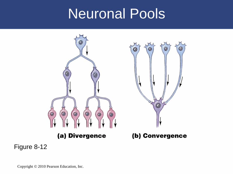
Neuronal Pools
Figure 8-12

Copyright © 2010 Pearson Education, Inc.
8-5 The brain and spinal cord are surrounded by three
layers of membranes called the meninges

Copyright © 2010 Pearson Education, Inc.
The Three Meningeal Layers
• The Dura Mater
– Tough and fibrous
– Cranially:
• Fuses with periosteum of occipital bone
– Caudally:
• Tapers to dense cord of collagen fibers
• Joins filum terminale in coccygeal ligament
– The epidural space:
• Between spinal dura mater and walls of vertebral canal
• Contains loose connective and adipose tissue
• Anesthetic injection site

Copyright © 2010 Pearson Education, Inc.
The Three Meningeal Layers
• The Arachnoid Mater
– Middle meningeal layer
– Arachnoid membrane:
• Simple squamous epithelia
• Covers arachnoid mater

Copyright © 2010 Pearson Education, Inc.
The Three Meningeal Layers
• The Pia Mater
– Is the innermost meningeal layer
– Is a mesh of collagen and elastic fibers
– Is bound to underlying neural tissue

Copyright © 2010 Pearson Education, Inc.
The Three Meningeal Layers
Figure 8-13

Copyright © 2010 Pearson Education, Inc.
8-6 The spinal cord contains gray matter surrounded by
white matter and connects to 31 pairs of spinal nerves

Copyright © 2010 Pearson Education, Inc.
Gross Anatomy of the Spinal Cord
• About 18 inches (45 cm) long
• 1/2 inch (14 mm) wide
• Ends between vertebrae L1 and L2
• Bilateral symmetry
– Grooves divide the spinal cord into left and right
– Posterior median sulcus: on posterior side
– Anterior median fissure: deeper groove on anterior side

Copyright © 2010 Pearson Education, Inc.
Gross Anatomy of the Spinal Cord
• Enlargements of the Spinal Cord
– Caused by:
• Amount of gray matter in segment
• Involvement with sensory and motor nerves of
limbs
– Cervical enlargement:
• Nerves of shoulders and upper limbs
– Lumbar enlargement:
• Nerves of pelvis and lower limbs

Copyright © 2010 Pearson Education, Inc.
Gross Anatomy of the Spinal Cord
• Gross Anatomy of the Spinal Cord
– The distal end:
• Conus medullaris:
– thin, conical spinal cord below lumbar enlargement
• Filum terminale:
– thin thread of fibrous tissue at end of conus medullaris
– attaches to coccygeal ligament
• Cauda equina:
– nerve roots extending below conus medullaris

Copyright © 2010 Pearson Education, Inc.
Gross Anatomy of the Spinal Cord
Figure 8-14
Copyright © 2010 Pearson Education, Inc.
Gross Anatomy of the Spinal Cord
• 31 Spinal Cord Segments
– Based on vertebrae where spinal nerves
originate
– Positions of spinal segment and vertebrae
change with age:
• Cervical nerves:
– are named for inferior vertebra
• All other nerves:
– are named for superior vertebra

Copyright © 2010 Pearson Education, Inc.
Gross Anatomy of the Spinal Cord
• Roots
– Two branches of spinal nerves:
• Ventral root:
– contains axons of motor neurons
• Dorsal root:
– contains axons of sensory neurons
– Dorsal root ganglia:
• contain cell bodies of sensory neurons

Copyright © 2010 Pearson Education, Inc.
Gross Anatomy of the Spinal Cord
• The Spinal Nerve
– Each side of spine:
• Dorsal and ventral roots join
• To form a spinal nerve
– Mixed nerves:
• Carry both afferent (sensory) and efferent (motor)
fibers

Copyright © 2010 Pearson Education, Inc.
Sectional Anatomy of the Spinal Cord
• White matter
– Is superficial
– Contains myelinated and unmyelinated axons
• Gray matter
– Surrounds central canal of spinal cord
– Contains neuron cell bodies, neuroglia, unmyelinated
axons
– Has projections (gray horns)

Copyright © 2010 Pearson Education, Inc.
Sectional Anatomy of the Spinal Cord
• Organization of White Matter
– Posterior white columns: lie between posterior gray
horns and posterior median sulcus
– Anterior white columns: lie between anterior gray
horns and anterior median fissure
• Anterior white commissure: area where axons cross from one
side of spinal cord to the other
– Lateral white columns: located on each side of
spinal cord between anterior and posterior columns

Copyright © 2010 Pearson Education, Inc.
Sectional Anatomy of the Spinal Cord
Figure 8-15a

Copyright © 2010 Pearson Education, Inc.
8-7 The brain has several principal structures, each with
specific functions

Copyright © 2010 Pearson Education, Inc.
An Introduction to the Brain
• The Adult Human Brain
– Ranges from 750 cc to 2100 cc
– Contains almost 97% of the body’s neural
tissue
– Average weight about 1.4 kg (3 lb)

Copyright © 2010 Pearson Education, Inc.
The Brain
• Six Regions of the Brain
– Cerebrum
– Diencephalon
– Midbrain
– Pons
– Medulla oblongata
– Cerebellum
3D Peel-Away of the Brain

Copyright © 2010 Pearson Education, Inc.
The Brain
• Cerebrum
– Largest part of brain
– Controls higher mental functions
– Divided into left and right cerebral
hemispheres
– Surface layer of gray matter (neural cortex)

Copyright © 2010 Pearson Education, Inc.
The Brain
• Diencephalon
– Located under cerebrum and cerebellum
– Links cerebrum with brain stem
– Three divisions:
• Left thalamus
• Right thalamus
• Hypothalamus

Copyright © 2010 Pearson Education, Inc.
The Brain
• Diencephalon
– Thalamus:
• Relays and processes sensory information
– Hypothalamus:
• Hormone production
• Emotion
• Autonomic function
– Pituitary gland:
• Major endocrine gland
• Connected to hypothalamus
• Via infundibulum (stalk)
• Interfaces nervous and endocrine systems

Copyright © 2010 Pearson Education, Inc.
The Brain
• The Brain Stem
– Processes information between:
• Spinal cord and cerebrum or cerebellum
– Includes:
• Midbrain
• Pons
• Medulla oblongata
Copyright © 2010 Pearson Education, Inc.
The Brain
• The Brain Stem
– Midbrain:
• Processes sight, sound, and associated reflexes
• Maintains consciousness
– Pons:
• Connects cerebellum to brain stem
• Is involved in somatic and visceral motor control

Copyright © 2010 Pearson Education, Inc.
The Brain
• The Brain Stem
– Medulla oblongata:
• Connects brain to spinal cord
• Relays information
• Regulates autonomic functions:
– heart rate, blood pressure, and digestion

Copyright © 2010 Pearson Education, Inc.
The Brain
• Cerebellum
– Second largest part of brain
– Coordinates repetitive body movements
– Two hemispheres
– Covered with cerebellar cortex

Copyright © 2010 Pearson Education, Inc.
The Brain
Figure 8-16a

Copyright © 2010 Pearson Education, Inc.
The Brain
Figure 8-16b

Copyright © 2010 Pearson Education, Inc.
The Brain
Figure 8-16c
Copyright © 2010 Pearson Education, Inc.
The Brain
• Ventricles of the Brain
– Each cerebral hemisphere contains one large lateral ventricle:
• Separated by a thin medial partition (septum pellucidum)
– Third ventricle:
• Ventricle of the diencephalon
• Lateral ventricles communicate with third ventricle:
– via interventricular foramen (foramen of Monro)
– Fourth ventricle:
• Extends into medulla oblongata
• Connects with third ventricle:
– aqueduct of midbrain

Copyright © 2010 Pearson Education, Inc.
The Ventricles of the Brain
Figure 8-17

Copyright © 2010 Pearson Education, Inc.
The Brain
• Cerebrospinal Fluid
– Subdural space:
• Between arachnoid mater and dura mater
– Subarachnoid space:
• Between arachnoid mater and pia mater
• Contains collagen/elastin fiber network (arachnoid trabeculae)
• Filled with cerebrospinal fluid (CSF)
– Cerebrospinal fluid (CSF):
• Carries dissolved gases, nutrients, and wastes
• Spinal tap: withdraws CSF

Copyright © 2010 Pearson Education, Inc.
CSF Circulation
Figure 8-18

Copyright © 2010 Pearson Education, Inc.
The Cerebrum
• The Cerebrum
– Is the largest part of the brain
– Controls all conscious thoughts and
intellectual functions
– Processes somatic sensory and motor
information

Copyright © 2010 Pearson Education, Inc.
The Cerebrum
• Gray matter
– In cerebral cortex and basal nuclei
• White matter
– Deep to basal cortex
– Around basal nuclei
Copyright © 2010 Pearson Education, Inc.
The Cerebrum
• Structures of the Cerebrum
– Gyri of neural cortex:
• Increase surface area (number of cortical neurons)
– Insula (island) of cortex:
• Lies medial to lateral sulcus
– Longitudinal fissure:
• Separates cerebral hemispheres
– Lobes:
• Divisions of hemispheres

Copyright © 2010 Pearson Education, Inc.
The Cerebrum
• Structures of the Cerebrum
– Central sulcus divides:
• Anterior frontal lobe from posterior parietal lobe
– Lateral sulcus divides:
• Frontal lobe from temporal lobe
– Parieto-occipital sulcus divides:
• Parietal lobe from occipital lobe

Copyright © 2010 Pearson Education, Inc.
The Cerebrum
• Motor and Sensory Areas of the Cortex
– Central sulcus separates motor and sensory
areas
– Motor areas:
• Precentral gyrus of frontal lobe:
– directs voluntary movements
• Primary motor cortex:
– is the surface of precentral gyrus
• Pyramidal cells:
– are neurons of primary motor cortex

Copyright © 2010 Pearson Education, Inc.
The Cerebrum
• Motor and Sensory Areas of the Cortex
– Sensory areas:
• Postcentral gyrus of parietal lobe:
– receives somatic sensory information (touch, pressure,
pain, vibration, taste, and temperature)
• Primary sensory cortex:
– surface of postcentral gyrus

Copyright © 2010 Pearson Education, Inc.
The Cerebrum
• Special Sensory Cortexes
– Visual cortex:
• Information from sight receptors
– Auditory cortex:
• Information from sound receptors
– Olfactory cortex:
• Information from odor receptors
– Gustatory cortex:
• Information from taste receptors

Copyright © 2010 Pearson Education, Inc.
The Cerebrum
Figure 8-19

Copyright © 2010 Pearson Education, Inc.
The Cerebrum
• Association Areas
– Sensory association areas:
• Monitor and interpret arriving information at sensory areas of
cortex
– Somatic motor association area (premotor
cortex):
• Coordinates motor responses (learned movements)

Copyright © 2010 Pearson Education, Inc.
The Cerebrum
• Sensory Association Areas
– Somatic sensory association area:
• Interprets input to primary sensory cortex (e.g., recognizes
and responds to touch)
– Visual association area:
• Interprets activity in visual cortex
– Auditory association area:
• Monitors auditory cortex

Copyright © 2010 Pearson Education, Inc.
The Cerebrum
• General Interpretive Area
– Also called Wernicke area
– Present in only one hemisphere
– Receives information from all sensory association
areas
– Coordinates access to complex visual and auditory
memories

Copyright © 2010 Pearson Education, Inc.
The Cerebrum
• Other Integrative Areas
– Speech center:
• Is associated with general interpretive area
• Coordinates all vocalization functions
– Prefrontal cortex of frontal lobe:
• Integrates information from sensory association
areas
• Performs abstract intellectual activities (e.g.,
predicting consequences of actions)

Copyright © 2010 Pearson Education, Inc.
The Cerebrum
• Hemispheric Lateralization
– Functional differences between left and right
hemispheres
– Each cerebral hemisphere performs certain
functions that are not ordinarily performed by
the opposite hemisphere

Copyright © 2010 Pearson Education, Inc.
The Cerebrum
• The Left Hemisphere
– In most people, the left brain (dominant hemisphere)
controls:
• Reading, writing, and math
• Decision making
• Speech and language
• The Right Hemisphere
– Right cerebral hemisphere relates to:
• Senses (touch, smell, sight, taste, hearing)
• Recognition (faces, voice inflections)

Copyright © 2010 Pearson Education, Inc.
Hemispheric Lateralization
Figure 8-20

Copyright © 2010 Pearson Education, Inc.
The Cerebrum
• Monitoring Brain Activity
– Brain activity is assessed by an
electroencephalogram (EEG):
• Electrodes are placed on the skull
• Patterns of electrical activity (brain waves) are
printed out

Copyright © 2010 Pearson Education, Inc.
The Cerebrum
• Four Categories of Brain Waves– Alpha waves:
• Found in healthy, awake adults at rest with eyes closed
– Beta waves:• Higher frequency
• Found in adults concentrating or mentally stressed
– Theta waves:• Found in children
• Found in intensely frustrated adults
• May indicate brain disorder in adults
– Delta waves:• During sleep
• Found in awake adults with brain damage

Copyright © 2010 Pearson Education, Inc.
Brain Waves
Figure 8-21

Copyright © 2010 Pearson Education, Inc.
The Cerebrum
• The Basal Nuclei
– Also called cerebral nuclei
– Are masses of gray matter
– Are embedded in white matter of cerebrum
– Direct subconscious activities

Copyright © 2010 Pearson Education, Inc.
The Cerebrum
• Structures of Basal Nuclei
– Caudate nucleus:
• Curving, slender tail
– Lentiform nucleus:
• Globus pallidus
• Putamen

Copyright © 2010 Pearson Education, Inc.
The Cerebrum
• Functions of Basal Nuclei
– Involved with:
• The subconscious control of skeletal muscle tone
• The coordination of learned movement patterns
(walking, lifting)

Copyright © 2010 Pearson Education, Inc.
The Basal Nuclei
Figure 8-22a

Copyright © 2010 Pearson Education, Inc.
The Basal Nuclei
Figure 8-22b

Copyright © 2010 Pearson Education, Inc.
The Limbic System
• The Limbic System
– Is a functional grouping that:
• Establishes emotional states
• Links conscious functions of cerebral cortex with autonomic
functions of brain stem
• Facilitates memory storage and retrieval

Copyright © 2010 Pearson Education, Inc.
The Limbic System
• Components of the Limbic System
– Amygdaloid body:
• Acts as interface between the limbic system, the
cerebrum, and various sensory systems
– Limbic lobe of cerebral hemisphere:
• Cingulate gyrus
• Dentate gyrus
• Parahippocampal gyrus
• Hippocampus

Copyright © 2010 Pearson Education, Inc.
The Limbic System
• Components of the Limbic System
– Fornix:
• Tract of white matter
• Connects hippocampus with hypothalamus
– Anterior nucleus of the thalamus:
• Relays information from mamillary body to
cingulate gyrus
– Reticular formation:
• Stimulation or inhibition affects emotions (rage,
fear, pain, sexual arousal, pleasure)

Copyright © 2010 Pearson Education, Inc.
The Limbic System
Figure 8-23

Copyright © 2010 Pearson Education, Inc.
The Diencephalon
• Integrates sensory information and motor
commands
• Thalamus, epithalamus, and hypothalamus
– The pineal gland:
• Found in posterior epithalamus
• Secretes hormone melatonin

Copyright © 2010 Pearson Education, Inc.
The Midbrain
• Two pairs of sensory nuclei (corpora quadrigemina)
– Superior colliculus (visual)
– Inferior colliculus (auditory)
• Cerebral peduncles
– Nerve fiber bundles on ventrolateral surfaces
– Contain:
• Descending fibers to cerebellum
• Motor command fibers

Copyright © 2010 Pearson Education, Inc.
The Pons
• Links cerebellum with mesencephalon,
diencephalon, cerebrum, and spinal cord
– Sensory and motor nuclei of cranial nerves V,
VI, VII, and VIII

Copyright © 2010 Pearson Education, Inc.
The Medulla Oblongata
• The Medulla Oblongata
– Allows brain and spinal cord to communicate
– Coordinates complex autonomic reflexes
– Controls visceral functions
– Nuclei in the medulla:
• Autonomic nuclei: control visceral activities
• Sensory and motor nuclei: of cranial nerves
• Relay stations: along sensory and motor pathways

Copyright © 2010 Pearson Education, Inc.
The Diencephalon and Brain Stem
Figure 8-24

Copyright © 2010 Pearson Education, Inc.
8-8 The PNS connects the CNS with the body’s external
and internal environments

Copyright © 2010 Pearson Education, Inc.
Cranial Nerves
• 12 pairs connected to brain
• Four Classifications of Cranial Nerves
– Sensory nerves: carry somatic sensory information,
including touch, pressure, vibration, temperature, and
pain
– Special sensory nerves: carry sensations such as
smell, sight, hearing, balance
– Motor nerves: axons of somatic motor neurons
– Mixed nerves: mixture of motor and sensory fibers

Copyright © 2010 Pearson Education, Inc.
Cranial Nerves
• Cranial nerves are classified by primary
functions
• May also have important secondary
functions
– Distributing autonomic fibers to peripheral
ganglia

Copyright © 2010 Pearson Education, Inc.
Cranial Nerves
Figure 8-25

Copyright © 2010 Pearson Education, Inc.
Cranial Nerves
• Olfactory Nerves (I)
– Primary function:
• Special sensory (smell)
• Optic Nerves (II)
– Primary function:
• Special sensory (vision)
• Oculomotor Nerves (III)– Primary function:
• Motor (eye movements)

Copyright © 2010 Pearson Education, Inc.
Cranial Nerves
• The Trochlear Nerves (IV)
– Primary function:
• Motor (eye movements)
• The Trigeminal Nerves (V)
– Primary function:
• Mixed (sensory and motor) to face
• The Abducens Nerves (VI)
– Primary function:
• Motor (eye movements)

Copyright © 2010 Pearson Education, Inc.
Cranial Nerves
• The Facial Nerves (VII)
– Primary function:
• Mixed (sensory and motor) to face
• The Vestibulocochlear Nerves (VIII)
– Primary function: special sensory:
• Vestibular branch:
– balance and equilibrium
• Cochlear branch:
– hearing

Copyright © 2010 Pearson Education, Inc.
Cranial Nerves
• The Glossopharyngeal Nerves (IX)
– Primary function:
• Mixed (sensory and motor) to head and neck
• The Vagus Nerves (X)
– Primary function:
• Mixed (sensory and motor)
• Widely distributed in thorax and abdomen

Copyright © 2010 Pearson Education, Inc.
Cranial Nerves
• The Accessory Nerves (XI)
– Primary function
• Motor to muscles of neck and upper back
• The Hypoglossal Nerves (XII)
– Primary function
• Motor (tongue movements)

Copyright © 2010 Pearson Education, Inc.

Copyright © 2010 Pearson Education, Inc.
Spinal Nerves and Plexuses
• Nerve Plexuses
– Complex, interwoven networks of nerve fibers
– Formed from blended fibers of ventral rami of
adjacent spinal nerves
– Control skeletal muscles of the neck and
limbs
3D Rotation of Peripheral Nerves and Nerve Plexuses

Copyright © 2010 Pearson Education, Inc.
Dermatomes
Figure 8-27

Copyright © 2010 Pearson Education, Inc.
Peripheral Nerves and Nerve Plexuses
Figure 8-26

Copyright © 2010 Pearson Education, Inc.
Peripheral Nerves and Nerve Plexuses
Figure 8-26

Copyright © 2010 Pearson Education, Inc.
Copyright © 2010 Pearson Education, Inc.
8-9 Reflexes are rapid, automatic responses to stimuli

Copyright © 2010 Pearson Education, Inc.
Reflexes
• Automatic responses coordinated within
spinal cord
• Through interconnected sensory neurons,
motor neurons, and interneurons
• Produce simple and complex reflexes

Copyright © 2010 Pearson Education, Inc.
Reflexes
• Neural Reflexes
– Rapid, automatic responses to specific stimuli
– Basic building blocks of neural function
– One neural reflex produces one motor response
– Reflex arc:
• The wiring of a single reflex
• Beginning at receptor
• Ending at peripheral effector
• Generally opposes original stimulus (negative feedback)

Copyright © 2010 Pearson Education, Inc.
Reflexes
• Five Steps in a Neural Reflex
– Step 1: Arrival of stimulus, activation of receptor:
• Physical or chemical changes
– Step 2: Activation of sensory neuron:
• Graded depolarization
– Step 3: Information processing by postsynaptic cell:
• Triggered by neurotransmitters
– Step 4: Activation of motor neuron:
• Action potential
– Step 5: Response of peripheral effector:
• Triggered by neurotransmitters

Copyright © 2010 Pearson Education, Inc.
Reflexes
Figure 8-28

Copyright © 2010 Pearson Education, Inc.
Spinal Reflexes
• Monosynaptic Reflexes
– A stretch reflex
– Have least delay between sensory input and motor
output:
• For example, stretch reflex (such as patellar reflex)
– Completed in 20–40 msec
– Receptor is muscle spindle

Copyright © 2010 Pearson Education, Inc.
A Stretch Reflex
Figure 8-29

Copyright © 2010 Pearson Education, Inc.
Spinal Reflexes
• Withdrawal Reflexes
– Move body part away from stimulus (pain or pressure)
• For example, flexor reflex:
– pulls hand away from hot stove
– Strength and extent of response:
• Depends on intensity and location of stimulus

Copyright © 2010 Pearson Education, Inc.
A Flexor Reflex
Figure 8-30

Copyright © 2010 Pearson Education, Inc.
Integration and Control of Spinal Reflexes
• Reflex behaviors are automatic
– But processing centers in brain can facilitate
or inhibit reflex motor patterns based in spinal
cord

Copyright © 2010 Pearson Education, Inc.
8-10 Separate pathways carry sensory and motor commands

Copyright © 2010 Pearson Education, Inc.
Sensory Pathways
• Deliver somatic and visceral sensory information
to their final destinations inside the CNS using
– Nerves
– Nuclei
– Tracts

Copyright © 2010 Pearson Education, Inc.
Sensory Pathways
• Somatic Sensory Pathways
– Posterior column pathway:
• Carries sensations of highly localized (“fine”)
touch, pressure, vibration, and proprioception
• Spinal tracts involved:
– left and right fasciculus gracilis
– left and right fasciculus cuneatus
Figure 15–5a

Copyright © 2010 Pearson Education, Inc.
Sensory Pathways
• Posterior Column Pathway
– Axons synapse
• On third-order neurons in one of the ventral nuclei
of the thalamus
• Nuclei sort the arriving information according to:
– the nature of the stimulus
– the region of the body involved
Figure 15–5a
Copyright © 2010 Pearson Education, Inc.
Sensory Pathways
• Posterior Column Pathway
– Processing in the thalamus:
• Determines whether you perceive a given sensation as fine
touch, as pressure, or as vibration
– Ability to determine stimulus:
• Precisely where on the body a specific stimulus originated
depends on the projection of information from the thalamus
to the primary sensory cortex
Figure 15–5a

Copyright © 2010 Pearson Education, Inc.
Sensory Pathways
• Posterior Column Pathway
– Sensory information:
• From toes arrives at one end of the primary
sensory cortex
• From the head arrives at the other:
– when neurons in one portion of your primary sensory
cortex are stimulated, you become aware of sensations
originating at a specific location
Figure 15–5a
Copyright © 2010 Pearson Education, Inc.
Sensory Pathways
• Posterior Column Pathway
– Sensory homunculus:
• Functional map of the primary sensory cortex
• Distortions occur because area of sensory cortex
devoted to particular body region is not
proportional to region’s size, but to number of
sensory receptors it contains

Copyright © 2010 Pearson Education, Inc.
The Posterior Column Pathway
Figure 8-31

Copyright © 2010 Pearson Education, Inc.
Motor Pathways
• The Corticospinal Pathway
– Sometimes called the pyramidal system
– Provides voluntary control over skeletal muscles:
• System begins at pyramidal cells of primary motor cortex
• Axons of these upper motor neurons descend into brain stem
and spinal cord to synapse on lower motor neurons that
control skeletal muscles
– Contains three pairs of descending tracts:
• Corticobulbar tracts
• Lateral corticospinal tracts
• Anterior corticospinal tracts

Copyright © 2010 Pearson Education, Inc.
Motor Pathways
• The Corticospinal Pathway
– Corticospinal tracts:
• As they descend, lateral corticospinal tracts are visible
along the ventral surface of medulla oblongata as pair of
thick bands, the pyramids
• At spinal segment it targets, an axon in anterior
corticospinal tract crosses over to the opposite side of the
spinal cord in anterior white commissure before synapsing
on lower motor neurons in anterior gray horns

Copyright © 2010 Pearson Education, Inc.
Motor Pathways
• The Corticospinal Pathway
– Motor homunculus:
• Primary motor cortex corresponds point by point with specific
regions of the body
• Cortical areas have been mapped out in diagrammatic form
• Homunculus provides indication of degree of fine motor
control available:
– hands, face, and tongue, which are capable of varied and
complex movements, appear very large, while trunk is relatively
small
– these proportions are similar to the sensory homunculus

Copyright © 2010 Pearson Education, Inc.
The Corticospinal Pathway
Figure 8-32

Copyright © 2010 Pearson Education, Inc.
Motor Pathways
• The Medial and Lateral Pathways
– Several centers in cerebrum, diencephalon, and brain
stem may issue somatic motor commands as result of
processing performed at a subconscious level
– These nuclei and tracts are grouped by their primary
functions:
• Components of medial pathway help control gross
movements of trunk and proximal limb muscles
• Components of lateral pathway help control distal limb
muscles that perform more precise movements

Copyright © 2010 Pearson Education, Inc.

Copyright © 2010 Pearson Education, Inc.
8-11 The autonomic nervous system, composed of the
sympathetic and parasympathetic divisions, is involved in the unconscious regulation of body functions

Copyright © 2010 Pearson Education, Inc.
An Introduction to the ANS
• Somatic Nervous System (SNS)
– Operates under conscious control
– Seldom affects long-term survival
– SNS controls skeletal muscles
• Autonomic Nervous System (ANS)
– Operates without conscious instruction
– ANS controls visceral effectors
– Coordinates system functions: cardiovascular, respiratory,
digestive, urinary, reproductive
The Organization of the Somatic and Autonomic Nervous Systems

Copyright © 2010 Pearson Education, Inc.
Somatic Nervous System
Figure 8-33a

Copyright © 2010 Pearson Education, Inc.
Autonomic Nervous System
Figure 8-33b

Copyright © 2010 Pearson Education, Inc.
Divisions of the ANS
• The autonomic nervous system
– Operates largely outside our awareness
– Has two divisions:
• Sympathetic division:
– increases alertness, metabolic rate, and muscular
abilities
• Parasympathetic division:
– reduces metabolic rate and promotes digestion

Copyright © 2010 Pearson Education, Inc.
Divisions of the ANS
• Sympathetic Division
– “Kicks in” only during exertion, stress, or emergency
– “Fight or flight”
• Parasympathetic Division
– Controls during resting conditions
– “Rest and digest”

Copyright © 2010 Pearson Education, Inc.
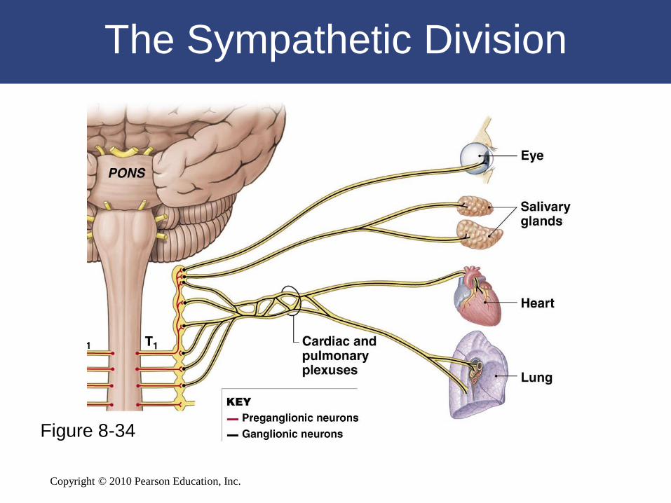
The Sympathetic Division
• Ganglionic Neurons
– Occur in three locations:
• Sympathetic chain ganglia
• Collateral ganglia
• Suprarenal medullae

Copyright © 2010 Pearson Education, Inc.
The Sympathetic Division
• Ganglionic Neurons
– Sympathetic chain ganglia:
• Are on both sides of vertebral column
• Control effectors:
– in body wall
– inside thoracic cavity
– in head
– in limbs

Copyright © 2010 Pearson Education, Inc.
The Sympathetic Division
• Ganglionic Neurons
– Collateral ganglia:
• Are anterior to vertebral bodies
• Contain ganglionic neurons that innervate tissues
and organs in abdominopelvic cavity

Copyright © 2010 Pearson Education, Inc.
The Sympathetic Division
• Ganglionic Neurons
– Suprarenal (adrenal) medullae:
• Very short axons
• When stimulated, release neurotransmitters into
bloodstream (not at synapse)
• Function as hormones to affect target cells
throughout body

Copyright © 2010 Pearson Education, Inc.
The Sympathetic Division
Figure 8-34
Copyright © 2010 Pearson Education, Inc.
The Sympathetic Division
Figure 8-34

Copyright © 2010 Pearson Education, Inc.
The Sympathetic Division
Figure 8-34

Copyright © 2010 Pearson Education, Inc.
The Sympathetic Division
Figure 8-34

Copyright © 2010 Pearson Education, Inc.
The Parasympathetic Division
• Autonomic Nuclei
– Are contained in the mesencephalon, pons,
and medulla oblongata:
• Associated with cranial nerves III, VII, IX, X
– In lateral gray horns of spinal segments S2–S4

Copyright © 2010 Pearson Education, Inc.
The Parasympathetic Division
• Ganglionic Neurons in Peripheral Ganglia
– Near target organ
– Embedded in tissues of target organ
– Usually paired

Copyright © 2010 Pearson Education, Inc.
The Parasympathetic Division
Figure 8-35

Copyright © 2010 Pearson Education, Inc.
The Parasympathetic Division
Figure 8-35
Copyright © 2010 Pearson Education, Inc.
The Parasympathetic Division
• Parasympathetic Activation
– Centers on relaxation, food processing, and
energy absorption
– Localized effects, last a few seconds at most

Copyright © 2010 Pearson Education, Inc.

Copyright © 2010 Pearson Education, Inc.

Copyright © 2010 Pearson Education, Inc.

Copyright © 2010 Pearson Education, Inc.
8-12 Aging produces various structural and functional changes in the nervous
system

Copyright © 2010 Pearson Education, Inc.
Aging and the Nervous System
• Anatomical and physiological changes
begin after maturity (age 30)
• Accumulate over time
• 85% of people over age 65 have changes
in mental performance and CNS function

Copyright © 2010 Pearson Education, Inc.
Aging and the Nervous System
• Reduction in Brain Size and Weight
• Reduction in Number of Neurons
• Decrease in Blood Flow to Brain
• Changes in Synaptic Organization of Brain
• Intracellular and Extracellular Changes in CNS
Neurons

Copyright © 2010 Pearson Education, Inc.
8-13 The nervous system is closely integrated with other
body systems

The Nervous System
in Perspective
Functional
the Nervous System and Other Systems
Copyright © 2010 Pearson Education, Inc.

The Integumentary System
provides sensations of touch,
pressure, pain, vibration, and
temperature; hair provides some
protection and insulation for skull
and brain; protects peripheral
nerves.
The Nervous System controls
contraction of arrector pili muscles
and secretion of sweat glands.
The Integumentary System
Copyright © 2010 Pearson Education, Inc.

The Skeletal System
The Skeletal System provides
calcium for neural function;
protects brain and spinal cord.
The Nervous System controls
skeletal muscle contractions that
produce bone thickening and
maintenance, and determine bone
position.
Copyright © 2010 Pearson Education, Inc.

The Muscular System
The Muscular System’s facial
muscles express emotional state;
intrinsic laryngeal muscles permit
communication; muscle spindles
provide proprioceptive sensations.
The Nervous System controls
skeletal muscle contractions;
coordinates respiratory and
cardiovascular activities.
Copyright © 2010 Pearson Education, Inc.

The Endocrine System
The Endocrine System’s
Many hormones affect CNS neural
metabolism; the reproductive
hormones and thyroid hormone
influence CNS development.
The Nervous System controls
pituitary gland and many other
endocrine organs; secretes ADH
and oxytocin.
Copyright © 2010 Pearson Education, Inc.

The Cardiovascular System
The Cardiovascular System’s
capillaries maintain the blood-brain
barrier when stimulated by
astrocytes; blood vessels (with
ependymal cells) produce CSF.
The Nervous System modifies
heart rate and blood pressure;
astrocytes stimulate maintenance
of blood-brain barrier.
Copyright © 2010 Pearson Education, Inc.

The Lymphoid System
The Lymphoid System defends
against infection and assists in
tissue repairs.
The Nervous System’s release of
neurotransmitters and hormones
affects sensitivity of immune
response.
Copyright © 2010 Pearson Education, Inc.

The Respiratory System
The Respiratory System provides
oxygen and eliminates carbon
dioxide.
The Nervous System controls the
pace and depth of respiration.
Copyright © 2010 Pearson Education, Inc.

The Digestive System
The Digestive System provides
nutrients for energy production and
neurotransmitter synthesis.
The Nervous System regulates
digestive tract movement and
secretion.
Copyright © 2010 Pearson Education, Inc.

The Urinary System
The Urinary System eliminates
metabolic wastes; regulates body
fluid pH and electrolyte
concentrations.
The Nervous System adjusts renal
pressure and controls urination.
Copyright © 2010 Pearson Education, Inc.

The Reproductive System
The Reproductive System’s
sex hormones affect CNS
development
and sexual behaviors.
The Nervous System controls
sexual behaviors and sexual
function.
Copyright © 2010 Pearson Education, Inc.