Embed Size (px)
Citation preview

THE MUSCULAR SYSTEM:
SKELETAL MUSCLE TISSUE
AND MUSCLE
ORGANIZATION

Skeletal Muscles
• Attach to bones
• Produce skeletal movement (voluntary)
• Maintain posture
• Support soft tissues
• Regulate entrances to the body
• Maintain body temperature

Properties of Skeletal Muscles
Electrical excitability
-ability to respond to stimuli by producing electrical signals
such as action potentials
-two types of stimuli: 1. autorhythmic electrical signals
2. chemical stimuli
Contractility
-ability to contract when stimulated by an AP
-isometric contraction: tension develops, length doesn’t change
-isotonic contraction: tension develops, muscle shortens
Extensibility
-ability to stretch without being damaged
-allows contraction even when stretched
Elasticity
-ability to return to its original length and shape

Classification • According to arrangement of fibers and fascicles
– Parallel muscles
• Parallel to long axis of muscle
– Convergent muscles
• Fibers converge on common attachment site
– Pennate muscles
• One or more tendons run through body of muscle
• Unipennate, bipennate, multipennate
– Circular muscles
• Fibers concentrically arranged

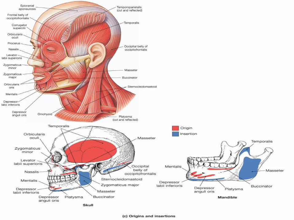
Origins and Insertions
• Origin remains stationary
– Typically proximal to insertion
• Insertion moves
• Muscles identified by
– Origin
– Insertion
– Primary action
• Classified as
– Prime mover (agonist)
– Synergist
– Antagonist
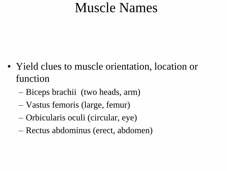
Muscle Names
• Yield clues to muscle orientation, location or
function
– Biceps brachii (two heads, arm)
– Vastus femoris (large, femur)
– Orbicularis oculi (circular, eye)
– Rectus abdominus (erect, abdomen)

Axial Musculature
• Arises from and inserts on the axial skeleton
• Positions the head and spinal column
• Moves the rib cage, assisting in breathing
• Axial musculature
– Originates and inserts on axial skeleton
• Appendicular musculature
– Stabilizes or moves components of the
appendicular skeleton


Gross Anatomy
•muscles are really groups of
fascicles
•the fascicles are groups of muscle
fibers = considered to be an
individual muscle cell

•the muscle fiber is made up
of protein filaments = myofibrils
•each myofibril is comprised of
repeating units = sarcomeres

Gross Anatomy
•muscle is wrapped in a protective fascia
-fascia = sheet of fibrous connective tissue that supports
and surrounds muscle or organs
•a superficial fascia separates muscle from the overlying skin
-also known as the subcutaneous layer
-made up of areolar tissue and adipose tissue
-provides support for blood vessel and nerves
-the adipose tissue stores most of the body’s triglycerides
and provides insulation
•muscles with similar functions are grouped and held together by layers
of deep fascia
-dense irregular connective tissue
-allow free movement of muscles, carries nerves, BVs

• three layers of connective tissue extend from the deep fascial layer
– Epimysium
– Perimysium
– Endomysium
• these layers further strengthen and protect muscle
• outermost layer = epimysium
– encircles the entire muscle
• next layer = perimysium
– surrounds groups of 10 to 100 individual muscle fibers
– separates them into bundles = fascicles
– give meat its “grain” because the fascicles are visible
– both epimysium and perimysium are dense irregular connective tissue
• penetrating the fasicles and separating them into individual muscle
fibers = endomysium (areolar connective tissue)

•generally muscles are supplied with one artery and two veins
•they accompany the nerve
•nerves that induce muscle contraction = somatic motor neurons (part
of the somatic division of the PNS)
•communication between muscle and these neurons
Neuromuscular junction (NMJ)
•all three of these connective tissue layers extend beyond the muscle
and attaches it to other structures
-called a tendon = cord of regular dense CT that attaches
a muscle to the periosteum of bone
•when the CT extends as a broad flat sheet = aponeurosis


Microanatomy of Skeletal Muscle
Fibers • New terminology
– Cell membrane = sarcolemma
– Cytoplasm = sarcoplasm
– Internal membrane system = sarcoplasmic reticulum
• Large, multinucleated cells
– embryonic development – stem cells (satellite cells) differentiate into immature myoblasts which begin to make the proteins of the myofilament
– These myoblasts mature into myocytes
– Multiple myocytes fuse to form the muscle cell (muscle fiber)
– once fused, these muscle cells lose the ability of undergo mitosis
– number of muscle cells predetermined before birth
– But satellite cells can repair damaged/dying muscle cells throughout adulthood

Muscle Cell Anatomy
• Transverse tubules
– Invaginations of sarcolemma
– Carry electrical impulses
• Myofibrils within sarcoplasm
– “skeleton” of protein filaments (myofilaments) organized as Sarcomeres
• Myofilaments form the myofibrils
– Thin filaments (actin, troponin, tropomyosin)
– Thick filaments (myosin)

Microanatomy of Skeletal Muscle
Fibers
•muscle fibers are bound by a plasma membrane = sarcolemma
•thousands of tiny invaginations in this sarcolemma called T or transverse
tubules - tunnel in toward the center of the cell
-T tubules are open to the outside of the fiber
- filled with interstitial fluid
- action potentials generated in the neuron travel along the sarcolemma
and the T tubules
- allows for the even and quick spread of an action potential deep into the cell
•the cytoplasm is called a sarcoplasm
-substantial amounts of glycogen - can be broken into glucose
-contains myoglobin - binds oxygen needed for muscle ATP
production

Microanatomy of Skeletal Muscle
Fibers
•contractile elements of the myofibrils = myofilaments
-2 microns in diameter
-comprised of primarily actin or myosin
-give the muscle its striated appearance
•Fibers also have a system of fluid-filled membranes = sarcoplasmic
reticulum
-encircles each myofibril
-similar to the ER
-have dilated end sacs = terminal cisterns
-stores calcium when at rest - releases it during contraction
-release is triggered by an AP

The Proteins of Muscle
• Myofibrils are built of 3 kinds of protein
– contractile proteins
• myosin and actin
– regulatory proteins which turn contraction on & off
• troponin and tropomyosin
– structural proteins which provide proper alignment,
elasticity and extensibility
• titin, myomesin, nebulin, actinin and dystrophin • Dystrophin – connects sarcomere to sarcolemma
– -transmits tension along muscle
• Actinin – part of Z-line
• Titin – connects myosin to Z-line and M-line
– Role in recovery after being stretched
• Nebulin – forms core of the actin chain/thin filament

Types of Muscle Fibers
• Fast fibers = glycolytic
• Slow fibers = oxidative
• Fibers of one motor unit all the same type
• Percentage of fast versus slow fibers is genetically
determined
• Proportions vary with the usual action of the muscle
- neck, back and leg muscles have a higher proportion of postural,
slow oxidative fibers
- shoulder and arm muscles have a higher proportion of fast
glycolytic fibers

Fast Fibers
• Large in diameter
• Contain densely packed myofibrils
• Large glycogen reserves
• Fast oxidative-glycolytic (fast-twitch A)
– red in color (lots of mitochondria, myoglobin & blood vessels)
– split ATP at very fast rate; used for walking and sprinting
• Fast glycolytic (fast-twitch B)
– white in color (few mitochondria & BV, low myoglobin)
– anaerobic movements for short duration; used for weight-lifting

• Slow fibers
– Half the diameter of fast fibers
– Three times longer to contract
– Continue to contract for long periods of time
• e.g. marathon runners

• Atrophy
– wasting away of muscles
– caused by disuse (disuse atrophy) or severing of the
nerve supply (denervation atrophy)
– the transition to connective tissue can not be reversed
• Hypertrophy
– increase in the diameter of muscle fibers
– resulting from very forceful, repetitive muscular
activity and an increase in myofibrils, SR &
mitochondria

Muscle Metabolism
• Production of ATP:
-contraction requires huge amounts of ATP
-muscle fibers produce ATP three ways:
1. Creatine phosphate
2. Aerobic metabolism
3. Anaerobic metabolism

Creatine Phosphate
• Muscle fibers at rest produce more ATP then they need for resting
metabolism
• Excess ATP within resting muscle used to form creatine phosphate
• By the enzyme creatine kinase
• Creatine phosphate: 3-6 times more plentiful than ATP within
muscle
• Its quick breakdown provides energy for creation of ATP
• Sustains maximal contraction for 15 sec (used for 100 meter dash).
• Athletes tried creatine supplementation
– gain muscle mass but shut down bodies own synthesis (safety?)

• sarcomere = regions of myosin (thick myofilament) and actin (thin myofilament)
• bounded by the Z line (actinin)
• actin filaments project out from Z line
• myosin filaments lie in center of sarcomere - overlap with actin and connect
via cross-bridges
• myosin only region = H zone
• myosin filaments are held in place by the M line proteins.
• actin only region = I band
• length of myosin filaments = A band
• contraction = “sliding filament theory”
-actin and myosin myofilaments slide over each other and sarcomere shortens
M line
Sarcomere
Structure

Contraction: The Sliding Filament Theory
• SF Theory:
– Explains how a muscle fiber exerts tension
– Four step process
• Active sites on actin
• Crossbridge formation
• Cycle of attach, pivot, detach, return
• Troponin and tropomyosin control contraction
• Contraction:
– Active process
– Elongation is passive
– Amount of tension produced is proportional to degree
of overlap of thick and thin filaments
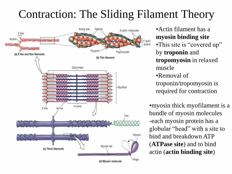
Contraction: The Sliding Filament Theory
•Actin filament has a
myosin binding site
•This site is “covered up”
by troponin and
tropomyosin in relaxed
muscle
•Removal of
troponin/tropomyosin is
required for contraction
•myosin thick myofilament is a
bundle of myosin molecules
-each myosin protein has a
globular “head” with a site to
bind and breakdown ATP
(ATPase site) and to bind
actin (actin binding site)
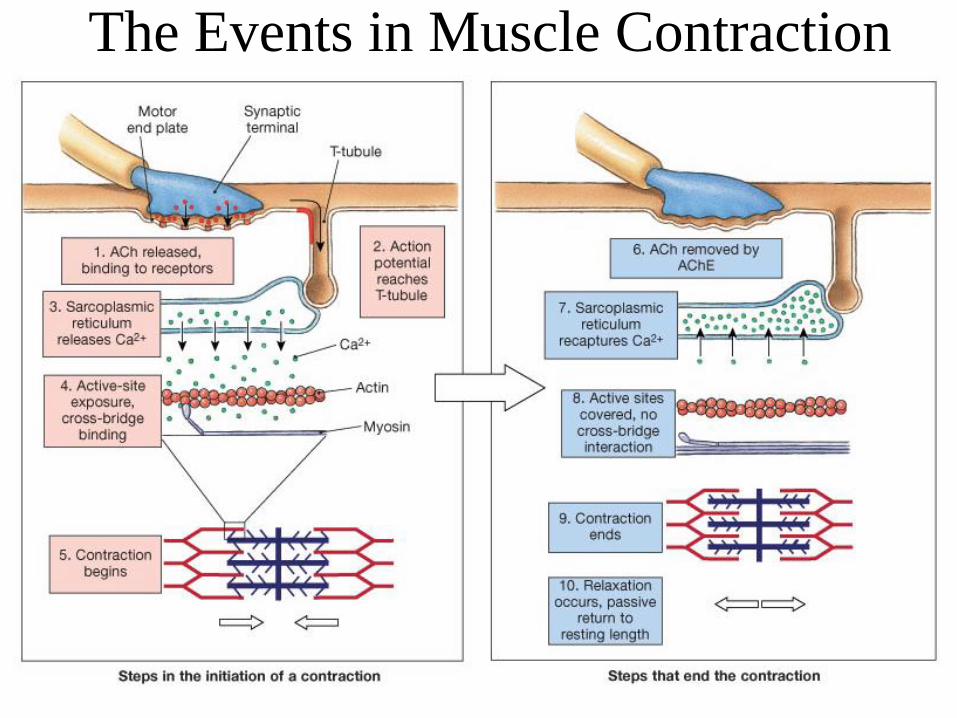
The Events in Muscle Contraction

Muscle Contraction: A summary
• ACh released from synaptic vesicles
• Binding of ACh to motor end plate
• Generation of electrical impulse in sarcolemma
• Conduction of impulse along T-tubules
• Release of Calcium ions by SR - binds to troponin
• Exposure of active sites on actin
• Cross-bridge formation and contraction
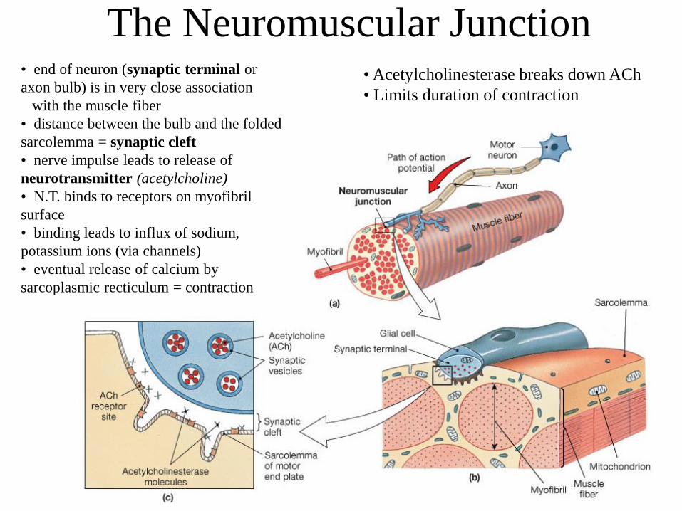
The Neuromuscular Junction • end of neuron (synaptic terminal or
axon bulb) is in very close association
with the muscle fiber
• distance between the bulb and the folded
sarcolemma = synaptic cleft
• nerve impulse leads to release of
neurotransmitter (acetylcholine)
• N.T. binds to receptors on myofibril
surface
• binding leads to influx of sodium,
potassium ions (via channels)
• eventual release of calcium by
sarcoplasmic recticulum = contraction
• Acetylcholinesterase breaks down ACh
• Limits duration of contraction
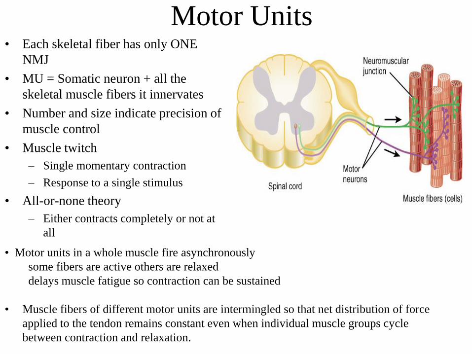
Motor Units • Each skeletal fiber has only ONE
NMJ
• MU = Somatic neuron + all the
skeletal muscle fibers it innervates
• Number and size indicate precision of
muscle control
• Muscle twitch
– Single momentary contraction
– Response to a single stimulus
• All-or-none theory
– Either contracts completely or not at
all
• Muscle fibers of different motor units are intermingled so that net distribution of force
applied to the tendon remains constant even when individual muscle groups cycle
between contraction and relaxation.
• Motor units in a whole muscle fire asynchronously
some fibers are active others are relaxed
delays muscle fatigue so contraction can be sustained

Axial muscles organized into four
groups
• Muscles of the head and neck
• Muscles of the vertebral column
• Oblique and rectus muscles
• Muscles of the pelvic floor

Muscles of the Vertebral Column • Longus capitus
• Longus colli
– Rotate and flex the neck
• Quadratus lumborum
muscles
– Flex the spine
– Depress the ribs

The Oblique and Rectus Muscles

The Diaphragm

Muscles of the Pelvic Floor
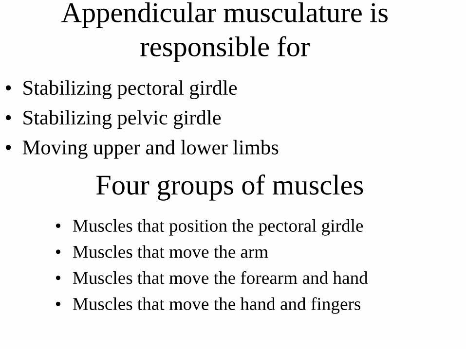
Appendicular musculature is
responsible for
• Stabilizing pectoral girdle
• Stabilizing pelvic girdle
• Moving upper and lower limbs
Four groups of muscles
• Muscles that position the pectoral girdle
• Muscles that move the arm
• Muscles that move the forearm and hand
• Muscles that move the hand and fingers


Extrinsic Muscles that Move the Hand and
Fingers

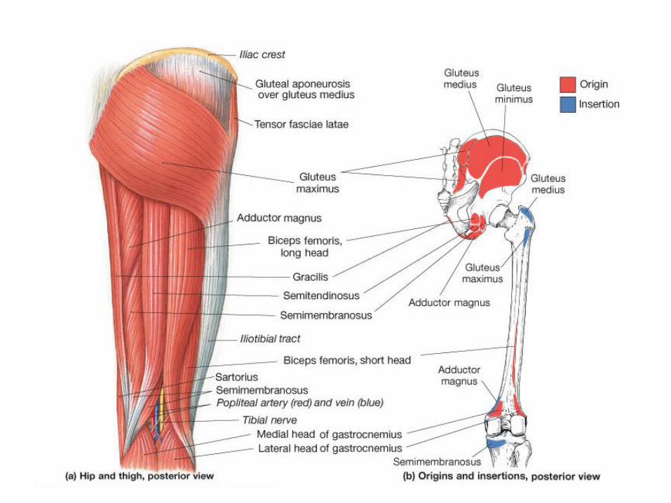
Muscles that Move the Thigh