Embed Size (px)
Citation preview

The Muscular SystemSashyel Altman Sofia Esteller Alex Hasson
Period 6

Functions and Types of Muscles
-All muscles in the human body, despite which type of muscle they are, has the ability to contract, or shorten. The human body has three types of muscles: smooth, cardiac, and skeletal. They all have contractile cells, which elongate, and are called muscle fibers.

Smooth Muscle • Smooth Muscle is located in the walls of hollow internal organs and
blood vessels. Its involuntary contractions moves material through organs, and regulates blood flow. Smooth muscle fibers are narrow, tapered rod shaped cells, and uninucleate. They arranged in parallel lines, forming sheets. It doesn’t have striations.

Cardiac Muscle • Cardiac muscle forms the heart wall. It’s fibers are uninucleated,
striated, tubular, and branched, which permits the fibers to connect at intercalated disks. Intercalated disks contain gap junctions, which allow contractions to spread quickly throughout the heart. Contraction of cardiac fibers is rhythmical and occurs without requiring outside nerve stimulation. Cardiac fibers relax completely between contractions, to prevent fatigue.

Skeletal Muscle • Skeletal muscle fibers are tubular, multinucleated, and striated. They
are the muscles attached to the skeleton. Skeletal muscle is voluntary because it’s contractions are always stimulated and controlled by the nervous system.


Connective Tissue Covering-Muscles are organs, and they contain other types of tissues such as nervous,
blood and connective tissue. Connective tissue is vital to the organization of the fibers in a muscle.
-First, each fiber is surrounded by a thin layer of areolar connective tissue called the endomysium. Blood capillaries and nerve fibers reach each muscle fiber by way of the endomysium.
-Second, the muscle fibers are grouped into bundles called fascicles. Fascicles have a sheath of connective tissue called perimysium.
-Finally, the muscle it’s self is covered by a connective tissue layer called the epimysium. Collagen fibers of the epimysium continue as a strong, fibrous tendon that attaches the muscle to the bone.

Functions of the Skeletal Muscles
• Skeletal muscle supports the body.
• Skeletal muscle makes bones and other parts of the body move.
•
• Skeletal muscle help maintain a constant body temperature.
• Skeletal muscle contractions assists movement in cardiovascular and lymphatic vessels.
• Skeletal muscles help protect bones and internal organs, also stabilize joints.

Microscopic Anatomy and Microscopic Anatomy and Contraction of Skeletal MuscleContraction of Skeletal Muscle
The skeletal muscle tissue alternates between light and dark bands, giving it a striated appearance. The electron microscope shows that these bands are due to the arrangement of protein filaments, called myofilaments, in a muscle fiber.

Muscle FiberMuscle Fiber A muscle fiber contains the usual cellular components, but special names
have been assigned to some of these components. To learn these names, we can apply our knowledge on medical terminology.
1. Sarcolemma: Plasma membrane of a muscle fiber that forms T Tubules.2.Sarcoplasm: Cytoplasm of a muscle fiber that contains organelles, including
myofibrils3.Glycogen: A polysaccharide that stores energy for muscle contraction4.Myoglobin: A red pigment that stores oxygen for muscle contraction5.T Tubule: Extension of the sarcolemma that extends intro the muscle fiber
and conveys nerve signals that couse Ca2+ to be released from the sarcoplasmic reticulum into the sarcoplasm.
6.Sarcoplasmic reticulum: The smooth ER of a muscle fiber that stores Ca2+ .The sarcoplasmic reticulum encases hundreds and sometimes even thousands of myofibrils, which are bundles of myofilaments. Myofibrils are the contractile portions of the muscle fibers. Any other organelles such as mitochondria, are found in the sarcoplasma between the myofibrils.
•7. Myofibril: A bundle of Myofilament that contracts
•8. Myofilament: Thick and thin filaments whose structure and functions account for muscle striations and contractions.

What are Myofibrils?What are Myofibrils?
Myofibrils are cylindrical in shape and run the length of the muscle fiber. Each myofibril is composed of numerous sarcomeres. Each sarcomere extends between two dark and vertical lines called Z lines.
The horizontal stripes or striations of skeletal muscle fibers are formed by the placement of myofilaments within the sarcomeres.
Each sarcomere contains two types of protein myofilaments:• -Thick Filaments• - Are composed of a single protein called myosin. • -Thin Filaments• -Are composed of three proteins: globular protein called, actin,
plus tropomyosin and troponin. • -Sliding Filaments• -When muscles are activated by motor nerves, impulses travel
down a T tubule, and calcium is released from the sarcoplasmic reticulum. The muscle fiber contracts as the sarcomeres within the myofibrils shorten.
• -The movement of actin filaments in relation to myosin filaments is called sliding filament theory.

Skeletal Muscle Skeletal Muscle ContractionContraction
Muscle fibers are innervated- that is, they are stimulated to contract by motor neurons whose axons are found in nerves.
• Axon terminals contain synaptic vesicles that are filler with the neurotransmitter acetylcholine (Ach).
1.When nerve signals travel down a motor neutro, they arrive at an axon terminal.
2.Then, the synaptic vesicles release neurotransmitter into the the synaptic cleft, which quickly diffuses accoss the cleft and binds to receptors on the sarcolemma.
3.Then, the sarcolemma genterates signals that spread over the sarcolemma and down T tubules to the sarcoplasmic reticulum, triggering calcium release.
4.The release of calcium from the sarcoplasmic reticulum causes the filaments within the sarcomeres to slide past one another.
5.Finally, the sarcomere contraction results in myofibril contraction, which in turn results in muscle fiber and becomes a muscle contraction.

The Role of Actin and The Role of Actin and MyosinMyosin
• There are several steps to reach the placement of the three proteins that comprise a thin filament.
• Each of the cross-bridges if myosin thick filament has two binding sites. One site binds to ATP and then functions as an ATPase enzyme, splitting ATP into ADP…
1. ATP is hydrolized when myosin head is unattached.2. ADP+P are bound to myosin as myosin head attaches to actin3. ADP+P release causes head to change position and actin
filament to move. 4. Binding of ATP causes myosin head to return to resting position.
• And then the cycle begins again, the thin filaments move nearer the center of the sarcomere each time the cycle is repeated.

Contraction of a Smooth Contraction of a Smooth MuscleMuscle
Smooth Muscle in involuntary. Like skeletal system, smooth muscle contains thick and thin filaments. In smooth muscle, filaments are not arranged into myofibrils that create visible
striations. Thin filaments in smooth muscles are anchored directly to the sarcolemma or to
protein molecules called dense bodies. When smooth muscle contracts, the elongated cells become shorter and wider. It occurs very slowly, but can last for long periods of time without causing fatigue.

Energy for Muscle Energy for Muscle ContractionContraction• Muscles acquire new ATP in different ways:
• Creatine phosphate breakdown: is a high-energy compound built up when a muscle is resting. Creatine phosphate cannot participate directly in muscle contraction.
• Cellular respiration: A muscle cell can use glucose from glycogen and fatty acids from fat as fuel to produce ATP if oxygen is available.
• Fermentation: supplies ATP without consuming oxygen. Thus, it is an anaerobic process. Fermentation produces ATP necessary for short bursts of exercise.
• During fermentation, glucose is broken down to lactate.

Oxygen DebtOxygen Debt An oxygen debt occurs when a muscle uses fermentation to
supply its energy needs Brain tissue cannot last nearly as long without oxygen as
muscles can. Repaying an oxygen debt requires replenishing creatine
phosphate supplies and disposing of lactic acid. Lactic Acid can be changed back to a compound called pyruvic
acid and metabolized completely in mitochondria, or it can just be sent to the liver to reconstruct glycogen.
•Important Fact: People who train rely more heavily on cellular respiration, than do people who do not train. Due to the increase of numbers of muscle mitochondria, so fermentation is not needed to produce ATP.
Muscle ResponsesMuscle Responses
• Contrast the responses of a muscle fiber and whole muscle in the laboratory with their responses in the body.
• Contrast slow-twitch and fast-twitch muscle fibers.
• Big Idea: Muscles can be studied in the laboratory in an effort to understand how they respond when in the body.

In the Laboratory
When a muscle fiber is isolated, placed on a microscope slide, and provided with ATP plus the various electrolytes it requires, it contracts completely along its entire length.This observation has resulted in the all-or-none law: A muscle fiber contracts completely or not at all.In contrast, a whole muscle (made up of many muscle fibers) shows degrees of contraction.To study whole muscle contraction in the laboratory, an isolated muscle is stimulated electrically, and the mechanical force of contraction is recorded as a visual pattern called a myogram.

Rigor mortis occurs as dying muscle cells deplete the last of their ATP energy reserve.It develops and reverses itself within a known time period of hours to days after death occurs.Forensic pathologists use body temperature and the presence or absence of rigor mortis to estimate time of death.
Rigor Mortis

In the LaboratoryWhen the strength of the stimulus is above a threshold level, the muscle contracts and then relaxes.This action–a single contraction that lasts only a fraction of a second–is called a muscle twitch.A muscle fiber in an intact muscle contracts when calcium leaves storage sacs in the sarcoplasmic reticulum and relaxes when calcium returns to storage sacs.Unlike the contraction of a muscle fiber, a muscle has degrees of contraction, and a twitch can vary in height (strength) depending on the degree of stimulation.A stronger stimulation causes more individual fibers to contract than before.

A myogram showing a single muscle twitch.
-Three stages:
Latent Period: period of time between stimulation and initiation of contraction
Contraction Period: when the muscle shortens
Relaxation Period: when the muscle returns to its former length

In the Laboratory
If a whole muscle is given a rapid series of stimuli, it can respond to the next stimulus without relaxing completely.Summation is increased muscle contraction until maximal sustained contraction, called a tetanic contraction, is achieved.

Myograms showing: a. a series of twitches,
b. summation, and c. a tetanic contraction.
The myogram no longer shows individual twitches;
rather, the twitches are fused and blended
completely into a straight line.
Tetanus continues until the muscle fatigues.

FatigueIn the body, fatigue is a gradual weakening that occurs after repetitive use.There are several reasons why muscles become fatigued.First, ATP is depleted during constant use of a muscle; the muscle essentially “runs out of energy.”Repetitive use causes production of lactic acid by fermentation, which lowers the pH of the sarcoplasm and inhibits muscle function.Also, the motor nerves that supply muscle can run out of their neurotransmitter, acetylcholine.People who train can exercise for longer periods without experiencing fatigue.

• In the body, muscles are innervated to contract by nerves.
• A nerve fiber together with all of the muscle fibers it innervates is called a motor unit.
• A motor unit obeys the all-or-none law.• All the muscle fibers in a motor unit are stimulated
at once, and they all either contract or do not contract.
• Tetanic contractions ordinarily occur in the body because, as the intensity of nervous stimulation increases, more and more motor units are activated.
• This phenomenon, known as recruitment, results in stronger and stronger muscle contractions.
• But while some muscle fibers are contracting, others are relaxing. Because of this, intact muscles rarely fatigue completely.
• Even when muscles appear to be at rest, they exhibit tone, in which some of their fibers are always contracting. Tone maintains posture.
In the Body

• Muscles that are not used or that are used for only very weak contractions decrease in size, or atrophy.
• Atrophy can occur when a limb is placed in a cast or when the nerve serving a muscle is damaged.
• If nerve stimulation is not restored, muscle fibers are gradually replaced by fat and fibrous tissue.
• Forceful muscular activity over a prolonged period causes muscle to increase in size as the number of myofibrils within the muscle fibers increases.
• Increase in muscle size, called hypertrophy, occurs only if the muscle contracts to at least 75% of its maximum tension.
Athletics and Muscle Contraction: Exercise and Size of Muscles

Slow-Twitch and Fast-Twitch Muscle Fibers
• All muscle fibers metabolize both aerobically (using oxygen during cellular respiration) and anaerobically (without oxygen, using fermentation or creatine phosphate breakdown).
• Some muscle fibers, however, utilize one method more than the other to provide myofibrils with ATP.
• Slow-twitch fibers tend to be aerobic, and fast-twitch fibers tend to be anaerobic.
• Slow-twitch fibers are also referred to as type I fibers and fast-twitch fibers are called type II fibers.

Slow-Twitch Muscle Fibers
• Contain a steadier tug and more endurance, despite having motor units with a smaller number of fibers.
• These muscle fibers are most helpful in sports such as long-distance running, biking, jogging, and swimming.
• Because they produce most of their energy aerobically, they tire only when their fuel supply is gone.
• Slow-twitch fibers have many mitochondria and are dark in color because they contain myoglobin, the respiratory pigment found in muscles.

Fast-Twitch Muscle Fibers
• These fibers tend to be anaerobic and seem to be designed for strength because their motor units contain many fibers.
• They provide explosions of energy and are most helpful in sports such as sprinting, weight lifting, swinging a golf club, or throwing a shot.
• Fast-twitch fibers are light in color because they have fewer mitochondria, little or no myoglobin, and fewer blood vessels than slow-twitch fibers do.
• However, their dependence on anaerobic energy leaves them vulnerable to an accumulation of lactic acid that causes them to fatigue quickly.

Slow- and fast-twitch fibers. If your muscles contain many slow-twitch fibers (dark color), you would probably do better at a sport like cross-country running. But if your muscles contain many fast-twitch fibers (light
color), you would probably do better at a sport like weight lifting.
Figure 7.9

Skeletal Muscles of the Body
Discuss how muscles work together to achieve the movement of a bone.
Give examples to show how muscles are named.
Describe the locations and actions of the major skeletal muscles of each body region.

Basic Principles• When a muscle contracts at a joint, one bone
remains fairly stationary, and the other one moves.
• The origin of a muscle is on the stationary bone, and the insertion of a muscle is on the bone that moves.
• A body part is moved by a group of muscles working together. Even so, one muscle does most of the work, and this muscle is called the prime mover.
• The assisting muscles are called the synergists.
• When muscles contract, they shorten. Therefore, muscles can only pull, not push. However, muscles have antagonists, and antagonistic pairs work opposite one another to bring about movement in opposite directions.

Figure 7.10
The origin of a muscle is on a bone that remains stationary, and the insertion of a muscle is on a bone that moves when a
muscle contracts. Two of the muscles shown here are antagonistic. a. When the biceps brachii contracts, the lower arm flexes. b.
When the triceps brachii contracts, the lower arm extends.

Naming Muscles• When learning the names of muscles,
considering what the name means will help you remember it. The names of the various skeletal muscles are often combinations of the following terms used to characterize muscles:
1.Size. maximus= large, minimus= small, vastus= huge, longus= long, brevis= short
2.Shape. deltoid= triangle, trapezius= trapezoid, latissimus= wide, teres= round
3.Direction of fibers. rectus= straight, oculi= circular, transverse= across, oblique= diagonal

Naming Muscles4.Location. frontalis= frontal bone, external= outside, pectoralis= chest, gluteus= buttock, brachii= arm, sub= beneath
5.Attachment. sternocleidomastoid= attached to sternum, clavicle, and mastoid process, brachioradialis= attached to brachium (arm) and the radius
6.Number of attachments. biceps brachii= two attachments, quadriceps femoris= four origins
7.Action. extensor digitorum extends the fingers or digits, adductor magnus is a large muscle that adducts the thigh, flexor= to flex, masseter= to chew, levator= to lift
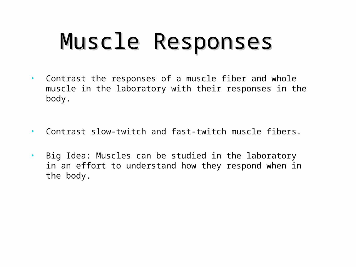
Figure 7.11 & Figure 7.12
Anterior view of
the body’s
superficial skeletal muscles.
Posterior view of the body’s
superficial
skeletal muscles
.
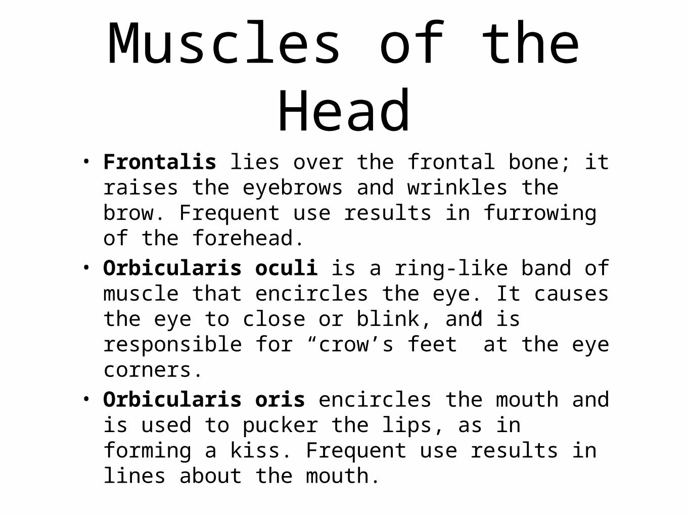
Muscles of the Head
• Frontalis lies over the frontal bone; it raises the eyebrows and wrinkles the brow. Frequent use results in furrowing of the forehead.
• Orbicularis oculi is a ring-like band of muscle that encircles the eye. It causes the eye to close or blink, and is responsible for “crow’s feet” at the eye corners.
• Orbicularis oris encircles the mouth and is used to pucker the lips, as in forming a kiss. Frequent use results in lines about the mouth.

Muscles of the Head
• Buccinator muscles are located in the cheek areas. When a buccinator contracts, the cheek is compressed, as when a person whistles or blows out air. It is used in swallowing.
• Zygomaticus extends from each zygomatic arch (cheekbone) to the corners of the mouth. It raises the corners of the mouth when a person smiles.

Muscles of the head and neck. Some of
these muscles account for our facial expressions and the ability to chew our
food; others move the head.

Muscles of Mastication
• We use the muscles of mastication when we chew food or bite something.
• Each masseter has its origin on the zygomatic arch and its insertion on the mandible. The masseter is a muscle of mastication (chewing) because it is a prime mover for elevating the mandible.
• Each temporalis is a fan-shaped muscle that overlies the temporal bone. It is also a prime mover for elevating the mandible. The masseter and temporalis are synergists.

Table 7.2 Muscles of the Head and Neck

Muscles of the Neck: Swallowing
• First, the tongue and the buccinators squeeze the food back along the roof of the mouth toward the pharynx.
• An important bone that functions in swallowing is the hyoid.
• The suprahyoid muscles pull the hyoid forward and upward toward the mandible. Small palatini muscles pull the soft palate backward, closing off the nasal passages.
• Pharyngeal constrictor muscles push the bolus of food into the pharynx, which widens when the suprahyoid muscles move the hyoid.
• The hyoid bone and larynx are returned to their original positions by the infrahyoid muscles.

Muscles that Move the Head
• Sternocleidomastoid muscles ascend obliquely from their origin on the sternum and clavicle to their insertion on the mastoid process of the temporal bone. When both sternocleidomastoid muscles contract, flexion of the head occurs.
• Each trapezius muscle is triangular, but together, they take a diamond or trapezoid shape. The origin of a trapezius is at the base of the skull. Its insertion is on a clavicle and scapula. The trapezius muscles help extend the head.

Muscles of the Thoracic Wall
• External intercostal muscles occur between the ribs; they originate on a superior rib and insert on an inferior rib. These muscles elevate the rib cage during the inspiration phase of breathing.
• The diaphragm is dome-shaped muscle that, as you know, separates the thoracic cavity from the abdominal cavity. Contraction of the diaphragm also assists inspiration.
• Internal intercostal muscles originate on an inferior rib and insert on a superior rib. These muscles depress the rib cage and contract only during a forced expiration. Normal expiration does not require muscular action.

Table 7.3 Muscles of the Trunk
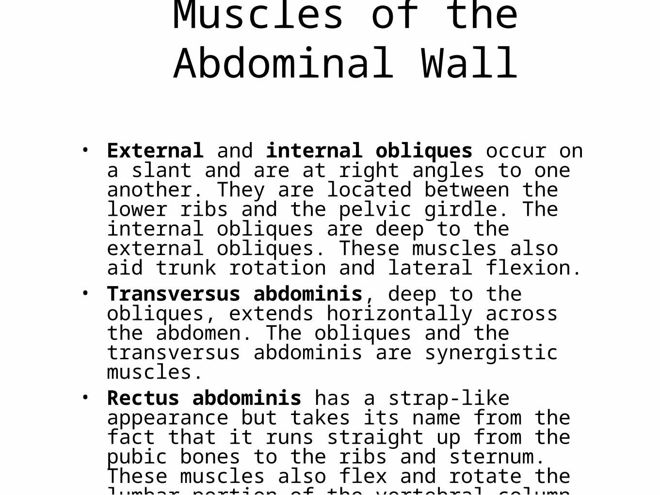
Muscles of the Abdominal Wall
• External and internal obliques occur on a slant and are at right angles to one another. They are located between the lower ribs and the pelvic girdle. The internal obliques are deep to the external obliques. These muscles also aid trunk rotation and lateral flexion.
• Transversus abdominis, deep to the obliques, extends horizontally across the abdomen. The obliques and the transversus abdominis are synergistic muscles.
• Rectus abdominis has a strap-like appearance but takes its name from the fact that it runs straight up from the pubic bones to the ribs and sternum. These muscles also flex and rotate the lumbar portion of the vertebral column.

Figure 7.14
Muscles of the anterior shoulder
and trunk. The right pectoralis
major is removed to show the deep
muscles of the chest.

Muscles that Move the Scapula
• The trapezius• Serratus anterior is located below
the axilla (armpit) on the lateral chest. It runs between the upper ribs and the scapula. It depresses the scapula and pulls it forward, as when we push something. Because this muscle causes a fast-forward jab of the arm, it is often called the boxer’s muscle. It also helps to elevate the arm above the horizontal level.

Muscles that Move the Arm
• Deltoid is a large, fleshy, triangular muscle that covers the shoulder and causes a bulge in the arm where it meets the shoulder. It runs from both the clavicle and the scapula of the pectoral girdle to the humerus. This muscle abducts the arm to the horizontal position.
• Pectoralis major is a large anterior muscle of the upper chest. It originates from a clavicle, but also from the sternum and ribs. It inserts on the humerus. The pectoralis major flexes the arm and adducts the arm, pulling it toward the chest.

• Latissimus dorsi is a large, wide, triangular muscle of the back. This muscle originates from the lower spine and sweeps upward to insert on the humerus. The latissimus dorsi extends and adducts the arm. This muscle is very important for swimming, rowing, and climbing a rope.
• Rotator cuff. There are four rotator cuff muscles. Three are located on the posterior scapula: supraspinatus, infraspinatus, and teres minor. The last rotator cuff muscle is the subscapularis muscle located on the anterior surface of the scapula.
The Muscles that Move the Arm

Muscles of the posterior
shoulder. The right
trapezius is removed to show deep
muscles that move the
scapula and the rotator
cuff muscles.

• Biceps brachii is a muscle of the proximal anterior arm that is familiar because it bulges when the forearm is flexed. It also supinates the hand when a doorknob is turned. The biceps brachii inserts on the radius.
• Brachialis originates on the humerus and inserts on the ulna.
• Triceps brachii is the only muscle of the posterior arm. It has three heads that attach to the scapula and humerus, and it inserts on the ulna. The triceps extends the forearm. The triceps is also used in tennis to do a backhand volley.
Muscles of the Arm


a. Muscles of the anterior arm and
shoulder. b. Muscles of the posterior arm
and shoulder. c. Muscles of the
anterior forearm. d. Muscles of the
posterior forearm.

• Flexor carpi and extensor carpi muscles originate on the bones of the forearm and insert on the bones of the hand. The flexor carpi flex the wrists and hands, and the extensor carpi extend the wrists and hands.
• Flexor digitorum and extensor digitorum muscles also originate on the bones of the forearm and insert on the bones of the hand. The flexor digitorum flexes the wrist and fingers, and the extensor digitorum extends the wrist and fingers.

• Iliopsoas originates at the ilium and the bodies of the lumbar vertebrae, and inserts on the femur anteriorly. This muscle is the prime mover for flexing the thigh and also the trunk, as when we bow. Also, it is important to the process of walking.
• Gluteus maximus is the largest muscle in the body and covers a large part of the buttock. It originates at the ilium and sacrum, and inserts on the femur. Used when walking or climbing stairs.
• Gluteus medius lies partly behind the gluteus maximus. It runs between the ilium and the femur, and functions to abduct the thigh.
• Adductor group muscles (pectineus, adductor longus, adductor magnus, gracilis) are located on the medial thigh. All of these muscles originate from the pubis and ischium, and insert on the femur. These muscles keep a rider on a horse.


• Quadriceps femoris group (rectus femoris, vastus lateralis, vastus medialis, vastus intermedius), also known as the “quads,” is found on the anterior and medial thigh. These muscles are the primary extensors of the leg, as when you kick a ball by straightening your knee.
• Sartorius is a long, strap-like muscle that has its origin on the iliac spine and then goes across the anterior thigh to insert on the medial side of the knee. It also rotates the thigh laterally, enabling us to sit cross-legged.
• Hamstring group (biceps femoris, semimembranosus, semitendinosus) is located on the posterior thigh. Their strong tendons can be felt behind the knee. They flex and rotate the leg medially, but they also extend the thigh.

Muscles of the
anterior right hip
and thigh.
Muscles of the
posterior right
hip and thigh.

• Gastrocnemius is a muscle of the posterior leg, where it forms a large part of the calf. It arises from the femur; distally, the muscle joins the strong calcaneal tendon, which attaches to the calcaneus bone. Aids in pushing the body forward during walking or running.
• Tibialis anterior is a long, spindle-shaped muscle of the anterior leg. It arises from the surface of the tibia and attaches to the bones of the ankle and foot.
• Fibularis muscles (fibularis longus, fibularis brevis) are found on the lateral side of the leg, connecting the fibula to the metatarsal bones of the foot.
• Flexor and extensor digitorum longus muscles are found on the lateral and posterior portion of the leg. They arise mostly from the tibia and insert on the toes. They flex and extend the toes, and assist in other movements of the feet.
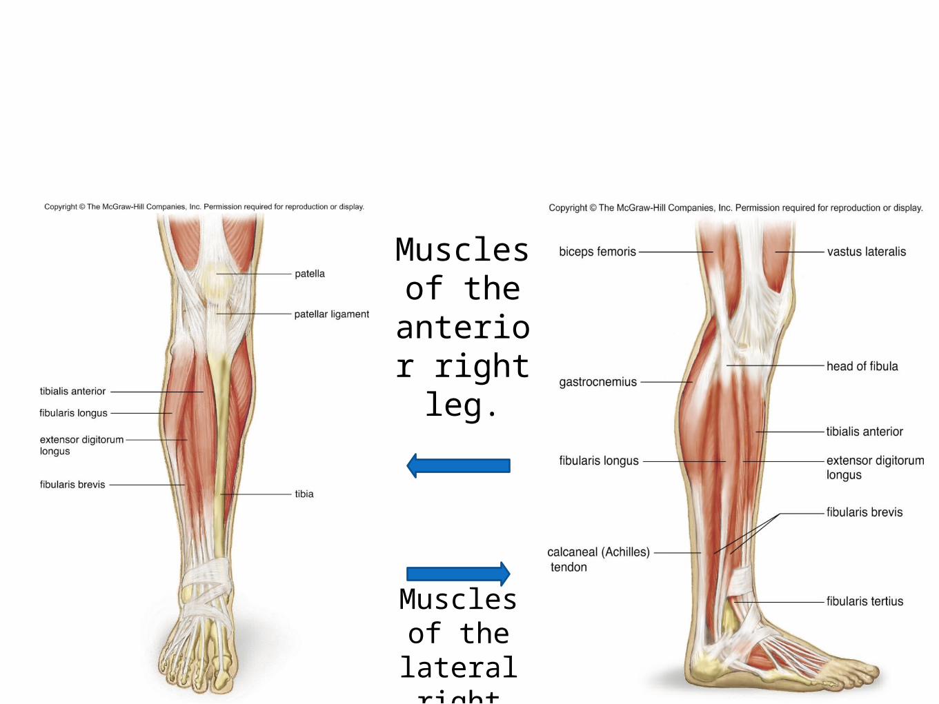
Muscles of the anterior
right leg.
Muscles of the lateral right leg.

In the BodyIn the body, muscles are innervated to contract by nerves.A nerve fiber together with all of the muscle fibers it innervates is called a motor unit.A motor unit obeys the all-or-none law.All the muscle fibers in a motor unit are stimulated at once, and they all either contract or do not contract.Tetanic contractions ordinarily occur in the body because, as the intensity of nervous stimulation increases, more and more motor units are activated.This phenomenon, known as recruitment, results in stronger and stronger muscle contractions.But while some muscle fibers are contracting, others are relaxing. Because of this, intact muscles rarely fatigue completely.Even when muscles appear to be at rest, they exhibit tone, in which some of their fibers are always contracting. Tone maintains posture.

Effects of Aging
• Muscle mass and strength tends to decrease as people age. Deteriorated muscle elements are replaced initially by connective tissue, and eventually fat. As we age degenerative changed happen in the mitochondria and endurance decreases.

Muscular Disorders-Spasms: Sudden, involuntary muscular contractions, usually accompanied by pain.
-Convulsions: Multiple spasms of skeletal muscles.
-Cramps: Strong painful spasms, especially of the leg and foot, usually due to athletic activity.
-Facial tics: Periodic eye blinking or grimacing.
-Strain: Twisting of a joint, leading to swelling and injury not only of muscles but also of ligaments, tendons, blood vessels and nerves.
-Tendinitis: The inflammation of a tendon due to repeated athletic activity.

Neuromuscular Diseases
• -Tetanus: Develops in persons who have not been properly immunized against the the toxin of the tetanus bacteria. Muscles become so stiff the patient cannot breathe or swallow.
• -Fibromyalgia: chronic condition whose symptoms include achy pain, tenderness and stiffness of muscles.
• -Muscular dystrophy: Progressive degeneration and weakening of muscles.
• -Duchenne muscular dystrophy: Most common type, inherited through flawed gene carried by the mother, where the person lacks a protein called dystrophin. When this occurs, calcium leaks into the cells and activate the enzyme that dissolves muscle fibers.
• -Myasthenia gravis: An autoimmune diseases characterized by weakness that especially affects the muscle of the eyelids, face, neck, and extremities.
• -Amyotrophic Lateral Sclerosis: Victims suffer from a gradual death of their motor neurons, thus loosing ability to walk, talk, or chew.
•

HomeostasisHomeostasis Muscle contraction provides heat to warm skin. Muscle moves skin. Muscle contraction causes bones to move joints. Muscles help protect bones Muscle contraction moves eyes, permits speech, creates facial
expresions. Muscles help protect glands. Muscle contraction keeps blood moving in heart and blood
vessels. Skeletal muscle contraction moves lymph, physical exercise
enhances immunity. Muscle contraction assist breathing; physical exercise increases
respiratory capacity. Smooth muscle contraction accounts for peristalsis, skeletal
muscles support and help protect abdominal organs

Smooth muscle contraction assists voiding of urine, skeletal muscles support and help protect urinary organs
Muscle contraction occurs during orgasm and moves gametes; abdominal and uterine muscle contraction occurs during childbirth.
•All these facts help our body maintain balance by utilizing the muscular system.