Embed Size (px)
Citation preview

1
The Msb3 GAP controls the activity of the Rab GTPases Vps21 and Ypt7 at endosomes and vacuoles
Jens Lachmann1, Francis A. Barr2, and Christian Ungermann1,3 1University of Osnabrück Department of Biology/Chemistry Biochemistry section Barbarastrasse 13 49076 Osnabrück, Germany 2 Department of Biochemistry University of Oxford South Parks Road OX1 3QU Oxford, United Kingdom 3 To whom correspondence should be addressed: Email: [email protected] Phone: +49-541-969-2752 (C.U.) Characters: (without spaces) 49,846 Abstract
Fusion of organelles in the endomembrane system depends on Rab GTPases that interact with
tethering factors prior to lipid bilayer mixing. In yeast, the Rab5 GTPase Vps21 controls fusion
and membrane dynamics between early and late endosomes. Here, we identify Msb3/Gyp3 as a
specific Vps21 GTPase activating protein (GAP). Loss of Msb3 results in an accumulation of
Vps21 and one of its effectors Vps8, a subunit of the CORVET complex, at the vacuole
membrane in vivo. In agreement, Msb3 forms a specific transition complex with Vps21, has the
highest activity of all recombinant GAPs for Vps21 in vitro, and is found at vacuoles despite its
predominant localization to bud tips and bud necks at the plasma membrane. Surprisingly, Msb3
also inhibits vacuole fusion, which can be rescued by the Ypt7 GEF, the Mon1-Ccz1 complex.
Consistently, msb3∆ vacuoles fuse more efficiently than wild-type vacuoles in vitro, suggesting
that the GAP can also act on Ypt7. Our data indicate that GAPs such as Msb3 can act on multiple
substrates in vivo at both ends of a trafficking pathway. This ensures specificity of the subsequent
GEF-mediated activation of the Rab that initiates the next transport event.
Key words: Endosome, vacuole, Msb3, Vps21, GAP, Rab GTPase, membrane fusion

2
Introduction
Transport of proteins and lipids in the secretory and endocytic transport requires vesicular
carriers that form at one membrane and fuse with the acceptor organelle. Fusion relies on a
conserved machinery that mediates the initial contact between the two membranes prior to bilayer
mixing mediated by membrane-anchored SNARE proteins. For each fusion event, a specific Rab
GTPase has been identified. Rabs are switch-like proteins, which exist in an inactive GDP- and
active GTP-form (Barr et al, 2010). As Rabs are incomplete enzymes, the transition between the
two forms depends on specific activating proteins. GDP-GTP exchange factors (GEFs) bind to
the GDP-Rab and promote exchange of the GDP for the much more abundant cellular GTP. Only
in their GTP-form, two specific regions within the Rab, termed switch I and II, are stabilized
such that interaction partners or effectors can bind the Rab-GTP. On the contrary, GAPs provide
a missing arginine to the active site of the Rab, which triggers the otherwise slow GTP hydrolysis
rate and inactivates the Rab. In their GDP form, Rabs can then be extracted from membranes by
the GDP dissociation inhibitor (GDI), which binds the GTPase domain and simultaneously
shields the hydrophobic C-terminal dual prenyl anchor (Goody et al, 2005).
The active Rab-GTP binds to several effectors. For fusion reactions, the tethering proteins
are the most important interactors. Tethering proteins can come in two forms, as long coiled-coil
proteins like the early endosome protein EEA1 (Vac1 in yeast), or as multisubunit tethering
complexes (MTCs) (Bröcker et al, 2010; Yu et al, 2010). Either tethering factor is thought to
initiate contact between membranes by binding the Rab GTP on one or both membranes that will
subsequently fuse, which is supported by our structural data on HOPS and Ypt7 (Bröcker et al,
2012). MTCs are thought to combine Rab binding and membrane tethering with the chaperoning
of SNAREs.

3
To this date, several GEFs and GAPs have been identified. Whereas GEFs have escaped
identification due to their variable structure, GAPs have a defined TBC (Tre2/Bub2/Cdc16)
domain, which contains the aforementioned ‘arginine’ and ‘glutamine’ finger, and could be
identified on the sequence level early on. Despite their early identification in yeast, it has been
challenging to match each GAP to its corresponding Rab, because the isolated TBC domain often
revealed overlapping activities for multiple Rabs (Albert et al, 1999a, b). However, functional
and in vivo studies identified Gyp7 as the GAP for Ypt7 and additional Rab-GAP pairs (Brett et
al, 2008; Gao et al, 2003; Du et al, 2001; Sciorra et al, 2005). A series of studies have identified
GAPs for mammalian Rabs regulating a range of trafficking pathways, including RabGAP5 in
endocytosis (Haas et al, 2005; Fuchs et al, 2007; Yoshimura et al, 2007).
We are interested in the control of the endocytic pathway and previously identified the
CORVET complex as a novel effector of the Rab5 GTPase Vps21 (Peplowska et al, 2007).
Vps21 is activated by the Rabex5 homolog Vps9 during endocytosis or on early endosomes
(Hama et al, 1999). We and others postulate that endosomal maturation will include an exchange
of Vps21 by the Rab7-like Ypt7, which is required for fusion of late endosomes with the vacuole
(Rink et al, 2005; Spang, 2009; Lachmann et al, 2011; Huotari et al, 2011; Abenza et al, 2010).
Such a cascade should include the inactivation of Vps21 by a GAP and its release from
membranes, once Ypt7 has been converted to the GTP-form. Indeed, evidence for such a cascade
exists for the exocytic pathway (Rivera-Molina et al, 2009). However, even though the
mammalian and the C.elegans Rab5 GAP is known, the equivalent in yeast is still lacking. Here,
we identify Msb3 as a specific Vps21 GAP, which binds Vps21, and is required for Vps21
recycling from the endosome. Msb3 is localized primarily to the plasma membrane and cytosol,
and has been previously assigned to the exocytic Rab Sec4 together with its homolog Msb4

4
(Albert et al, 1999b; 2000; Gao et al, 2003). As Msb3 can also inactivate Ypt7, our combined
data argue for an additional role of Msb3 in the endocytic pathway.
Results
Identification of the Vps21 GAP
Yeast contains 11 Rabs and eight GAPs, indicating a mismatch and potential overlapping
functions of the respective GAPs. By sequence comparison, three yeast GAPs, Mdr1/Gyp2,
Msb3/Gyp3 and Msb4/Gyp4, fall in the same clade as mammalian RabGAP-5 and C.elegans
TBC-2 and could be potential homologs that act on Vps21 (Gao et al, 2008). Previous
biochemical studies used often truncated GAPs or proteins extracted from yeast to determine
specificity. However, the isolated TBC of Gyp1 is, for instance, an efficient GAP of many Rabs
including Ypt7 (Eitzen et al, 2000), which indicates the challenge in defining Rab-GAP pairs.
We thus devised an assay to identify the Vps21-specific Rab. We reasoned that a
permanently activated Rab should not be extracted by Gdi1 and thus migrate to the next
organelle, i.e. the vacuole for Vps21 (Figure 1A). As predicted, the GTP-locked Vps21 Q66L
indeed accumulated on the vacuole rim, whereas GFP- or RFP-tagged wild-type Vps21 was
present in multiple endosomal dots (Markgraf et al, 2009)(Figure 1B). Similarly, we speculated
that Vps21 should be found on the vacuole if the specific GAP would be missing. We therefore
analyzed all GAP deletions. Only in the msb3∆ strain, Vps21 was found on the vacuole (Figure
1C), thus mimicking the GTP-locked Vps21 version analyzed before (Figure 1B). The shift of
Vps21 to the vacuole, either in msb3∆ or in the Vps21 Q66L cells, did not affect the vacuole
localization of Ypt7 (Figure 1D). Loss of Msb3 thus results in a compartment with two active
Rabs, which points to a missing inactivation of the upstream Vps21 (Figure 1E).

5
Msb3 and its homolog Msb4 have been assigned to the exocytic Rab Sec4 at the plasma
membrane (Gao et al, 2003). We therefore used an alternative assay to determine the specific
interaction of Msb3 with Vps21 (Figure 1F). During activation of GTP-hydrolysis the Rab binds
to the GAP via the arginine-finger motif (Pan et al, 2006). For the Ras-GTPase, this interaction
has been previously stabilized by the addition of GDP and AlF3, which mimicks a transition state
in the hydrolysis reaction, and has been sufficiently strong to crystallize a Ras-GAP and Rab-
GAP complex (Scheffzek et al, 1997; Pan et al, 2006). We then incubated immobilized GST-
Vps21 in the GDP form with different recombinantly purified GAPs either in the absence or
presence of AlF3, and analyzed the corresponding eluates. Importantly, Msb3 was strongly
retained with Vps21-GDP only in the presence of AlF3, whereas neither its homolog Msb4 nor
Gyp7 were binding significantly to Vps21 (Figure 1F).
Vps21-GTP on vacuoles retains its effector Vps8
Our data did not clarify whether Vps21 on the vacuole was indeed active. Along the endocytic
pathway, Vps21-GTP interacts with the early endosomal Vac1 protein and the CORVET subunits
Vps8 and Vps3 (Tall et al, 1999; Plemel et al, 2011; Markgraf et al, 2009). In the past, we
observed that elevated Vps21 levels affected the localization of Vps8, but not Vps3 (Markgraf et
al, 2009). We therefore asked, if any of the two interactors would shift to the vacuole with Vps21
(that we assume to be in its GTP-loaded state) due to a prolonged binding. We thus expressed
Vps8 as an additional GFP-tagged copy in msb3∆ or Vps21 Q66L cells, and could observe that
Vps8 clearly followed Vps21 to the vacuole (Figure 2A). We previously showed that the GEF of
Ypt7, the Mon1-Ccz1 complex, is mislocalized in cells lacking Vps21 and Vps8 (Nordmann et
al, 2010). This observation suggested a crosstalk between Vps21 and the Mon1-Ccz1 complex as
the activator of the downstream Ypt7 Rab in the process of endosome maturation. When we
analyzed functional GFP-tagged Mon1, we observed more protein on vacuoles of msb3∆ and

6
Vps21 Q66L expressing cells (Figure 2B). This indicates that active Vps21 also affects Mon1-
Ccz1 localization, either by directly binding Mon1-Ccz1 or by affecting the endosomal surface
composition, to relocate Vps21 to the vacuole, in agreement with our model (Nordmann et al,
2010).
In contrast, the localization of the endosomal Vps21 effector Vac1 was unaffected,
probably due to the restricted binding of Vac1 to endosomal phosphoinositole-3-phosphate via its
FYVE domain (Figure 2C). Likewise, endosomes as marked by Pep12 did not change in msb3∆
cells. These controls point to a functional recycling of endosomal factors and no increased
unspecific fusion of the endosome with the vacuole (Figure 2D). Interestingly, the loss of Msb3
had no effect on Vps3 (Figure 2E), even though it is part of the CORVET complex (Peplowska et
al, 2007), also interacts with Vps21-GTP in vitro (Plemel et al, 2011), and requires Vps21 for
localization (our unpublished results). This suggests that the functional cooperation of the two
Rab-specific subunits Vps3 and Vps8 with the Rab5 Vps21 differs in vivo.
Msb3 requires its active arginine to affect Vps21 localization
To analyze Msb3 in vivo, we first examined the functionality of the tagged protein using the
Vps21 localization (Figure 1A,B) as a read-out. As shown in Figure 2F, Msb3 tagged at either N-
or C-terminus with GFP localized to the plasma membrane, the bud neck, and cytosol, in
agreement with previous studies (Gao et al, 2003; Tcheperegine et al, 2005). Moreover, Vps21
localized as in wild-type cells if Msb3 was tagged, indicating that tagged Msb3 is functional.
Msb3 binds to many proteins of the polarisome as well as to regulators of cytoskeleton
organization (Tcheperegine et al, 2005). We therefore asked whether the msb3 deletion could
have a secondary effect on Vps21 localization and therefore generated active-site point mutants.
A replacement of the arginine finger is considered to be sufficient to disrupt GAP activity (Haas
et al, 2005). For Msb3, a mutation of R282 to lysine or phenylalanine yielded an Msb3 variant

7
that was correctly localized, but behaved as msb3∆ for Vps21 localization (Figure 2G). We thus
conclude that the GAP-activity of Msb3 is required for correct Vps21 localization by inactivation
and recycling of the Rab GTPase during maturation.
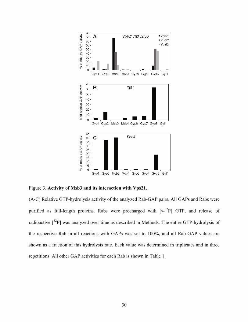
In vitro activity of yeast GAPs on individual Rabs
We next tested if we could find the same specificity of Msb3 for Vps21 in vitro. For this purpose,
we isolated all recombinant full-length Rabs and GAPs (except of Gyp5) to determine their
relative specificity in an in vitro GAP assay that monitors phosphate release after GTP
hydrolysis. Here, we focus on the three relevant Rabs for our study: Vps21, Sec4, and Ypt7
(Figure 3). All GAP-Rab data are summarized in Table 1. To compare the specificity of all GAPs
for a particular Rab, we set the entire hydrolysis rate within the assay as 100%, and plotted all
GAP-Rab hydrolysis rates in relation to this value. Within our data set, we could largely confirm
the previously reported GAP values (Figure 3C)(Albert et al, 2000)(see also Brett et al, 2008, and
references therein). Importantly, the most active GAP for Vps21/Ypt51 and its homolog Ypt52
was again Msb3 (Figure 3A), whereas the homologous Ypt53 protein was unaffected by the same
GAP. Moreover, our MBP-Gyp8 construct showed very high activity on many Rabs and had the
highest relative activity on Ypt7 (Figure 3B), but did act poorly on Vps21, in support of a
specific Msb3-Vps21 interaction for Vps21 inactivation. Our data nicely fit with role of Msb3 in
Vps21 localization and its interaction in vitro (Figure 1), and is also in good agreement with a
previous study (Albert et al, 1999a). As also noticed before, GAPs can act on multiple substrates,
and Msb3 shows also strong activity for Sec4, but none for Ypt7 (Figure 3B, C). Gyp2 acts
equally well as Msb3 on Sec4 (Figure 3C), whereas Msb4 shows no activity in our assays.
Surprisingly, the identified GAP of Ypt7, Gyp7, also does not show any activity for Ypt7 in our
in vitro assay. It has been reported that the full-length Gyp7 is in general of poor activity and
therefore truncated versions were prefered for in vitro assays (Albert et al, 1999a; Eitzen et al,

8
2000). It should also be noted that Gyp8 has a transmembrane domain and it thought to be an ER
protein, which would restrict it to Rabs that can bind to the ER. We thus consider it likely that
some GAPs require the right membrane context for Rab inactivation.
Msb3 interacts with Vps21 in vivo
We were surprised that a deletion or mutation of Msb3 caused such a strong defect on
localization of Vps21 and its effector Vps8. In wild-type cells, the majority of Msb3 is localized
to the plasma membrane, and here in particular to the bud tip and neck (Figure 4A), even though
we also detect a large cytosolic portion in vivo (Figure 4A, B, C). It was possible that Msb3 was
transiently present on FM4-64 positive endosomal structures. We thus followed cells labeled with
this lipophilic dye, which passes through the endocytic pathway to the vacuole, but did not detect
any significant co-labeling. Likewise, the accumulation of MVBs in the vps4 mutant, which leads
to an accumulation of several endocytic proteins such as Vps21 or ESCRT subunits, did not
affect Msb3 localization (Figure 4B). Similar results were obtained for Msb4. Interestingly, a
control GAP protein, Gyp2, was found in FM4-64 positive structures (Figure 4A), and partially
accumulated in endosomes of the vps4 mutant (Figure 4B), which suggests a role in endosomal
transport, most likely in recyling from endosomes to the Golgi as described previously
(Lafourcade et al, 2003).
Our data did, however, not exclude an additional role of Msb3 in the endocytic pathway
due to a transient interaction with Vps21. We therefore selected two additional approaches. First,
we examined cells with GFP-tagged GAPs by subcellular fractionation (Figure 4C). Most of the
GAPs were found in the cytosol, consistent with our in vivo analysis, though a small fraction of
each GAP was also recovered in the vacuole-containing P13 fraction (Figure 4C). Gyp2 was in
addition enriched in the P100 fraction, indicative of an endosomal pool (Figure 4B). For the P13
fraction, we can not exclude the plasma membrane as one origin of the detected Msb3, since we

9
found the marker protein Sso2 in this fraction as well. However, Msb3 is present in large
cytosolic pool of Msb3, which could have access to the endocytic pathway. To examine this in
more detail, we isolated vacuoles and vacuole associated organelles by gradient flotation, and
analyzed protein fractions for the presence of selected GAPs. As shown in Figure 4D, Msb3, and
Msb4 were co-purified with vacuoles, suggesting that a portion of Msb3 is associated with
vacuoles or vacuole-associated endosomes. For Gyp2, we observed strong degradation in the
vacuole-enriched fraction (not shown). However, due to the impurity of the preparation with
portions of the plasma membrane, we also detected Sso2 in the floated vacuole fraction.
Therefore, we also analyzed the same vacuoles by fluorescence microscopy and found Msb3 at
the vacuole membrane (Figure 4E). This clearly indicates that the endosomal and vacuole
population of Msb3 is likely masked by the cytosolic background and the predominant plasma
membrane pool, when we localized Msb3 in vivo (Figure 2F, G) or biochemically (Figure 4C, D).
In contrast, GFP-Msb4 was found in the vacuolar lumen, possibly due to its degradation in the
vacuole. Secondly, we used the split-YFP system (Sung et al, 2007) to ask if Vps21 encounters
Msb3 in vivo. When either protein was fused to the YFP-half, no fluorescent signal was observed.
However, co-expression of both resulted in a clear signal close to the plasma membrane (Figure
4F), suggesting that Vps21 and Msb3 can interact in vivo. We were surprised to find this
interaction at the plasma membrane in patches where Msb3 normally localizes, instead of seeing
it relocalized to the endosomes. To exclude unspecific binding, we used a Vps39 construct fused
to one half of YFP, which interacts efficiently with the Ypt7 that carries the opposite half (our
unpublished observation). When this Vps39 construct was expressed together with tagged Msb3
we observed no interaction (Figure 4F), indicating that the Vps21-Msb3 interaction is specific.
We then asked if the Split-YFP would stabilize the interaction of Vps21 and Msb3 and therefore
cluster the endosomal compartment at the plasma membrane. When we co-localized the Msb3-

10
Vps21 interaction site with Vac1, we observed a clear separation, indicating that no unspecific
recruitment of Vps21 together with endosomes occurred (Figure 4G). We conclude that Msb3
and Vps21 do interact transiently in vivo, and that an active fraction of Msb3, but not Msb4, is
found on endosomes and vacuoles.
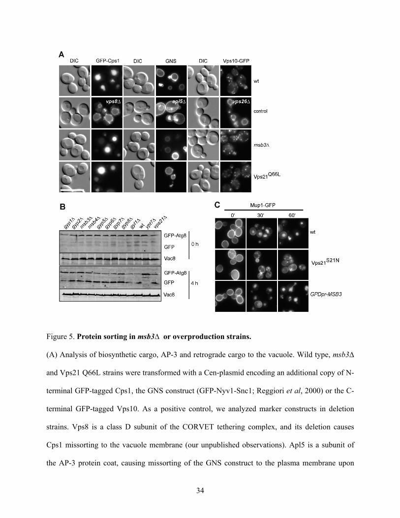
Msb3 deletion does not affect protein trafficking to the vacuole
The Msb3 deletion has a clear effect on the relocalization of Vps21 to the vacuole, though it had
not been detected in any previous screen as a protein involved in endocytic trafficking. We
decided to readdress this issue by analyzing central trafficking reactions. Initially, we followed
sorting of GFP-tagged Cps1, a substrate of the ESCRT pathway at the endosome, which is sorted
to the vacuole lumen in wild-type cells. In ESCRT deletions (Odorizzi et al, 1998), but also in
vps21∆ cells (our unpublished observations), GFP-Cps1 is found on the vacuole rim. However,
neither in msb3∆ cells nor in cells expressing only activated Vps21 Q66L did we detect any
defect in sorting (Figure 5A). Similarly, the AP-3 pathway as marked by the GFP-Nyv1-Snc1
fusion protein (Reggiori et al, 2000) or the retrograde transport from the Golgi, which we
followed by tagging the CPY-receptor Vps10 with GFP was unaffected in both mutants (Figure
5A). In addition, we observed no defect for autophagy, which we followed by GFP-Atg8
processing (Figure 5B). The latter assay relies on the clipping of Atg8 to free GFP, which is
observed in wt, but not in the absence of the vacuolar Ypt7 Rab GTPase (Figure 5B).
Interestingly, GAP proteins have been implicated in mammalian autophagy (Behrends et al,
2010), though we did not find evidence so far.
We finally analyzed trafficking of the methionine transporter Mup1 from the plasma
membrane. In the absence of methinone, the transporter is stable at the plasma membrane, and is
sorted to the vacuole in the presence of methionine (Figure 5C). This transport is defective if
Vps21 is inactive, as shown for the Vps21 S21N mutant (Figure 5C). If Msb3 were

11
overexpressed, we expected a similar effect. However, sorting appeared unaffected if Msb3
expression was driven from the GPD promoter, suggesting that Vps21 was not completely
inactivated under these conditions. We thus conclude that the mislocalization of Vps21 to the
vacuole in msb3∆ cells does not affect protein trafficking to the vacuole, and that Msb3
overexpression does not affect the overall Vps21 function.
Msb3 acts on both Ypt7 and Vps21
During our in vivo analyses, we noticed that vacuoles in msb3∆ and Vps21 Q66L mutants
were enlarged and often showed very small structures adjacent to the main vacuole structure,
reminiscent of the described class F phenotype (Raymond et al, 1992). Whereas wild-type
vacuoles are normally stretched toward the daughter cell during inheritance, both mutants
displayed fragmented inheritance structures, though the process is functional per se (Figure 6A).
The alteration in morphology suggested thus a shift in the fusion and fission equilibrium and a
potential role of Msb3 on the vacuole biogenesis.
In agreement with this observation in the msb3 mutant, it was previously observed that
overexpression of Msb3 or Msb4 interferes with vacuole morphology (Brett et al, 2008), which
we could reproduce when Msb3 was placed under the control of the strong GAL1 promoter
(Figure 6B). We then wondered how we could explain that the overexpression of Msb3 caused a
vacuole morphology defect, and considered the possibility that Msb3 might also affect Ypt7. We
therefore titrated Msb3 into the vacuole fusion reaction, which employs isolated vacuoles from
two tester strains. Lumenal mixing of the two vacuole types results in activation of immature
alkaline phosphatase present in one type by the protease of the other type (Cabrera et al, 2008).
Interestingly, Msb3 addition strongly inhibited vacuole fusion, whereas similar amounts of highly
active Gyp2 (Figure 3B) were far less active in this assay (Figure 6C). We asked if it was indeed
Ypt7 that was inactivated by Msb3. As shown in Figure 6D, the Ypt7-specific GEF, the Mon1-

12
Ccz1 complex, rescued fusion activity of vacuoles that were pretreated with Msb3, showing that
Msb3 acts on Ypt7. This was surprising as Msb3 had almost no activity on Ypt7 in vitro (Figure
3)(Brett et al, 2008). We therefore consider it likely that GAPs such as Msb3 act in a context-
specific manner on organellar membranes and might require activation to act on Ypt7. If Msb3
were a GAP for both Ypt7 and Vps21, we would expect that vacuoles fuse better in its absence.
This was indeed observed (Figure 6E). Vacuoles obtained from msb3∆ cells were at least 30%
more fusogenic than the corresponding wild-type vacuoles, and both vacuole types were sensitive
to the general fusion inhibitor Gyp1-46. Our data thus suggest that Msb3 can act on Vps21 and
Ypt7 along the endocytic pathway to dampen Rab activity and promote membrane fusion
specificity and dynamics.
We finally asked if we also could observe an interaction of Msb3 and Ypt7 in vivo. We
reasoned that an excess of Ypt7 should affect the relative localization of Msb3. Indeed, we could
shift a portion of GFP-tagged Msb3 to the vacuole by overexpressed Ypt7 (Figure 6F). To
analyze the interaction of both proteins further, we used the Split-YFP assay. As for Vps21, we
observed an interaction of the two proteins at the plasma membrane (Figure 6G). In addition,
both proteins colocalized at some vacuoles during inheritance (Figure 6H), though inheritance
was not affected in the msb3 mutant, as discussed above. In sum, we take the combined findings
as an indication of an encounter of Msb3 GAP and two Rabs, Vps21 and Ypt7, within the
endocytic pathway, which is consistent with our in vitro interactions (Figure 1) and in vivo
localization of Vps21 in msb3∆ (Figure 2).
Discussion
Based on an extensive mapping of all yeast full-length GAPs versus all Rabs, and a comparison
of Vps21 localization in all GAP deletion strains, we were able to identify Msb3, but not its
homolog Msb4, as a Vps21-specific GAP. We demonstrate the specificity at several levels. First,

13
Msb3 interacts with Vps21-GDP in the presence of AlF3, which stabilizes the transition state of
GTP hydrolysis. Second, Msb3 binds Vps21 in vivo. Third, in the absence of Msb3, activated
Vps21 remains on endosomes, is translocated to the vacuole and interacts with its effector Vps8.
This is a specific effect that was only observed for the Msb3 GAP deletion. Finally, Msb3 can
also inactivate Ypt7 on isolated vacuoles (Figure 6), and presumably in vivo (Figure 6B, (Brett et
al, 2008)), suggesting that the Rab GAPs have a broader specificity in the cell than previously
anticipated. In agreement with our study, Merz and colleagues have identified Msb3
independently as the Vps21 GAP, using a completely different approach of endosomal signaling
activity as a read-out (Nickerson et al, submitted to MBoC).
Msb3 takes part in the organization of polarized growth on several levels by regulating
exocytosis via its activity on Sec4, interacting with and possibly coordinating Rho GTPases,
components of the polarisome, and the actin cytoskeleton (Bi et al, 2000; Gao et al, 2003;
Tcheperegine et al, 2005). Msb3 was thus not our primary candidate for a Vps21 GAP. Like its
homolog Msb4, Msb3 is primarily found at the plasma membrane, and here in particular at the
growing bud tip and bud neck (Gao et al, 2003)(this study). The predominant localization of
Msb3 at the plasma membrane suggested that it also acts predominantly there, which is most
likely true for the Sec4 inactivation (Gao et al, 2003). The situation is likely different for Vps21
and Ypt7. Msb3 is partially cytosolic, suggesting that it could be transiently recruited to selected
organelles such as endosomes to inactivate Vps21. Such activities are known as ‘moonlighting’
and reflect additional functions of a specific enzyme, and likely apply to Msb3. Consistent with
this notion, we identified a portion of Msb3 on isolated vacuoles, though the primary Vps21-
Msb3 interaction as measured by the split-YFP assay occurred proximal to the plasma membrane.
At present, we cannot be certain that the interaction of Vps21 or Ypt7 with Msb3 as detected by
Split-YFP corresponds to the enzymatic Rab-GAP intermediate. Such a complex should form

14
only very transiently, consistent with the very weak interaction of Rabs and GAPs with affinities
in the micromolar range (Albert et al, 1999a,b). As such, the split-YFP interaction of Msb3 and
Vps21 or Ypt7 at the plasma membrane may correspond to a prolonged interaction of the Rab
and GAP mediated by the two halfs of the fluorophore. With regard to this, the Rab GTPase may
then follow Msb3 to the plasma membrane, before being recycled very slowly. It is thus possible
that this finding does not represent the physiologic place of interaction. How Msb3 then diverts
its activities also to the late endosome and vacuole is not yet clear.
Our data on the GAP localization is not without precedence. Until now, no data of the
direct physical interaction of a Rab GTPase with its GAP in living cells is available and, along
the same line, some Rab - GAP pairs seem to localize at different compartments. For instance, the
mammalian Rab43 localizes to the Golgi and ER to regulate retrograde transport from the
endocytic pathway to the Golgi, and has an additional function in the effective uptake of Shigella
toxin at the plasma membrane (Haas et al, 2007; Fuchs et al, 2007). Interestingly, the Rab43-
specific GAP RN-tre localizes mainly to the cell surface, possibly implicating a similar
mechanism. RN-tre has also been proposed to be a GAP for Rab5 (Lanzetti et al, 2004), which
then would raise exactly the same problem of the Rab5-GAP interaction site.
We noticed that only massive overproduction of Msb3 resulted in impaired vacuole
biogenesis, and overproduction from a constitutive promoter did not interfere with the
endocytosis of the methionine transporter Mup1. At this stage, we can exclude a redundant
function of the Vps21 paralogs Ypt52 and Ypt53, since we analyzed endocytosis after Msb3
overproduction also in a double deletion mutant (data not shown). It is possible that an additional
factor that regulates Msb3 localization is thus limiting to locally inactivate Vps21.
Our data suggest that Msb3 is not the only GAP that acts in the endocytic pathway. Gyp2
is also found in the same clade of RabGAP-5 like proteins with Msb3 and Msb4 (Gao et al,
15
2008), affects vacuole morphology if overproduced (Brett et al, 2008), and accumulates at late
endosomes in vps4∆ mutants. However, the inhibition of Gyp2 of in vitro vacuole fusion was by
far less efficient than the activity of Msb3. It is thus possible that Gyp2 cooperates with Msb3
along the endocytic pathway, even though we were not able to identify a specific transport defect
in gyp2∆ cells. Moreover, Gyp2 has been described previously as a GAP for Ypt31 and Ypt6 in
vivo, again pointing to a redundant function of GAPs in vivo (Sciorra et al, 2005; Lafourcade et
al, 2003).
It is assumed that a Rab cascade, in which the downstream Rab (such as Ypt7) replaces
the previous one (Vps21) on the same organelle, requires the recruitment of the respective Vps21
GAP at a specific time point. Such a scenario has been postulated for the forward reaction as
well, in that the GEF of the downstream Rab would be recruited by the upstream Rab
(Hutagalung et al, 2011; del Conte-Zerial et al, 2008). Indeed, Vps21-GTP seems to affect the
Mon1-Ccz1 localization (Figure 2). Several studies on yeast exocytosis (Wang et al, 2002;
Rivera-Molina et al, 2009; Mizuno-Yamasaki et al, 2010) and the endocytic pathway (Rink et al,
2005; Poteryaev v, 2010; Nordmann et al, 2010; Abenza et al, 2010) also support such models,
though details on the exact mechanism are still lacking. A Rab cascade model would be an
attractive explanation to explain consecutive fusion reactions along the endomembrane system.
For Msb3, we also asked if we could detect an interaction between Ypt7 and Msb3, and detected
a signal proximal to the plasma membrane (Figure 6). The relevance of this interaction is
presently not clear. It is possible that Msb3 act primarily, but not exclusively, on Ypt7 during
vacuole inheritance.
How do we explain that Msb3 has such a broad substrate specificity also in vivo? We
favor a model, in which Rab GAPs mark both ends of a trafficking pathway to sharpen the
specificity of the active Rab (Figure 7). As such, Rabs may act in a membrane-specific manner in

16
addition to their substrate-specificity. This would explain the influence of multiple GAPs in the
endocytic pathway observed by us and others, and why so little data on a specific in vivo GAP
activity is available in general. Whereas the GAP system in yeast may reveal more redundancies,
higher specificity is expected in mammalian cells. However, also in mammalian cells the more
than 60 Rabs face about 30 TBC-domain proteins, demonstrating an obvious mismatch. In the
case of Msb3, the predominant plasma membrane localization would inactivate the Rab GTPase
Sec4, which was initially present on exocytotic vesicles. Furthermore, Msb3 could also act on
Vps21-GTP that might be sorted to the plasma membrane via recycling vesicles from the
endocytic compartment. Since a portion of Msb3 also localizes to the late endosomal/vacuolar
compartment (Figure 4E, Figure 6F), it would promote turn-over of Vps21, and perhaps of Ypt7.
In this manner, it helps to regulate exocytosis and define the boundaries of Vps21 activity along
the endosomal pathway. In this context, Msb3 seems to be the primary GAP of Vps21, as Vps21
accumulates at the vacuole in its absence, though we cannot exclude additional contributions by
Gyp2, for instance. As such, Msb3 would be specific enough to keep maintain a constant Rab
turn-over and dampen the Rab-GTP pool. In turn, this strengthens the importance of the Vps21
and Ypt7 GEF, and consequently sharpens organelle boundaries (Figure 7). Having overlapping
GAP activities may be advantageous as long as the GEF activity is defined and specific enough.
Msb3 may thus be a paradigm of GAP function along one defined pathway.
Methods
Yeast strains and molecular biology
S. cerevisiae strains used in this study are summarized in Table S1. Deletion of genes, promotor
exchange and tagging were done by homologous recombination with PCR-amplified fragments
(Janke et al, 2004). Vps21 was genomically tagged at the N-terminus as described (Markgraf et
al, 2009), or expressed as a dsRED tagged version under the control of the PHO5 promotor from

17
a pRS411 plasmid. Mutant versions of VPS21 (Markgraf et al, 2009) were expressed from a
genomically reintegrated pRS406 or pRS403 vector under the control of the NOP1 promotor with
or without a N-terminal GFP fusion. TPI1pr-mCherry-Ypt7, NOP1pr-VPS8-GFP, NOP1pr-
VPS3-GFP and PHO5pr-dsRED-Pep12 were expressed from a pRS-based CEN vector. Plasmids
with pGNS416 expressing GFP-NYV1-SNC1 and pCu-GFP-ATG8 were kind gifts from Fulvio
Reggiori (University Medical Center Utrecht, The Netherlands). For Split-YFP, VPS21 or YPT7
were set under the control of the CET1 promotor, and N-terminally tagged with the VC half of
YFP (Sung et al, 2007). Then, MSB3 or VPS39 was placed under the control of the CET1
promoter and tagged with the VN half at its N-terminus. Point mutations of the arginine 282 of
Msb3 were generated using the quickchange mutagenesis kit (QuickChange; Agilent
Technologies). All mutations were confirmed by sequencing. Mutants of Msb3 or the wild type
version were subcloned into a pRS406 vector and genomically reintegrated in a deletion
background. Expression was performed under the control of the NOP1 promotor.
Rab GTPases and GAPs were amplified from S. cerevisiae genomic DNA using the Pfu
polymerase (Stratagene), and cloned into a pCRII-TOPO vector (Invitrogen). GYP8 was
amplified as a N-terminal fragment of the first 426 amino acids lacking the transmembrane
domain. The Rab GTPases were subcloned into the T7 polymerase hexahistidine–GST
expression vector pFAT2 or a modified pET24d. The GAPs were subcloned into a pQE32 vector
(Qiagen) except of GYP2, GYP6 and GYP8 (1-426), which were introduced into the pMalC2
vector (New England Biolabs).
Microscopy
Cells were grown to mid–logarithmic phase in yeast extract peptone medium (YP) containing 2%
glucose (YPD) or galactose (YPG). To maintain plasmids, cells were grown in synthetic

18
complete medium lacking selected amino acids or nucleotides (SDC). They were harvested by
centrifugation, washed once with synthetic complete medium supplemented with all amino acids,
and visualized at room temperature. For FM4-64 (Invitrogen) labeling of the vacuoles, cells were
treated as described before (LaGrassa et al, 2005). To follow Mup1-GFP sorting, cells were
grown in SDC-Met to mid–logarithmic phase. 20 µg/ml methionine were added and images were
taken at indicated time points. Images were acquired using a Leica DM5500 B microscope
(Leica, Mannheim, Germany) with a SPOT Pursuit-XS camera (Diagnostic Instruments, Sterling
Heights, MI, USA) using filters for GFP, dsRED, mCherry, FM4-64 and YFP. Where indicated,
the data was subjected to a 2D deconvolution with the AutoQuant X Software (Media
Cybernetics, Bethesda, MD, USA) to improve resolution.
Total protein Extraction from Yeast
The cells were grown in YPD and 1 OD600 unit was lysed in 0.25 M NaOH, 140 mM
β-mercaptoethanol, and 3 mM PMSF on ice. The samples were subjected to TCA precipitation
(13 % final concentration of TCA), centrifuged and washed with 1 ml of ice-cold acetone. The
pellet was resuspended in SDS sample buffer and equal amounts of protein extracts were
analyzed by SDS-PAGE and Western blotting.
Protein expression and purification from bacteria
Plasmids were transformed into E. coli BL21 (DE3) or JM109 strains and protein expression was
induced with 0.5 mM of IPTG over night at 18 °C. Cells were harvested via centrifugation, and
resuspended in lysis buffer containing 20 mM Tris-HCl, pH 8.0, 300 mM NaCl, 5 mM
β-mercaptoethanol, 1 mM PMSF and 0.1 fold Protease inhibitor cocktail (LaGrassa et al, 2005).
Lysis was performed by sonification or with the help of a Microfluidizer (Microfluidics). Lysates

19
were then centrifuged, and the supernatants were incubated for 1 hour with Ni-NTA agarose
(Qiagen, Hilden, Germany) or Amylose resin (New England Biolabs, Frankfurt, Germany) at
4 °C. Beads were washed with 10 column volumes of lysis buffer with or without 20 mM
imidazole. The proteins were eluted with buffer containing 0.2 M imidazole or 10 mM maltose.
Proteins were then dialyzed over night, or desalted via a PD-10 column (GE Healthcare) against
storage buffer containing 50 mM Tris-HCl (pH7.4), 150 mM NaCl, 2 mM DTT and 10 %
glycerole. Aliquots were frozen in liquid nitrogen and stored at -80 °C.
GST-Rab pull-down
1 µmol of recombinant GST-Vps21 or GST-Ypt7 were incubated in 500 μl of 20 mM
HEPES/NaOH, pH 7.4, 20 mM EDTA, 100 mM NaCl, 5 mM β-mercaptoethanol, and 1 mM
GDP. After an incubation for 20 minutes at 30 °C, MgCl2 was added to a final concentration of
27 mM. The samples were then loaded onto 50 μl of prewashed GSH Sepharose (GE Healthcare),
and incubated for 1 h at 4°C in a buffer containing 20 mM HEPES/NaOH, pH 7.4, 100 mM
NaCl, 2 mM MgCl2, 5 mM β-mercaptoethanol, 5 mg/ml BSA, and 0.1 % NP-40. After
incubation, the loaded beads were pelleted by centrifugation, and washed twice with the same
buffer. The beads were then incubated with 40-150 µg of Msb3, Msb4, or Gyp7 in a total volume
of 375 µl in the presence of 1 mM GDP, 0.5 mM AlCl3 and 50 mM NaF. As a control, the
incubation was performed without AlCl3 and NaF. After 1 hour at 4 °C, the beads were harvested
by centrifugation and washed three times with or without AlCl3 and NaF. Proteins were then
eluted by boiling in SDS-sample buffer, and analyzed by SDS-PAGE and Western Blot.

20
GTP hydrolysis assay
The assay was performed similar as described (Haas et al, 2005). The Rab GTPases were
preloaded by mixing 10 μl of assay buffer, 74 μl H2O, 5 μl 10 mM EDTA, pH 8.0, 5 μl of 1 mM
GTP, 1 μl γ-[32P]GTP (10 mCi/ml; 5,000 Ci/mmol; GE Healthcare), and 100 nmol Rab protein
on ice. The loading reaction was then performed for 15 minutes at 30 °C, and stored on ice. The
GAP reactions were started by adding 10 pmol to 7 nmol of the GAP. Reactions were then
incubated for 60 minutes at 30 °C. 2.5 µl of the reaction were directly placed into a scintillation
counter to measure the specific activity in counts per minute/picomole GTP. A duplicate sample
of 5 μl was added to 795 µl activated charcoal (5 % in 50 mM NaH2PO4; Sigma Aldrich), and
stored on ice for 1 hour. The charcoal was pelleted by centrifugation, and 400 µl of the
supernatant were counted. The amount of hydrolyzed GTP was calculated from the specific
activity of the reaction mixture.
Isolation of Yeast vacuoles and in vitro fusion
Vacuoles were purified from the tester strains BJ3505 and DKY6281 with or without Msb3, or
from BY4728 as described previously (Cabrera et al, 2008). Purified vacuoles were used in the
vacuole fusion assay, analyzed by Western blotting, or by microscopy. Fusion reactions
containing 3 μg of each vacuole type were done in fusion reaction buffer (10 mM PIPES/KOH,
pH 6.8, 5 mM MgCl2, 125 mM KCl, 0.2 M sorbitol), containing 10 µM CoA, 10 µg of His-Sec18,
and an ATP-regenerating system (0.5 mM ATP, 40 mM creatine phosphate, 0.1 mg/ml creatine
kinase). Reactions were incubated for 90 min at 26°C, and then developed (LaGrassa et al, 2005).
Purified Mon1-Ccz1 (300 nM) (Nordmann et al, 2010), and a recombinant fragment of Gyp1
(Gyp1-46; 150 nM) were added to the reaction where indicated.
Membrane fractionation

21
Fractionation was performed as described previously (LaGrassa et al, 2005). After lysis of
spheroblasts, the samples were centrifuged for 15 minutes at 10,000 g at 4°C. The supernatant
was then centrifuged for 1 hour at 100,000 g, resulting in a P100 pellet and a S100 supernatant
fraction. The pellet fractions and the S100 fractions were subjected to TCA precipitation, washed
with acetone and resuspended in SDS sample buffer. Proteins were subsequently analyzed by
SDS-PAGE and Western blotting.
Acknowledgements
This work was supported by the SFB 944 (project P11) and by the Hans-Mühlenhoff foundation
(to C.U.). F.B. is a Cancer Research UK fellow.
References
Abenza JF, Galindo A, Pantazopoulou A, Gil C, de Los Ríos V,, Peñalva MA (2010). Aspergillus RabB Rab5 integrates acquisition of degradative identity with the long distance movement of early endosomes. Mol Biol Cell 21, 2756–2769.
Albert S, Gallwitz D (1999a). Two new members of a family of Ypt/Rab GTPase activating proteins. Promiscuity of substrate recognition. J Biol Chem 274, 33186–33189.
Albert S, Will E, Gallwitz D (1999b). Identification of the catalytic domains and their functionally critical arginine residues of two yeast GTPase-activating proteins specific for Ypt/Rab transport GTPases. EMBO J 18, 5216–5225.
Albert S, Gallwitz D (2000). Msb4p, a protein involved in Cdc42p-dependent organization of the actin cytoskeleton, is a Ypt/Rab-specific GAP. Biol Chem 381, 453–456.
Barr F, Lambright DG (2010). Rab GEFs and GAPs. Curr Opin Cell Biol 22, 461-470.
Behrends C, Sowa ME, Gygi SP, Harper JW (2010). Network organization of the human autophagy system. Nature 466, 68-76.
Bi E, Chiavetta JB, Chen H, Chen GC, Chan CS, Pringle JR (2000). Identification of novel, evolutionarily conserved Cdc42p-interacting proteins and of redundant pathways linking Cdc24p and Cdc42p to actin polarization in yeast. Mol Biol Cell 11, 773–793.
Brett CL, Plemel RL, Lobinger BT, Vignali M, Fields S, Merz AJ (2008). Efficient termination of vacuolar Rab GTPase signaling requires coordinated action by a GAP and a protein kinase. J Cell Biol 182, 1141–1151.
Bröcker C, Engelbrecht-Vandré S, Ungermann C (2010). Multisubunit tethering complexes and

22
their role in membrane fusion. Curr Biol 20, R943–52.
Bröcker C, Kuhlee A, Gatsogiannis C, Kleine Balderhaar HJ, Hönscher C, Engelbrecht-Vandré S, Ungermann C, Raunser S (2012). Molecular architecture of the multisubunit homotypic fusion and vacuole protein sorting (HOPS) tethering complex. Proc Natl Acad Sci 109, 1991–1996.
Cabrera M, Ungermann C (2008). Purification and in vitro analysis of yeast vacuoles. Meth. Enzymol. 451, 177–196.
del Conte-Zerial P, Brusch L, Rink JC, Collinet C, Kalaidzidis Y, Zerial M, Deutsch A (2008). Membrane identity and GTPase cascades regulated by toggle and cut-out switches. Mol Syst Biol 4, 206-209.
Du LL, Novick P (2001). Yeast rab GTPase-activating protein Gyp1p localizes to the Golgi apparatus and is a negative regulator of Ypt1p. Mol Biol Cell 12, 1215–1226.
Eitzen G, Will E, Gallwitz D, Haas A, Wickner W (2000). Sequential action of two GTPases to promote vacuole docking and fusion. EMBO J 19, 6713–6720.
Fuchs E, Haas AK, Spooner RA, Yoshimura S-I, Lord JM, Barr FA (2007). Specific Rab GTPase-activating proteins define the Shiga toxin and epidermal growth factor uptake pathways. J Cell Biol 177, 1133–1143.
Gao X, Jin C, Xue Y, Yao X (2008). Computational analyses of TBC protein family in eukaryotes. Prot Pept Lett 15, 505–509.
Gao X-D, Albert S, Tcheperegine SE, Burd CG, Gallwitz D, Bi E (2003). The GAP activity of Msb3p and Msb4p for the Rab GTPase Sec4p is required for efficient exocytosis and actin organization. J Cell Biol 162, 635–646.
Goody R, Rak A, Alexandrov K (2005). The structural and mechanistic basis for recycling of Rab proteins between membrane compartments. Cell Mol Life Sci 62, 1657–1670.
Haas AK, Fuchs E, Kopajtich R, Barr FA (2005). A GTPase-activating protein controls Rab5 function in endocytic trafficking. Nat Cell Biol 7, 887–893.
Haas AK, Yoshimura S-I, Stephens DJ, Preisinger C, Fuchs E, Barr FA (2007). Analysis of GTPase-activating proteins: Rab1 and Rab43 are key Rabs required to maintain a functional Golgi complex in human cells. J Cell Sci 120, 2997–3010.
Hama H, Tall G, Horazdovsky B (1999).Vps9p is a guanine nucleotide exchange factor involved in vesicle- mediated vacuolar protein transport. J Biol Chem 274, 15284–15291.
Huotari J, Helenius A (2011). Endosome maturation. EMBO J 30, 3481–3500.
Hutagalung AH, Novick PJ (2011). Role of Rab GTPases in Membrane Traffic and Cell Physiology. Physiological Reviews 91, 119–149.

23
Janke C, Magiera M, Rathfelder N, Taxis C, Reber S, Maekawa H, Moreno-Borchart A, Doenges G, Schwob E, Schiebel E, Knop M (2004). A versatile toolbox for PCR-based tagging of yeast genes: new fluorescent proteins, more markers and promoter substitution cassettes. Yeast 21, 947–962.
Lachmann J, Ungermann C, Engelbrecht-Vandré S (2011). Rab GTPases and tethering in the yeast endocytic pathway. Small GTPases 2, 182–186.
Lafourcade C, Galan J-M, Peter M (2003). Opposite roles of the F-box protein Rcy1p and the GTPase-activating protein Gyp2p during recycling of internalized proteins in yeast. Genetics 164, 469–477.
LaGrassa TJ, Ungermann C (2005). The vacuolar kinase Yck3 maintains organelle fragmentation by regulating the HOPS tethering complex. J Cell Biol 168, 401–414.
Lanzetti L, Palamidessi A, Areces L, Scita G, Di Fiore PP (2004). Rab5 is a signalling GTPase involved in actin remodelling by receptor tyrosine kinases. Nature 429, 309–314.
Markgraf DF, Ahnert F, Arlt H, Mari M, Peplowska K, Epp N, Griffith J, Reggiori F, Ungermann C (2009). The CORVET subunit Vps8 cooperates with the Rab5 homolog Vps21 to induce clustering of late endosomal compartments. Mol Biol Cell 20, 5276–5289.
Mizuno-Yamasaki E, Medkova M, Coleman J, Novick P (2010). Phosphatidylinositol 4-Phosphate Controls Both Membrane Recruitment and a Regulatory Switch of the Rab GEF Sec2p. Dev Cell 18, 828–840.
Nordmann M, Cabrera M, Perz A, Bröcker C, Ostrowicz CW, Engelbrecht-Vandré S, Ungermann C (2010). The Mon1-Ccz1 complex is the GEF of the late endosomal Rab7 homolog Ypt7. Curr Biol 20, 1654–1659.
Odorizzi G, Babst M, Emr S (1998). Fab1p PtdIns(3)P 5-kinase function essential for protein sorting in the multivesicular body. Cell 95, 847–858.
Pan X, Eathiraj S, Munson M, Lambright D (2006). TBC-domain GAPs for Rab GTPases accelerate GTP hydrolysis by a dual-finger mechanism. Nature 442, 303–306.
Peplowska K, Markgraf DF, Ostrowicz CW, Bange G, Ungermann C (2007). The CORVET tethering complex interacts with the yeast Rab5 homolog Vps21 and is involved in endo-lysosomal biogenesis. Dev Cell 12, 739–750.
Plemel RL, Lobingier BT, Brett CL, Angers CG, Nickerson DP, Paulsel A, Sprague D, Merz AJ (2011). Subunit organization and Rab interactions of Vps-C protein complexes that control endolysosomal membrane traffic. Mol Biol Cell 22, 1353-1363.
Poteryaev D, Datta S, Ackema K, Zerial M, Spang A (2010). Identification of the Switch in Early-to-Late Endosome Transition. Cell 141, 497–508.
Raymond C, Howald-Stevenson I, Vater C, Stevens T (1992). Morphological classification of the

24
yeast vacuolar protein sorting mutants: evidence for a prevacuolar compartment in class E vps mutants. Mol Biol Cell 3, 1389–1402.
Reggiori F, Black M, Pelham H (2000). Polar transmembrane domains target proteins to the interior of the yeast vacuole. Mol Biol Cell 11, 3737–49.
Rink J, Ghigo E, Kalaidzidis Y, Zerial M (2005). Rab conversion as a mechanism of progression from early to late endosomes. Cell 122, 735–749.
Rivera-Molina FE, Novick PJ (2009). A Rab GAP cascade defines the boundary between two Rab GTPases on the secretory pathway. Proc Nat Acad Sci 106, 14408–14413.
Scheffzek K, Ahmadian MR, Kabsch W, Wiesmüller L, Lautwein A, Schmitz F, Wittinghofer A (1997). The Ras-RasGAP complex: structural basis for GTPase activation and its loss in oncogenic Ras mutants. Science 277, 333–338.
Sciorra VA, Audhya A, Parsons AB, Segev N, Boone C, Emr SD (2005). Synthetic genetic array analysis of the PtdIns 4-kinase Pik1p identifies components in a Golgi-specific Ypt31/rab-GTPase signaling pathway. Mol Biol Cell 16, 776–793.
Spang A (2009). On the fate of early endosomes. Biol Chem 390, 753–759.
Sung M-K, Huh W-K (2007). Bimolecular fluorescence complementation analysis system for in vivo detection of protein-protein interaction in Saccharomyces cerevisiae. Yeast 24, 767–775.
Tall G, Hama H, DeWald D, Horazdovsky B (1999) The phosphatidylinositol 3-phosphate binding protein Vac1p interacts with a Rab GTPase and a Sec1p homologue to facilitate vesicle-mediated vacuolar protein sorting. Mol Biol Cell 10, 1873–1889.
Tcheperegine SE, Gao X-D, Bi E (2005). Regulation of cell polarity by interactions of Msb3 and Msb4 with Cdc42 and polarisome components. Mol Cell Biol 25, 8567–8580.
Wang W, Ferro-Novick S (2002). A Ypt32p exchange factor is a putative effector of Ypt1p. Mol Biol Cell 13, 3336–3343.
Yoshimura S-I, Egerer J, Fuchs E, Haas AK, Barr FA (2007). Functional dissection of Rab GTPases involved in primary cilium formation. J Cell Biol 178, 363–369.
Yu I-M, Hughson FM (2010). Tethering Factors as Organizers of Intracellular Vesicular Traffic. Annu Rev Cell Dev Biol 26, 137–156.

25
Table1: Summary of in vitro GAP assays
Rab/GAP Gyp1 Gyp2 Msb3 Msb4 Gyp6 Gyp7 Gyp8 Gyl1 Ypt1 6.1 0.6 6.2 6.5 4.8 0 48 0 Ypt6 0 9.5 7.4 0 100/129 0 25 67 Ypt7 3.5 3.1 0 100/118 20 7 25 0 Ypt10 100/349 17 35 68 0 100/432 0 0 Ypt11 0 6.3 16 8.8 0 0.5 0 52 Ypt31 0 1.1 4.3 3.3 0 0 5.4 0 Ypt32 0 2.4 9.0 7.7 0 0 12 0 Vps21/Ypt51 13 1.0 30 1.9 0 4.2 8.8 0 Ypt52 0 2.3 34 0 0 3.0 85 0 Ypt53 4.1 0.5 0 0 0 0 3.0 32 Sec4 13 100/1952 100/2200 32 38 7.6 100/999 0 Tem1 1.2 0 0 0 n.d. 0 0 100/5
26
Figure legends

27
Figure 1. Identification of Msb3 as a Vps21-specific GAP.
(A) A screen to identify a Vps21-specific GAP in yeast. In wild-type Vps21 is found in
endosomes, whereas it is expected to shift to the vacuole if maintained in the GTP-form. See text
for details.
(B) Localization of GTP-locked Vps21 to the vacuole. GFP-tagged Vps21 and its Q66L variant
were introduced into a vps21∆ strain by integration of a linearized plasmid, in which Vps21 is
under the control of the NOP1-promotor. Yeast cells were monitored by fluorescence
microscopy. DIC, differential interference contrast.
(C) Localization of Vps21 in all GAP deletion strains. The indicated strains were transformed
with a single copy (CEN) plasmid encoding an additional copy of N-terminal dsRED-tagged
Vps21 and analyzed as in (A).
(D) Ypt7-localization. mCherry-tagged Ypt7 was expressed as an additional copy in wild type,
msb3∆ and Vps21Q66L strains, and analyzed as in (A).
(E) Colocalization of GFP-Vps21 and mCherry-Ypt7 in the indicated strains by fluorescence
microscopy. The gray scale pictures were 2D deconvolved with the AutoQuant X software to
reduce background fluorescence, and were combined to an overlay with the GFP signal in the
green channel and the mCherry signal in the red and blue channel. Merged signals can be
observed in white.
(F) In vitro interaction of Vps21 with GAPs. Purified His-Msb3, His-Msb4 or His-Gyp7 were
incubated in the presence or absence of AlF3 with GST-Vps21, which was preloaded with GDP
and coupled to GSH agarose. Bound proteins were eluted by boiling, resolved on SDS-PAGE and
detected with antibodies to the His-tag (see Methods for details). A coomassie stained loading
control of GST-Vps21 is shown.
28
Figure 2. Effect of msb3 deletion and mutants on the localization of Vps21 and effectors.
(A-E) Wild type, msb3∆ and Vps21 Q66L strains were transformed with a single copy CEN
plasmid expressing Vps8-GFP under the control of a NOP1 promotor (A), dsRED-Pep12 under
the control of a PHO5 promotor (D) and Vps3-GFP under the control of a NOP1 promotor (E).
Mon1 (B) and Vac1 (C) were genomically tagged with GFP or tomato, respectively. All strains
were analyzed by fluorescence microscopy (see Methods).
(F) Localization of GFP-tagged Msb3 and dsRED-tagged Vps21 was analyzed by fluorescence
microsopy.
29
(G) Localization of Vps21 in Msb3 active site mutants. Variations of MSB3 were generated by
quickchange mutagenesis and stably integrated in the yeast genome of msb3∆ strains via a pRS
shuttle vector. The proteins were expressed as GFP-fusions under the control of the NOP1
promotor. dsRED-Vps21 was expressed from a single copy CEN plasmid under the control of the
PHO5 promotor. Cells were analyzed by fluorescence microscopy as before.
30
Figure 3. Activity of Msb3 and its interaction with Vps21.
(A-C) Relative GTP-hydrolysis activity of the analyzed Rab-GAP pairs. All GAPs and Rabs were
purified as full-length proteins. Rabs were precharged with [γ-32P] GTP, and release of
radioactive [32P] was analyzed over time as described in Methods. The entire GTP-hydrolysis of
the respective Rab in all reactions with GAPs was set to 100%, and all Rab-GAP values are
shown as a fraction of this hydrolysis rate. Each value was determined in triplicates and in three
repetitions. All other GAP activities for each Rab is shown in Table 1.

31

32
Figure 4. Co-localization of Msb3 and Vps21 in vivo.
(A) Co-localization of GAPs with endosomal membranes. Cells expressing the indicated GFP-
tagged GAPs were labeled with FM4-64, washed and the dye was monitored after 10 and 60 min
by fluorescence microscopy.
(B) Localization of GAPs in the vps4 deletion. FM4-64 labeling and microscopy of the indicated
strains was done as before.
(C) Subcellular fractionation of cells expressing GFP-tagged Gyp2, Msb3 and Msb4. Cells of the
indicated strains were lysed as described in material and methods, and proteins were seperated
into a low speed (P13) and a high speed (P100) pellet. The pellets and the supernatants of the
high speed pellets (S100) were subjected to the TCA-precipitation, and analyzed by Western
Blotting against the GFP-tag. The vacuolar membrane protein Vac8 and the plasma membrane
marker Sso2 served as a control. We assume that the additional bands of Msb4 and Msb3
represent degradation products that appear due to the preparation, though cannot exclude possible
posttranslational modifications.
(D-E) Analysis of Msb3 and Msb4 in isolated vacuole fractions. Vacuoles were purified from BY
strains carrying GFP-tagged Msb3 and Msb4 (see Methods), and 40 µg of the isolated vacuoles
were analyzed by SDS-PAGE and Western blotting against the GFP-tag (D). In addition,
vacuoles were directly analyzed by fluorescence microscopy (E).
(F) Split-YFP analysis of Vps21 and Msb3. Vps21 and Msb3 were N-terminally tagged with the
C-terminal (VC) or N-terminal (VN) Venus fragment, respectively (see Methods). Individually
expressed proteins as well as both in the same strain were monitored by fluorescence microscopy.
N-terminally tagged VC-Vps39 together with VN-Msb3 was included as a control for unspecific
binding.

33
(G) Co-localization of the Vps21-Msb3 signal with genomically tomato-tagged Vac1 at its C-
terminus by fluorescence microscopy.
34
Figure 5. Protein sorting in msb3∆ or overproduction strains.
(A) Analysis of biosynthetic cargo, AP-3 and retrograde cargo to the vacuole. Wild type, msb3∆
and Vps21 Q66L strains were transformed with a Cen-plasmid encoding an additional copy of N-
terminal GFP-tagged Cps1, the GNS construct (GFP-Nyv1-Snc1; Reggiori et al, 2000) or the C-
terminal GFP-tagged Vps10. As a positive control, we analyzed marker constructs in deletion
strains. Vps8 is a class D subunit of the CORVET tethering complex, and its deletion causes
Cps1 missorting to the vacuole membrane (our unpublished observations). Apl5 is a subunit of
the AP-3 protein coat, causing missorting of the GNS construct to the plasma membrane upon

35
deletion (Reggiori et al, 2000), and Vps10-GFP is missorted if retrograde transport is interrupted
as in the vps26 mutant, a subunit of the retromer.
(B) Effect of GAP deletions on macroautophagy. GAP deletions were transformed with a Cen
plasmid harboring GFP-Atg8, and the cells were grown to mid-exponential phase in a synthetic
full medium lacking uracil. The cells were shifted to synthetic starvation medium, harvested at
indicated time points, and protein extract were analyzed on a Western Blot using anti-GFP
antibodies.
(C) Sorting of endocytic cargo to the vacuole lumen. The methionine transporter Mup1 was GFP
tagged in wt cells, cells expressing inactive Vps21 S22N, or those carrying MSB3 under the
control of the strong GPD promoter (Janke et al, 2004). Cells were grown in the absence of
methionine (0’). Methionine was added to a final concentration of 20 µg/ml, and Mup1-GFP was
monitored 30 or 60 min thereafter.

36
37
Figure 6. Influence of Msb3 on vacuole morphology and fusion.
(A) Typical vacuole morphology and morphological structures during inheritance. Vacuoles of
wild type, msb3∆ and Vps21 Q66L strains from a BY background (vacuole morphology) and a
BJ background (inheritance) were labeled with FM4-64 as before and analyzed by fluorescence
microscopy. BJ vacuoles have consistently large vacuoles and thus facilitate the scoring of
possible inheritance defects.
(B) Vacuole morphology upon Msb3 overexpression. Cells without (YPD) or with overexpressed
(YPG) Msb3 were stained with FM4-64 and monitored by fluorescence microscopy.
(C) Titration of purified GAPs into the vacole fusion reaction. Vacuoles from the two tester
strains were isolated as described in Methods, and incubated in the presence of ATP and the
indicated amounts of recombinant Gyp2 or Msb3. Fusion was determined after 90 min at 26°C
(see Methods).
(D) Recovery of fusion activity by Mon1-Ccz1. Fusion of wild-type vacuoles was done as in (C).
Where indicated, 0.5 µM Msb3 was added, and Mon1-Ccz1 (300 nM, Nordmann et al, 2010) was
included in the presence of the same amounts of Msb3.
(E) Fusion activity of msb3∆ vacuoles. Fusion of wild-type tester vacuoles was performed in
parallel to tester vacuoles without Msb3. Gyp1-46 (0.5 µM) was added, where indicated.
(F) Localization of Msb3 in the absence and presence of overproduced Ypt7. Ypt7 was
overproduced from the GAL1-promoter in cells expressing GFP-tagged Msb3. Cells were
monitored by fluorescence microscopy.
(G) Split-YFP analysis of Ypt7 and Msb3. Ypt7 and Msb3 were N-terminally tagged with the C-
terminal (VC) or N-terminal (VN) Venus fragment, respectively (see Methods). Individually
expressed proteins as well as both in the same strain were monitored by fluorescence microscopy.

38
(H) Co-localization of the Ypt7-Msb3 signal with vacuoles. Vacuoles of a strain expressing both,
VC-Ypt7 andVN-Msb3 were labeled with FM4-64 as before and analyzed by fluorescence
microscopy.
39
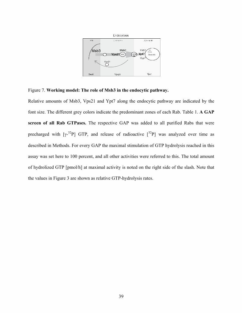
Figure 7. Working model: The role of Msb3 in the endocytic pathway.
Relative amounts of Msb3, Vps21 and Ypt7 along the endocytic pathway are indicated by the
font size. The different grey colors indicate the predominant zones of each Rab. Table 1. A GAP
screen of all Rab GTPases. The respective GAP was added to all purified Rabs that were
precharged with [γ-32P] GTP, and release of radioactive [32P] was analyzed over time as
described in Methods. For every GAP the maximal stimulation of GTP hydrolysis reached in this
assay was set here to 100 percent, and all other activities were referred to this. The total amount
of hydrolized GTP [pmol/h] at maximal activity is noted on the right side of the slash. Note that
the values in Figure 3 are shown as relative GTP-hydrolysis rates.