Embed Size (px)
Citation preview
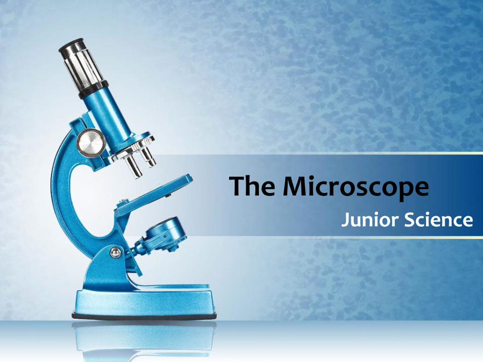
The MicroscopeJunior Science
Lesson Objectives
• identify, and understand the functions of the main parts of a light microscope and use it to examine an animal cell and a plant cell.
• prepare a slide from plant tissue and sketch the cells under magnification.
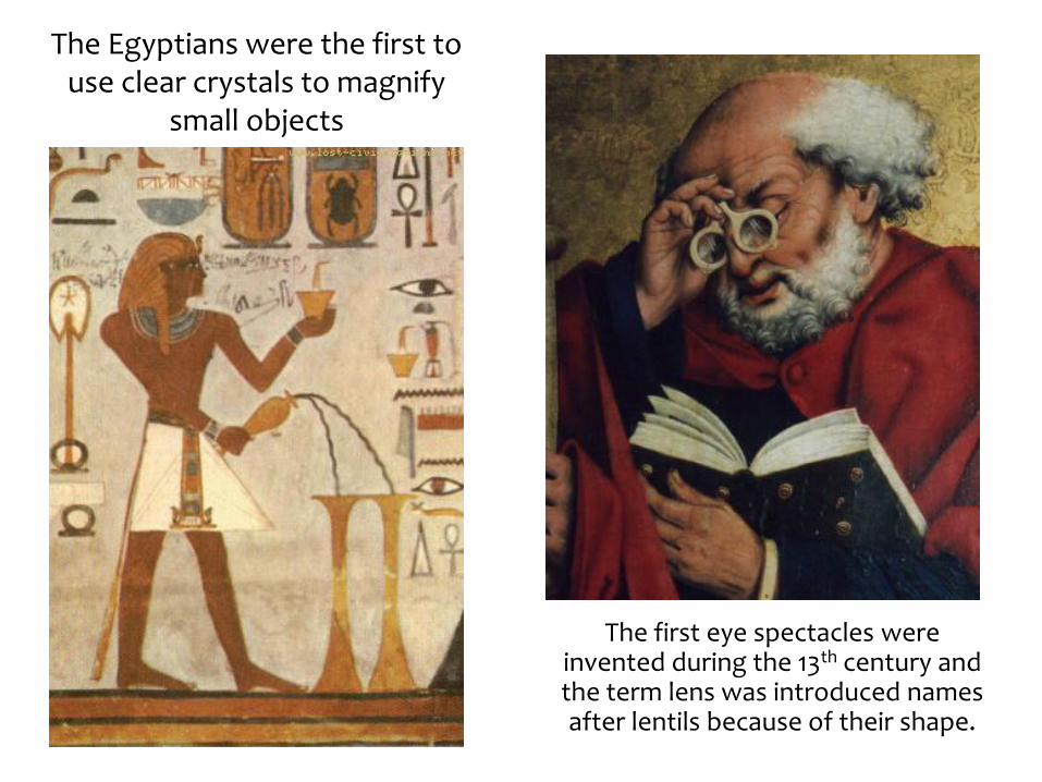
The Egyptians were the first to use clear crystals to magnify
small objects
The first eye spectacles were invented during the 13th century and the term lens was introduced names after lentils because of their shape.
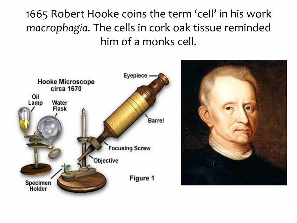
1665 Robert Hooke coins the term ‘cell’ in his work macrophagia. The cells in cork oak tissue reminded
him of a monks cell.
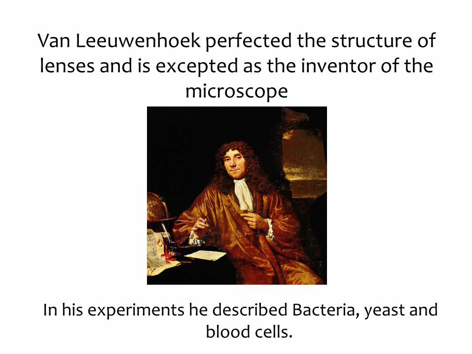
Van Leeuwenhoek perfected the structure of lenses and is excepted as the inventor of the
microscope
In his experiments he described Bacteria, yeast and blood cells.
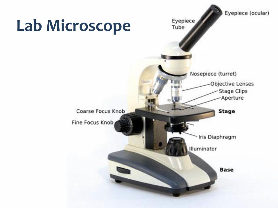
Lab Microscope
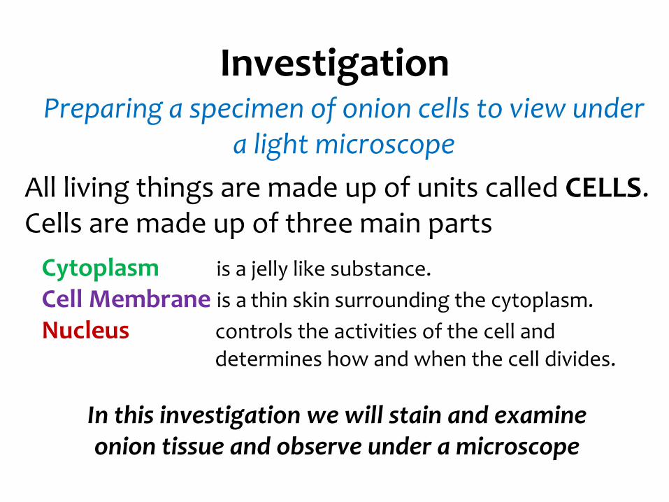
Investigation
All living things are made up of units called CELLS.Cells are made up of three main parts
Cytoplasm is a jelly like substance.
Cell Membrane is a thin skin surrounding the cytoplasm.
Nucleus controls the activities of the cell and determines how and when the cell divides.
In this investigation we will stain and examine onion tissue and observe under a microscope
Preparing a specimen of onion cells to view under a light microscope
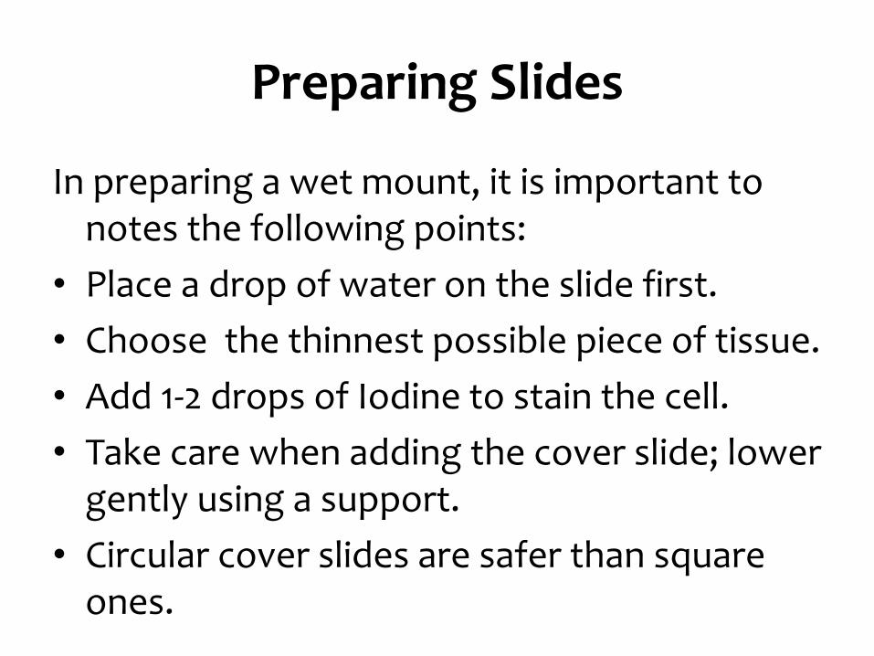
Preparing Slides
In preparing a wet mount, it is important to notes the following points:
• Place a drop of water on the slide first.
• Choose the thinnest possible piece of tissue.
• Add 1-2 drops of Iodine to stain the cell.
• Take care when adding the cover slide; lower gently using a support.
• Circular cover slides are safer than square ones.
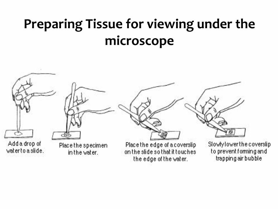
Preparing Tissue for viewing under the microscope
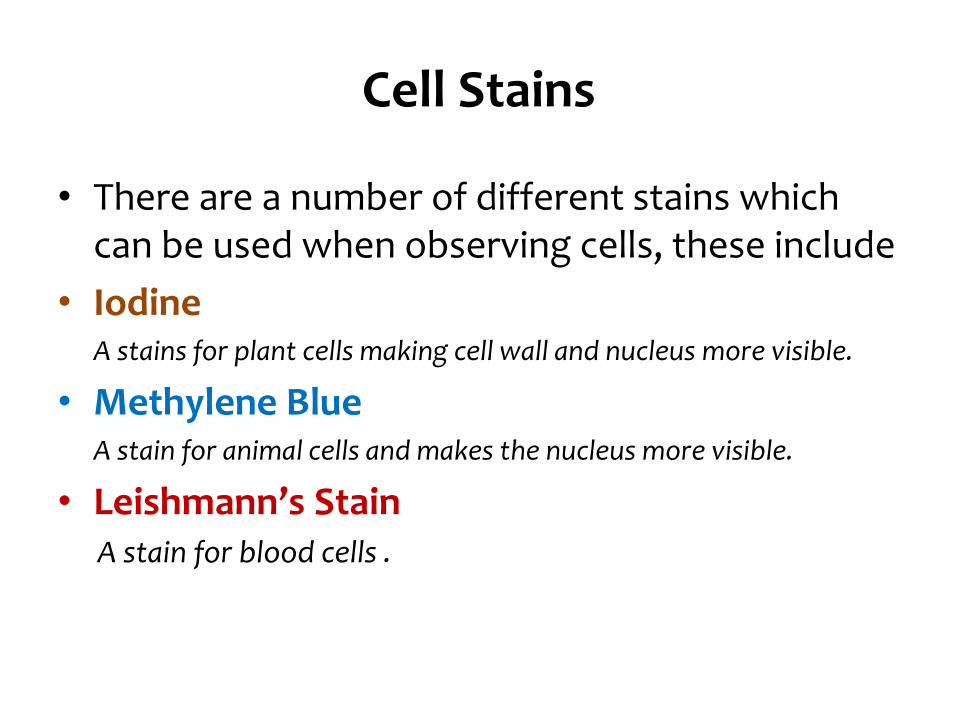
Cell Stains
• There are a number of different stains which can be used when observing cells, these include
• IodineA stains for plant cells making cell wall and nucleus more visible.
• Methylene BlueA stain for animal cells and makes the nucleus more visible.
• Leishmann’s StainA stain for blood cells .
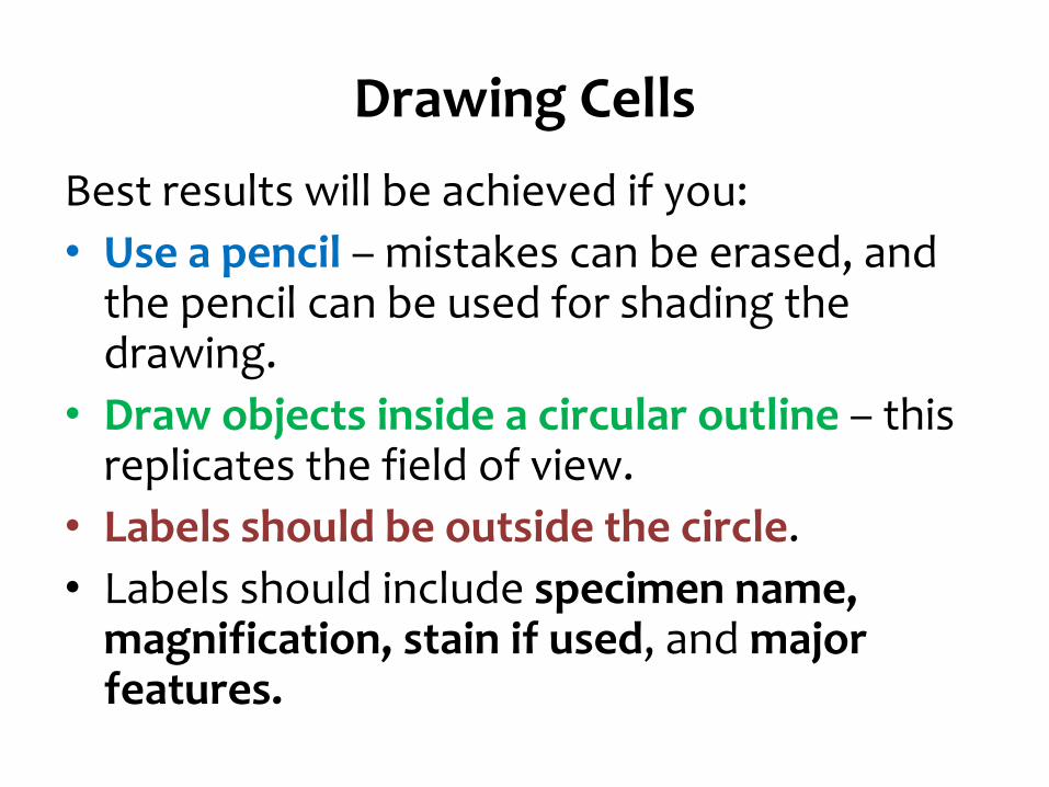
Drawing Cells
Best results will be achieved if you:
• Use a pencil – mistakes can be erased, and the pencil can be used for shading the drawing.
• Draw objects inside a circular outline – this replicates the field of view.
• Labels should be outside the circle.
• Labels should include specimen name, magnification, stain if used, and major features.
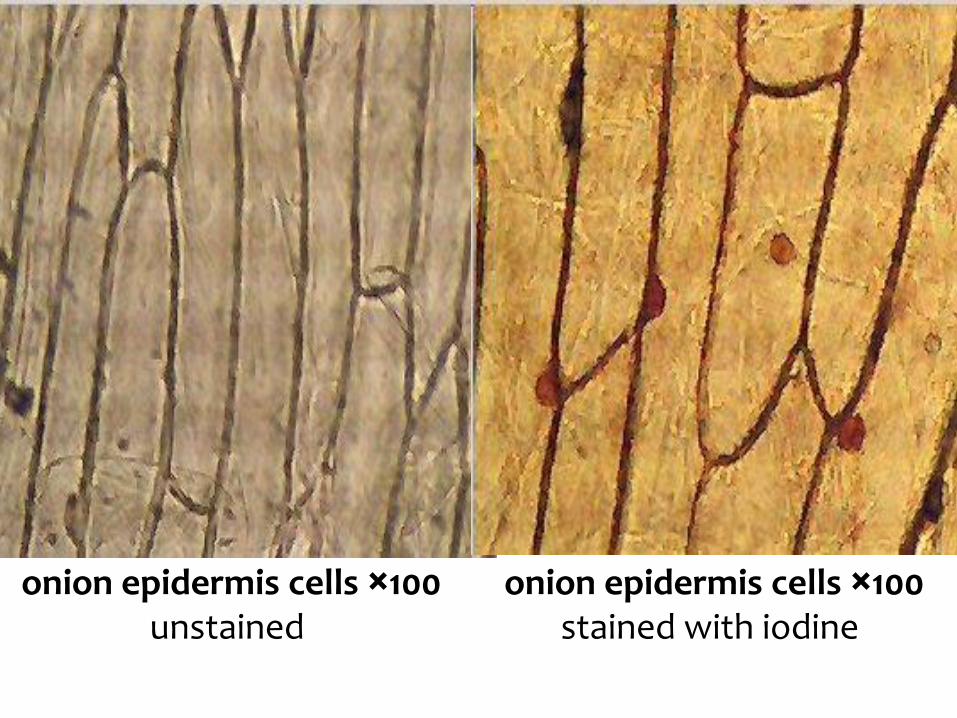
Onion Cells
onion epidermis cells ×100unstained
onion epidermis cells ×100stained with iodine
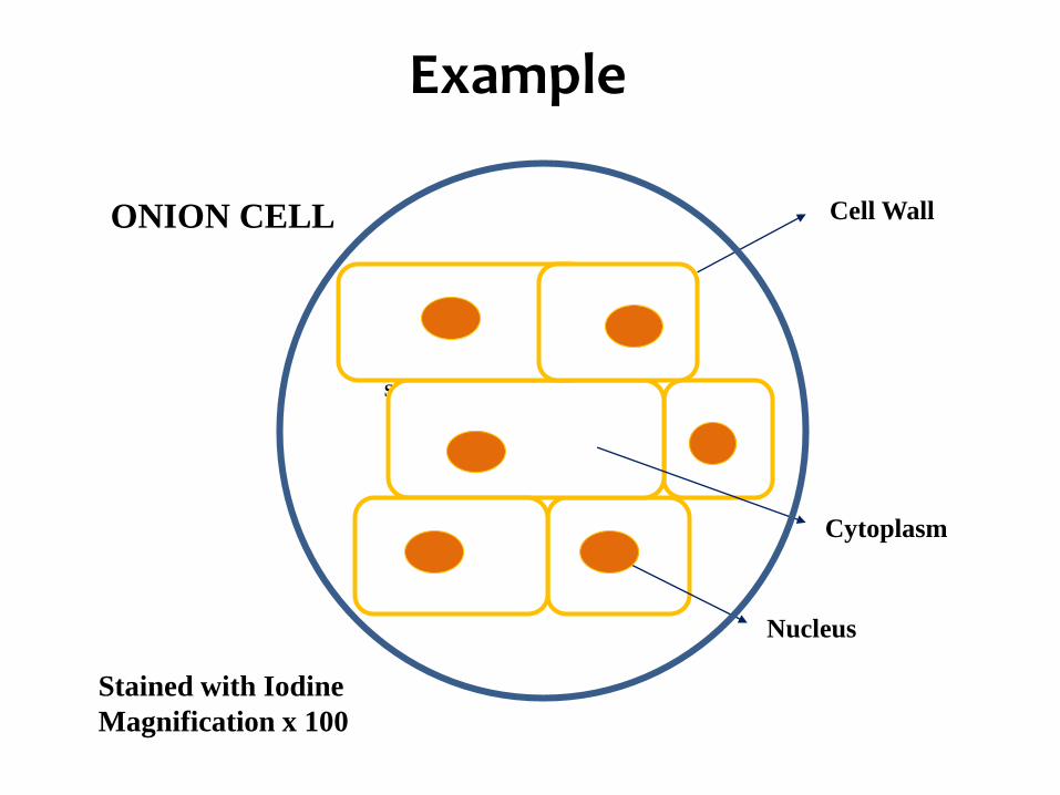
Specimen clearly drawn in pencil
Cell WallONION CELL
Example
Stained with Iodine
Magnification x 100
Nucleus
Cytoplasm
Onion Cell
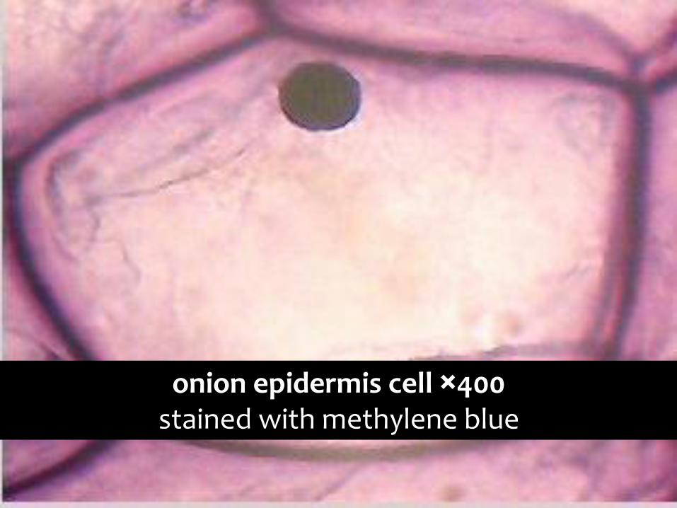
onion epidermis cell ×400stained with methylene blue
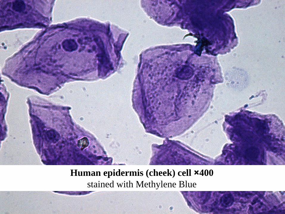
Human Cheek Cells
Human epidermis (cheek) cell ×400
stained with Methylene Blue
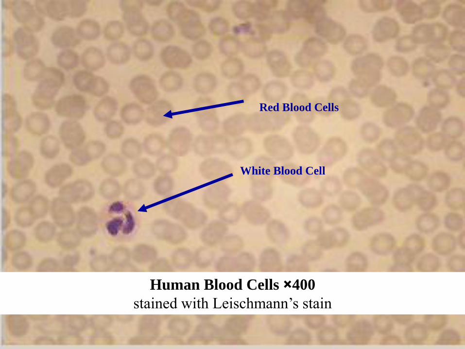
Blood Cells
Human Blood Cells ×400
stained with Leischmann’s stain
White Blood Cell
Red Blood Cells
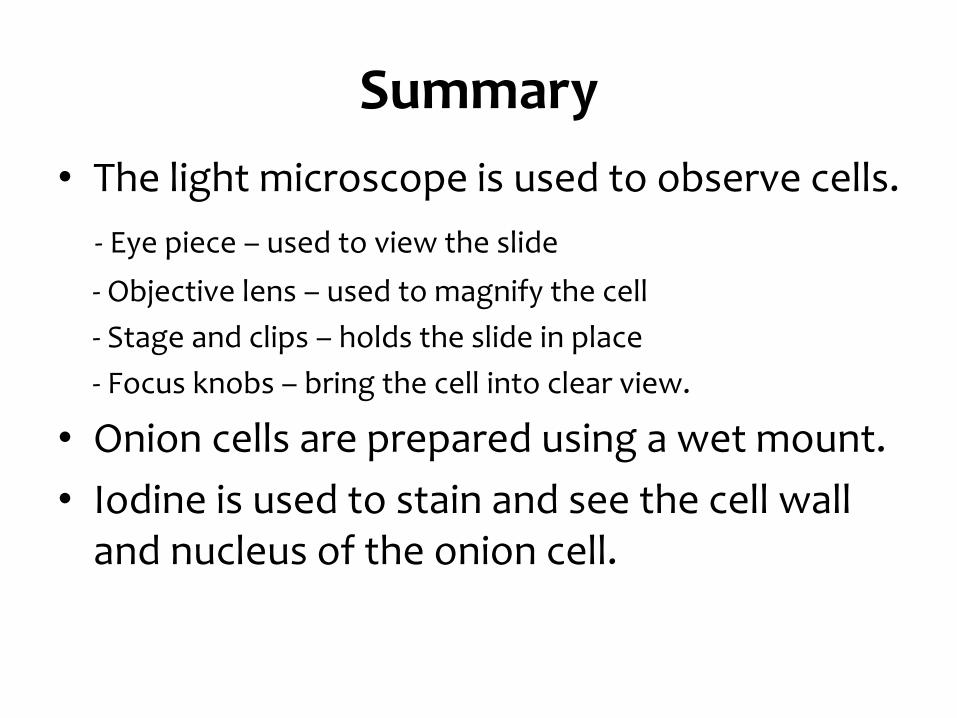
Summary
• The light microscope is used to observe cells.
- Eye piece – used to view the slide
- Objective lens – used to magnify the cell
- Stage and clips – holds the slide in place
- Focus knobs – bring the cell into clear view.
• Onion cells are prepared using a wet mount.
• Iodine is used to stain and see the cell wall and nucleus of the onion cell.