Embed Size (px)
Citation preview

Recent advances in the culture-independent investi gation of microbial community structure1,2 and function2–4, including advances in sequencing technologies and the development of bioinformatic tools, have spawned a veri-table ‘microbiome renaissance’. Owing to these advances, the microbiota is now often referred to as the ‘forgotten organ’ because of our understanding and appreciation of its contributions to host physiology, metabolism and disease5.
The gut microbiota is a diverse and dense microbial community, unparalleled when compared with other body habitats. It is estimated that the gut microbiota is composed of more than 100 trillion cells and 5 million unique genes, outnumbering our own cells and genes6. Although the gut microbiota is composed of thousands of species, the majority of them belong to six bacte-rial phyla: Firmicutes, Bacteroidetes, Actinobacteria, Proteobacteria, Fusobacteria and Verrucomicrobia7. In addition to bacteria, the gut microbiota includes microscopic fungi, archaea, protozoa and viruses. The gut microbiota is highly dynamic and demonstrates substan-tial inter-individual and intra-individual vari ation. The structure of this microbial community is tightly linked to environmental factors, such as diet and drug intake3,8 (discussed below), but has also been associated with many additional factors, such as age9 and host genetics10.
The microbiota has key functions, such as the break-down of plant polysaccharides (that is, fibre) that are indigestible to the host, the biosynthesis of essential vita-mins and amino acids, the detoxification of xenobiotics, resistance against pathogens and the development of the host immune system. Indeed, the microbiome has
now been linked to many areas that were previously considered unrelated to microorganisms, from the cir-cadian rhythm11 to neuroscience12–14, cancer biology15,16, forensics17,18 and metabolic disease19,20.
Despite this exponential increase in microbiome research, the links between the microbiota and pharma-cology remain largely underexplored. The discovery that microorganisms in the human gut can metabolize drugs dates back nearly a century21. Experiments with the sulf-onamide antibiotic prontosil, the first broad-spectrum and commercially available antibiotic, demonstrated a lack of antibacterial activity in vitro22. The discrepancy between the in vivo and in vitro activities of this drug is due to the fact that gut microorganisms activate pront-osil by reducing its azo bond. This biotransformation affects a wide range of compounds, from azo dyes23, which are commonly used as additives in foods, to sulf-asalazine, which is used to treat ulcerative colitis and rheumatoid arthritis24,25. Soon after these discoveries, it became apparent that the biotransformation of drugs by the gut microbiota might be far more widespread than previously appreciated, but limited mechanistic insights were uncovered owing, in part, to the difficulties in ana-lysing complex gut microbial communities using tra-ditional culture-based techniques. To date, the fields of pharmacogenetics and pharmacogenomics still largely focus on variations in the human genome, rather than on the genes encoded by the microbiome26.
Several reviews have highlighted the role of the gut microbiome in pharmacology and precision medicine27–32. In this Review, we focus on recent studies that provide insight into the microbial and molecular
Department of Microbiology & Immunology, George Williams Hooper Foundation, University of California San Francisco, 513 Parnassus Avenue, San Francisco, California 94143, USA.Correspondence to P.J.T. [email protected]
doi:10.1038/nrmicro.2016.17Published online 14 Mar 2016
MicrobiomeThe combined genetic material and metabolic activities of the microbiota.
MicrobiotaThe collection of all microorganisms (archaea, bacteria, microscopic fungi, parasites and viruses) found in a given body habitat.
XenobioticsCompounds that are foreign to a biological system. For humans, these include drugs, dietary bioactive compounds, food additives and environmental toxins.
Azo bondA chemical bond composed of N=N.
The microbial pharmacists within us: a metagenomic view of xenobiotic metabolismPeter Spanogiannopoulos, Elizabeth N. Bess, Rachel N. Carmody and Peter J. Turnbaugh
Abstract | Although the importance of human genetic polymorphisms in therapeutic outcomes is well established, the role of our ‘second genome’ (the microbiome) has been largely overlooked. In this Review, we highlight recent studies that have shed light on the mechanisms that link the human gut microbiome to the efficacy and toxicity of xenobiotics, including drugs, dietary compounds and environmental toxins. Continued progress in this area could enable more precise tools for predicting patient responses and for the development of a new generation of therapeutics based on, or targeted at, the gut microbiome. Indeed, the admirable goal of precision medicine may require us to first understand the microbial pharmacists within.
M I C R O B I O M E
NATURE REVIEWS | MICROBIOLOGY ADVANCE ONLINE PUBLICATION | 1
REVIEWS
© 2016
Macmillan
Publishers
Limited.
All
rights
reserved.

PharmacogeneticsThe study of how genetic factors influence therapeutic outcomes.
PharmacogenomicsThe use of sequencing-based genomic methods to analyse the links between genetics and therapeutic outcomes.
BioavailabilityThe proportion of an administered compound that reaches systemic circulation and thus has the potential to influence the intended target.
First-pass metabolismThe metabolism of orally ingested compounds before reaching general circulation.
Biliary excretionThe transfer of xenobiotics and other compounds from the plasma to bile through hepatocytes, which is followed by the release of the compounds into the gut lumen.
Enterohepatic circulationThe circulation of xenobiotics and endogenous compounds that are absorbed from the intestines, transported to the liver, and then re-enter the intestine through the bile ducts, where they may be reabsorbed or metabolized by the gut microbiota.
ReductionA chemical reaction in which the oxidation state of a chemical bond is reduced. For example, a carbon–carbon bond modified to a carbon–hydrogen bond is a reductive transformation.
HydrolysisA chemical reaction in which a chemical bond is cleaved using a water molecule, which acts as the nucleophile.
Cytochrome P450 enzymes(CYPs) A family of enzymes that is responsible for the oxidative biotransformation of xenobiotics and other compounds.
ProdrugsDrugs that are administered in an inactive form and become active when metabolized.
FolateA B vitamin that is essential for DNA synthesis, DNA repair and other biological reactions.
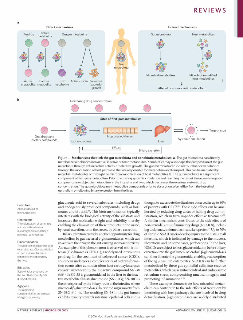
mechanisms that are relevant to the prevention and treat-ment of human disease. Gut microorganisms can affect drug therapy through various different mechanisms that can generally be grouped into direct effects or indirect effects (FIG. 1). Direct mechanisms include the biotrans-formation of drugs or their metabolites into products with altered bioactivities. Indirect mechanisms involve more complex host–microbial interactions that modulate host pathways for xenobiotic metabolism or transport. We also discuss other classes of xeno biotics, including dietary compounds, food additives and environmen-tal toxins. Finally, we briefly highlight the immediate translational implications of this research and discuss early progress towards microbiome-based diagnostics and co-therapies.
The gut microbiota and pharmaceuticalsThe gut microbiota can influence the metabolism of dozens of pharmaceuticals, in many cases changing their efficacy and/or side effect profiles. In this section, we highlight key examples of the direct and indirect mechanisms by which the gut microbiota influences drug therapy.
Microbial metabolism of drugs and their metabolites. The bioavailability of orally administered drugs depends on the extent of first-pass metabolism by intestinal and hepatic enzymes before reaching systemic circulation33. However, oral drugs may encounter the gut microbiota before reaching host tissues, representing another impor-tant site of first-pass metabolism (FIG. 1). In fact, there is already in vitro and/or in vivo evidence for the metabo-lism of 50 drugs by the gut microbiota28 (Supplementary information S1 (table)). This number is probably an underestimate given the lack of any systematic analyses of the microbial metabolism of drugs in the gut and the vast genetic diversity within the microbiome34. Furthermore, the rate of absorption probably has an important role in determining the extent of microbial metabolism owing to the fact that the density of gut microorganisms increases substantially in the distal small intestine (also known as the ileum) and colon. Drugs and their metabolites can also re-encounter the gut microbiota by biliary excretion, at which point they can be further metabolized and reabsorbed through enterohepatic circulation.
Despite the diversity of chemical structures among the drugs that are known to be subject to gut microbial metabolism, two broad chemical transformation patterns have been repeatedly observed — reduction and hydrolysis (FIG. 2; Supplementary information S1 (table)). These two reactions may reflect the energetic demands of the gut microbiota. The gut is largely anaerobic; consequently, microorganisms in the gut cannot rely on oxygen as a ter-minal electron acceptor for respiration35. Reductive xeno-biotic metabolism may facilitate anaerobic respiration by increasing the range of alternative electron acceptors that are available for respiration. Conversely, hydrolysis directly provides substrates for microbial growth. For example, many dietary components are glycosylated and their hydrolysis liberates sugars that could be shunted into glycolysis36.
The commonality of these two reaction types (reduc-tion and hydrolysis) may also imply that there are core microbial species or gene families that affect a wide range of small molecules37. If so, the identification of the major players could act as the basis for predicting the man-ner in which a novel drug will be modified by the gut micro biota. Such knowledge is likely to revolutionize drug development and precision medicine, similar to the advances that followed the discovery that cytochrome P450 enzymes (CYPs) are expressed in the intestine and liver, where they metabolize several xenobiotics38,39. Chemical functional groups that are subject to microbial metabolism could be removed through rational design or used to control drug delivery.
The therapeutic effects of several prodrugs that contain azo bonds require bioactivation by gut microorganisms. Following oral administration, the azo bond is reduced by microbial azoreductases, which liberate the biolog-ically active compound. For example, the anti bacterial drug prontosil is cleaved by the gut micro biota, which results in the production of triamino benzene and sulf-anilamide21, a bacteriostatic antibiotic that inhibits folate metabolism. Based on these findings, azo bonds have been used in drug development. For example, sulf-asalazine was strategically designed to treat rheumatoid arthritis, which at the time was thought to be the result of bacterial infections, by linking the sulfon amide sulf-apyridine with the anti-inflammatory drug sali cylic acid by an azo bond40,41 (FIG. 2). Intact sulfasalazine can be recovered from the stool of antibiotic- treated or germ-free rats, but not in conventionally raised animals42. Furthermore, a simplified gut microbiota composed of four bacterial strains (Bacteroides sp., Enterococcus faecalis and two Lactobacillus sp.) is sufficient to restore sulfasalazine metabolism in gnotobiotic rats, and the in vitro incubation of sulfasalazine with bacterial isolates from these animals results in drug cleavage42.
Azoreductases are widespread across several bacte-rial phyla found in the human gut28 and possess broad substrate compatibility43,44; however, they metabolize azo compounds at different rates depending on the broader chemical structure of the molecule45. Gut microorgan-isms can also metabolize the downstream metabolites of azo reductions. For example, the bioactive component of sulfasalazine, 5-aminosalicylic acid, is inactivated by microbial arylamine N-acetyltransferases. The activity of these enzymes can vary up to tenfold between indivi-duals46, highlighting the considerable inter- individual dif-ferences in gut microbial metabolism that may contribute to variations in drug efficacy.
β-glucuronidases are another generalist enzyme family expressed by human-associated gut bacteria that influence the biological activity and toxicity of a wide range of drugs, dietary components and endogenous metabolites47. Recent studies have discovered the role of β-glucuronidases in the toxicity of drugs that are used to treat cancer and inflammation. In these exam-ples, gut bacteria metabolize and interfere with drug metabolites that are generated by the host detoxification pathway of glucuronidation. Uridine diphosphate (UDP)-glucuronosyltransferases expressed in the liver add
R E V I E W S
2 | ADVANCE ONLINE PUBLICATION www.nature.com/nrmicro
© 2016
Macmillan
Publishers
Limited.
All
rights
reserved. ©
2016
Macmillan
Publishers
Limited.
All
rights
reserved.
Nature Reviews | Microbiology
Activemetabolite
Activemetabolite
Inactivemetabolite
Toxicmetabolite
Antimicrobial
Gut microbiota
Intestinal epitheliumOral drugs anddietary compounds
Decreasing drug concentration
Liver
EffluxBiliary excretion
Target tissue
Systemic circulation
Selectivebacterialgrowth
Prodrug Drug or metabolite
Gutmicrobiota
Direct mechanisms
a
b
Sites of first-pass metabolism
Indirect mechanisms
Host metabolites
Altered host xenobiotic metabolism
Microbiota-modified host metabolites
Microbial metabolites
Gut microbiota
Germ-freeAnimals devoid of microorganisms.
GnotobioticThe colonization of germ-free animals with individual microorganisms or defined microbial communities.
GlucuronidationThe addition of glucuronic acid to a substrate. Glucuronidation is used as a mechanism of xenobiotic metabolism by the host.
Bile acidsSteroid acids produced by the liver that emulsify fats during digestion.
AglyconeThe remaining compound after the removal of a glycosyl moiety.
glucuronic acid to several substrates, including drugs and endogenously produced compounds, such as hor-mones and bile acids48. This biotransformation typically interferes with the biological activity of the substrate and increases the molecular weight and solubility, thereby enabling the elimination of these products in the urine, by renal excretion, or in the faeces, by biliary excretion.
Biliary excretion provides another opportunity for drug metabolism by gut bacterial β-glucuronidases, which can re-activate the drug in the gut causing increased toxicity. An example of this phenomenon is observed with irino-tecan (also known as CPT-11), a widely used intravenous prodrug for the treatment of colorectal cancer (CRC). Irinotecan undergoes a complex series of biotransforma-tion events after administration: host carboxylesterases convert irinotecan to the bioactive compound SN-38 (REF. 49); SN-38 is glucuronidated in the liver to the inac-tive metabolite SN-38 glucuronide (SN-38G); SN-38G is then transported by the biliary route to the intestine where microbial β-glucuronidases liberate the sugar moiety from SN-38G (FIG. 2). The resulting SN-38 in the gut lumen exhibits toxicity towards intestinal epithelial cells and is
thought to exacerbate the diarrhoea observed in up to 80% of patients with CRC50,51. These side effects can be ame-liorated by reducing drug doses or halting drug admin-istration, which in turn impedes effective treatment52. A similar mechanism contributes to the side effects of non-steroidal anti- inflammatory drugs (NSAIDs), includ-ing diclofenac, indomethacin and ketoprofen53. Up to 70% of chronic NSAID users develop injury to the distal small intestine, which is indicated by damage to the mucosa, ulcerations and, in some cases, perforations. In the liver, NSAIDs are subject to host glucuronidation before biliary excretion into the gut lumen. Microbial β-glucuronidases can then liberate the glucuronide, enabling reabsorption of the aglycone into enterocytes. NSAIDs can be further metabolized by these gut epithelial cells into reactive metabolites, which cause mitochondrial and endo plasmic reticulum stress, compromising mucosal integrity and promoting inflammation51,54.
These examples demonstrate how microbial metab-olism can contribute to the side effects of treatment by interfering with host pathways that are involved in drug detoxification. β-glucuronidases are widely distributed
Figure 1 | Mechanisms that link the gut microbiota and xenobiotic metabolism. a | The gut microbiota can directly metabolize xenobiotics into active, inactive or toxic metabolites. Xenobiotics may also shape the composition of the gut microbiota through antimicrobial activity or selective growth. The gut microbiota can indirectly influence xenobiotics through the modulation of host pathways that are responsible for metabolism and transport. This can be mediated by microbial metabolites or through the microbial modification of host metabolites. b | The gut microbiota is a significant component of first-pass metabolism. Prior to entering systemic circulation and reaching the target tissue, orally ingested compounds are subject to metabolism in the intestine and liver, which decreases the eventual systemic drug concentration. The gut microbiota may metabolize compounds prior to absorption, after efflux from the intestinal epithelium or following biliary excretion from the liver.
R E V I E W S
NATURE REVIEWS | MICROBIOLOGY ADVANCE ONLINE PUBLICATION | 3
© 2016
Macmillan
Publishers
Limited.
All
rights
reserved. ©
2016
Macmillan
Publishers
Limited.
All
rights
reserved.

Nature Reviews | Microbiology
Reduction Hydrolysis
Dru
g co
ncen
trat
ion
Dru
g co
ncen
trat
ion
Time
a b
c d e
Time
Therapeuticwindow
Germ-free animal
Microbiota increases bioavailability Microbiota decreases bioavailability Microbiota increases toxicity
Activation Inactivation Toxicity
Colonized animal Germ-free animal Colonized animal Germ-free animal Colonized animal
Time Time
Subtherapeutic
Tox
in c
once
ntra
tion
Time Time
Toxicitythreshold
Examples: SulfasalazineProntosil
Examples: DigoxinMethotrexate
Examples: SN-38GNSAIDsMelamine
HN
NH
F
O
O
HO
HO
OH
O
NH2
5-Fluorouracil Levodopa
Digoxin
SulindacSulfasalazine
OH
OH
H
HO
Triglycoside
O O
H
O
HO
F
S
O
CH3
N
N
HO
HOO
S
HN
N
OO
O
OH
OHO
O
HO
N
N
O
HOO
O
SN-38G
O
OH
HO
NHO
NH
O
O
Br
Sorivudine
O
OO
OHO
N
N
N
N
H3C
NH2N
NH2
O O
HNOH
O
HO
Methotrexate
Lovastatin
across many gut bacterial species, including members of the Proteobacteria, Firmicutes and Actinobacteria phyla55–60. However, it remains unclear whether all of these enzymes exhibit a similarly broad substrate scope or whether they are specialized for distinct niches (physical or occupational) in the gastrointestinal tract. Further work is necessary to determine whether the abundance and/or activity of these enzymes can explain inter-individual variations in drug toxicity.
Microbial metabolism can also interfere with the bioavailability of drugs. A classic example of this phen-omenon comes from the cardiac glycoside digoxin,
which is used for the treatment of cardiac arrhythmia (an irregular heartbeat) and heart failure26. The use of digoxin is challenging owing to its exceedingly narrow therapeutic range (0.5–2 ng ml–1), which can make even minor changes to its concentration clinically relevant. Approximately 10% of patients excrete high levels of an inactive metabolite of digoxin, dihydrodigoxin, which results from the reduction of the α,β-unsaturated lactone ring by bacteria61,62 (FIG. 2). In some cases, more than 50% of the administered drug is inactivated63, which leads to a substantial decrease in systemic drug concentra-tion. Seminal studies that were conducted in the 1980s
Figure 2 | Major reaction types catalysed by the gut microbiota and their pharmacological consequences. The majority of known microbial biotransformations can be segregated into one of two reaction classes: reduction, in which compounds gain electrons from electron donors (part a), and hydrolysis, in which chemical bonds are cleaved through the addition of water (part b). The sites of modifications are highlighted in orange for reduction reactions and in blue for hydrolysis reactions. For a comprehensive list of drug biotransformations see Supplementary information S1 (Table). The microbial metabolism of pharmaceuticals can lead to their activation (part c), inactivation (part d) or result in the production of toxic compounds (part e); this is illustrated by the differential effects of drugs in germ-free animals, compared with colonized animals. Activation refers to the conversion of a prodrug to its bioactive form, contributing to therapeutic concentrations. Examples of activation include the anti-inflammatory drug sulfasalazine and the antibiotic prontosil. Inactivation refers to the conversion of an active metabolite into a downstream metabolite with reduced bioactivity. Examples of inactivation include the cardiac drug digoxin and the anti-inflammatory drug methotrexate. Toxicity occurs owing to the production of metabolites by microorganisms that are harmful to the host. Examples include the hydrolysis of the anti-cancer drug metabolite SN-38 glucuronide (SN-38G), the hydrolysis of glucuronidated non-steroidal anti-inflammatory drugs (NSAIDs), which are used to treat pain, and the metabolism of the dietary contaminant melamine to cyanuric acid.
R E V I E W S
4 | ADVANCE ONLINE PUBLICATION www.nature.com/nrmicro
© 2016
Macmillan
Publishers
Limited.
All
rights
reserved. ©
2016
Macmillan
Publishers
Limited.
All
rights
reserved.
Serum metabolomeThe collection of all metabolites found in serum.
suggested that only a single bacterial species, Eggerthella lenta, reduces digoxin64, but unfortunately neither the presence nor the abundance of this species predicts the successful reduction of this drug64–66. This discrepancy seems to be driven by strain-level variations in the E. lenta population67. In E. lenta DSM2243, digoxin induces the expression of a two-gene operon, referred to as the cardiac glycoside reductase (cgr) operon. The proteins encoded by this operon, Cgr1 and Cgr2, are homologous to enzymes that are involved in electron transport. Cgr1 shows similarity to cytochrome c reductases, which are membrane-bound proteins that are involved in shut-tling electrons from quinones to an electron reductase partner. Cgr2, which shows similarity to flavin adenine dinucleo tide (FAD)-binding fumarate reductases, is pre-dicted to interact and accept electrons from Cgr1 and, in turn, reduce the lactone ring of digoxin37. The cgr operon is not found in the genomes of E. lenta strains that lack the ability to reduce digoxin, which suggests that the cgr operon is necessary for the reduction of digoxin and provides an explanation for the difficulty in predicting the reduction of digoxin based only on the presence of E. lenta species. Furthermore, the reduction of digoxin is enhanced in the presence of a complex gut microbiota and suppressed by dietary protein67, which suggests that microbial drug metabolism is sensitive to both microbial and environmental interactions.
Microbial control of xenobiotic metabolism and absorp-tion. Compounds that resist microbial metabolism can still be influenced by the gut microbiota through several mechanisms (BOX 1). Comparisons between germ-free and colonized mice have revealed that the
microbiota affects the expression of several host genes that are involved in drug metabolism and transport68,69. This influence on gene expression in the host by the gut microbiota can be local68,70 (in gut tissues) or distant, which includes affecting the most vital organ involved in drug metabolism — the liver69,71. In the liver, more than 100 genes are differentially expressed between germ-free and colonized mice69. One of the largest groups of differ-entially expressed genes is the one encoding CYPs. An example that illustrates the importance of the microbiota to xenobiotic metabolism in the liver mediated by CYPs is the anaesthetic pentobarbital (also known as pento-barbitone). Pentobarbital is administered intravenously and is metabolized by CYPs in the liver. Germ-free animals, which show increased expression of CYPs compared with colonized animals, are more efficient at metabolizing pentobarbital than colonized animals69.
A recent RNA-sequencing-based study confirmed the differential expression of several genes that are involved in xenobiotic metabolism in the liver of germ-free and colonized mice72. Furthermore, this study reported a substantial increase in the expression of the xenobiotic- sensing transcription factors aryl hydrocarbon receptor (AhR), constitutive androstane receptor (CAR), perox-isome proliferator- activated receptor-α (PPARα) and nuclear factor erythroid 2-related factor 2 (NRF2) in germ-free mice. However, additional work is necessary to elucidate the mechanisms responsible for these differ-ences in gene expression.
The microbiota also substantially changes the serum metabolome. Comparisons between germ-free and col-onized mice revealed that the gut microbiota not only alters the abundance of endogenous metabolites, with
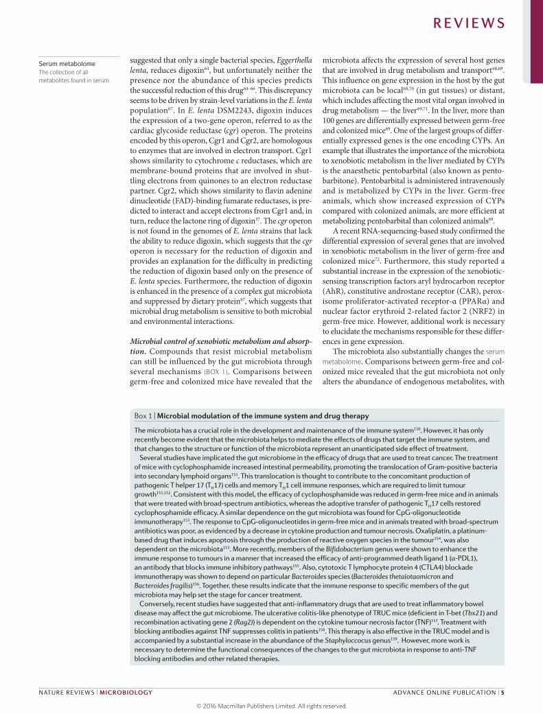
Box 1 | Microbial modulation of the immune system and drug therapy
The microbiota has a crucial role in the development and maintenance of the immune system150. However, it has only recently become evident that the microbiota helps to mediate the effects of drugs that target the immune system, and that changes to the structure or function of the microbiota represent an unanticipated side effect of treatment.
Several studies have implicated the gut microbiome in the efficacy of drugs that are used to treat cancer. The treatment of mice with cyclophosphamide increased intestinal permeability, promoting the translocation of Gram-positive bacteria into secondary lymphoid organs151. This translocation is thought to contribute to the concomitant production of pathogenic T helper 17 (TH17) cells and memory TH1 cell immune responses, which are required to limit tumour growth151,152. Consistent with this model, the efficacy of cyclophosphamide was reduced in germ-free mice and in animals that were treated with broad-spectrum antibiotics, whereas the adoptive transfer of pathogenic TH17 cells restored cyclophosphamide efficacy. A similar dependence on the gut microbiota was found for CpG-oligonucleotide immunotherapy153. The response to CpG-oligonucleotides in germ-free mice and in animals treated with broad-spectrum antibiotics was poor, as evidenced by a decrease in cytokine production and tumour necrosis. Oxaliplatin, a platinum- based drug that induces apoptosis through the production of reactive oxygen species in the tumour154, was also dependent on the microbiota153. More recently, members of the Bifidobacterium genus were shown to enhance the immune response to tumours in a manner that increased the efficacy of anti-programmed death ligand 1 (α-PDL1), an antibody that blocks immune inhibitory pathways155. Also, cytotoxic T lymphocyte protein 4 (CTLA4) blockade immunotherapy was shown to depend on particular Bacteroides species (Bacteroides thetaiotaomicron and Bacteroides fragilis)156. Together, these results indicate that the immune response to specific members of the gut microbiota may help set the stage for cancer treatment.
Conversely, recent studies have suggested that anti-inflammatory drugs that are used to treat inflammatory bowel disease may affect the gut microbiome. The ulcerative colitis-like phenotype of TRUC mice (deficient in T-bet (Tbx21) and recombination activating gene 2 (Rag2)) is dependent on the cytokine tumour necrosis factor (TNF)157. Treatment with blocking antibodies against TNF suppresses colitis in patients158. This therapy is also effective in the TRUC model and is accompanied by a substantial increase in the abundance of the Staphyloccocus genus159. However, more work is necessary to determine the functional consequences of the changes to the gut microbiota in response to anti-TNF blocking antibodies and other related therapies.
R E V I E W S
NATURE REVIEWS | MICROBIOLOGY ADVANCE ONLINE PUBLICATION | 5
© 2016
Macmillan
Publishers
Limited.
All
rights
reserved. ©
2016
Macmillan
Publishers
Limited.
All
rights
reserved.

ConjugationThe addition of a chemical unit (for example, glucuronic acid or glutathione) to xenobiotics, increasing the solubility and molecular weight of the parent compound and facilitating elimination from the body.
Metabolic syndromeA collection of physiological and biochemical conditions, defined as a combination of high blood pressure, increased blood sugar levels, excess fat and abnormal cholesterol levels. This syndrome increases the risk of heart disease, stroke and diabetes.
10% of shared metabolites differing in abundance by at least 50%, but also contributes unique microbial compounds to systemic circulation73,74. Some of these microbial metabolites are processed by the host in a man-ner analogous to xenobiotics (that is, conjugation)74. This overlap between the host response to drugs and microbial metabolites may have implications during drug therapy; for example, it may result in increased toxicity or half-life of xenobiotics owing to competition between the drug and microbial metabolites for the same host enzymes that are involved in drug detoxification or elimination. An example of this type of interaction can be observed with acetaminophen (also known as paracetamol), which is one of the most widely used drugs worldwide. Overdose of acetamino phen can lead to severe and sometimes fatal hepato toxicity75, and both drug metabolism and toxicity vary between individuals76,77. Acetaminophen is metabolized in the liver and the predominant metabolites that result, acetaminophen sulfate and acetaminophen glucuronide, are inactive. However, a minor metabo-lite, N-acetyl-p-benzoquinone imine (NAPQI), causes toxicity in the liver. Based on these findings, a metabo-lomic study aimed to determine whether pre-dose uri-nary metabolite profiles could predict acetaminophen metabolism in humans78. Pre-dose levels of p-cresol sulfate were found to be inversely associated with the ratio of acetaminophen sulfate to acetaminophen glu-curonide. The microbial metabolite p-cresol is an end- product of tyrosine and phenyl alanine metabolism and has been demonstrated to be produced by several micro-organisms, including those belonging to the Firmicutes (Clostridium difficile), Bacteroidetes, Actinobacteria and Fusobacteria phyla79,80. Following absorption and cir-culation, p-cresol is metabolized in the liver to p- cresol sulfate. p-cresol and acetaminophen are both substrates of the human cytosolic sulfotransferase 1A1 (SULT1A1)81 (FIG. 3). This competition probably impedes the ability of the host to detoxify acetaminophen, which increases the likelihood of accumulating the toxic metabolite NAPQI.
Host–microbial interactions may also influence drug efficacy. Statins, which are cholesterol-lowering drugs prescribed for coronary artery disease, have substantial inter-individual variations in efficacy, with up to 33% of patients failing to reach lipid-lowering targets82. In humans, the response to treatment with simvastatin (indicated by low-density lipoprotein (LDL) cholesterol levels) is positively associated with the pre-treatment levels of three secondary bile acids produced by the microbiota: lithocholic acid, tauro-lithocholic acid and glycolithocholic acid83. Although the mechanism, or mechanisms, responsible for this association remains unknown, one intriguing hypoth-esis is that primary bile acids may compete for the same intestinal transporters that enable the absorption of statins (FIG. 3). Therefore, microbial bile acid metabo-lism may decrease this competition, priming the host for more effective statin therapy.
A similar type of interaction may influence the effi-cacy of tempol, an antioxidant that protects against diet-induced obesity in animal models84. Treatment of mice with tempol altered the relative abundance of the
two dominant bacterial phyla in the distal gut, increas-ing the abundance of Bacteroidetes and decreasing the abundance of Firmicutes85. In the Firmicutes phylum, members of the genus Lactobacillus were substantially reduced (FIG. 3). Several members of the Lactobacillus genus encode bile salt hydrolases, which produce free bile acids by deconjugating taurine- conjugated bile acids86. Consistent with these findings, tempol increases the intestinal concentration of several taurine- conjugated bile acids, including tauro-β- muricholic acid (Tβ-MCA). Tβ-MCA is an antagonist of the farnesoid X receptor (FXR), a master regulator of lipid, glucose and bile acid metabolism85,87,88. Tempol does not further reduce adiposity in intestinal-specific FXR-null mice85, consistent with the hypothesis that changes in microbial bile acid metabolism and sub sequent signalling by the FXR pathway contribute to the mechanism of action of tempol. It remains to be determined whether tempol has direct antimicrobial activity against members of the gut microbiota or whether the observed changes in micro-bial community structure are mediated through drug interactions with the host.
The gut microbiota and dietary xenobioticsIn addition to influencing drugs, the gut microbiota can metabolize numerous xenobiotic compounds found in our diet, including natural products and chemical addi-tives. In some cases, these compounds have beneficial health effects that depend on microbial bioactivation. In other instances, the gut microbiota can produce toxic metabolites. In this section we highlight key examples of how gut microbial metabolism affects the health effects of the foods that we consume.
Diet-derived bioactive compounds. Our diet is rich in small molecules that have important consequences for human health and disease. Many of these ‘diet- derived bioactive compounds’ are metabolized by the gut micro-biota (FIG. 4; Supplementary information S2 (table); Supplementary information S3 (table)) and some are dependent on this transformation for activation and/or absorption47. Below, we highlight examples with consid-erable recent evidence elucidating their interaction with, and dependence on, the gut microbiota.
Clinical and epidemiological studies suggest that dietary polyphenols, such as anthocyanins (ACNs) and proanthocyanidins (PACs), protect against metabolic syndrome89,90. Supplementation of high-fat diets with ACNs or PACs has been shown to suppress the expression of genes that are involved in fatty acid and triacylglycerol synthesis, the regulation of lipogenesis and cholesterol biosynthesis, and the assembly of very low-density lipo-proteins90–92. ACNs and PACs have also been argued to stop the development of insulin resistance by increasing insulin signalling, glycogen accumulation and the secre-tion of adiponectin in the presence of free fatty acids93. Intriguingly, studies in rodents indicate that just 6–12% of radiolabeled polyphenols are metabolized and enter cir-culation during their passage through the gut94,95, which raises the question of how these compounds confer their protective effects. Recent studies on polyphenol extracts
R E V I E W S
6 | ADVANCE ONLINE PUBLICATION www.nature.com/nrmicro
© 2016
Macmillan
Publishers
Limited.
All
rights
reserved. ©
2016
Macmillan
Publishers
Limited.
All
rights
reserved.

O
O
OH
S
Nature Reviews | Microbiology
a b
cp-cresol sulfate
Acetaminophen sulfate (inactive)
Tempol
Host transcriptionPrimary bile acids
Gut microbiota
Gut microbiota p-cresol
Acetaminophen (active)
Secondarybile acids
Host transporters
Increasedlevels ofsimvastatin
Regulation of metabolism (lipid, glucose, bile acids)
OH O O
O
OH
S
OHHN
O
OHN
O
Lactobacillus spp.Bile salt hydrolase
β-muricholic acid
Tauro-β-muricholic acid
FXR
N
OH
O–
Host SULT
MetforminAn oral medication used to treat type 2 diabetes.
isolated from grapes20 and cranberries96 provide support for a mechanism that acts through the gut microbiota. Mice fed high-fat diets that were supplemented with polyphenols showed a reduction in diet-induced weight gain and adiposity, improved insulin sensitivity, and diminished markers of intestinal inflammation and oxi-dative stress compared with controls20,96. These improve-ments were coupled with substantial sevenfold-to-tenfold blooms of Akkermansia muciniphila, a mucin-degrading bacterium that is argued to have an important role in the preservation of the integrity of the gut mucus layer, thus limiting the risk of systemic inflammation97. The abundance of A. muciniphila has been linked to reduced weight gain, adiposity, insulin resistance, and/or inflam-matory markers in many contexts, including during pregnancy98, following gastric bypass surgery19,99, in prebiotic or metformin treatment experiments100,101, and in other polyphenol feeding experiments involving green or black tea102,103 or a grape juice and red wine mixture103. Furthermore, the administration of live (but not heat-killed) A. muciniphila was sufficient to reduce host adi-posity, inflammatory markers and insulin resistance in diet-induced obese mice97,101. Further work is required to establish how polyphenols promote the growth of A. muciniphila and whether this effect is direct or medi-ated through changes in host physiology. However,
a recent in vitro study reported that the exposure of a complex human faecal microbial community to black tea or grape- derived polyphenols can directly increase the abundance of A. muciniphila, which suggests a limited dependence on host factors in this process103.
Fruit-derived ellagitannins are thought to provide pro-tective properties for the plant by preventing microbial decay104. The hydrolysis of ellagitannins releases ellagic acid, which can be metabolized by the gut microbiota into several structurally related urolithins that can reach high concentrations locally, in the colon, and systemically105. Several in vitro studies have shown that urolithins have antioxidant, anti cancer, anti- inflammatory and anti-microbial properties; however, there are currently a limited number of in vivo and mechanistic studies on urolithins. A recent study found that ellagic acid metab-olism varied substantially between individuals but could be generally grouped into three categories depending on the metabolites generated, including a subset of individ-uals that did not produce any urolithins106,107. This implies that the composition of the gut microbiota in an indivi-dual is a key determinant of whether beneficial products in a diet can be extracted or activated. Bacterial isolates from the gut that are capable of metabolizing ellagitan-nins have been identified, including members of the Gordonibacter genus in the Actinobacteria phylum108,109.
Figure 3 | Host–microbiota interactions shape therapeutic outcomes. a | Levels of the drug simvastatin in the host positively correlate with levels of secondary bile acids. The metabolism of bile acids by gut bacteria possibly contributes to the absorption of simvastatin by modulating the expression of host transporters or by directly competing with host transporters. b | The protective effects of tempol on diet-induced obesity are mediated through the gut microbiota. Treatment with tempol reduces the abundance of Lactobacillus spp., which are involved in deconjugating taurine- conjugated bile acids into free bile acids through the action of bile salt hydrolases. A reduction in the abundance of Lactobacillus spp. results in increased levels of taurine-conjugated bile acids, such as tauro-β-muricholic acid, a known antagonist of the metabolic regulator farnesoid X receptor (FXR). c | Microbial metabolites compete with drugs for host xenobiotic metabolism enzymes. The microbial product p-cresol, a product of tyrosine metabolism, and acetaminophen both act as substrates for the same enzyme, the host sulfotransferase SULT1A1. Therefore, increased levels of p-cresol inhibit the conversion of acetaminophen (the active form) to acetaminophen sulfate (the inactive form).
R E V I E W S
NATURE REVIEWS | MICROBIOLOGY ADVANCE ONLINE PUBLICATION | 7
© 2016
Macmillan
Publishers
Limited.
All
rights
reserved. ©
2016
Macmillan
Publishers
Limited.
All
rights
reserved.

Nature Reviews | Microbiology
Melamine
Pinoresinol
Lariciresinol
Secoisolariciresinol
Didemethylsecoisolariciresinol
Enterolactone (active)
Enterodiol (active)
a b
d
c
Kidney stones
Lactonifactor longoviformis
4-hydroxyl reduction
Eggerthella lenta Clostridium scindens
Blautia producta Eubacterium limosum
Cyclization
Glycyl radical enzymes
Flavin monooxygenases
Several bacterial taxa carrying uidA gene
Several bacterialtaxa carrying cut gene cluster
Lipases Transferases
β-glucuronidases
UDP-glucuronosyltransferase
Benzyl ether reduction
Benzyl ether reduction
Demethylation
Eggerthella lentaEnterococcus faecalis
Eggerthella lenta
2-amino-3-methylimidazo[4,5-f]quinoline (IQ) (active)
Phosphatidylcholine
TMA
Choline
IQ-5-O-glucuronide (inactive)
IQ (active) Re-enters circulation
TMAO
Atherosclerosis
Cyanuric acid
HO
O
CH3
O
OOH
O
H3C
HO
O
CH3
O
OHOH
O
H3C
HO
O
CH3
HO
OH
OH
O
H3C
HO
HO
HO
OHOH
OH
HO
HO OH
OH
HO OHOO
N
NN
CH3
NH2
N
NN
CH3
NH2
O
OH
OH
OH
OHO
HON+
CH3
H3CCH3
CH3
NCH3H3C
O–
N+
H3C CH3CH3
N
N
N
NH2
NH2H2N
HN
NH
NH
O O
O
N
N
N
N
NH2N
HH
H
H
H
O
O
N NH
HN O
Klebsiella terrigena
Figure 4 | Microbial metabolism of dietary compounds. a | The plant-derived dietary lignans pinoresinol and secoisolariciresinol are metabolized by several bacteria into the cancer-protective compounds enterodiol and enterolactone. b | The microbiota is responsible for the reactivation of the heterocyclic amine 2-amino-3-methylimidazo[4,5-f]quinoline (IQ) after hepatic inactivation, which leads to the delayed excretion of the carcinogenic compound. c | The microbial production of trimethylamine (TMA) from choline-containing compounds represents a crucial link between dietary phosphatidylcholine and the atherosclerotic metabolite trimethylamine N-oxide (TMAO). d | The metabolism of melamine by the gut microbiota leads to kidney stones. Klebsiella terrigena converts melamine to cyanuric acid, which complexes with melamine to form insoluble aggregates in the kidney. uidA, β-glucuronidase. cut, choline utilization cluster; UDP, uridine diphosphate.
R E V I E W S
8 | ADVANCE ONLINE PUBLICATION www.nature.com/nrmicro
© 2016
Macmillan
Publishers
Limited.
All
rights
reserved. ©
2016
Macmillan
Publishers
Limited.
All
rights
reserved.

Two phytoestrogen classes, isoflavones and lignans, represent plant-derived chemicals that are metabolized by a diverse range of gut bacteria, such as members of the Actinobacteria, Bacteroidetes and Firmicutes phyla, to molecules that bind to oestrogen receptors and may elicit protective effects against breast cancer110–112. One such isoflavone, daidzin, is a glycosidic isoflavone that is pre-dominantly found in soy products and is metabolized to equol by several species of bacteria residing in the gut (for example, Enterococcus faecium, Lactobacillus mucosae, Bifidobacterium sp. and Eggerthella sp.) by glycosidic cleavage and reduction of an α,β-unsaturated ketone113. Facile absorption introduces equol into systemic cir-culation in which it demonstrates a high affinity for oestrogen receptor-β (ERβ). The biological effect of the affinity of equols for ERβ may be particularly apparent in Asian populations, who traditionally consume diets that are rich in phytoestrogens, with the consumption of approximately 10 mg of isoflavones per day being associ-ated with a lower incidence of breast cancer (risk reduced by 12%)114. This may be attributed to both a higher con-centration of isoflavones (equol precursors) in the gut and the presence of gut bacteria that are able to generate equol115. By contrast, women in western populations, who typically consume a much smaller quantity of isoflavones (approximately 0.3 mg per day), demonstrated no associ-ation between isoflavone consumption and breast cancer risk. Although there are results that both support116 and discount117 that individuals producing equol may have a decreased risk of developing breast cancer, further con-sideration and characterization of the equol-producing bacteria that are present in the microbiota will help to explain these results.
The protective effects against breast cancer that are associated with the consumption of plant lignans (found in flaxseed, sesame seeds, legumes, grains, berries, cru-ciferous vegetables and tea) are similarly dependent on metabolism by bacteria in the gut118,119. In a multi-step pathway involving several gut bacteria (includ-ing E. faecalis, E. lenta, Blautia producta, Eubacterium limosum, Clostridium scindens and Lactonifactor longo‑viformis), lignans, such as pino resinol and secoisolari-ciresinol, are metabolized to the bioactive ‘mammalian lignans’ enterodiol and entero lactone120 (FIG. 4). The protective effects of enterodiol and enterolactone in a chemically induced breast cancer model were assessed when germ-free rats or germ-free rats that had been colonized with a bacterial consortium that was demon-strated to convert secoisolariciresinol to enterodiol and entero lactone (composed of Clostridium saccharogumia, E. lenta, B. producta and L. longoviformis) were fed a flax-seed-rich diet121. For the colonized group, the number of breast tumours was 2.5 times lower and the tumour size and weight were ~2 times lower than was observed for the germ-free group. These findings highlight the impor-tance of gut bacteria for actualizing the protective effects of lignans against breast cancer.
Conversely, microbial biotransformation may exac-erbate the effect of harmful compounds that are derived from the diet. For example, the activity of microbial β-glucuronidases may contribute to associations between
the risk of CRC and the intake of heterocyclic amines, which are compounds formed during the charring of meat. Several carcinogenic heterocyclic amines are detoxified through hepatic glucuronidation, including prevalent compounds that are derived from the diet, such as 2-amino- 3-methylimidazo[4,5-f]quinoline (IQ), 2-amino- 1-methyl-6-phenylimidazo[4,5-b] pyridine (PhIP) and 2-amino-3,8-dimethylimidazo[4,5-f]-quinoxaline (MeIQx)122 (FIG. 4). The glucuronidated com-pounds are excreted into the intestinal lumen through bile, at which point microbial β-glucuronidases could theoretically release the conjugate group, reactivating the toxic compound and thereby augmenting its genotoxic-ity. To this end, studies on IQ have repeatedly observed more DNA damage and DNA adducts in conventional mice than in germ-free mice123,124. Importantly, the geno-toxicity of IQ was assessed directly in gnotobiotic rats that were mono ssociated with isogenic Escherichia coli strains either carrying or deficient in the gene uidA, which encodes β-glucuronidase125. The E. coli β-glucuronidase increased the colonic genotoxicity of IQ threefold and led to several peaks in urinary and faecal excretion of this compound, consistent with enterohepatic circulation.
In addition, analysis of a large clinical cohort recently found an association between the risk of cardiovascular disease and microbial metabolites of choline-containing compounds, which are liberated in the intestine through the breakdown of dietary phosphatidylcholine medi-ated by lipases. In the colon, choline-containing com-pounds undergo metabolism by microbial glycyl radical enzymes126 to form the intermediate gas trimethylamine (TMA; FIG. 4). In turn, TMA is absorbed and oxidized by hepatic flavin monooxygenases to form TMA N-oxide (TMAO), a metabolite linked to the accumulation of cholesterol in macrophages and foam cell deposition127, and a higher risk of a major adverse cardiac event128. A similar mechanism seems to be responsible for the link between atherosclerosis and dietary l-carnitine, a compound abundant in red meat129. The microbial choline utilization (cut) gene clusters that are respon-sible for the production of TMA have been detected in 20 members of the human gut microbiota, including representatives of the major Firmicutes, Proteobacteria, and Actinobacteria (but not Bacteroidetes) phyla126. As the capability to utilize choline is unevenly distributed across the gut microbiota, inter-individual differences in gut microbial community composition could potentially act as biomarkers for the strength of the linkage between diet and cardiovascular outcomes.
Artificial sweeteners and emulsifiers. Many processed foods contain chemical additives that are meant to enhance flavour or maximize shelf-life without any con-sequence for the consumer. However, several recent stud-ies have begun to suggest that these dietary additives may have deleterious interactions with the gut microbiota.
Non-caloric artificial sweeteners (NAS) are widely used food additives that are designed to be resistant to host metabolism and provide a sweet flavour with-out increasing caloric intake. However, in some cases, the gut microbiota is still capable of modifying these
R E V I E W S
NATURE REVIEWS | MICROBIOLOGY ADVANCE ONLINE PUBLICATION | 9
© 2016
Macmillan
Publishers
Limited.
All
rights
reserved. ©
2016
Macmillan
Publishers
Limited.
All
rights
reserved.
compounds, converting them to bioactive metabolites of which cyclamate is a classic example. Many intesti-nal microorganisms, including those belonging to the genera Enterococcus, Clostridium, Corynebacterium, Campylobacter and Escherichia, can convert cyclamate to cyclohexylamine130, which has exhibited toxicity in animals. A small number of animals that were dosed with high levels of cyclamate developed bladder tumours and, as a result, this compound has been banned from being included in any food or drug in the US and UK since 1970 (REF. 131). Recent studies suggest that several other NAS alter the gut microbiota, including xylitol132 and saccharin133. Chronic consumption of NAS in mice was shown to affect gut microbial community struc-ture133, resulting in an increase in the abundance of bac-teria belonging to the Bacteroides genus and members of the Clostridiales order. These differences seem to have a functional consequence, as germ-free mice that are colonized with gut micro organisms from NAS-treated mice or that are colonized with faecal microorganisms that were exposed to NAS ex vivo have impaired glu-cose homeo stasis. Preliminary results in humans suggest that saccharin may only affect a subset of individuals133, which potentially explains why large-scale epidemiolog-ical analyses have failed to link the consumption of NAS to diabetes134. Further work is required to determine the mechanisms through which NAS shapes the structure and function of the gut microbiota, and whether these changes have implications for glucose homeostasis in the host.
The gut microbiota may also be affected by emulsify-ing agents. These additives are used in processed foods, such as ice cream, to enable them to be stored for long periods of time without particles falling out of suspen-sion. However, these compounds have detergent-like properties and may have an effect on the composition of the gut microbiota and on the integrity of host tissue. Controlled feeding of two emulsifying agents, carboxy-methylcellulose and polysorbate-80 (Tween 80), to mice resulted in a reduction in the thickness of intestinal mucus and, as a result, microbial cells showed increased encroachment towards epithelial cells135. The compo-sition of the gut microbiota was also affected, with a decrease in the abundance of Bacteroidales (an order in the Bacteroidetes phylum) and an increase in the abundance of mucolytic bacteria, such as Ruminococcus gnavus135. Although the overall mucus layer decreases in thickness in treated animals, the increase in the abun-dance of mucolytic bacteria may reflect increased accessi-bility to mucin by bacterial penetration. These shifts in microbial community structure were accompanied by low-grade inflammation, increased gut permeability, increased weight and adiposity, and the development of metabolic syndrome. Interestingly, emulsifiers failed to have this effect on germ-free mice. However, the trans-fer of the microbiota from animals that were treated with emulsifiers to germ-free recipients was sufficient to induce the same symptoms, even in the absence of further emulsifier feeding. This suggests that the com-position of the gut microbiota is a key driver of metabolic syndrome and low-grade inflammation. Furthermore, inflammation may be exacerbated among individuals
that are predisposed to intestinal conditions, such as colitis, as feeding emulsifiers to genetically sensitized animals that are prone to inflammation (interleukin-10 (Il‑10)-deficient and Toll-like receptor 5 (Tlr5)-deficient mice) promoted a colitis phenotype.
Toxicity caused by dietary contaminants. The indus-trial compound melamine and its microbial metabolite cyanuric acid form an insoluble complex that interferes with kidney function, which can lead to severe renal toxicity136. In China in 2008, milk that was contami-nated with melamine caused more than 50,000 infant hospitalizations and six deaths due to renal failure137, provoking scientific inquiry into the mechanisms responsible. Administration of melamine in combi-nation with broad-spectrum antibiotics resulted in decreased kidney damage in animal models138, poten-tially due to the decreased microbial conversion of mel-amine to cyanuric acid139,140. The colonization of animals that were fed melamine with Klebsiella terrigena, which produces cyanuric acid, led to increased kidney dam-age138 (FIG. 4). Interestingly, K. terrigena is sparsely dis-tributed in the gut of healthy individuals, being present in approximately 1% of the population141. Therefore, additional work is necessary to determine whether the gut microbiota is a major contributor to inter- individual differences in the toxicity of melamine and other dietary contaminants.
Moving towards microbiome-based medicineThe emerging appreciation that the gut microbiota influences pharmacology and nutrition has begun to reveal the immediate translational potential of this research28,30,142. Continued progress in this area could lead to approaches to improve drug outcomes by alter-ing the gut microbiota, and to predict drug outcomes by metabolite or genetic screening of the gut microbiota. In this section, we highlight recent studies that provide a proof-of-principle demonstration for each of these goals (FIG. 5). Furthermore, these studies may also pro-vide additional information about the microbiome that can be used to harvest new drugs (BOX 2).
Targeting the microbiome for therapeutic benefit. Despite the numerous undesirable biotransformations that are catalysed by the gut microbiota, our ability to manipu-late gut microbial metabolism in a targeted fashion to prevent these biotransformations remains in its infancy. One approach would be to develop small- molecule inhib-itors that target the microbial enzymes that are respon-sible for undesirable xenobiotic transformations (FIG. 5). However, the complexity of the gut microbiota and its many redundant enzymes raises questions about whether these targets will truly be druggable. The answer seems to be yes for the bacterial β-glucuronidases, for which sev-eral inhibitors now exist that have minimal effects on the mammalian homologue143–146. This specificity towards the bacterial enzymes was revealed with the assistance of crystallography and bio informatics, which demon-strated that these inhibitors interact with a ‘bacterial loop’ that is highly conserved and well distributed across the
R E V I E W S
10 | ADVANCE ONLINE PUBLICATION www.nature.com/nrmicro
© 2016
Macmillan
Publishers
Limited.
All
rights
reserved. ©
2016
Macmillan
Publishers
Limited.
All
rights
reserved.

Nature Reviews | Microbiology
a
b
c
Gut microbiota
Species or strains Genes
Biomarkers
Co-therapies
Gut microbiota
Bacteria-specificβ-glucuronidase
inhibitor
β-GlucuronidasesSN-38
(active)SN-38G
(inactive)
Drugs
Enzymes Metabolites
E. lenta
ArginineProtein
cgr12Dihydrodigoxin
(inactive)Digoxin(active)
Abundance ofFaecalibacterium
prausnitzii
cgr operoncopy number
p-cresol levels
Species Strains or genes Metabolites
Tacrolimus efficacy Digoxin bioavailability Acetaminophenmetabolism
Increasedtoxicity
Decreasedbioavailability
enzymes in gut bacteria, but is absent from the mamma-lian enzyme146. However, not all bacterial glucuronidases contain this loop, including those from members of the Bacteroidetes phylum60.
Notably, β-glucuronidase inhibitors are capable of rescuing mice from drug toxicity. Mice receiving irino-tecan along with a β-glucuronidase inhibitor had a sub-stantially lower incidence of diarrhoea and less damage to the gastrointestinal epithelium than mice receiving irinotecan alone146 (FIG. 5). Similarly, in animals exposed to the NSAIDs diclofenac, indomethacin and keto profen, co-administration of the β-glucuronidase inhibitor reduced mucosal injury and enteropathy compared with control mice that were not receiving the inhibitor54,147. These inhibitors may also be useful for minimizing the toxicity that is induced by other bacterial deglucuroni-dation events, such as the colonic reactivation of the heterocyclic amine IQ125.
Dietary intervention represents an alternative strat-egy to control the microbial biotransformation of drugs (FIG. 5), as diet has been shown to rapidly and repro-ducibly alter the gut microbiota in humans and animal models8,148. Research into the cardiac drug digoxin has provided an initial proof-of-principle for this approach. The amino acid arginine prevents digoxin inactiva-tion by E. lenta in vitro, decreasing the expression and activity of the genes responsible (the cgr operon)67. In these experiments, germ-free mice were colonized with E. lenta and fed a similar diet that only differed in the amount of total protein. Following the administration of digoxin, mice that were fed a high-protein diet showed substantially increased serum and urinary digoxin levels compared with controls. Furthermore, dietary protein did not have an effect on mice colonized with a strain of E. lenta that lacks the cgr operon67. Therefore, these results highlight the potential to revise the nutritional guidelines for drugs based on their interaction with the gut microbiota.
Developing microbiome-based diagnostics. Another emerging area of interest is the development of diag-nostic biomarkers that predict the optimal drug or dos-age based on the gut microbiome (FIG. 5). Although this could theoretically be used for any microbial metabo-lite, species or gene family linked to the drug respon-sible (or even in an unbiased fashion), we highlight three examples of more targeted tests: the pain- reliever acetaminophen, the cardiac drug digoxin and the immunosuppressant tacrolimus.
As discussed above, the pre-dose levels of p -cresol sulfate were found to be inversely associated with the ratio of acetaminophen sulfate to acetaminophen glu-curonide. Therefore, it has been suggested that the con-centration of p-cresol sulfate could act as a predictive biomarker for drug detoxification, helping to minimize liver damage78 (FIG. 5).
Similarly, the variation in metabolic activity between distinct strains of E. lenta suggests a potential microbiome- based genetic test for drug bioavailabil-ity. Strains of this species vary as to whether they carry the genes responsible for digoxin reduction — the cgr
Figure 5 | Translational implications of microbiome research in pharmacology. a | Metagenomic and metabolomic approaches enable the dissection of microbial communities at several scales from complex communities to individual metabolites. This information can be used to find biomarkers, to develop co-therapies that target the microbiota or to identify novel drugs. b | Inhibiting microbial enzymes in the gut. Such examples include using small molecules to inhibit the activity of bacterial β-glucuronidases (left panel) and the dietary inhibition of cardiac drug inactivation by Eggerthella lenta (right panel). Bacterial β-glucuronidases convert the compound SN-38 glucuronide (SN-38G) to SN-38, which results in increased toxicity. Therefore, inhibiting bacterial enzymatic activity can lower toxicity. Strains of E. lenta that express the cardiac glycoside reductase (cgr) operon can catalyse the transformation of digoxin into the inactive metabolite dihydrodigoxin. However, the microbial conversion of digoxin is inhibited by feeding mice a high-protein diet, which restores the activity of digoxin. c | Microbiome-based diagnostics. Microbial species, strains, genes, enzymes or metabolites could also be used for diagnostics. For example, measuring the abundance of specific bacterial species, such as Faecalibacterium prausnitzii, could be used to predict the efficacy of tacrolimus. Similarly, the microbiota could be analysed for the presence of genes that are associated with the bioavailability of digoxin, such as the cgr operon. The levels of specific microbial metabolites, such as p-cresol (which is associated with acetaminophen metabolism), could also be assessed.
R E V I E W S
NATURE REVIEWS | MICROBIOLOGY ADVANCE ONLINE PUBLICATION | 11
© 2016
Macmillan
Publishers
Limited.
All
rights
reserved. ©
2016
Macmillan
Publishers
Limited.
All
rights
reserved.

PharmacopoeiaA manual for the preparation and use of medicinal drugs. The name is derived from the Greek words pharmakon (drug) and -poios (making).
operon67. Using quantitative PCR, human faecal samples were evaluated for their cgr ratio (the proportion of cgr abundance normalized to the abundance of the E. lenta species). Notably, the cgr ratio could be used to effectively discriminate between microbial communities exhibiting low digoxin reduction versus high digoxin reduction (FIG. 5). Such diagnostics might enable physicians to distinguish a priori which patients are likely to respond favourably to digoxin therapy.
The immunosuppressant drug tacrolimus has a very narrow therapeutic range and a small number of patients who receive this therapy require an increase in dosing. A study examining kidney transplant recipients found a positive correlation between recipients who required an increase in tacrolimus dosing and the abundance of the gut bacterium Faecalibacterium prausnitzii 149 (FIG. 5). Although the reason for the observed correlation between F. prausnitzii and tacrolimus dosing is unknown, the abundance of this bacterium may still act as a useful biomarker for increased dosing requirements.
Box 2 | Mining the microbiome for new drugs
In addition to influencing drug outcomes, the gut microbiome may provide a rich source of novel therapeutics. A recent analysis of 2,430 reference genomes from human-associated microorganisms identified more than 14,000 biosynthetic gene clusters that are predicted to synthesize diverse small molecules from saccharides, non-ribosomally encoded peptides, polyketides, and ribosomally encoded and post-translationally modified peptides160. The gut and oral cavity represented the richest sources of gene clusters, with considerable variation in the number of gene clusters between individuals. Of note, gene clusters that encode antibacterial thiopeptides were found at every body site. A new class of thiopeptide, named lactocillin, was isolated from the vaginal isolate Lactobacillus gasseri JV-V03. Interestingly, lactocillin had broad activity against Gram-positive pathogens, consistent with the activity observed for other thiopeptides, but lactocillin had no activity against other vaginal Lactobacillus isolates160.
Bile acid metabolizing bacteria, or their metabolites, could represent another source of new drugs. Broad-spectrum antibiotics that are used in clinical practice can provide an opportunity for infection by enteric pathogens161. In patients who are undergoing bone marrow transplantation and in mice exposed to a panel of antibiotics, the abundance of Clostridium scindens was inversely associated with infection with Clostridium difficile162. C. scindens was sufficient to protect mice from infection following antibiotic treatment owing to its unique ability to generate the secondary bile acids deoxycholate and lithocholate, which inhibit the growth of C. difficile162–164. Therefore, bacteria that are able to metabolize bile acids, or the metabolites that result from these reactions, could represent a novel treatment regimen for infection with C. difficile.
Finally, an alternative approach may be to identify surrogate biomarkers (for example, proteins, metabolites or nucleic acids) in the blood or urine that predict the abundance of specific strains of F. prausnitzii, E. lenta or other clinically relevant microorganisms, which would enable the rapid and routine stratification of patients according to their predicted therapeutic outcomes.
OutlookThe studies discussed throughout this Review empha-size that the human gut microbiome has an important role in xenobiotic metabolism, influencing the efficacy and toxicity of drugs, dietary compounds and environ-mental toxins. Gut microorganisms have evolved numer-ous enzymes that enable them to directly metabolize xenobiotics and their metabolites, as well as ill-defined mechanisms for controlling host xenobiotic metabolism and transport.
Given the recent resurgence in microbiome research, it is now an opportune time to consider a more compre-hensive view of pharmacology that includes the mem-bership, structure and function of our resident microbial communities and a deeper understanding of their inter-actions with each other, their host habitat and the nutri-tional milieu of the gastrointestinal tract. Continued progress will require concerted efforts to expand the scope of metagenomic and metabolomic studies, while also developing complementary experimental and com-putational approaches to model gut microbial metabo-lism along the entire length of the gastrointestinal tract. This work will provide fundamental insights into poorly studied, but clinically relevant, microbial taxa and enable the more complete annotation of the genetic dark matter of the micro biome. Studies on xenobiotic metabolism, and microbial metabolism in general, will be essential for the microbiome field to move beyond simply describ-ing ‘who is there’ to interpreting ‘what they are doing’. The translational implications of this work are already becoming apparent, whether through the discovery of gut microbial signatures that predict drug outcomes, co-therapies that precisely target members of the gut microbiota or new drugs harvested from the micro biome (BOX 2). Together, these results emphasize that the micro-biome will be a key component of a twenty-first century pharmacopoeia, as it provides a modifier, target and source for the drugs of the future.
1. Faith, J. J. et al. The long-term stability of the human gut microbiota. Science 341, 1237439 (2013).
2. Kuczynski, J. et al. Experimental and analytical tools for studying the human microbiome. Nat. Rev. Genet. 13, 47–58 (2012).
3. Maurice, C. F., Haiser, H. J. & Turnbaugh, P. J. Xenobiotics shape the physiology and gene expression of the active human gut microbiome. Cell 152, 39–50 (2013).This study is the first to develop methods to define the metabolically active set of gut bacteria and demonstrate that xenobiotics shape the structure and physiology of these bacteria.
4. Maurice, C. F. & Turnbaugh, P. J. Quantifying the metabolic activities of human-associated microbial communities across multiple ecological scales. FEMS Microbiol. Rev. 37, 830–848 (2013).
5. O’Hara, A. M. & Shanahan, F. The gut flora as a forgotten organ. EMBO Rep. 7, 688–693 (2006).
6. Qin, J. et al. A human gut microbial gene catalogue established by metagenomic sequencing. Nature 464, 59–65 (2010).
7. Eckburg, P. B. et al. Diversity of the human intestinal microbial flora. Science 308, 1635–1638 (2005).
8. David, L. A. et al. Diet rapidly and reproducibly alters the human gut microbiome. Nature 505, 559–563 (2014).
9. Yatsunenko, T. et al. Human gut microbiome viewed across age and geography. Nature 486, 222–227 (2012).
10. Goodrich, J. K. et al. Human genetics shape the gut microbiome. Cell 159, 789–799 (2014).
11. Thaiss, C. A. et al. Transkingdom control of microbiota diurnal oscillations promotes metabolic homeostasis. Cell 159, 514–529 (2014).
12. Diaz Heijtz, R. et al. Normal gut microbiota modulates brain development and behavior. Proc. Natl Acad. Sci. USA 108, 3047–3052 (2011).
13. Foster, J. A. & McVey Neufeld, K. A. Gut–brain axis: how the microbiome influences anxiety and depression. Trends Neurosci. 36, 305–312 (2013).
14. Hsiao, E. Y. et al. Microbiota modulate behavioral and physiological abnormalities associated with neurodevelopmental disorders. Cell 155, 1451–1463 (2013).
15. Arthur, J. C. et al. Intestinal inflammation targets cancer-inducing activity of the microbiota. Science 338, 120–123 (2012).
16. Kostic, A. D. et al. Fusobacterium nucleatum potentiates intestinal tumorigenesis and modulates the tumor-immune microenvironment. Cell Host Microbe 14, 207–215 (2013).
17. Fierer, N. et al. Forensic identification using skin bacterial communities. Proc. Natl Acad. Sci. USA 107, 6477–6481 (2010).
R E V I E W S
12 | ADVANCE ONLINE PUBLICATION www.nature.com/nrmicro
© 2016
Macmillan
Publishers
Limited.
All
rights
reserved. ©
2016
Macmillan
Publishers
Limited.
All
rights
reserved.
18. Franzosa, E. A. et al. Identifying personal microbiomes using metagenomic codes. Proc. Natl Acad. Sci. USA 112, E2930–E2938 (2015).
19. Liou, A. P. et al. Conserved shifts in the gut microbiota due to gastric bypass reduce host weight and adiposity. Sci. Transl. Med. 5, 178ra141 (2013).
20. Roopchand, D. E. et al. Dietary polyphenols promote growth of the gut bacterium Akkermansia muciniphila and attenuate high-fat diet-induced metabolic syndrome. Diabetes 64, 2847–2858 (2015).This study suggests that the beneficial effects of dietary polyphenols may be mediated by the gut microbiome.
21. Fuller, A. T. Is p-aminobenzenesulphonamide the active agent in protonsil therapy? Lancet 229, 194–198 (1937).
22. Colebrook, L., Buttle, G. A. H. & O’Meara, R. A. Q. The mode of action of p-aminobenzene sulphonamide and prontosil in hemolytic streptococcal infections. Lancet 228, 1323–1326 (1936).
23. Radomski, J. L. & Mellinger, T. J. The absorption, fate and excretion in rats of the water-soluble azo dyes, FD&C Red No. 2, FD&C Red No. 4, and FD&C Yellow No. 6. J. Pharmacol. Exp. Ther. 136, 259–266 (1962).
24. Klotz, U., Maier, K., Fischer, C. & Heinkel, K. Therapeutic efficacy of sulfasalazine and its metabolites in patients with ulcerative colitis and Crohn’s disease. N. Engl. J. Med. 303, 1499–1502 (1980).
25. Plosker, G. L. & Croom, K. F. Sulfasalazine: a review of its use in the management of rheumatoid arthritis. Drugs 65, 1825–1849 (2005).
26. Rocco, T. P. & Fang, J. C. in Goodman & Gilman’s The Pharmacological Basis of Therapeutics (eds Brunton, L. L., Lazo, J. S. & Parker, K. L.) (McGraw-Hill, 2011).
27. Grundmann, O. The gut microbiome and pre-systemic metabolism: current state and evolving research. J. Drug Metab. Toxicol. 1, 1–7 (2010).
28. Haiser, H. J. & Turnbaugh, P. J. Developing a metagenomic view of xenobiotic metabolism. Pharmacol. Res. 69, 21–31 (2013).
29. Li, H. & Jia, W. Cometabolism of microbes and host: implications for drug metabolism and drug-induced toxicity. Clin. Pharmacol. Ther. 94, 574–581 (2013).
30. Nicholson, J. K., Holmes, E. & Wilson, I. D. Gut microorganisms, mammalian metabolism and personalized health care. Nat. Rev. Microbiol. 3, 431–438 (2005).
31. Saad, R., Rizkallah, M. R. & Aziz, R. K. Gut pharmacomicrobiomics: the tip of an iceberg of complex interactions between drugs and gut-associated microbes. Gut Pathog. 4, 16 (2012).
32. Tralau, T., Sowada, J. & Luch, A. Insights on the human microbiome and its xenobiotic metabolism: what is known about its effects on human physiology? Expert Opin. Drug Metab. Toxicol. 11, 411–425 (2015).
33. Pond, S. M. & Tozer, T. N. First-pass elimination. Basic concepts and clinical consequences. Clin. Pharmacokinet. 9, 1–25 (1984).
34. Backhed, F., Ley, R. E., Sonnenburg, J. L., Peterson, D. A. & Gordon, J. I. Host–bacterial mutualism in the human intestine. Science 307, 1915–1920 (2005).
35. Arkhipova, O. V. & Akimenko, V. K. Unsaturated organic acids as terminal electron acceptors for reductase chains of anaerobic bacteria. Microbiology 76, 725–737 (2005).
36. Novel, G., Didier-Fichet, M. L. & Stoeber, F. Inducibility of β-glucuronidase in wild-type and hexuronate-negative mutants of Escherichia coli K-12. J. Bacteriol. 120, 89–95 (1974).
37. Haiser, H. J., Seim, K. L., Balskus, E. P. & Turnbaugh, P. J. Mechanistic insight into digoxin inactivation by Eggerthella lenta augments our understanding of its pharmacokinetics. Gut Microbes 5, 233–238 (2014).
38. de Groot, M. J. Designing better drugs: predicting cytochrome P450 metabolism. Drug Discov. Today 11, 601–606 (2006).
39. Zanger, U. M. & Schwab, M. Cytochrome P450 enzymes in drug metabolism: regulation of gene expression, enzyme activities, and impact of genetic variation. Pharmacol. Ther. 138, 103–141 (2013).
40. Bachrach, W. H. Sulfasalazine: I. An historical perspective. Am. J. Gastroenterol. 83, 487–496 (1988).
41. Svartz, N. Sulfasalazine: II. Some notes on the discovery and development of salazopyrin. Am. J. Gastroenterol. 83, 497–503 (1988).
42. Peppercorn, M. A. & Goldman, P. The role of intestinal bacteria in the metabolism of salicylazosulfapyridine. J. Pharmacol. Exp. Ther. 181, 555–562 (1972).
43. Chen, H., Wang, R. F. & Cerniglia, C. E. Molecular cloning, overexpression, purification, and characterization of an aerobic FMN-dependent azoreductase from Enterococcus faecalis. Protein Expr. Purif. 34, 302–310 (2004).
44. Morrison, J. M., Wright, C. M. & John, G. H. Identification, isolation and characterization of a novel azoreductase from Clostridium perfringens. Anaerobe 18, 229–234 (2012).
45. Sousa, T. et al. On the colonic bacterial metabolism of azo-bonded prodrugs of 5-aminosalicylic acid. J. Pharm. Sci. 103, 3171–3175 (2014).
46. Delomenie, C. et al. Identification and functional characterization of arylamine N-acetyltransferases in eubacteria: evidence for highly selective acetylation of 5-aminosalicylic acid. J. Bacteriol. 183, 3417–3427 (2001).
47. Carmody, R. N. & Turnbaugh, P. J. Host–microbial interactions in the metabolism of therapeutic and diet-derived xenobiotics. J. Clin. Invest. 124, 4173–4181 (2014).
48. Wells, P. G. et al. Glucuronidation and the UDP-glucuronosyltransferases in health and disease. Drug Metab. Dispos. 32, 281–290 (2004).
49. Wiseman, L. R. & Markham, A. Irinotecan. A review of its pharmacological properties and clinical efficacy in the management of advanced colorectal cancer. Drugs 52, 606–623 (1996).
50. Stein, A., Voigt, W. & Jordan, K. Chemotherapy-induced diarrhea: pathophysiology, frequency and guideline-based management. Ther. Adv. Med. Oncol. 2, 51–63 (2010).
51. Mani, S., Boelsterli, U. A. & Redinbo, M. R. Understanding and modulating mammalian–microbial communication for improved human health. Annu. Rev. Pharmacol. Toxicol. 54, 559–580 (2014).
52. Rothenberg, M. L. et al. Phase II trial of irinotecan in patients with progressive or rapidly recurrent colorectal cancer. J. Clin. Oncol. 14, 1128–1135 (1996).
53. Higuchi, K. et al. Present status and strategy of NSAIDs-induced small bowel injury. J. Gastroenterol. 44, 879–888 (2009).
54. Saitta, K. S. et al. Bacterial β-glucuronidase inhibition protects mice against enteropathy induced by indomethacin, ketoprofen or diclofenac: mode of action and pharmacokinetics. Xenobiotica 44, 28–35 (2014).This study demonstrates that the toxicity associated with NSAIDs can be alleviated by inhibiting bacterial enzyme activity with small-molecule inhibitors.
55. Beaud, D., Tailliez, P. & Anba-Mondoloni, J. Genetic characterization of the β-glucuronidase enzyme from a human intestinal bacterium, Ruminococcus gnavus. Microbiology 151, 2323–2330 (2005).
56. Dabek, M., McCrae, S. I., Stevens, V. J., Duncan, S. H. & Louis, P. Distribution of β-glucosidase and β-glucuronidase activity and of β-glucuronidase gene gus in human colonic bacteria. FEMS Microbiol. Ecol. 66, 487–495 (2008).
57. Flores, R. et al. Association of fecal microbial diversity and taxonomy with selected enzymatic functions. PLoS ONE 7, e39745 (2012).
58. Roy, D. & Ward, P. Rapid detection of Bifidobacterium dentium by enzymatic hydrolysis of β-glucuronide substrates. J. Food Protect. 55, 291–295 (1992).
59. Russell, W. M. & Klaenhammer, T. R. Identification and cloning of gusA, encoding a new β-glucuronidase from Lactobacillus gasseri ADH. Appl. Environ. Microbiol. 67, 1253–1261 (2001).
60. Wallace, B. D. et al. Structure and inhibition of microbiome β-glucuronidases essential to the alleviation of cancer drug toxicity. Chem. Biol. 22, 1238–1249 (2015).
61. Lindenbaum, J. et al. Inactivation of digoxin by the gut flora: reversal by antibiotic therapy. N. Engl. J. Med. 305, 789–794 (1981).
62. Matzuk, M. M., Shlomchik, M. & Shaw, L. M. Making digoxin therapeutic drug monitoring more effective. Ther. Drug Monit. 13, 215–219 (1991).
63. Peters, U., Falk, L. C. & Kalman, S. M. Digoxin metabolism in patients. Arch. Intern. Med. 138, 1074–1076 (1978).
64. Saha, J. R., Butler, V. P., Neu, H. C. & Lindenbaum, J. Digoxin-inactivating bacteria: identification in human gut flora. Science 220, 325–327 (1983).
65. Mathan, V. I., Wiederman, J., Dobkin, J. F. & Lindenbaum, J. Geographic differences in digoxin inactivation, a metabolic activity of the human anaerobic gut flora. Gut 30, 971–977 (1989).
66. Rowland, I. R. Factors affecting metabolic activity of the intestinal microflora. Drug Metab. Rev. 19, 243–261 (1988).
67. Haiser, H. J. et al. Predicting and manipulating cardiac drug inactivation by the human gut bacterium Eggerthella lenta. Science 341, 295–298 (2013).This study is the first to show that the bacterial inactivation of drugs can be predicted with a genetic marker and can be prevented using dietary intervention.
68. Hooper, L. V. et al. Molecular analysis of commensal host–microbial relationships in the intestine. Science 291, 881–884 (2001).
69. Bjorkholm, B. et al. Intestinal microbiota regulate xenobiotic metabolism in the liver. PLoS ONE 4, e6958 (2009).
70. Lundin, A. et al. Gut flora, Toll-like receptors and nuclear receptors: a tripartite communication that tunes innate immunity in large intestine. Cell. Microbiol. 10, 1093–1103 (2008).
71. Claus, S. P. et al. Colonization-induced host–gut microbial metabolic interaction. mBio 2, e00271-10 (2011).
72. Selwyn, F. P., Cui, J. Y. & Klaassen, C. D. RNA-seq quantification of hepatic drug processing genes in germ-free mice. Drug Metab. Dispos. 43, 1572–1580 (2015).
73. Claus, S. P. et al. Systemic multicompartmental effects of the gut microbiome on mouse metabolic phenotypes. Mol. Syst. Biol. 4, 219 (2008).
74. Wikoff, W. R. et al. Metabolomics analysis reveals large effects of gut microflora on mammalian blood metabolites. Proc. Natl Acad. Sci. USA 106, 3698–3703 (2009).This study demonstrates that the microbiome has a substantial role in the signature and abundance of circulating metabolites in mammalian blood.
75. Hodgman, M. J. & Garrard, A. R. A review of acetaminophen poisoning. Crit. Care Clin. 28, 499–516 (2012).
76. Court, M. H. et al. Interindividual variability in acetaminophen glucuronidation by human liver microsomes: identification of relevant acetaminophen UDP-glucuronosyltransferase isoforms. J. Pharmacol. Exp. Ther. 299, 998–1006 (2001).
77. Harrill, A. H. et al. Mouse population-guided resequencing reveals that variants in CD44 contribute to acetaminophen-induced liver injury in humans. Genome Res. 19, 1507–1515 (2009).
78. Clayton, T. A., Baker, D., Lindon, J. C., Everett, J. R. & Nicholson, J. K. Pharmacometabonomic identification of a significant host–microbiome metabolic interaction affecting human drug metabolism. Proc. Natl Acad. Sci. USA 106, 14728–14733 (2009).This study is the first to demonstrate the potential of using levels of microbial metabolites as predictive biomarkers for drug metabolism.
79. Bone, E., Tamm, A. & Hill, M. The production of urinary phenols by gut bacteria and their possible role in the causation of large bowel cancer. Am. J. Clin. Nutr. 29, 1448–1454 (1976).
80. Selmer, T. & Andrei, P. I. p-hydroxyphenylacetate decarboxylase from Clostridium difficile. A novel glycyl radical enzyme catalysing the formation of p-cresol. Eur. J. Biochem. 268, 1363–1372 (2001).
81. Gamage, N. et al. Human sulfotransferases and their role in chemical metabolism. Toxicol. Sci. 90, 5–22 (2006).
82. Mangravite, L. M., Thorn, C. F. & Krauss, R. M. Clinical implications of pharmacogenomics of statin treatment. Pharmacogenom. J. 6, 360–374 (2006).
83. Kaddurah-Daouk, R. et al. Enteric microbiome metabolites correlate with response to simvastatin treatment. PLoS ONE 6, e25482 (2011).
84. Mitchell, J. B. et al. A low molecular weight antioxidant decreases weight and lowers tumor incidence. Free Radic. Biol. Med. 34, 93–102 (2003).
85. Li, F. et al. Microbiome remodelling leads to inhibition of intestinal farnesoid X receptor signalling and decreased obesity. Nat. Commun. 4, 2384 (2013).
86. Jiang, J. et al. Diversity of bile salt hydrolase activities in different lactobacilli toward human bile salts. Ann. Microbiol. 60, 81–88 (2010).
87. de Wit, N. J. et al. The role of the small intestine in the development of dietary fat-induced obesity and insulin resistance in C57BL/6J mice. BMC Med. Genom. 1, 14 (2008).
R E V I E W S
NATURE REVIEWS | MICROBIOLOGY ADVANCE ONLINE PUBLICATION | 13
© 2016
Macmillan
Publishers
Limited.
All
rights
reserved. ©
2016
Macmillan
Publishers
Limited.
All
rights
reserved.

88. Sayin, S. I. et al. Gut microbiota regulates bile acid metabolism by reducing the levels of tauro-β-muricholic acid, a naturally occurring FXR antagonist. Cell. Metab. 17, 225–235 (2013).
89. Tsuda, T. Regulation of adipocyte function by anthocyanins; possibility of preventing the metabolic syndrome. J. Agr. Food Chem. 56, 642–646 (2008).
90. Quesada, H. et al. Grape seed proanthocyanidins correct dyslipidemia associated with a high-fat diet in rats and repress genes controlling lipogenesis and VLDL assembling in liver. Int. J. Obes. (Lond.) 33, 1007–1012 (2009).
91. Tsuda, T., Horio, F., Uchida, K., Aoki, H. & Osawa, T. Dietary cyanidin 3-O-β-d-glucoside-rich purple corn color prevents obesity and ameliorates hyperglycemia in mice. J. Nutr. 133, 2125–2130 (2003).
92. Baiges, I., Palmfeldt, J., Blade, C., Gregersen, N. & Arola, L. Lipogenesis is decreased by grape seed proanthocyanidins according to liver proteomics of rats fed a high fat diet. Mol. Cell. Proteom. 9, 1499–1513 (2010).
93. Cefalu, W. T. et al. Botanicals and the metabolic syndrome. Am. J. Clin. Nutr. 87, 481S–487S (2008).
94. Felgines, C. et al. Radiolabelled cyanidin 3-O-glucoside is poorly absorbed in the mouse. Br. J. Nutr. 103, 1738–1745 (2010).
95. Abia, R. & Fry, S. C. Degradation and metabolism of 14C-labelled proanthocyanidins from carob (Ceratonia siliqua) pods in the gastrointestinal tract of the rat. J. Sci. Food Agr. 81, 1156–1165 (2001).
96. Anhe, F. F. et al. A polyphenol-rich cranberry extract protects from diet-induced obesity, insulin resistance and intestinal inflammation in association with increased Akkermansia spp. population in the gut microbiota of mice. Gut 64, 872–883 (2015).
97. Everard, A. et al. Cross-talk between Akkermansia muciniphila and intestinal epithelium controls diet-induced obesity. Proc. Natl Acad. Sci. USA 110, 9066–9071 (2013).
98. Santacruz, A. et al. Gut microbiota composition is associated with body weight, weight gain and biochemical parameters in pregnant women. Br. J. Nutr. 104, 83–92 (2010).
99. Zhang, H. et al. Human gut microbiota in obesity and after gastric bypass. Proc. Natl Acad. Sci. USA 106, 2365–2370 (2009).
100. Everard, A. et al. Responses of gut microbiota and glucose and lipid metabolism to prebiotics in genetic obese and diet-induced leptin-resistant mice. Diabetes 60, 2775–2786 (2011).
101. Shin, N. R. et al. An increase in the Akkermansia spp. population induced by metformin treatment improves glucose homeostasis in diet-induced obese mice. Gut 63, 727–735 (2014).This study suggests that the anti-diabetic drug metformin may act, in part, by altering the gut microbiome.
102. Axling, U. et al. Green tea powder and Lactobacillus plantarum affect gut microbiota, lipid metabolism and inflammation in high-fat fed C57BL/6J mice. Nutr. Metab. (Lond.) 9, 105 (2012).
103. Kemperman, R. A. et al. Impact of polyphenols from black tea and red wine/grape juice on a gut model microbiome. Food Res. Int. 53, 659–669 (2013).
104. Haslam, E. Natural polyphenols (vegetable tannins) as drugs: possible modes of action. J. Nat. Prod. 59, 205–215 (1996).
105. Nunez-Sanchez, M. A. et al. Targeted metabolic profiling of pomegranate polyphenols and urolithins in plasma, urine and colon tissues from colorectal cancer patients. Mol. Nutr. Food. Res. 58, 1199–1211 (2014).
106. Garcia-Munoz, C. & Vaillant, F. Metabolic fate of ellagitannins: implications for health, and research perspectives for innovative functional foods. Crit. Rev. Food Sci. Nutr. 54, 1584–1598 (2014).
107. Tomas-Barberan, F. A., Garcia-Villalba, R., Gonzalez-Sarrias, A., Selma, M. V. & Espin, J. C. Ellagic acid metabolism by human gut microbiota: consistent observation of three urolithin phenotypes in intervention trials, independent of food source, age, and health status. J. Agr. Food Chem. 62, 6535–6538 (2014).
108. Garcia-Villalba, R., Beltran, D., Espin, J. C., Selma, M. V. & Tomas-Barberan, F. A. Time course production of urolithins from ellagic acid by human gut microbiota. J. Agr. Food Chem. 61, 8797–8806 (2013).
109. Selma, M. V., Beltran, D., Garcia-Villalba, R., Espin, J. C. & Tomas-Barberan, F. A. Description of urolithin production capacity from ellagic acid of two
human intestinal Gordonibacter species. Food Funct. 5, 1779–1784 (2014).
110. Adlercreutz, H. Lignans and human health. Crit. Rev. Clin. Lab Sci. 44, 483–525 (2007).
111. Lampe, J. W. Is equol the key to the efficacy of soy foods? Am. J. Clin. Nutr. 89, 1664S–1667S (2009).
112. Patisaul, H. B. & Jefferson, W. The pros and cons of phytoestrogens. Front. Neuroendocrinol. 31, 400–419 (2010).
113. Setchell, K. D. & Clerici, C. Equol: history, chemistry, and formation. J. Nutr. 140, 1355S–1362S (2010).
114. Wu, A. H., Yu, M. C., Tseng, C. C. & Pike, M. C. Epidemiology of soy exposures and breast cancer risk. Br. J. Cancer 98, 9–14 (2008).
115. Setchell, K. D., Brown, N. M. & Lydeking-Olsen, E. The clinical importance of the metabolite equol-a clue to the effectiveness of soy and its isoflavones. J. Nutr. 132, 3577–3584 (2002).
116. Duncan, A. M., Merz-Demlow, B. E., Xu, X., Phipps, W. R. & Kurzer, M. S. Premenopausal equol excretors show plasma hormone profiles associated with lowered risk of breast cancer. Cancer Epidemiol. Biomarkers Prev. 9, 581–586 (2000).
117. Virk-Baker, M. K., Barnes, S., Krontiras, H. & Nagy, T. R. S-(–)equol producing status not associated with breast cancer risk among low isoflavone-consuming US postmenopausal women undergoing a physician-recommended breast biopsy. Nutr. Res. 34, 116–125 (2014).
118. Mazur, W. & Adlercreutz, H. Natural and anthropogenic environmental oestrogens: the scientific basis for risk assessment. Pure Appl. Chem. 70, 1759–1776 (1998).
119. Penalvo, J. L., Haajanen, K. M., Botting, N. & Adlercreutz, H. Quantification of lignans in food using isotope dilution gas chromatography/mass spectrometry. J. Agr. Food Chem. 53, 9342–9347 (2005).
120. Clavel, T., Borrmann, D., Braune, A., Dore, J. & Blaut, M. Occurrence and activity of human intestinal bacteria involved in the conversion of dietary lignans. Anaerobe 12, 140–147 (2006).
121. Mabrok, H. B. et al. Lignan transformation by gut bacteria lowers tumor burden in a gnotobiotic rat model of breast cancer. Carcinogenesis 33, 203–208 (2012).This study determines that the microbial production of bioactive metabolites from dietary sources mediates anticancer effects for the host.
122. Kaderlik, K. R. et al. Glucuronidation of N-hydroxy heterocyclic amines by human and rat liver microsomes. Carcinogenesis 15, 1695–1701 (1994).
123. Hirayama, K. et al. Effects of human intestinal flora on mutagenicity of and DNA adduct formation from food and environmental mutagens. Carcinogenesis 21, 2105–2111 (2000).
124. Kassie, F. et al. Intestinal microflora plays a crucial role in the genotoxicity of the cooked food mutagen 2-amino-3-methylimidazo[4,5-f]quinoline. Carcinogenesis 22, 1721–1725 (2001).
125. Humblot, C. et al. β-glucuronidase in human intestinal microbiota is necessary for the colonic genotoxicity of the food-borne carcinogen 2-amino-3-methylimidazo[4,5-f]quinoline in rats. Carcinogenesis 28, 2419–2425 (2007).This study shows that microbial glucuronidation activity in the gut contributes to the carcinogenic effect of heterocyclic amines from charred meat.
126. Craciun, S. & Balskus, E. P. Microbial conversion of choline to trimethylamine requires a glycyl radical enzyme. Proc. Natl Acad. Sci. USA 109, 21307–21312 (2012).
127. Wang, Z. et al. Gut flora metabolism of phosphatidylcholine promotes cardiovascular disease. Nature 472, 57–63 (2011).This study demonstrates that the metabolism of dietary lipids by gut bacteria contributes to heart disease.
128. Tang, W. H. et al. Intestinal microbial metabolism of phosphatidylcholine and cardiovascular risk. N. Engl. J. Med. 368, 1575–1584 (2013).
129. Koeth, R. A. et al. Intestinal microbiota metabolism of l-carnitine, a nutrient in red meat, promotes atherosclerosis. Nat. Med. 19, 576–585 (2013).
130. Drasar, B. S., Renwick, A. G. & Williams, R. T. The role of the gut flora in the metabolism of cyclamate. Biochem. J. 129, 881–890 (1972).
131. Legator, M. S., Palmer, K. A., Green, S. & Petersen, K. W. Cytogenetic studies in rats of cyclohexylamine, a metabolite of cyclamate. Science 165, 1139–1140 (1969).
132. Tamura, M., Hoshi, C. & Hori, S. Xylitol affects the intestinal microbiota and metabolism of daidzein in adult male mice. Int. J. Mol. Sci. 14, 23993–24007 (2013).
133. Suez, J. et al. Artificial sweeteners induce glucose intolerance by altering the gut microbiota. Nature 514, 181–186 (2014).
134. Brown, R. J., de Banate, M. A. & Rother, K. I. Artificial sweeteners: a systematic review of metabolic effects in youth. Int. J. Pediatr. Obes. 5, 305–312 (2010).
135. Chassaing, B. et al. Dietary emulsifiers impact the mouse gut microbiota promoting colitis and metabolic syndrome. Nature 519, 92–96 (2015).This research shows that dietary supplements can disrupt mucus–bacterial interactions, promoting gut inflammation.
136. Hau, A. K., Kwan, T. H. & Li, P. K. Melamine toxicity and the kidney. J. Am. Soc. Nephrol. 20, 245–250 (2009).
137. Ingelfinger, J. R. Melamine and the global implications of food contamination. N. Engl. J. Med. 359, 2745–2748 (2008).
138. Zheng, X. et al. Melamine-induced renal toxicity is mediated by the gut microbiota. Sci. Transl. Med. 5, 172ra122 (2013).This study attributes the toxicity of dietary contaminants to gut microbial metabolism.
139. Shelton, D. R., Karns, J. S., McCarty, G. W. & Durham, D. R. Metabolism of melamine by Klebsiella terragena. Appl. Environ. Microbiol. 63, 2832–2835 (1997).
140. Jutzi, K., Cook, A. M. & Hutter, R. The degradative pathway of the s-triazine melamine. The steps to ring cleavage. Biochem. J. 208, 679–684 (1982).
141. Podschun, R. Isolation of Klebsiella terrigena from human feces: biochemical reactions, capsule types, and antibiotic sensitivity. Zentralbl. Bakteriol. 275, 73–78 (1991).
142. Sonnenburg, J. L. & Fischbach, M. A. Community health care: therapeutic opportunities in the human microbiome. Sci. Transl. Med. 3, 78ps12 (2011).
143. Ahmad, S. et al. A high throughput assay for discovery of bacterial β-glucuronidase inhibitors. Curr. Chem. Genom. 5, 13–20 (2011).
144. Ahmad, S., Hughes, M. A., Yeh, L. A. & Scott, J. E. Potential repurposing of known drugs as potent bacterial β-glucuronidase inhibitors. J. Biomol. Screen 17, 957–965 (2012).
145. Roberts, A. B., Wallace, B. D., Venkatesh, M. K., Mani, S. & Redinbo, M. R. Molecular insights into microbial β-glucuronidase inhibition to abrogate CPT-11 toxicity. Mol. Pharmacol. 84, 208–217 (2013).
146. Wallace, B. D. et al. Alleviating cancer drug toxicity by inhibiting a bacterial enzyme. Science 330, 831–835 (2010).This is the first study to show that the toxicity associated with the microbial metabolism of cancer drugs in the gut can be mitigated using bacterial enzyme-specific small-molecule inhibitors.
147. LoGuidice, A., Wallace, B. D., Bendel, L., Redinbo, M. R. & Boelsterli, U. A. Pharmacologic targeting of bacterial β-glucuronidase alleviates nonsteroidal anti-inflammatory drug-induced enteropathy in mice. J. Pharmacol. Exp. Ther. 341, 447–454 (2012).
148. Carmody, R. N. et al. Diet dominates host genotype in shaping the murine gut microbiota. Cell Host Microbe 17, 72–84 (2015).
149. Lee, J. R. et al. Gut microbiota and tacrolimus dosing in kidney transplantation. PLoS ONE 10, e0122399 (2015).
150. Hooper, L. V., Littman, D. R. & Macpherson, A. J. Interactions between the microbiota and the immune system. Science 336, 1268–1273 (2012).
151. Viaud, S. et al. The intestinal microbiota modulates the anticancer immune effects of cyclophosphamide. Science 342, 971–976 (2013).
152. Viaud, S. et al. Cyclophosphamide induces differentiation of TH17 cells in cancer patients. Cancer Res. 71, 661–665 (2011).
153. Iida, N. et al. Commensal bacteria control cancer response to therapy by modulating the tumor microenvironment. Science 342, 967–970 (2013).
154. Ozben, T. Oxidative stress and apoptosis: impact on cancer therapy. J. Pharm. Sci. 96, 2181–2196 (2007).
155. Sivan, A. et al. Commensal Bifidobacterium promotes antitumor immunity and facilitates anti-PD-L1 efficacy. Science 350, 1084–1089 (2015).
R E V I E W S
14 | ADVANCE ONLINE PUBLICATION www.nature.com/nrmicro
© 2016
Macmillan
Publishers
Limited.
All
rights
reserved. ©
2016
Macmillan
Publishers
Limited.
All
rights
reserved.

156. Vetizou, M. et al. Anticancer immunotherapy by CTLA-4 blockade relies on the gut microbiota. Science 350, 1079–1084 (2015).
157. Garrett, W. S. et al. Communicable ulcerative colitis induced by T-bet deficiency in the innate immune system. Cell 131, 33–45 (2007).
158. Neurath, M. F. New targets for mucosal healing and therapy in inflammatory bowel diseases. Mucosal Immunol. 7, 6–19 (2014).
159. Rooks, M. G. et al. Gut microbiome composition and function in experimental colitis during active disease and treatment-induced remission. ISME J. 8, 1403–1417 (2014).
160. Donia, M. S. et al. A systematic analysis of biosynthetic gene clusters in the human microbiome reveals a common family of antibiotics. Cell 158, 1402–1414 (2014).This study reveals the potential of the human microbiome as a rich source of bioactive natural products, including antibiotics.
161. Rupnik, M., Wilcox, M. H. & Gerding, D. N. Clostridium difficile infection: new developments in epidemiology and pathogenesis. Nat. Rev. Microbiol. 7, 526–536 (2009).
162. Buffie, C. G. et al. Precision microbiome reconstitution restores bile acid mediated resistance to Clostridium difficile. Nature 517, 205–208 (2015).
163. Sorg, J. A. & Sonenshein, A. L. Inhibiting the initiation of Clostridium difficile spore germination using analogs of chenodeoxycholic acid, a bile acid. J. Bacteriol. 192, 4983–4990 (2010).
164. Wilson, K. H. Efficiency of various bile salt preparations for stimulation of Clostridium difficile spore germination. J. Clin. Microbiol. 18, 1017–1019 (1983).
AcknowledgementsThe authors apologize to all of those colleagues whose work could not be included in this Review owing to space con-straints. The authors also thank the reviewers for their
comments and suggestions. This work was supported by the US National Institutes of Health (R01AT008618, R01HL122593 and F32DK101154), the Young Investigator Grant for Probiotics Research, the George Williams Hooper Research Foundation and the University of California San Francisco (UCSF) Department of Microbiology & Immunology. P.J.T. is a Nadia’s Gift Foundation Innovator supported, in part, by the Damon Runyon Cancer Research Foundation (DRR-42-16).
Competing interests statementThe authors declare competing interests: see Web version for details.
SUPPLEMENTARY INFORMATIONSee online article: S1 (table) | S2 (table) | S3 (table)
ALL LINKS ARE ACTIVE IN THE ONLINE PDF
R E V I E W S
NATURE REVIEWS | MICROBIOLOGY ADVANCE ONLINE PUBLICATION | 15
© 2016
Macmillan
Publishers
Limited.
All
rights
reserved. ©
2016
Macmillan
Publishers
Limited.
All
rights
reserved.


![Building an Information Infrastructure to Support Microbial Metagenomic Sciences " Presentation for the Microbe Project Interagency Team []](https://img.dokumen.tips/doc/110x75/5514a10f550346f06e8b5a4c/building-an-information-infrastructure-to-support-microbial-metagenomic-sciences-presentation-for-the-microbe-project-interagency-team-wwwmicrobeprojectgov.jpg)