Embed Size (px)
Citation preview

Pharo et al. BMC Evolutionary Biology 2012, 12:80http://www.biomedcentral.com/1471-2148/12/80
RESEARCH ARTICLE Open Access
The mammary gland-specific marsupial ELP andeutherian CTI share a common ancestral geneElizabeth A Pharo1,2*, Alison A De Leo1,2, Marilyn B Renfree1,3, Peter C Thomson2,4, Christophe M Lefèvre1,2,5 andKevin R Nicholas1,2,5
Abstract
Background: The marsupial early lactation protein (ELP) gene is expressed in the mammary gland and the protein issecreted into milk during early lactation (Phase 2A). Mature ELP shares approximately 55.4% similarity with thecolostrum-specific bovine colostrum trypsin inhibitor (CTI) protein. Although ELP and CTI both have a single bovinepancreatic trypsin inhibitor (BPTI)-Kunitz domain and are secreted only during the early lactation phases, theirevolutionary history is yet to be investigated.
Results: Tammar ELP was isolated from a genomic library and the fat-tailed dunnart and Southern koala ELP genescloned from genomic DNA. The tammar ELP gene was expressed only in the mammary gland during latepregnancy (Phase 1) and early lactation (Phase 2A). The opossum and fat-tailed dunnart ELP and cow CTI transcriptswere cloned from RNA isolated from the mammary gland and dog CTI from cells in colostrum. The putative matureELP and CTI peptides shared 44.6%-62.2% similarity. In silico analyses identified the ELP and CTI genes in the otherspecies examined and provided compelling evidence that they evolved from a common ancestral gene. In addition,whilst the eutherian CTI gene was conserved in the Laurasiatherian orders Carnivora and Cetartiodactyla, it hadbecome a pseudogene in others. These data suggest that bovine CTI may be the ancestral gene of the Artiodactyla-specific, rapidly evolving chromosome 13 pancreatic trypsin inhibitor (PTI), spleen trypsin inhibitor (STI) and the fiveplacenta-specific trophoblast Kunitz domain protein (TKDP1-5) genes.
Conclusions: Marsupial ELP and eutherian CTI evolved from an ancestral therian mammal gene before thedivergence of marsupials and eutherians between 130 and 160 million years ago. The retention of the ELP gene inmarsupials suggests that this early lactation-specific milk protein may have an important role in the immunologicallynaïve young of these species.
BackgroundMarsupials and eutherians diverged between 130 and 160million years ago [1-3] and evolved very differentreproductive strategies [4-6]. Marsupials have an ultra-short gestation ranging from 10.7 days for the stripe-faceddunnart (Smithopsis macroura) [7] to 38 days for the long-nosed potoroo (Potorous tridactylus) [8] and deliver an al-tricial young [5].Organogenesis is completed after birth supported by a
long and physiologically complex lactation, during whichthere is an increase in maternal mammary gland size and
* Correspondence: [email protected] of Zoology, The University of Melbourne, Melbourne, Victoria3010, Australia.2Cooperative Research Centre for Innovative Dairy ProductsFull list of author information is available at the end of the article
© 2012 Pharo et al.; licensee BioMed Central LCommons Attribution License (http://creativecreproduction in any medium, provided the or
milk production, and there are dramatic changes in milkcomposition [5,9-13]. In contrast, eutherians have a longpregnancy during which maternal investment is high[14,15]. During eutherian lactation, milk compositionremains relatively constant apart from the initial produc-tion of colostrum 24–36 hr postpartum (pp) [16].The tammar wallaby (Macropus eugenii) has a 26.5-day
pregnancy after embryonic diapause [17]. After givingbirth, the tammar produces milk for ~300 days until theyoung is weaned. Phase 1 of lactation is comprised ofmammary development during pregnancy and lactogen-esis around parturition. At birth, the altricial young(~400 mg) attaches to one of the four teats [5,9,13,18].Lactation proceeds only in the sucked gland, whilst theremaining three glands regress [5,9]. The young remainspermanently attached to the teat from the day of birth
td. This is an Open Access article distributed under the terms of the Creativeommons.org/licenses/by/2.0), which permits unrestricted use, distribution, andiginal work is properly cited.

Pharo et al. BMC Evolutionary Biology 2012, 12:80 Page 2 of 21http://www.biomedcentral.com/1471-2148/12/80
until day 100 pp (Phase 2A) followed by detachmentfrom the teat and a period of intermittent sucking whileconfined in the pouch between days 100–200 pp (Phase2B) [5,13,18]. The final phase is from day 200 to at leastday 300 when the young suckles variably and begins tograze as well as maintaining a milk intake (Phase 3) [18].These phases are highly correlated with changes in milkcomposition and mammary gland gene expression[10,13,19]. Milk protein genes such as α-lactalbumin, β-lactoglobulin (LGB), α-casein, β-casein and κ-casein areinduced at parturition and expressed throughout lacta-tion, whilst others are expressed and secreted in a phase-specific manner [13]. Early lactation protein (ELP) isexpressed during Phase 2A only [13,20,21], whey acidicprotein (WAP) is Phase 2B-specific [22] and late lacta-tion protein A and B are characteristic to late Phase 2B/Phase 3 and Phase 3 respectively [23,24].The ELP gene was first identified in an Australian mar-
supial, the brushtail possum (Trichosurus vulpecula)[25]. ELP encodes a small precursor protein with a singlebovine pancreatic trypsin inhibitor (BPTI)-Kunitz do-main characteristic to serine protease inhibitors. ELP issecreted in milk in multiple isoforms, which include an~8 kDa peptide and a heavily N-glycosylated protein(~16 kDa) [25]. ELP was later identified in the tammar[13,20,21,26], the stripe-faced and fat-tailed dunnarts(Sminthopsis macroura and Sminthopsis crassicaudatarespectively) and the South American grey short-tailedopossum (Monodelphis domestica) [27] (Refer to Add-itional file 1: Table S1 for the species in which the puta-tive functional ELP/CTI gene, transcript and proteinhave been identified). Marsupial ELP expression is lim-ited to the early phase of lactation [13,20,21,27,28] at thetime the mother produces milk for an immunologicallynaïve young [29,30]. During this period, the tammaryoung is permanently attached to the teat and protectedby humoral (passive) immunity acquired from itsmother’s milk and its own innate immunity [18,30].Whilst an ELP orthologue is yet to be identified in
eutherians, tammar and possum ELP share ~37% similar-ity with bovine colostrum trypsin inhibitor (CTI) [20,25].CTI was discovered by chance in bovine colostrum over60 years ago [31]. Putative CTI proteins with trypsin in-hibitor activity were subsequently isolated from colos-trum of the pig [32], cat, sheep, goat, dog, reindeer,ferret and Blue fox [33], but were not found in equinecolostrum [34]. These glycosylated proteins inhibitedserine endopeptidases such as trypsin, pepsin andchymotrypsin [31,32,35]. However, of these putative CTIproteins, only bovine CTI has been sequenced (Add-itional file 1: Table S1) and found to contain a Kunitz do-main which generally indicates serine protease inhi-bitor activity (see below) [36]. Laskowski and Laskowski[31] hypothesised that bovine CTI protected
immunoglobulins against proteolysis during the crucialperiod of immunoglobulin transfer from cow to calf viacolostrum. However, its function is yet to be determined.Although CTI and ELP are expressed in early milk, bo-vine CTI secretion is brief (~1-2 days) [31,37], but mar-supial ELP expression is prolonged (up to 100 days pp)[20,21,25,28]. However, their secretion in milk is corre-lated with the period of immuno-incompetence in theyoung [29,31].The Kunitz domain was thought to have evolved over
500 million years ago [38] and is now ubiquitous inmammals, reptiles, birds, plants, insects, nematodes,venoms from snakes, spiders, cone snails and sea ane-mones and in viruses and bacteria [39-42]. The arche-typal protein of the Kunitz domain and the BPTI-Kunitzfamily I2, clan IB of serine endopeptidase inhibitors inthe MEROPS database [43,44] is the much studied bo-vine pancreatic trypsin inhibitor, also known as aprotinin(reviewed in [45]). The Kunitz domain is characterisedby six conserved cysteine residues which form three di-sulphide bonds, producing a compact, globular proteinof α+ β folds [43,46,47]. Serine endopeptidase inhibitionoccurs through the binding of the P1 reactive site residuewithin the ‘binding loop’ of the Kunitz domain to aserine residue within the catalytic cleft of the protease[47,48]. This is a reversible, tight-binding, 1:1 interaction[44,48]. Furthermore, the Kunitz domain P1 residuedetermines protease-specificity [39,47].Since its evolution, the Kunitz domain has been incor-
porated into many different genes [43,44]. In general,each domain is encoded by a single exon [43,49]. Somegenes encode proteins with a single Kunitz domain, e.g.ELP, CTI, PTI, spleen trypsin inhibitor (STI), the fivetrophoblast Kunitz domain protein genes (TKDP1-5) andserine protease inhibitor Kunitz-type-3 (SPINT3) andSPINT4. These genes, apart from the TKDPs, have 3exons. The first exon encodes the signal- and pro-pep-tide, the second, a single Kunitz domain and the third, ashort C-terminus. However, the TKDPs have a variablenumber of unique N domains inserted between the sig-nal peptide and the Kunitz domain-encoding exon[50,51]. Genes that encode multiple Kunitz domains in-clude: hepatocyte growth factor activator inhibitor 1 and2, also known as SPINT1 and SPINT2 respectively (twodomains), tissue factor pathway inhibitor 1 and 2 (threedomains); with up to 12 domains in the Ac-KPI-1 Inematode (Ancylostoma caninum) protein [38,43,44]. Inaddition, the Kunitz domain has been integrated intomulti-domain proteins, some of which include: the colla-gen α3(VI), α1(VII) and α1(XXVIII) chains, WFDC6 andWFDC8, amyloid beta A4 protein, α1-microglobulin/bikunin precursor (AMBP), SPINLW1 [serine peptidaseinhibitor-like, with Kunitz and WAP domains 1 (eppin)]and the WAP, follistatin/kazal, immunoglobulin, Kunitz

Pharo et al. BMC Evolutionary Biology 2012, 12:80 Page 3 of 21http://www.biomedcentral.com/1471-2148/12/80
and netrin domain containing (WFIKKN)1 and 2 pro-teins [39]. Furthermore, each domain within a multi-Kunitz domain protein, may exhibit different proteaseactivity, such as for the three tandemly repeated domainswithin both tissue factor pathway inhibitor 1 and 2[43,44,52].The early lactation/colostrum-specific expression of
ELP/CTI suggests these Kunitz domain-encoding genesmay play an important role in the neonate. The sequen-cing of the tammar genome [53], in addition to the avail-ability of numerous vertebrate genomes including oneother marsupial, the opossum, a monotreme, the platy-pus, many eutherians, birds (chicken, Zebra finch), fish(Zebrafish, Japanese medaka, Three-spine stickleback,Tiger and Green spotted puffers), amphibian (Africanclawed frog) and reptile (Green anole lizard), provides aninvaluable resource with which to investigate the evolu-tion of these genes. We used a comparative genomics ap-proach based upon bioinformatics and PCR-basedcloning of cDNA and genomic DNA to characterise themarsupial ELP and eutherian CTI genes and investigatetheir evolutionary history.
ResultsELP/CTI evolved from a common ancestral geneTo determine whether the marsupial ELP gene waspresent in other species, we used multiple approaches.We cloned the ELP genes of the koala and fat-tailed dun-nart and isolated tammar ELP from a genomic library.ELP/CTI transcripts were cloned from the mammarygland of the cow, opossum and fat-tailed dunnart andthe dog CTI transcript was cloned from epithelial cellsisolated from canine colostrum. We performed BLASTsearches of genomic databases (Ensembl, Release 62,April 2011 [49], NCBI GenBank nr and WGS [54] andUCSC [55]), using a cut-off of E-value ≤ 1e-8 (nucleo-tides) and E-value ≤ 1e-17 (proteins). To further refinethe identification of ELP/CTI orthologues based uponprotein sequence, we also compared gene structures(where possible) to identify genes with a similar three-exon structure to ELP/CTI. Based upon these methods,no genes orthologous to marsupial ELP/eutherian CTIwere present in fish (Zebrafish, Tiger and green spottedpuffers, Three-spined stickleback), birds (chicken, zebrafinch), amphibian (African clawed frog), reptile (Greenanole lizard), monotreme (platypus), nor sea squirts, fruitfly, nematode (Caenorhabditis elegans) or yeast. How-ever, many of the current genomes available provide onlylow sequence coverage (e.g. anole lizard, 2x; green spot-ted pufferfish, 2.5x; chicken, zebra finch and platypus,6x; elephant, 7x). Many assemblies are also incomplete(contain gaps) and may contain incorrect assemblies.Hence it is possible that ELP/CTI orthologues may be
identified within these genomes with future improve-ments in sequence coverage and assemblies.The CTI gene was present in the Laurasiatherian orders
Cetartiodactyla (cow, pig, common bottle-nosed dolphin)and Carnivora (dog, cat, Giant panda). However, basedupon current genome assemblies, it is a pseudogene inAfrotheria, Xenarthra, Euarchontoglires and the Laura-siatherian orders Chiroptera and Perissodactyla.The mammalian ELP/CTI gene was composed of 3
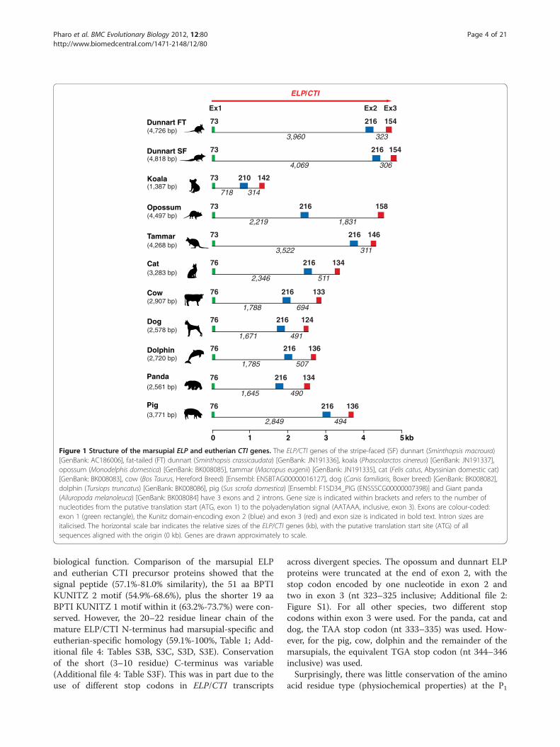
exons and 2 introns (Figure 1). The marsupial ELP generanged from ~1.4 kb for the koala to ~4.8 kb for the stripefaced dunnart, whilst eutherian ELP spanned from ~2.5 kbfor the panda to ~3.8 kb for the pig. ELP exon 1 and 2sizes respectively were highly conserved across all mam-mals (Figure 1). Exon 1 encoded the putative signal pep-tide and the first four amino acids at the N-terminus ofthe protein. The 216 bp exon 2 (with the exception of thekoala, 210 bp) encoded the remainder of the N-terminalregion, plus a single BPTI-Kunitz domain towards its 3'-end. ELP/CTI exon 3 differed most and encoded amaximum of seven amino acids. The ELP/CTI transcripts(putative translation start site to the polyadenylation sig-nal, inclusive) were short. Marsupial ELP and eutherianCTI transcripts ranged from 425–447 bp and 416–428 bprespectively and shared 56.1%-63.6% similarity at the nu-cleotide level (Additional file 2: Figure S1; Additional file3: Tables S2A, S2B). A highly conserved marsupial-specificregion (87%-100%) was also identified within the ELP 3'-UTR (nt 420–475, Additional file 2: Figure S1; Additionalfile 3: Table S2C).Based upon signal peptide analysis [56], the putative
ELP/CTI peptides identified in this study were predictedto be secreted in milk, as for tammar and possum ELPand bovine CTI [20,25,26,31]. The mature ELP and CTIpeptides shared 44.6%-62.2% similarity (Table 1; Add-itional file 4: Table S3A). In addition, the conservation ofthe two Kunitz domain motifs in all species suggestedthey may inhibit the S1 family of serine endopeptidaseslike many other members of the BPTI-Kunitz family[43,44]. The BPTI KUNITZ 2 motif [C1-C6, C2-C4 andC3-C5, Prosite: PS00280] indicates the 3 disulphidebonds which determine the structure of the domain (Fig-ure 2). This motif spanned the entire 51 amino acidKunitz domain (aa 23–73, C23-C73, C32-C56 and C48-C69, Figure 2). The second shorter motif BPTI KUNITZ1 [F-x(2)-{I}-G-C-x(6)-[FY]-x(5)-C; where x representsany residue, those within square brackets are permitted,but those within curly brackets are not, Prosite:PS00280] was located within BPTI KUNITZ 2 (aa 51–69,Figure 2). A putative trypsin interaction site within theKunitz domain (from KU NCBI cd00109) [57], is alsodepicted (aa 30–34, 36, Figure 2).Conserved amino acid residues within a protein pro-
vide an indication of sites essential for its structure and
Pig 76 216
2,849 494
136(3,771 bp)
Koala
Opossum
Dog
Cow
Tammar
Cat
Dolphin
76
73
73
73
76
76
76 216
216
216
216
216
216
210 142
158
718 314
2,219 1,831
146
3,522 311
2,346 511
134
133
1,671 491
136
124
1,788 694
1,785 507
ELP/CTI
0 2 4 531 kb
Ex1 Ex2 Ex3
(1,387 bp)
(4,497 bp)
(4,268 bp)
(3,283 bp)
(2,907 bp)
(2,578 bp)
(2,720 bp)
Dunnart FT 73 216
73 216
154
154
3233,960
4,069 306
Dunnart SF
(4,726 bp)
(4,818 bp)
Panda
1,645 490
76 216 134(2,561 bp)
Figure 1 Structure of the marsupial ELP and eutherian CTI genes. The ELP/CTI genes of the stripe-faced (SF) dunnart (Sminthopsis macroura)[GenBank: AC186006], fat-tailed (FT) dunnart (Sminthopsis crassicaudata) [GenBank: JN191336], koala (Phascolarctos cinereus) [GenBank: JN191337],opossum (Monodelphis domestica) [GenBank: BK008085], tammar (Macropus eugenii) [GenBank: JN191335], cat (Felis catus, Abyssinian domestic cat)[GenBank: BK008083], cow (Bos Taurus, Hereford Breed) [Ensembl: ENSBTAG00000016127], dog (Canis familiaris, Boxer breed) [GenBank: BK008082],dolphin (Tursiops truncatus) [GenBank: BK008086], pig (Sus scrofa domestica) [Ensembl: F1SD34_PIG (ENSSSCG00000007398)] and Giant panda(Ailuropoda melanoleuca) [GenBank: BK008084] have 3 exons and 2 introns. Gene size is indicated within brackets and refers to the number ofnucleotides from the putative translation start (ATG, exon 1) to the polyadenylation signal (AATAAA, inclusive, exon 3). Exons are colour-coded:exon 1 (green rectangle), the Kunitz domain-encoding exon 2 (blue) and exon 3 (red) and exon size is indicated in bold text. Intron sizes areitalicised. The horizontal scale bar indicates the relative sizes of the ELP/CTI genes (kb), with the putative translation start site (ATG) of allsequences aligned with the origin (0 kb). Genes are drawn approximately to scale.
Pharo et al. BMC Evolutionary Biology 2012, 12:80 Page 4 of 21http://www.biomedcentral.com/1471-2148/12/80
biological function. Comparison of the marsupial ELPand eutherian CTI precursor proteins showed that thesignal peptide (57.1%-81.0% similarity), the 51 aa BPTIKUNITZ 2 motif (54.9%-68.6%), plus the shorter 19 aaBPTI KUNITZ 1 motif within it (63.2%-73.7%) were con-served. However, the 20–22 residue linear chain of themature ELP/CTI N-terminus had marsupial-specific andeutherian-specific homology (59.1%-100%, Table 1; Add-itional file 4: Tables S3B, S3C, S3D, S3E). Conservationof the short (3–10 residue) C-terminus was variable(Additional file 4: Table S3F). This was in part due to theuse of different stop codons in ELP/CTI transcripts
across divergent species. The opossum and dunnart ELPproteins were truncated at the end of exon 2, with thestop codon encoded by one nucleotide in exon 2 andtwo in exon 3 (nt 323–325 inclusive; Additional file 2:Figure S1). For all other species, two different stopcodons within exon 3 were used. For the panda, cat anddog, the TAA stop codon (nt 333–335) was used. How-ever, for the pig, cow, dolphin and the remainder of themarsupials, the equivalent TGA stop codon (nt 344–346inclusive) was used.Surprisingly, there was little conservation of the amino
acid residue type (physiochemical properties) at the P1
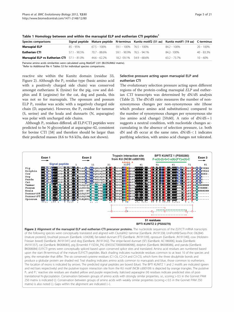
Table 1 Homology between and within the marsupial ELP and eutherian CTI peptides1
Species comparisons Signal peptide Mature peptide N-terminus Kunitz motif2 (51 aa) Kunitz motif1 (19 aa) C-terminus
Marsupial ELP 85 - 95% 67.5 - 100% 59.1 - 100% 76.5 - 100% 84.2 - 100% 20 - 100%
Eutherian CTI 57.1 - 90.5% 70.7 - 88.6% 59.1 - 90.9% 76.5 - 94.1% 84.2- 100% 40 - 83.3%
Marsupial ELP vs Eutherian CTI 57.1 - 81.0% 44.6 - 62.2% 18.2 -59.1% 54.9 - 68.6% 63.2 - 73.7% 10 - 60%
Pairwise amino acids similarities were calculated using MatGAT 2.01 (BLOSUM62 matrix).1Refer to Additional file 4: Tables S3 for individual species comparisons.
Pharo et al. BMC Evolutionary Biology 2012, 12:80 Page 5 of 21http://www.biomedcentral.com/1471-2148/12/80
reactive site within the Kunitz domain (residue 33,Figure 2). Although the P1 residue type (basic amino acidwith a positively charged side chain) was conservedamongst eutherians: K (lysine) for the pig, cow and dol-phin and R (arginine) for the cat, dog and panda, thiswas not so for marsupials. The opossum and possumELP P1 residue was acidic with a negatively charged sidechain (D, aspartate). However, the P1 residue for tammar(S, serine) and the koala and dunnarts (N, asparagine)was polar with uncharged side chains.Although P1 residues differed, all ELP/CTI peptides were
predicted to be N-glycosylated at asparagine-42, consistentfor bovine CTI [58] and therefore should be larger thantheir predicted masses (8.6 to 9.6 kDa, data not shown).
1 11 21
Exon 1 Exon 2
+-----
-- -----
PigDolphinCow MDogPandaCat MKFSLFLALCFPTammar MKFT-Possum MKFT-Koala MKFT-IVALCFALSLAGLTSSEKLSD--DunnartSF MKFT-DunnartFT MKFT-Opossum MKFT-
**:: :** : * *::* : :: .: :* *
----
LLPP
MKLSLSLALCLTLCLPGMASSGKTLASLKQEASQELFQTPPALCQLPPVMKLSRLLALCLTLCLVGLASSGKTSANLQQEASQELLQTPPALCQLPAV
MKFSPFLALCFLLCLVGISSSEKASAHLKHEAPRELSQALPAMCQLRPAFCLVGIASSEKTSAHLEREAPQELLQTLPALCRLPPV
IIALCLALSLVGMTSSEKLLDRIRANSLENLSRLVPSLCLLPSGHVNSLENPYQLVPSLCLLSPA
IIALCFAFSLAGMTSSEKLLDQIPVNSLENPSRLVPALCQLSPQIIALFFAFSLAGMTSSEKLLDQIPMNSLENPSRLVPALCQLSPQ
IVALYFALSLAGMTSSEKCLDQIQVNSLENLSLLVPSLCLLPPV
MKFSLFLALCFLLGLVGITSLEKASAHLRQEAFQELSQTLPVLCQLPPG
IVALCFALGLAGITSSEEVLEQNPLNTQENPVPLVLPLC
KLSCLLALCLTPCLVGLASSGETSDNLKQEASQDLFQTPPDLCQLPQA
C
Signal peptide
Trypsfrom K
Figure 2 Alignment of the marsupial ELP and eutherian CTI precursorof the following species were conceptually translated and aligned with Clu(mature protein)], brushtail possum [GenBank: U34208], fat-tailed dunnart (FFriesian breed) [GenBank: JN191341] and dog [GenBank: JN191342]. The strJN191337], cat [GenBank: BK008083], pig [Ensembl: F1SD34_PIG (ENSSSCT00BK008084] ELP/CTI genes were conceptually spliced based upon conservedupon the start (N-terminus) of the mature ELP/CTI peptides. Black shadinggrey, the remainder that differ. The six conserved cysteine residues (C1-C6,produce a globular protein are shaded red. Teal shading indicates amino aThe location of exons is indicated by arrows. The predicted signal peptidesand red bars respectively) and the putative trypsin interaction site from theP1 and P1' reactive site residues are shaded yellow and purple respectively.translational N-glycosylation. Conservation between groups of amino acids250 matrix is indicated (:). Conservation between groups of amino acids wimatrix) is also noted (.). Gaps within the alignment are indicated (−).
Selective pressure acting upon marsupial ELP andeutherian CTIThe evolutionary selection pressure acting upon differentregions of the protein-coding marsupial ELP and euther-ian CTI transcripts was determined by dN/dS analysis(Table 2). The dN/dS ratio measures the number of non-synonymous changes per non-synonymous site (thosewhich produce amino acid substitutions) compared tothe number of synonymous changes per synonymous site(no amino acid change) [59,60]. A ratio of dN/dS = 1suggests a neutral condition, with nucleotide changes ac-cumulating in the absence of selection pressure, i.e. bothdN and dS occur at the same rates. dN/dS< 1 indicatespurifying selection, with amino acid changes not tolerated.
31 41 51 61 81
: *:*: ** * * **.** *:*:: * : *.
BPTI KUNITZ 2 (PS50279)51 residues
#
P1- 'P1
Exon 3BPTI KUNITZ 1 (PS00280)
F-x(2)-{I}-G-C-x(6)-[FY]-x(5)-C
--------------------------------------------+
--- #-------------- ------- ------------ --- --
----+-----------------------+
--
+--------------------+----
FYNSTSSACEP -------------
-
---------------------
* * :*ERGNCDSLNLRY
GGPCKASLRRYRGPCKASLHRY
KGPCRGLFYRYEGPCRGRFYRY
RGNCDSQILRYRGNCNSQTLRYRGNCNDNIRRYRGNCNDNIRRY
RGNCSSQILHY
KGPCRGRFYRYRGPCKAALLRY
C C C C
--
C
in interaction siteU (NCBI cd00109)
71
FYNSTSIECEPFTYGGCQGNANNFETTEICVRVCKPPETKVKSSFTYGGCQGNDNNFETTEMCLRICQPPETEDKS
FYNSTSSECEHFIYGGCQGNANNFETTEICLKICKPPETR
FYNTTSRTCETFIYSGCNGNRNNFNSEEYCLKTCRRNKNRNNNNFYNATSHTCEVFLYSGCNGNGNNFDSLECCLKTCRLNKYRNNNFYNTTSRTCEAFIYSGCHGNGNNFDSLQCCLKTCRPNKNRNDNN
YYNTTSRICEEFIYTGCNGNGNNFDSVECCLKTCKLN
FYNSTSAECELFMYGGCQGNANNFETTAICRRVCNPPDTKVKNG
FYNSTSRLCEAFIYSGCNGNGNNFDTVECCLKTCRPN
YYNTTSRICEEFIYTGCNGNGNNFDSVECCLKTCKLN
FSNSTSSECEHFTYGGCQGNANDFETTEICSRICKPPETGFYNSTAHECEHFTYGGCRGNANNFETTEMCLKVCKPPGTR
proteins. The nucleotide sequences of the ELP/CTI mRNA transcriptsstalW2: tammar [GenBank: JN191338; UniProtKB/Swiss-Prot: O62845T) [GenBank: JN191339], opossum [GenBank: JN191340], cow (Holstein-ipe-faced dunnart (SF) [GenBank: AC186006], koala [GenBank:000008098)], dolphin [GenBank: BK008086], and panda [GenBank:splice sites and translated. Amino acid residues are numbered basedindicates nucleotide residues common to at least 10 of the species andC2-C4 and C3-C5), which form the three disulphide bonds andcids common to marsupials and blue, those common to eutherians.are boxed (blue). The BPTI KUNITZ 1 and 2 motifs are indicated (greenKU motif (NCBI cd00109) is depicted by orange triangles. The putativeItalicised asparagine (N) residues indicate predicted sites of post-with strongly similar properties, i.e., scoring> 0.5 in the Gonnet PAMth weakly similar properties (scoring< 0.5 in the Gonnet PAM 250

Pharo et al. BMC Evolutionary Biology 2012, 12:80 Page 6 of 21http://www.biomedcentral.com/1471-2148/12/80
In contrast, dN/dS> 1 is indicative of positive Darwinianselection for amino acid changes [59,61].The protein-coding marsupial ELP and eutherian CTI
transcripts and regions within them generally exhibited atrend towards purifying selection, with a dN/dS ratio <1(Table 2). However, based upon codon-based Z-tests, onlythe eutherian CTI BPTI KUNITZ 1 motif (57 nt encoding19 amino acids) was found to be undergoing purifying se-lection (p< 0.05). Although the regions encoding the mar-supial BPTI KUNITZ 1 motif (p = 0.103) and themarsupial and eutherian BPTI KUNITZ 2 motifs(p= 0.101 and p= 0.105 respectively) exhibited a strongtrend towards purifying selection, the test values (dN< dS)were not significant. This tendency was also consistent forthe putative trypsin interaction site. In contrast, threeregions of the ELP/CTI transcripts showed a trend towardspositive selection (dN/dS> 1). These included the regionsencoding the ELP/CTI N-terminus and the eutherian CTIsignal peptide. However, based upon codon-based Z-tests(dN> dS), only the eutherian CTI signal peptide (p< 0.05)was undergoing positive selection.
Marsupial ELP and eutherian CTI share common flankinggenesIn order to confirm that the marsupial ELP and eutherianCTI genes were orthologous, we characterised the locationand arrangement of ELP/CTI and its flanking genes. Weused fluorescence in situ hybridisation to map tammar ELP
Table 2 Average rates of synonymous (dS) and non-synonymeutherian CTI
ELP/CTI protein-codingregion
dN SE dS SE
Precursor protein Marsupials 0.145 0.022 0.190 0.0
Eutherians 0.194 0.026 0.225 0.0
Mature protein Marsupials 0.166 0.026 0.185 0.0
Eutherians 0.186 0.028 0.242 0.0
Signal peptide Marsupials 0.071 0.029 0.226 0.0
Eutherians 0.225 0.072 0.165 0.0
N-terminus Marsupials 0.240 0.064 0.116 0.0
Eutherians 0.242 0.050 0.224 0.0
BTPI KUNITZ 2# Marsupials 0.146 0.031 0.224 0.0
Eutherians 0.162 0.035 0.243 0.0
BPTI KUNITZ 1~ Marsupials 0.095 0.030 0.223 0.0
Eutherians 0.066 0.026 0.264 0.1
Trypsin interaction site^ Marsupials 0.230 0.136 0.323 0.1
Eutherians 0.175 0.093 0.228 0.1#PS50279 153 nt, 51 aa.~PS00280 57 nt, 19 aa.^18 nt, 6 aa site from KU (NCBI cd00109).+Codon based Z-tests in MEGA5.*p< 0.05.{ NS not significant.
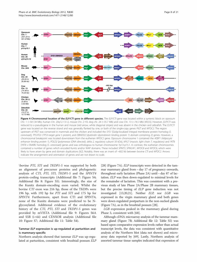
to chromosome 1q (Figure 3). The ELP/CTI gene waslocated on a syntenic segment in the marsupial (stripe-faceddunnart [27] and opossum) and eutherian genomes [49,55]and was generally flanked by one or both of the single-copygenes phosphatidyl inositol glycan, class T (PIGT) and WAPfour disulphide core domain 2 (WFDC2), confirming theywere true orthologues (Figure 4).The PIGT-WFDC2 region of bovine chromosome 13
(~74.51-75.14 Mb) was unique. Bovine CTI was adjacent toPIGT, but there was an insertion of ~602 kb between theCTI and WFDC2 genes [49,55] (data not shown). This re-gion included 7 Artiodactyla-specific Kunitz domain-en-coding genes including PTI, STI, plus the five placenta-specific TKDP1-TKDP5 genes inclusive [50,63]. Further-more, the SPINLW1 gene which contains both a Kunitzand a WAP domain and the eutherian-specific SPINT4gene were located a further ~38 kb and ~90 kb respectivelydownstream from WFDC2 [49,55] (data not shown). Asmentioned previously, these genes, with the exception ofSPINLW1 and the TKDPs, share a similar 3-exon structure.However, the TKDPs differ due to the likely “exonisation”of an intron and its subsequent duplication to produce avariable number of tripartite N-domains between the exonencoding the signal peptide and the Kunitz domain [50,51].
CTI has been lost in some eutheriansUsing the canine sequence as the basis for mVISTAcomparative analysis [64], the region between the PIGT
ous (dN) substitutions occurring in marsupial ELP and
dN/dSRatio (a) Neutralselection test(dN 6¼dS)+*
(b) Purifyingselection test(dN<dS)+*
(c) Positiveselection test(dN>dS)+*
33 0.763 0.256 (NS{) 0.117 (NS) 1.000 (NS)
33 0.862 0.232 (NS) 0.472 (NS) 1.000 (NS)
36 0.897 0.653 (NS) 0.334 (NS) 1.000 (NS)
39 0.786 0.273 (NS) 0.130 (NS) 1.000 (NS)
94 0.314 0.133 (NS) 0.064 (NS) 1.000 (NS)
69 1.36 0.451 (NS) 1.000 (NS) 0.224 (NS)
48 2.07 0.064 (NS) 1.000 (NS) 0.041*
65 1.08 0.842 (NS) 1.000 (NS) 0.424 (NS)
52 0.651 0.215 (NS) 0.101 (NS) 1.000 (NS)
54 0.667 0.200 (NS) 0.105 (NS) 1.000 (NS)
98 0.426 0.212 (NS) 0.103 (NS) 1.000 (NS)
10 0.250 0.122 (NS) 0.046* 1.000 (NS)
81 0.712 0.740 (NS) 0.363 (NS) 1.000 (NS)
31 0.768 0.689 (NS) 0.345 (NS) 1.000 (NS)

1q
1q
Figure 3 Localisation of the tammar ELP gene to Macropuseugenii chromosome 1q using FISH.
Pharo et al. BMC Evolutionary Biology 2012, 12:80 Page 7 of 21http://www.biomedcentral.com/1471-2148/12/80
and WFDC2 genes was examined using the availablegenome assemblies - which have variable sequencecoverage, contain gaps and may contain misassembledsequences. Whilst the ELP/CTI gene was present insome mammals, it appeared to have become a disruptedpseudogene in others such as the African Savanna ele-phant and human (Figure 5). Exon 1 of the elephant andhuman CTI genes (signal- and pro-peptide) was present,but exon 2 (Kunitz domain) and exon 3 (C-terminus)were absent (red boxes, Figure 5), suggesting they hadbeen excised or transposed, whilst the horse and mouseCTI genes initially appeared intact.A closer examination of the nucleotide sequence be-
tween PIGT and WFDC2 in these and other species usingthe Ensembl and UCSC genome databases revealed thatdifferent mutations had most likely disrupted the CTI gene.Exon 1 was disrupted in the elephant, Hoffmann's two-toed sloth (Choloepus hoffmanni), armadillo (Dasypusnovemcinctus), human and other primates and horse, withexon 2 (Kunitz domain) also excised for these species,apart from the horse. Additional file 5: Figure S2A (i)depicts a nucleotide alignment of the functional/protein-coding dog CTI exon 1 compared with the putative dis-rupted CTI exon 1 of the elephant, sloth, human andhorse. Additional file 5: Figure S2A (ii) shows the trans-lated sequences to highlight mutations and/or deletionswithin the signal peptide region of CTI. The deletion oftwo nucleotides within human CTI exon 1 would producea frame-shift (as depicted by the +1 and +2 reading
frames). CTI exon 2 of the mouse, rat, large flying fox(Pteropus vampyrus) and horse also appeared to have beendisrupted by deletions resulting in frame-shifts when com-pared to the functional/protein-coding dog CTI exon 2.The disruption of the protein-coding region of equine CTIexons 1 and 2 by at least one mutation and one deletionrespectively would produce a frame-shift, suggested thesewere a recent occurrence (Additional file 5: Figure S2B(ii)).
Transposable elements within the ELP/CTI genesTransposable elements integrate randomly into the gen-ome, so the probability of the same element(s) integrat-ing independently into orthologous positions in differentspecies is extremely low. They therefore act as geneticmarkers and can be used to determine the phylogeneticrelationship between genes and species [65]. Further evi-dence that marsupial ELP and eutherian CTI evolvedfrom a common ancestral gene was provided by CEN-SOR retrotransposon analysis [66] (Additional file 6: Fig-ure S3). Retroelements of conserved fragment size andorientation were located within the PIGT-ELP/CTI re-gion. However, the elephant and human which appear tohave lost CTI exons 2 and 3, had also lost retrotranspo-sons in the corresponding region, but gained a MER5Aelement.
Bovine CTI, PTI, STI and the TKDPs share a commonancestral geneThe location of the 8 Kunitz-domain encoding genes (in-cluding CTI) on bovine chromosome 13 between the PIGTand WFDC2 genes and the Artiodactyla-specific distribu-tion of PTI, STI and TKDP1-5 (cow and sheep [51,63]) sug-gested they may have evolved from CTI. This hypothesiswas supported by phylogenetic analysis of the protein-cod-ing regions of the mammalian ELP/CTI, bovine PTI, STIand TKDP1-5 transcripts, with bovine SLPI used as an out-group root (SLPI omitted, Figure 6). Several different meth-ods in PHYLIP were used to determine the evolutionaryrelationships. These included the character-based max-imum-likelihood (with/without a molecular clock) andmaximum parsimony, as well as distance-based analysis(Fitch-Margoliash tree method using the Kimura distancemodel of nucleotide substitution). Trees were evaluatedusing the bootstrap method (100 replicates). Of the algo-rithms used, the maximum likelihood method using a mo-lecular clock assumption, which assumes a constantevolutionary rate for all species, produced a tree with thehighest bootstrap values. Huttley and colleagues [67] haveshown that the eutherian nucleotide substitution rates are~30% slower than for marsupials. However, all methodsproduced consensus trees which consistently separated the19 sequences into the two groups depicted (Figure 6). Thehypothesis that bovine CTI was the ancestral gene for
OpossumChr. 1(501.25 to501.67Mb)
DogChr. 24(35.64 to35.89 kb)
CowChr. 13(74.48 to75.20 kb)
HumanChr. 7p13-p15.1
HumanChr. 20q12-q13.12
MouseChr. 2H3
ChickenChr. 20 (-)(5.08 to4.93 Mb)
ZebrafishChr. 23(12.27 to12.72 Mb)
SPINT3
WFDC8LOC
611069 CE10
LOC100130157
RPL5P2 WFDC6SYS1-DBNDD2
Gm14317
HSPD1P21PIGT WFDC2
TP53TG5SYS1
DBNDD2
PigTTp53tg5Sys1 Dbndd2 Spint3 Spinlw1Wfdc6aWfdc2
PIGT CTITP53TG5SYS1 DBNDD2 WFDC2
PIGT CTITP53TG5SYS1 DBNDD2 WFDC2
LOC611096
SPINLW1
SPINLW1
PIGT WFDC2ELP AEBP1 POLD2 MYL7TP53TG5SYS1 DBNDD2 GCK YKT6
AEBP1 POLD2 MYL7 GCK YKT6
~602 kbp
PIGTTP53TG5 DBNDD2SYS1LOC419
182 SRC BLCAP EMILIN3MANBAL GHRH
PIGT snx21 haus8wfdc2si:zfos-452g4.1sycp2 phactr3appplr3da
LOC100128997
CTI
Cti
Figure 4 Chromosomal location of the ELP/CTI gene in different species. The ELP/CTI gene was located within a syntenic block on opossumChr. 1 (~501.34 Mb), human Chr. 20q12-13.12, mouse Chr. 2 H3, dog chr. 24 (~35.7 Mb) and cow Chr. 13 (~74.5 Mb) [49,55]. However, ELP/CTI wasreduced to a pseudogene in the human and mouse (red arrow, white diagonal stripes) and was absent in the chicken and zebrafish. The ELP/CTIgene was located on the reverse strand and was generally flanked by one, or both of the single-copy genes PIGT and WFDC2. The regionupstream of PIGT was conserved in mammals and the chicken and included the SYS1 [Golgi-localized integral membrane protein homolog (S.cerevisiae)], TP53TG5 (TP53-target gene 5 protein), and DBNDD2 [dysbindin (dystrobrevin binding protein 1) domain containing 2] genes. However, achromosomal breakpoint was located downstream from the eutherian WFDC2 gene. Opossum chromosome 1 contained the AEBP1 (Adipocyteenhancer binding protein 1), POLD2 [polymerase (DNA directed), delta 2, regulatory subunit 50 kDa], MYL7 (myosin, light chain 7, regulatory) and YKT6[YKT6 v-SNARE homolog (S. cerevisiae)] genes and was orthologous to human chromosome 7p13-p15.1. In contrast, the eutherian chromosomescontained a number of genes which encoded Kunitz and/or WAP domains. These included SPINT3, SPINLW1, WFDC8 and WFDC6, which werelikely to have arisen by gene and domain duplications [62]. Notably, there was an insert of ~602 kb between bovine CTI and WFDC2. Arrowsindicate the arrangement and orientation of genes and are not drawn to scale.
Pharo et al. BMC Evolutionary Biology 2012, 12:80 Page 8 of 21http://www.biomedcentral.com/1471-2148/12/80
\bovine PTI, STI and TKDP1-5 was supported by bothan alignment of precursor proteins and phylogeneticanalysis of CTI, PTI, STI, TKDP1-5 and the SPINT4protein-coding transcripts (Additional file 7: Figure S4;Additional file 8: Figure S5). Interestingly, the size ofthe Kunitz domain-encoding exon varied. Whilst thebovine CTI exon was 216 bp, those of the TKDPs were196 bp, with 192 bp for PTI and STI and 175 bp forSPINT4. Furthermore, apart from CTI and SPINT4,none of the Kunitz domains were predicted to be N-glycosylated. Additional evidence of the evolutionaryhistory of the CTI, PTI, STI and TKDP1-5 genes wasprovided by mVISTA (Additional file 9: Figures S6Aand S5B (i-viii) and CENSOR analysis (Additional file10: Figure S7; Additional file 11: Table S4).
Tammar ELP expression is up-regulated at parturition andis mammary-specificNorthern analysis showed that tammar ELP was up-regu-lated at parturition, consistent with brushtail possum ELP
[28] (Figure 7A). ELP transcripts were detected in the tam-mar mammary gland from~day 17 of pregnancy onwards,throughout early lactation (Phase 2A) until ~day 87 of lac-tation. ELP was then down-regulated to minimal levels forthe remainder of lactation. This was consistent with a pre-vious study of late Phase 2A/Phase 2B mammary tissues,but the precise timing of ELP gene induction was notinvestigated [13,20,21]. Neither ELP, nor LGB wasexpressed in the virgin mammary gland and both geneswere down-regulated postpartum in the non-sucked glands(Figure 7A), as in the brushtail possum [28].LGB expression peaked in the mammary gland during
Phase 3, consistent with [68].Although cDNA microarray analysis of the tammar mam-
mary gland (Figure 7B; Additional file 12: Table S5) wasbased upon comparative expression levels rather than actualtranscript levels, the data was consistent with quantitativeanalysis of the Northern blot (data not shown) and micro-array data reported by [69]. Lastly, Northern analysis ofassorted tammar tissue samples indicated that expression of

Dog
Cow
Elephant
Horse
Mouse
Human
Opossum
WFDC2
35,682 35,684 35,686 35,688 35,690 35,692 35,694 35,696 kbp
gene exon UTRCNS
Dog
Cow
Elephant
Horse
Mouse
Human
Opossum
100%
10%100%
10%
100%
10%
100%
10%100%
10%100%
10%
100%
10%100%
10%
100%
10%
100%
10%100%
10%100%
10%
35,698 35,700 35,702 35,704 35,706 35,708 35,710 35,712 kbp
~602 kbp
PIGT ELP/CTI
Figure 5 VISTA plot of pairwise alignments for selected mammals in the region containing the PIGT, ELP/CTI and WFDC2 genes.Sequence homology within the PIGT-ELP/CTI-WFDC2 region of the dog, cow, elephant, horse, human, mouse and opossum genomes wasdetermined with mVISTA [64]. The dog sequence was used as the reference sequence (horizontal axis, dog chromosome 24 numbering). Greyhorizontal arrows indicate gene location and direction of transcription. Blue rectangles indicate coding exons and untranslated regions (UTRs) ofthe gene are depicted by light green rectangles. Exon 1 of canine WFDC2 was missing (gap in the current assembly) from the dog genome and isindicated by a blue rectangle with diagonal white stripes. The right axis indicates the percentage identity within a 100 bp window for eachpairwise comparison, ranging from 10% to 100%. Regions sharing greater than 25% identity are shaded and the black horizontal line indicates70% identity. The region containing the Kunitz domain-encoding ELP/CTI exon 2 was conserved in the cow, horse, mouse and opossum, but wasabsent in the elephant and human CTI genes (red boxes).
Pharo et al. BMC Evolutionary Biology 2012, 12:80 Page 9 of 21http://www.biomedcentral.com/1471-2148/12/80
ELP and LGB was mammary gland-specific (Figure 8), un-like the ubiquitously expressed cystatin C (CST3) gene(data not shown).
DiscussionELP was originally thought to be a marsupial-specific gene[19]. However, we have shown that the marsupial ELP andeutherian CTI genes evolved from a common therian an-cestral gene (Figure 9). Mammalian ELP/CTI was generallyflanked by one or both of the single copy PIGT andWFDC2 genes in a region that was syntenic to that ofother mammals. The conserved genomic structure of 3exons and 2 introns and homologous transposable elementfragments confirmed that ELP and CTI were true ortholo-gues. CTI was also identified as the putative ancestral geneof the ruminant-specific PTI, STI and TKDP1-5 genes.Based upon current genome sequencing and assemblies,ELP/CTI was not found in birds, fish, reptiles, nor amphi-bians, suggesting the gene was present in the therian
ancestor before the divergence of marsupials and euther-ians at least 130 million years ago [1,2,70].
Mammalian ELP/CTI and the evolution of bovine PTI, STIand the TKDPsThe Kunitz-type inhibitor domain has been duplicatedmany times throughout evolutionary history [38]. Thiswas no more evident than for the region of bovinechromosome 13 on which CTI and the 7 CTI-like geneswere located. The PTI, STI and TKDP1-5 genes werespecific to the order Cetartiodactyla, sub-order Rumi-nantia [50,51,63,72], strong evidence they evolved fromCTI after the divergence of the Ruminantia ~25-35 MYA[1]. The CTI, PTI and STI genes had a similar 3-exonstructure and conserved regions within both coding andnon-coding segments. The PTI and STI genes and pro-teins were homologous and almost certainly arose bygene duplication [73]. However, the TKDP1-5 geneshad one or more additional exons inserted between
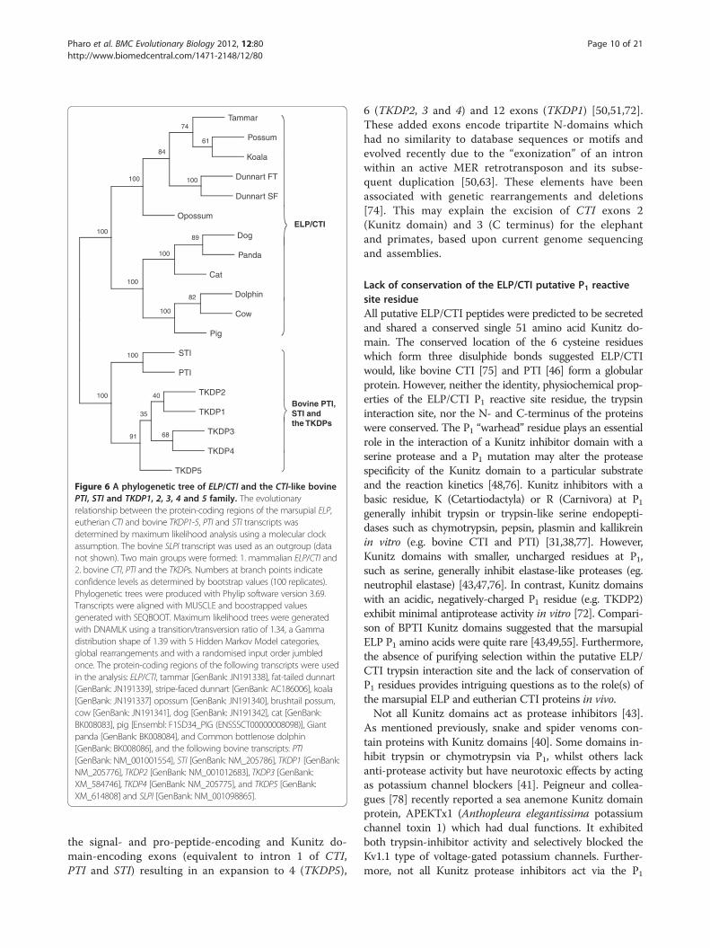
Tammar
Possum
Koala
Dunnart FT
Dunnart SF
Opossum
Dog
Panda
Cat
Dolphin
Cow
Pig
STI
PTI
TKDP2
TKDP1
TKDP3
TKDP4
TKDP5
74
61
84
100
100
100
100
100
91
35
40
68
89
100
100
82
100
ELP/CTI
Bovine PTI,STI andthe TKDPs
Figure 6 A phylogenetic tree of ELP/CTI and the CTI-like bovinePTI, STI and TKDP1, 2, 3, 4 and 5 family. The evolutionaryrelationship between the protein-coding regions of the marsupial ELP,eutherian CTI and bovine TKDP1-5, PTI and STI transcripts wasdetermined by maximum likelihood analysis using a molecular clockassumption. The bovine SLPI transcript was used as an outgroup (datanot shown). Two main groups were formed: 1. mammalian ELP/CTI and2. bovine CTI, PTI and the TKDPs. Numbers at branch points indicateconfidence levels as determined by bootstrap values (100 replicates).Phylogenetic trees were produced with Phylip software version 3.69.Transcripts were aligned with MUSCLE and boostrapped valuesgenerated with SEQBOOT. Maximum likelihood trees were generatedwith DNAMLK using a transition/transversion ratio of 1.34, a Gammadistribution shape of 1.39 with 5 Hidden Markov Model categories,global rearrangements and with a randomised input order jumbledonce. The protein-coding regions of the following transcripts were usedin the analysis: ELP/CTI, tammar [GenBank: JN191338], fat-tailed dunnart[GenBank: JN191339], stripe-faced dunnart [GenBank: AC186006], koala[GenBank: JN191337] opossum [GenBank: JN191340], brushtail possum,cow [GenBank: JN191341], dog [GenBank: JN191342], cat [GenBank:BK008083], pig [Ensembl: F1SD34_PIG (ENSSSCT00000008098)], Giantpanda [GenBank: BK008084], and Common bottlenose dolphin[GenBank: BK008086], and the following bovine transcripts: PTI[GenBank: NM_001001554], STI [GenBank: NM_205786], TKDP1 [GenBank:NM_205776], TKDP2 [GenBank: NM_001012683], TKDP3 [GenBank:XM_584746], TKDP4 [GenBank: NM_205775], and TKDP5 [GenBank:XM_614808] and SLPI [GenBank: NM_001098865].
Pharo et al. BMC Evolutionary Biology 2012, 12:80 Page 10 of 21http://www.biomedcentral.com/1471-2148/12/80
the signal- and pro-peptide-encoding and Kunitz do-main-encoding exons (equivalent to intron 1 of CTI,PTI and STI) resulting in an expansion to 4 (TKDP5),
6 (TKDP2, 3 and 4) and 12 exons (TKDP1) [50,51,72].These added exons encode tripartite N-domains whichhad no similarity to database sequences or motifs andevolved recently due to the “exonization” of an intronwithin an active MER retrotransposon and its subse-quent duplication [50,63]. These elements have beenassociated with genetic rearrangements and deletions[74]. This may explain the excision of CTI exons 2(Kunitz domain) and 3 (C terminus) for the elephantand primates, based upon current genome sequencingand assemblies.
Lack of conservation of the ELP/CTI putative P1 reactivesite residueAll putative ELP/CTI peptides were predicted to be secretedand shared a conserved single 51 amino acid Kunitz do-main. The conserved location of the 6 cysteine residueswhich form three disulphide bonds suggested ELP/CTIwould, like bovine CTI [75] and PTI [46] form a globularprotein. However, neither the identity, physiochemical prop-erties of the ELP/CTI P1 reactive site residue, the trypsininteraction site, nor the N- and C-terminus of the proteinswere conserved. The P1 “warhead” residue plays an essentialrole in the interaction of a Kunitz inhibitor domain with aserine protease and a P1 mutation may alter the proteasespecificity of the Kunitz domain to a particular substrateand the reaction kinetics [48,76]. Kunitz inhibitors with abasic residue, K (Cetartiodactyla) or R (Carnivora) at P1generally inhibit trypsin or trypsin-like serine endopepti-dases such as chymotrypsin, pepsin, plasmin and kallikreinin vitro (e.g. bovine CTI and PTI) [31,38,77]. However,Kunitz domains with smaller, uncharged residues at P1,such as serine, generally inhibit elastase-like proteases (eg.neutrophil elastase) [43,47,76]. In contrast, Kunitz domainswith an acidic, negatively-charged P1 residue (e.g. TKDP2)exhibit minimal antiprotease activity in vitro [72]. Compari-son of BPTI Kunitz domains suggested that the marsupialELP P1 amino acids were quite rare [43,49,55]. Furthermore,the absence of purifying selection within the putative ELP/CTI trypsin interaction site and the lack of conservation ofP1 residues provides intriguing questions as to the role(s) ofthe marsupial ELP and eutherian CTI proteins in vivo.Not all Kunitz domains act as protease inhibitors [43].
As mentioned previously, snake and spider venoms con-tain proteins with Kunitz domains [40]. Some domains in-hibit trypsin or chymotrypsin via P1, whilst others lackanti-protease activity but have neurotoxic effects by actingas potassium channel blockers [41]. Peigneur and collea-gues [78] recently reported a sea anemone Kunitz domainprotein, APEKTx1 (Anthopleura elegantissima potassiumchannel toxin 1) which had dual functions. It exhibitedboth trypsin-inhibitor activity and selectively blocked theKv1.1 type of voltage-gated potassium channels. Further-more, not all Kunitz protease inhibitors act via the P1

ELP
RNA
PREGNANCY LACTATION
LGB
⎨ ⎧⎧10
P
17P
18P
21P
26P
20L
70L
76L
80L
87L
127L
130L
163L
151L
180L
240L
260L
13P
15P
16P
20P
22P
25P
VIR
GIN
1L 2L 2L 2L N
S
3L N
S
4L N
S
10L
15L
40L
168L
Phase 1 Phase 2A Phase 2B Phase 3
⎨ ⎧⎧ ⎪ ⎪A
Lactation stage (days)
Phase 1
Day 2
Day 260
Day 35 Day 210Day 158
Ave
rag
e g
ene
exp
ress
ion
0
1.0
2.0
3.0
4.0
5.0
6.0
7.0
8.0
20 40 60 80 100 120 140 160 180 200 220 240 2600-20 280
Phase 2A Phase 2B Phase 3Virgin gland
Day 95
9.0
Day 25fetus
B
ELP LGB GAPDH
Figure 7 ELP expression in the tammar mammary gland throughout the reproductive cycle. A. Northern analysis of total RNA (10 μg)extracted from the mammary glands of tammar wallabies during pregnancy (P, Phase 1) and lactation (L, Phase 2A, Phase 2B and Phase 3), fromnon-sucked (NS) glands and from a virgin female (~220 days of age). Tammar ELP expression was undetected in the virgin gland, minimal duringpregnancy (Phase 1) and then induced at parturition and expressed during early lactation (Phase 2A). ELP was down-regulated at mid-lactation(Phase 2B), consistent with [13,20,21]. ELP transcripts were not detected in Phase 3. ELP expression also declined postpartum in non-sucked glands.Tammar LGB was used as a positive control for lactation and exhibited a similar expression pattern to ELP, but with LGB expression increased (butnot significantly so) during Phases 2B and 3, as reported previously [13,68,69]. Ribosomal RNA bands indicate RNA integrity and loading. B.Microarray analysis of the tammar mammary gland [ArrayExpress: E-MTAB-1057] supported the quantitative analysis of Northern blot (data notshown) and microarray data reported by [69]. Expression of the ELP and LGB milk protein genes and the housekeeping gene GAPDH(glyceraldehyde 3-phosphate dehydrogenase) is depicted as average normalised raw intensity based upon the expression n = 3, 7 and 2 clones oneach microarray respectively ± SEM (Additional file 12: Table S5). Whilst ELP (red) and LGB (blue) expression differed during the reproductive cycle,GAPDH (green) expression was constant.
Pharo et al. BMC Evolutionary Biology 2012, 12:80 Page 11 of 21http://www.biomedcentral.com/1471-2148/12/80
residue. The tick anticoagulant peptide (TAP) inhibits Fac-tor X, Factor Xa and thrombin but the reactive site islocated towards the N-terminus of the protein, rather thanat the P1 residue of the Kunitz domain [79].
ELP/CTI – a conserved N-glycosylation site predictedwithin the Kunitz domainAll ELP/CTI proteins shared a putative conserved N-glycosylation site within the Kunitz domain at asparagine-42 (as-
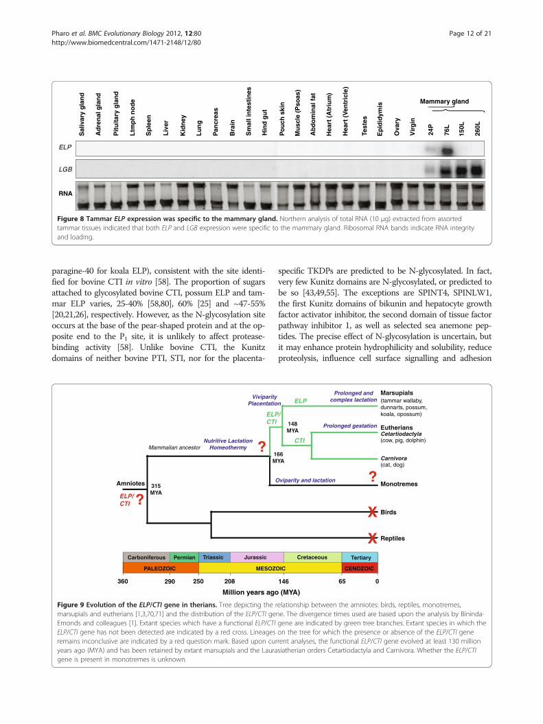
Mu
scle
(P
soas
)
Test
es
Ep
idid
ymis
Vir
gin
Ab
do
min
al f
at
Hea
rt (
Atr
ium
)
Hea
rt (
Ven
tric
le)
Ova
ry
Po
uch
ski
n
24P
76L
150L
260L
Ad
ren
al g
lan
d
Sp
leen
Liv
er
Lu
ng
Pit
uit
ary
gla
nd
Ltm
ph
no
de
Kid
ney
Sal
ivar
y g
lan
d
Pan
crea
s
Bra
in
Sm
all i
nte
stin
es
Hin
d g
ut
Mammary gland
ELP
LGB
RNA
⎨ ⎧⎧
Figure 8 Tammar ELP expression was specific to the mammary gland. Northern analysis of total RNA (10 μg) extracted from assortedtammar tissues indicated that both ELP and LGB expression were specific to the mammary gland. Ribosomal RNA bands indicate RNA integrityand loading.
Pharo et al. BMC Evolutionary Biology 2012, 12:80 Page 12 of 21http://www.biomedcentral.com/1471-2148/12/80
paragine-40 for koala ELP), consistent with the site identi-fied for bovine CTI in vitro [58]. The proportion of sugarsattached to glycosylated bovine CTI, possum ELP and tam-mar ELP varies, 25-40% [58,80], 60% [25] and ~47-55%[20,21,26], respectively. However, as the N-glycosylation siteoccurs at the base of the pear-shaped protein and at the op-posite end to the P1 site, it is unlikely to affect protease-binding activity [58]. Unlike bovine CTI, the Kunitzdomains of neither bovine PTI, STI, nor for the placenta-
Million years ago
PALEOZOIC MESOZO
JurassicTriassicPermianCarboniferous
250 208360 290
Amniotes
1M
ViviparityPlacentatio
Nutritive LactationHomeothermy
315MYA
Mammalian ancestor
O
ELPCTI
?
?ELP/CTI
Figure 9 Evolution of the ELP/CTI gene in therians. Tree depicting the rmarsupials and eutherians [1,3,70,71] and the distribution of the ELP/CTI geEmonds and colleagues [1]. Extant species which have a functional ELP/CTIELP/CTI gene has not been detected are indicated by a red cross. Lineagesremains inconclusive are indicated by a red question mark. Based upon curyears ago (MYA) and has been retained by extant marsupials and the Lauragene is present in monotremes is unknown.
specific TKDPs are predicted to be N-glycosylated. In fact,very few Kunitz domains are N-glycosylated, or predicted tobe so [43,49,55]. The exceptions are SPINT4, SPINLW1,the first Kunitz domains of bikunin and hepatocyte growthfactor activator inhibitor, the second domain of tissue factorpathway inhibitor 1, as well as selected sea anemone pep-tides. The precise effect of N-glycosylation is uncertain, butit may enhance protein hydrophilicity and solubility, reduceproteolysis, influence cell surface signalling and adhesion
(MYA)
CENOZOICIC
TertiaryCretaceous
065146
Eutherians
Marsupials
66YA
148MYA
n
Prolonged gestation
(tammar wallaby,dunnarts, possum,koala, opossum)
Monotremes
Birds
Reptiles
viparity and lactation
Cetartiodactyla(cow, pig, dolphin)
Carnivora(cat, dog)
ELP
CTI
/
X
X
?
Prolonged andcomplex lactation
elationship between the amniotes: birds, reptiles, monotremes,ne. The divergence times used are based upon the analysis by Bininda-gene are indicated by green tree branches. Extant species in which theon the tree for which the presence or absence of the ELP/CTI generent analyses, the functional ELP/CTI gene evolved at least 130 millionsiatherian orders Cetartiodactyla and Carnivora. Whether the ELP/CTI

Pharo et al. BMC Evolutionary Biology 2012, 12:80 Page 13 of 21http://www.biomedcentral.com/1471-2148/12/80
and affect protein folding, turnover and quality control [81-83]. Furthermore, oligosaccharides may act as soluble re-ceptor analogues for bacterial and viral pathogens, prevent-ing them from attaching to the wall of the intestines,thereby stopping their passage through the gastrointestinaland urinary tracts of the young [84,85].The lack of conservation of the ELP/CTI N- and
C-terminus was intriguing, particularly the positiveDarwinian selection (p< 0.05) acting upon the coil-like marsupial ELP N-terminus. In contrast, the eu-therian CTI N-terminus tended towards neutral se-lection. The N- and C-termini of proteins have beenassociated with sub-cellular targeting, protein-proteinand protein-lipid interactions and macromolecularcomplex formation [86]. The marsupial- and euther-ian-specific homology of the mature ELP/CTI N-terminus suggested these regions may have differentactivities. However, the lack of conservation of theELP/CTI C-terminus suggested these areas may havespecies-specific effects. Interestingly, the conservationof the TGA codon used by the tammar, koala, pig,dolphin and cow for all species but the cat (CGA)suggested it was the ancestral ELP/CTI stop codon,with more recent mutations producing a shortenedELP/CTI C-terminus in some species. Furthermore, aconserved marsupial-specific region within the 3'UTR may regulate ELP gene transcription.ELP/CTI is expressed and secreted in milk during the
early lactation/colostrogenesis period only [this study,[20,21,25-28,31,36,37]]. Furthermore, all mammalian neo-nates have an innate immune system but an immatureadaptive immune system and a gut which is yet to undergomaturation or ‘closure’ and is therefore permeable tomacromolecules [16,29,87-89]. For the calf, gut maturationoccurs 24–36 hr pp [16], whereas for the tammar, thisprocess does not occur until ~200 days pp [87]. Therefore,maternal milk immunoglobulins such as IgG can be pas-sively transferred via colostrum and Phase 2A/2B milk tothe gut of the young calf and tammar, respectively, wherethey are absorbed by the intestines and enter the circulatorysystem [16,89]. Hence ELP/CTI may enhance the survivalof the young by preventing the proteolytic degradation ofmaternal immunoglobulins [31], or by protecting the youngagainst pathogens [25]. Although sequence comparisonspredict the ELP/CTI peptides are likely to inhibit serineendopeptidases, their true function(s) will only be deter-mined through in vitro and/or in vivo studies.The importance of local control mechanisms in the regu-
lation of the tammar mammary glands and ELP were high-lighted in this study. Whilst ELP expression proceeds in thesucked gland, the gene is down-regulated and milk produc-tion ceases in the non-sucked glands, as for the possum[28]. However, this partitioning of mammary glands andlactation does not occur in eutherians [6]. Marsupial ELP/
eutherian CTI expression was specific to the mammarygland and lactation (Figure 8), unlike the genes that mostlikely evolved from bovine CTI. PTI and STI are producedin mast cells, which have a protective role and are distribu-ted throughout the body to tissues such as the duodenum,pancreas, lung, pituitary gland, spleen and chondrocytes[90]. In contrast, the five bovine TKDPs are differentiallyexpressed in trophoblast cells of the ruminant placenta onlyduring the peri-implantation period, suggesting they havean important role in the maintenance of the conceptus andpregnancy [51,63,72]. Hence, the bovine PTI, STI andTKDP1-5 genes have undergone positive (adaptive) selec-tion, changes in tissue-specific expression and functioncompared to the putative CTI ancestral gene, consistentwith gene duplication and neofunctionalisation [91,92].The location of the CTI gene in a rapidly evolving region
of the eutherian chromosome [51,62] may explain the con-version of CTI into a putative pseudogene in Afrotheria(elephant), Xenarthra (sloth, armadillo), Euarchontoglires(humans, primates, rodents) and in selected Laurasiather-ians such as the horse and flying fox.This region included many additional genes with Kunitz
and WAP 4-DSC domains [62], unlike for marsupials. It ispossible that the role of CTI is fulfilled by one of thesegenes and hence the loss of the CTI gene is tolerated. Alter-natively, CTI function may have become non-essential dueto physiological changes in selected species. Notably, milkprotein gene loss is not common amongst mammals, asgenes involved in milk production are generally under nega-tive selection [93]. However, the conservation of the ELP/CTI gene in marsupials and Laurasiatherian orders Carniv-ora (dog, cat, dolphin, panda) and Cetartiodactyla (cow, pig)suggests ELP/CTI has an important role in these species.
ConclusionsMarsupial ELP and eutherian CTI evolved from a commonancestral gene and encode a milk protein with a singleBPTI-Kunitz serine protease inhibitor domain. AlthoughCTI was identified as the putative ancestral gene of PTI,STI and the placenta-specific trophoblast TKDP1-5 genefamily, the origin of the ELP/CTI gene is inconclusive. ELP/CTI expression in the postpartum mammary gland is brief(~24-48 hrs) in eutherians but prolonged in the tammarand other marsupials (up to 100 days). However, this periodcorrelates with the provision of milk to an immuno-incom-petent young, suggesting ELP/CTI may play a vital role inimmune protection of the young at this time.
MethodsAnimalsTammar wallabies (Macropus eugenii) were providedfrom two different marsupial colonies: VIAS (VictorianInstitute of Animal Science), DPI (Department of PrimaryIndustries), Attwood, Victoria and The University of

Pharo et al. BMC Evolutionary Biology 2012, 12:80 Page 14 of 21http://www.biomedcentral.com/1471-2148/12/80
Melbourne, Victoria. Animals were kept in open grassyyards with ad libitum access to food, water and shelter,using standard animal husbandry conditions in accord-ance with the National Health and Medical ResearchCouncil guidelines [94]. All experiments were approvedby the Animal Experimentation Ethics Committees of theDepartment of Primary Industries and The University ofMelbourne.
TissuesTissues (salivary gland, adrenal gland, pituitary gland,lymph node, spleen, liver, kidney, lung, pancreas, brain,small intestines, hind gut, muscle, heart, ovaries) were col-lected from adult female tammars (n= 2). Mammaryglands were also collected from adult females at differentstages of pregnancy and lactation (n= 60). Mammaryglands from virgin females were collected from tammarpouch young (~220 days of age, n = 3). Testes and epididy-mides were collected from adult tammar males (n= 2).Tissue samples derived from ear-tagging of a population ofkoalas (Phascolarctos cinereus) located on French Island,Victoria, were donated by Dr. Kath Handasyde and Dr.Emily Hynes from the Department of Zoology, The Uni-versity of Melbourne. Total RNA extracted from a greyshort-tailed opossum (Monodelphis domestica) mammarygland from day 15 of lactation (early-lactation) was pro-vided by Dr Denijal Topcic (The University of Melbourne)from animals provided by Professor Norman Saunders(The University of Melbourne). Dr Peter Frappell (LatrobeUniversity) provided fat-tailed dunnart mammary glandtissue from day 37 of lactation (Phase 2) and liver tissue.Dr Amelia Brennan (The University of Melbourne) pro-vided total RNA isolated from the mammary gland of alate-pregnant (~8 months) Holstein-Friesian cow. A smallquantity of dog colostrum (~20 μL) from a late-pregnant(~2 weeks prepartum) Labrador in its first pregnancy wasalso kindly donated by Cate Pooley (The University ofMelbourne). All samples were snap frozen in liquid nitro-gen and stored at −80°C until use, with the exception ofthe koala ear punches, which were stored at 4°C.
RNA extraction and northern analysisTotal RNA was extracted from tissues using the QiagenRNeasy Midi Kit (Qiagen) and from cells isolated fromcolostrum using RNAWIZ (Ambion). RNA extractedfrom cells shed into milk during the lactation processprovides a good representation of gene expression in themammary gland [95] and therefore eliminates the needfor destructive tissue sampling. RNA was electrophor-esed through a 1% agarose, low-formaldehyde (1.1%) gelwith 1X MOPS [3(N-Morpholino) Propane SulfonicAcid] buffer at 4°C and then transferred to Zeta-ProbeGT Blotting Membrane (BioRad) in 20X SSC (3.0 M
sodium chloride, 0.3 M trisodium citrate, pH 7.0)overnight.Membranes were rinsed in 2X SSC, UV crosslinked at
1200 J (Stratagene UV Stratalinker1800) and hybridizedin 25 mL [30% deionised formamide, 5 X SSC, 50 mMsodium acetate, herring sperm DNA (100 μg/μL), 5 mLDenhart’s 50X stock solution, 0.1% SDS] with an [α-32P]dCTP-labelled probe [DECAprime II Random PrimingDNA Labelling Kit (Ambion)] and incubated for ~16 hrat 42°C. The tammar ELP, RsaI digested LGB (to detectboth LGB transcripts [96]) and CST3 probes were eitheramplified by RT-RCR from tammar mammary glandtotal RNA or sourced from clones in a tammar mam-mary gland EST library held by the CooperativeResearch Centre for Innovative Dairy Products [19], withplasmid DNA isolated and the cDNA insert amplified byPCR. Membranes were washed (0.1X SSC, 0.1% SDS)twice for 15 min at 60°C, wrapped in cling film, sealedinto plastic pockets and exposed to a General PurposeStorage Phosphor screen and scanned on a Typhoon8600 Scanner (Molecular Dynamics/GE Healthcare).Membranes were stripped of probes by incubation withboiling (100°C) 1X SSC, 0.1% SDS on a shaking platformfor two 15 min periods, then rinsed with RT 1X SSC,0.1% SDS.
RT-PCR and cloning of ELP/CTIcDNA was generated using Superscript III Reverse Tran-scriptase (Invitrogen), oligo(dT)20 primer (50 μM;Sigma-Proligo) and 5 μg of total RNA isolated frommammary tissue or cells separated from milk. PCR wasperformed using 2 μL (10%) of the first strand reaction,the proof-reading Platinum Taq DNA Polymerase HighFidelity (Invitrogen), plus the appropriate forward andreverse primers and conditions to amplify ELP/CTI tran-scripts (Table 3). PCR products were cloned into thepGEM-T Easy Vector System I (Promega) andsequenced. Full protein-coding ELP/CTI transcripts werecloned from total RNA extracted from the fat-tailed dun-nart, cow and opossum mammary gland tissues and fromcells in canine colostrum.
Genomic DNA isolation and cloningGenomic DNA was isolated from koala and fat-taileddunnart tissues as described [97]. The ELP/CTI geneswere amplified by PCR (Table 3) using Platinum TaqDNA Polymerase and ~200 ng of genomic DNA tem-plate, cloned into pGEM-T Easy and sequenced.
Isolation of the tammar ELP gene from a genomic libraryA tammar genomic library (liver) in the E. coli phagevector lambda EMBL3 T7/SP6 was screened withtammar ELP cDNA and a positive clone isolated. Theclone was SalI digested and the ~14.7 kb genomic
Table 3 Primer sequences and conditions used to amplify ELP/CTI genes and transcripts
ELP/CTI gene/transcript
Name Primer Sequence15' 3' PCR ProductSize (bp)
Primer Conditions
FT dunnarttranscript
FT_ELP_F GTCAAGTGTTATCTACTGGCAGCACCATG 488 94°C for 2 min; 35 cycles of 94°C for 30 sec;59°C for 30 sec; 68°C for 1 min; 68°C for 10 min
FT_ELP_R CCCAAAGTGCTGTTAATGCTTTATTGTAGC
Opossum transcript mELP_NheI_F GCTAGCAAGGTTTTCTCTCAGTGCCATC 488 94°C for 2 min; 35 cycles of 94°C for 30 sec;60°C for 30 sec; 68°C for 30 sec; 68°C for 10 min
mELP_BamHI_R GGATCCTGTTAATGCTTTATTGTACCAG
Tammar transcript tELP_NheI_F GCTAGCAAGTGTAGTCTACCAGTGGCACC 479 94°C for 2 min; 35 cycles of 94°C for 30 sec;58°C for 30 sec; 68°C for 30 sec; 68°C for 10 min
tELP_BamHI_R GGATCCTGTTAATGCTTTATTGTACCAG
Dogtranscript
Dog_ELP_Ex1_F GCCTAGAACATTCAGCTATTGGCACC 449 94°C for 2 min; 35 cycles of 94°C for 30 sec;55°C for 30 sec; 68°C for 1 min; 68°C for 10 min
Dog_ELP_Ex3_R TGAATGTTTTATTGACCTAGACCTGGAGG
Cow transcript bELP_NheI_F GCTAGCAACTCACAGCTCCTCACACCATG 463 94°C for 2 min; 35 cycles of 94°C for 30 sec;58°C for 30 sec; 68°C for 30 sec; 68°C for 10 min
bELP_BamHI_R GGATCCGAACACTTTATTGACCCAGTCCTG
FT dunnart gene FT_ELP_F GTCAAGTGTTATCTACTGGCAGCACCATG 4771 94°C for 2 min; 35 cycles of 94°C for 30 sec;55°C for 30 sec; 68°C for 6 min; 68°C for 10 min
FT_ELP_R CCCAAAGTGCTGTTAATGCTTTATTGTAGC
Koala gene tELP_Ex1_F GGTAGCAAGTGTAGTCTACCAGTGGCACC 1428 94°C for 2 min; 35 cycles of 94°C for 30 sec;52°C for 30 sec; 68°C for 4 min; 68°C for 10 min
tELP_BamHI_R GGATCCTGTTAATGCTTTATTGTACCAG
Tammar gene(6.2 kbpromoter)
T7 TAATACGACTCACTATAGGG 6326 94°C for 2 min; 35 cycles of 94°C for 30 sec;57°C for 30 sec; 68°C for 8 min; 68°C for 10 min
tELP_Prom_R GACTGATCAGACCAATATAAGCTT
Tammar gene(7.9 kbpromoter)
T7 TAATACGACTCACTATAGGG 8044 94°C for 2 min; 35 cycles of 94°C for 30 sec;57°C for 30 sec; 68°C for 8 min; 68°C for 10 min
tELP_Ex1_R GAGGGCCAACGATGGTAAATTTCAT1Restriction enzyme sites are indicated in bold, italicised text.
Pharo et al. BMC Evolutionary Biology 2012, 12:80 Page 15 of 21http://www.biomedcentral.com/1471-2148/12/80
DNA fragment cloned into a modified pBeloBACIIplasmid vector. Digestion of pBeloBACII-14.7kbtELPwith SalI and HindIII yielded three fragments, 6.2 kbSalI/HindIII, 5.2 kb HindIII/HindIII and 3.3 kb SalI/HindIII. These fragments were sub-cloned into pBlue-script SK and the latter two clones sequenced by theAustralian Research Genome Facility (Australia). Theremaining 6.2 kb was sequenced (Department of Path-ology, The University of Melbourne), providing thefull sequence of the genomic clone (14.704 kb).BLAST [98] searches of the NCBI Macropus eugeniiWGS (Whole Genome Shotgun) trace archives andassembly of hits with CAP3 [99,100] produced a con-tig of 54,363 bp which included ELP and the first 2exons of WFDC2.
Fluorescence in situ hybridisation (FISH)Metaphase spreads were prepared from the tammar andFISH performed as described [101]. The 14.7 kb tammarELP genomic clone was used as a probe. Slides wereexamined using a Zeiss Axioplan microscope and imagescaptured using the Spot Advance software package. Pic-tures were processed with Confocal Assistant, Image J,Adobe Illustrator and Adobe Photoshop. Chromosomallocation of ELP was verified by at least ten metaphasespreads that had at least three or four signals out of amaximum of four.
cDNA microarray analysis of tammar ELP gene expressionELP gene expression in the tammar mammary glandwas investigated by analysing a microarray database[69,102-104] produced from custom-made cDNAmicroarray slides and total RNA collected from glandsat each phase of the lactation cycle [69,102-104]. Glassmicroarray slides were printed by the Peter MacCallumCancer Centre Microarray Core Facility, Melbourne,Australia and contained 10,368 tammar cDNA spotswhich were derived from a commercially prepared (LifeTechnologies, Rockville, MD, USA), normalised 15,001tammar mammary gland EST (expressed sequence tag)library. The library was prepared using tammar mam-mary gland total RNA pooled from various time pointsin pregnancy (P), lactation (L) and involution (I). Theseincluded: day 26P, d55L, d87L, d130L, d180L, d220L,d260L and d5I (tissue from a d45L female 5 days afterremoval of the pouch young (RPY)) [19]. Gene expres-sion changes in the tammar mammary gland duringthe reproductive cycle were investigated by a large-scale microarray experiment involving 36 comparisons(72 slides including dye swaps, 144 channels in total)[69,102-104].Sixteen different time points were used in the experi-
ment: virgin female ~ 300 days old (n = 3), pregnancy(Phase 1: d5P, d25P, d26P; n = 1 per time point), lactation(Phase 2A: d1L, d5L, d80L; Phase 2B: d130L, d168L,d180L; Phase 3: d213L, d220L, d260L; n = 1 per time

Pharo et al. BMC Evolutionary Biology 2012, 12:80 Page 16 of 21http://www.biomedcentral.com/1471-2148/12/80
point) and involution (pouch young were removed atd264L and mammary tissue sampled 1, 5 and 10 daysafter RPY; n = 1 per time point). Microarray probes wereprepared from total RNA (50 μg per sample) using atwo-step procedure which involved incorporation of ami-noallyl-modified dUTP and then coupling with eitherCy3 or Cy5 fluorescent dye [102,104]. Slides were hybri-dised overnight (14–16 hr) in a humidified chamber[102,104], scanned (Agilent scanner) and the images ana-lysed with Versarray software (Bio-Rad).Quantile-quantile normalisation within and between
microarray slides was implemented using the LimmaPackage of Bioconductor [105]. The complete data setwas analysed simultaneously using a large-scale, linearmixed-model, which included random effects to accountfor the microarray experiment design, plus gene effectsand gene-contrast effects [102,106]. For each time pointduring pregnancy and lactation, there were a total of 4different microarray comparisons made; 8 including theCy3/Cy5 dye swap experiments. For the virgin tissues,there were a total of 12 comparisons, with these valuescombined for each gene and the average determined.The relative gene expression levels were determined byexponentiation of the gene effects values. The expressionlevels of the ELP and LGB milk protein genes and thehousekeeping gene glyceraldehyde 3-phosphate dehydro-genase (GAPDH) were based upon the average expres-sion of n = 3, 7 and 2 non-identical clones on eachmicroarray respectively ± SEM. Microarray experimentdata (E-MTAB-1057) was submitted to the EBI ArrayExpress Archive [107].
Sequence analysisELP/CTI genes and pseudogenes were identified byBLAST searches of the NCBI GenBank nr and WGS tracearchives and BLAST searches of the Ensembl Release 62,April 2011 [49] and UCSC [55] genome databases. Weused an Expect-value≤ 1e-8 as a cut-off for orthologueidentification for nucleotide comparisons and gene struc-ture comparison and an E-value≤ 1e-17 for protein com-parisons. Contigs were assembled with CAP3. Thefollowing ELP/CTI genes and transcripts were submittedto GenBank: the ELP gene of the tammar (14.704 kb)[GenBank: JN191335], Southern koala [GenBank:JN191337] and fat-tailed dunnart [GenBank: JN191336],the ELP transcripts of the tammar [GenBank: JN191338],fat-tailed dunnart [GenBank: JN191339] and South Ameri-can opossum [GenBank: JN191340] and CTI transcripts ofthe cow (Holstein-Friesian breed) [GenBank: JN191341]and dog (Labrador breed) [GenBank: JN191342]. Thirdparty annotations of the ELP/CTI gene were also submit-ted to GenBank for the cat: [GenBank: BK008083], dog:[GenBank: BK008082], dolphin [GenBank: BK008086],
opossum [GenBank: BK008085] and panda [GenBank:BK008084].The genomic regions encompassing the PIGT, ELP/CTI
and WFDC2 genes in different species were sourced fromeither the Ensembl or UCSC genome databases for se-quence comparisons using mVISTA [64]. These included:dog build CanFam2 chr24: 35680293–35758485, elephantbuild loxAfr3:SuperContig_scaffold_19:44809970–44903157, horse
build EquCab2 chr22: 34,465,586-34568786, humanbuild hg19 chr20: 436717–510935, mouse build mm9/NCBI37 chr2: 164320020–164401749, opossum buildMonDom5 chr1: 501309327–501453154 and cow buildBtau_4.0 chr13: 74506302–74550554 (included the PIGTand CTI genes) and 75064658–75139756 (included theWFDC2 gene). The tammar genome sequences used forcomparisons included the incomplete PIGT gene in tam-mar build Meug_1.0 GeneScaffold_3597: 2268–20682,and a 54,363 bp contig which included tammar ELP andthe first 2 exons of WFDC2. The contig was compiledby BLAST searches of the NCBI Macropus eugeniiWGS trace archives with the tammar ELP gene andassembly with CAP3. The following bovine chromo-some 13 genes were also extracted for comparisons:CTI (74530701–74533686), PTI (75011365–75016221),STI (75065067–75069211), TKDP1 (74843274–74860062), TKDP2 (74913592–74923363), TKDP3(74567402–74577188), TKDP4 (74874966–74883256),and TKDP5 (74976879–74983345). The web-basedCENSOR tool [108] was used to mask sequences andidentify transposable elements by comparison to theRepbase database of repeat elements [66]. Putativeexons, transcripts and proteins within genomicsequences were predicted using GENSCAN [109].However, the third exon of ELP/CTI was incorrectlypredicted by GENSCAN and was therefore determinedby manual comparison to known ELP/CTI splice sites.Splice site location was confirmed by comparison oftranscripts and putative proteins. Masked sequenceswere analysed with mVISTA [64]. Specifications usedfor each analysis are described in the relevant figurelegends.The ELP/CTI, PTI, STI, SPINT4 (bovine SPINT3 has
not been detected) and TKDP family of proteins weresubjected to a Prosite database scan [110] to identify pu-tative conserved motifs and post-translational modifica-tions. Putative leader sequences (indicative of secretedproteins) and N-glycosylation sites based upon the NX(S/T) motif were predicted by SignalP 3.0 and NetNGlyc1.0 Server, respectively, using the Center for BiologicalSequence analysis Prediction Servers [56]. Sequenceswere aligned with CLUSTALW2 [111] and homologywithin ELP/CTI transcripts and proteins assessed withMatGAT (Matrix Global Alignment Tool) 2.01 software

Pharo et al. BMC Evolutionary Biology 2012, 12:80 Page 17 of 21http://www.biomedcentral.com/1471-2148/12/80
[112]. MatGAT produces pairwise alignments only anddetermines homology between each sequence pair basedupon the BLOSUM50, BLOSUM62 (used for this study)or PAM250 matrix.
dN/dS analysisSelection pressures acting upon different regions of themarsupial ELP and eutherian CTI precursor proteins weredetermined by dN/dS analysis with MEGA5 software [60].The protein-coding regions of the marsupial and eutheriantranscripts were analysed separately. For each region, theaverage transition/transversion ratio was calculated usingthe Maximum Composite Likelihood estimate of the pat-tern of nucleotide substitution based upon the Tamura-Neimodel [113] and then used in the subsequent dN/dS ana-lysis. All codon positions were used, but positions withinthe alignment containing gaps were eliminated from theanalysis. In pairwise comparisons, dN (number of non-syn-onymous changes per non-synonymous site) and dS (num-ber of synonymous changes per synonymous site) wereestimated using the Nei-Gojobori method [114] with modi-fied Jukes-Cantor correction [115] and their variancesdetermined by boostrapping (1000 replications). Codon-based Z-tests for positive (dN> dS), purifying (dN< dS)and neutral (dN=dS) selection were carried out using theModified Nei-Gojobori method with Jukes-Cantor correc-tion in MEGA5.
Phylogenetic analysisThe phylogenetic relationship between the protein-codingregions of the marsupial ELP, eutherian CTI, bovine TKDP1-5, PTI and STI transcripts was investigated using PHYLIPsoftware version 3.69 [116]. Bovine secretory leukocyte prote-ase inhibitor (SLPI, GenBank: NM_001098865) was used asan outgroup for the analysis.Transcripts were aligned with MUSCLE [117] and then
100 bootstrapped alignments generated with SEQBOOT(PHYLIP). The phylogenetic relationship between thesequences was determined using different methods in-cluding the character-based maximum likelihood andmaximum parsimony methods, as well as distance-basedmethods. Maximum likelihood trees were generated withDNAMLK which uses a molecular clock assumption. Atransition/transversion ratio of 1.34 and a coefficient ofvariation for the rate of substitution among sites of 0.848(based upon a gamma distribution with a shape of 1.39)were also specified for the analysis. These values werederived from a Maximum Likelihood test of best fit for24 different nucleotide substitution models withMEGA5. A Hidden Markov Model using 5 categories,global rearrangements and a randomized input orderjumbled once were also used for the DNAMLK analysis.A consensus tree was generated with CONSENSE speci-fying SLPI as an outgroup root, redrawn with RETREE
and plotted with DRAWGRAM. Bootstrapped trees werealso generated without the molecular clock assumption(DNAML) and using maximum parsimony (DNAPARS).Distance-based analysis on bootstrapped alignments wascarried out with DNADIST using the Kimura [118]model of nucleotide substitution. The values used fortransition/transversion ratio and gamma distributionwere the same as for the maximum likelihood analysis.Trees were generated with the FITCH joining method[119] using global rearrangements, a randomized inputorder jumbled 10 times and SLPI as an outgroup root.The bovine CTI, TKDP1-5, PTI, STI, SPINT4 and SLPIprotein-coding transcripts were also analysed with PHY-LIP as described above. However, a transition/transver-sion ratio of 1.39 and a coefficient of variation for therate of substitution among sites of 0.913 were used.
Additional files
Additional file 1: Table S1. Characterisation of the putative functionalELP/CTI gene, transcript and protein.
Additional file 2: Figure S1. Alignment of the marsupial ELP andeutherian CTI transcripts. Nucleotide sequences of the tammar [GenBank:JN191338], fat-tailed dunnart (FT) [GenBank: JN191339], opossum[GenBank: JN191340], cow (Holstein-Friesian breed) [GenBank: JN191341],dog (Labrador breed) [GenBank: JN191342] and brushtail possum, plusthe transcripts predicted from the ELP genes of the stripe-face (SF)dunnart [GenBank: AC186006], koala [GenBank: JN191337], cat [GenBank:BK008083], pig [Ensembl: F1SD34_PIG (ENSSSCT00000008098)], Giantpanda [GenBank: BK008084], and Common bottlenose dolphin [GenBank:BK008086] were aligned with ClustalW2. Black shading indicate nucleotideresidues common to at least 10 of the species and grey, those that differ.Teal shading indicates nucleotides common to marsupials only, whilstthose common to eutherians are shaded blue. The putative translationstart site (ATG) is shaded green, the predicted stop codons red, and thepolyadenylation signal (AATAAA) is indicated by red text. Nucleotideswhich encode the signal peptide are indicated by a blue arrow and theKunitz domain motifs (BPTI KUNITZ 2, Prosite: PS50279 and BPTI KUNITZ 1,Prosite: PS00280) are indicated by red and green lines, respectively. Thecodons which encode the 6 cysteine residues that form the 3 disulphidebonds of the 51 amino acid Kunitz domain are boxed red. The putativeP1-P1' reactive site residues are shaded yellow and purple respectively.Black arrows indicate the location of ELP exons and gaps within thealignment are indicated (−).
Additional file 3: Table S2. Percentage similarity between and withinthe marsupial ELP and eutherian CTI transcripts. Pairwise similarities weredetermined using MatGAT2.01 software [112] based upon alignment ofsequence pairs using the BLOSUM62 matrix. A. ELP/CTI transcripts(translation start, ATG, to the polyadenylation signal, AATAAA inclusive), B.ELP/CTI transcripts (translation start, ATG, to the stop codon inclusive), C.Marsupial ELP 3'-UTR (untranslated region).
Additional file 4: Table S3. Percentage similarity between and withinthe marsupial ELP and eutherian CTI peptides. Pairwise similarities weredetermined using MatGAT2.01 software [112]. A. ELP/CTI signal peptide,B. ELP/CTI mature peptide, C. ELP/CTI N-terminus, D. ELP/CTI Kunitzdomain motif 2 (51 amino acids), E. ELP/CTI Kunitz domain motif 1 (19amino acids) and F. ELP/CTI C-terminus.
Additional file 5: Figure S2. Exon 1 and 2 mutations within selectedputative eutherian CTI pseudogenes. A (i). ClustalW2 alignment of CTIexon 1 of the sloth, elephant, human and horse compared to dog exon 1revealed different putative mutations and deletions. For sloth and horseCTI there was a point mutation within the putative translation start site(nt 1–3, methionine codon, ATG). However, human CTI exon 1 wasdisrupted by the deletion of 2 nucleotides (nt 26–27)

Pharo et al. BMC Evolutionary Biology 2012, 12:80 Page 18 of 21http://www.biomedcentral.com/1471-2148/12/80
which would produce a frame-shift (A (ii)). The predicted GT splice site(nt 77–78, orange box) was also disrupted for both the elephant andhorse CTI sequences. Interestingly, the mutation in the elephant GT splicesite would produce a putative protein-coding open reading frame of(279 bp). If this region was transcribed and translated, a precursor proteinof 92 amino acids would be secreted. Furthermore, SignalP analysissuggested a mature secreted protein of 70 residues would be produced(data not shown). Nucleotides common to at least four species are boxedblack and the remainder, grey. A (ii). ClustalW2 alignment of thetranslated exon 1 region of the functional canine CTI protein revealedmutations in the methionine codon (translation start site) for horse andsloth CTI. In addition, the predicted deletion of 2 nucleotides in humanCTI would produce a frame-shift. This is shown by the +1 and +2 readingframes of human CTI (2 yellow boxes). Amino acid residues identical tothose of the dog are shaded black, with similar amino acid types in grey.B (i). ClustalW2 alignment of the functional canine CTI exon 2 with thoseof the horse, mouse and rat revealed multiple deletions within rodent CTI,but a single nucleotide deletion (nt 186) in equine CTI. Putative splicesites are indicated (orange boxes). Mutations were also present in therodent intron 1 AG splice site (nt 20–21), whilst the GT splice site wasintact (nt 243–244). The location of the BPTI KUNITZ 1 and 2 motifswithin canine CTI exon 2 are indicated by green and reds barsrespectively. Nucleotides that encode the cysteine residues of the Kunitzdomain are shaded red. Mutations in the cysteine encoding nucleotidesof C1 were detected for the rat and mouse and also in C2 of murine CTI.Nucleotides common to at least four species are shaded black and thefifth grey. B (ii). ClustalW2 alignment of the protein encoded by thefunctional dog CTI exon 2 with the putative horse, mouse, and ratproteins revealed multiple mutations and frame-shifts. Equine CTI wasdisrupted by a frame-shift, as shown by alignment of the +2 and +3reading frames (2 yellow boxes). In contrast, rat CTI has been disruptedby multiple deletions, as evident from the comparison of the +1, +2 and+3 reading frames. This was also true for murine CTI (+1 and +2 readingframes only shown). Gaps have been added to the dog CTI sequence toassist with the alignment (.). Amino acid residues identical to those of thedog are shaded black, with similar residue types shaded grey.
Additional file 6: Figure S3. Transposable elements and simple repeatslocated within the PIGT and ELP/CTI genes and flanking regions.Conserved transposable elements in the region containing the PIGT andELP/CTI genes of the opossum, tammar, dog, horse, human, elephant andcow were identified using CENSOR [66,108]. The horizontal axis indicatesthe relative sizes of the regions compared. Green and red arrows indicatethe PIGT and ELP/CTI genes respectively, whilst red arrows with diagonalwhite stripes indicate the putative horse, human and elephant CTIpseudogenes. Exons are indicated by red rectangles. There was a gap inthe tammar genome assembly between PIGT and ELP and the last exonof PIGT was missing (red dashed rectangle). Coloured rectangles indicatethe different retroelement classes: Transposable elements: DNAtransposon (maroon), LTR (long terminal repeat) retrotransposons(brown), Endogenous retrovirus (orange), Non-LTR retrotransposons(blue), interspersed repeat (black) and simple repeat (green). White spaceindicates the absence of retroelements. Solid lines indicate elementsconserved between adjacent species as depicted. Dashed lines indicateelements not present in the adjacent species, but that are preserved inothers. Conserved elements are shown in coloured text and those thatdiffer are indicated by black text. Selected retroelements are identified.
Additional file 7: Figure S4. Alignment of the bovine CTI, PTI, STI,TKDP1-5 and SPINT4 precursor proteins. ClustalW2 alignment of thebovine CTI [GenBank: JN191341], PTI [GenBank: P00974], STI [GenBank:NP_991355], TKDP1 [GenBank: NP_991345], TKDP2 [GenBank: AF241777],TKDP3 [GenBank: DAA23071], TKDP4 [GenBank: AAF61250], TKDP5[GenBank: XP_614808] and SPINT4 [GenBank: XP_614808] precursorproteins. Amino acid residues are numbered based upon the translationstart of the precursor proteins and indicated on the right hand side ofthe alignment. The signal peptides were predicted by SignalP and boxed(blue). The region encoded by the Kunitz domain exon is also boxed(red). The six conserved cysteine residues (C1-C6, C2-C4 and C3-C5),which form the three disulphide bonds that produce a globular proteinare shaded red. Notably, C2 and C4 are absent from the TKDP3 andTKDP4 proteins [63]. The BPTI KUNITZ 1 and 2 motifs are indicated (green
and red bars respectively) and the putative trypsin interaction (TI) sitefrom the KU motif (NCBI cd00109) is shown by orange triangles. Theputative P1 reactive site is indicated. Bold, italicised asparagine (N)residues indicate predicted sites of post-translational N-glycosylation. OnlyCTI and SPINT4 were predicted to be N-glycosylated within the Kunitzdomain. Amino acid residues that overlap splice sites are shown in redtext. Conservation between groups of amino acids with strongly similarproperties, i.e., scoring> 0.5 in the Gonnet PAM 250 matrix is indicated (:).Conservation between groups of amino acids with weakly similarproperties (scoring< 0.5 in the Gonnet PAM 250 matrix) is also noted (.).Gaps within the alignment are indicated (−).
Additional file 8: Figure S5. Relationship between bovine CTI, PTI, STI,TKDP1-5 and SPINT4. The evolutionary history of the protein-codingregions of the bovine CTI, PTI, STI, SPINT4 and TKDP1-5 transcripts wasdetermined by maximum likelihood analysis based upon a molecularclock assumption using PHYLIP. Bovine SLPI was used as an outgroup(data not shown). Numbers at branch points indicate confidence levels asdetermined by bootstrap values (100 replicates). Transcripts were alignedwith MUSCLE and bootstrapped values generated with SEQBOOT. Treeswere generated with DNAMLK using a transition/transversion ratio of1.39, a coefficient of variation for the rate of substitution among sites of0.913, 5 Hidden Markov Model categories, global rearrangements and arandomised input order jumbled once. The protein-coding regions of thefollowing bovine transcripts were used in the analysis: CTI [GenBank:JN191341], PTI [GenBank: NM_001001554], STI [GenBank: NM_205786],TKDP1 [GenBank: NM_205776], TKDP2 [GenBank: NM_001012683], TKDP3[GenBank: XM_584746], TKDP4 [GenBank: NM_205775], TKDP5 [GenBank:XM_614808], SPINT4 [Ensembl: ENSBTAT00000039210] and SLPI [GenBank:NM_001098865].
Additional file 9: Figure S6. Genomic arrangement and mVISTA plot ofpairwise alignments for the bovine CTI, PTI, STI and TKDP1-5 genes. A.Arrangement and orientation of the bovine chromosome 13 CTI, PTI, STI,TKDP1 TKDP2, TKDP3, TKDP4 and TKDP5 genes. B. (i-viii) Homologybetween the CTI PTI, STI and TKDP1-5 genes as determined by mVISTApairwise sequence alignment. Grey horizontal arrows indicate genes,coding exons are indicated by blue boxes and UTRs of the gene as lightgreen rectangles. The right axis indicates the percentage identity for eachpairwise comparison within a 100 bp window, ranging from 10% to100%. Regions sharing greater than 25% identity are shaded and theblack horizontal line indicates 70% identity. The horizontal axis indicatesthe size of the reference sequence used for each comparison: (i) BovineCTI, (ii) PTI, (iii) STI, (iv) TKDP1, (v) TKDP2, (vi) TKDP3, (vii) TKDP4, and (viii)TKDP5. The CTI Kunitz domain was most similar to that of PTI, STI, andTKDP3, whilst PTI and STI homology was greatest within the TKDP genefamily.
Additional file 10: Figure S7. Transposable elements located withinthe bovine CTI, PTI, STI and TKDP1-5 genes. Conserved transposableelements within the CTI, PTI, STI, TKDP1, TKDP2, TKDP3, TKDP4 and TKDP5genes (translation start to polyadenylation site, inclusive) were identifiedusing CENSOR [66,108]. The TKDP N-domain-encoding exons locatedbetween exon 1 (signal- and pro-peptide) and the Kunitz domain-encoding exon (light blue rectangle) were most likely to have arisen dueto the “exonisation” of an intron [50]. Their evolutionary history includingphylogenetic and dN/dS analysis is discussed in detail [50,51,63]. Thesecond exon of TKDP5 (A) which is adjacent to a MER21 element may bethe ancestral exon of the unique 3-exon N-domains. The ancestral TKDP5gene was probably then duplicated to produce either the ancestralTKDP4, TKDP3 or TKDP2 gene. Within this copied gene, the retroelement(and the exonised intron) was most likely duplicated a further two times(B) and (C), producing a tripartite N-domain of 3 exons: C, B and A(yellow bar 1). This gene subsequently underwent 3 rounds ofduplication, resulting in four genes with one N-domain, i.e. three exonswhich encode the N-domain. Three of these genes, TKDP4, TKDP3 andTKDP2, retained the original N-domain. However, for the fourth (TKDP1)the tripartite N-domain was replicated twice (yellow bar 2 and yellow bar3). The horizontal axis indicates the relative sizes of the regionscompared, with all genes transcribed from left to right. Exons areindicated by red rectangles, with the exception of the Kunitz domain-encoding exon which is shown as a blue rectangle. Coloured rectanglesindicate the different retroelement classes: Transposable elements: DNA

Pharo et al. BMC Evolutionary Biology 2012, 12:80 Page 19 of 21http://www.biomedcentral.com/1471-2148/12/80
transposon (maroon), LTR (long terminal repeat) retrotransposons(brown), Endogenous retrovirus (orange), Non-LTR retrotransposons(blue), interspersed repeat (black) and simple repeat (green). White spaceindicates the absence of transposable elements. Coloured lines linkelements conserved between genes. Selected retroelements areidentified, with the conserved fragment size and orientation shown(d = direct, c = complementary). Conserved elements are indicated bycoloured text and those that differ, by black text. Arrows indicate therelative orientation of each gene on bovine chromosome 13.
Additional file 11: Table S4. Location, identity and orientation oftransposable elements within the bovine CTI, PTI, STI and TKDP1-5 genes.CENSOR [66,108] output tables showing the predicted identity, locationand orientation of retroelement fragments within the bovine CTI, PTI, STI,TKDP1, TKDP2, TKDP3, TKDP4 and TKDP5 genes.
Additional file 12: Table S5. Tammar wallaby mammary gland cDNAmicroarray data presented in Figure 7. Global normalised Cy3/Cy5 geneexpression data for (i) ELP, (ii) LGB and (iii) GAPDH - throughout thelactation cycle derived from custom-made tammar mammary glandcDNA microarrays. Stages of the reproductive cycle investigated includedthe virgin female mammary gland, Phase 1 (pregnancy), Phase 2A (earlylactation), Phase 2B (mid-lactation) and Phase 3 (late lactation). The EBIArrayExpress Accesion number (E-MTAB-1057) and GenBank Accessionnumbers for each clone (microarray spot) are provided, plus the averageexpression of ELP, CTI and LGB and the associated standard deviation andstandard errors.
Abbreviations4-DSC: Four disulphide core; aa: Amino acid; AMBP: α1-microglobulin/bikuninprecursor; bp: Base pairs; Da: Daltons; EST: Expressed sequence tag; LTR: Longterminal repeat; MER: Medium Reiterated frequency repeat; MYA: Millionyears ago; nt: Nucleotide; pp: Postpartum; PY: Pouch young; RPY: Removal ofpouch young; SPINLW1: Serine peptidase inhibitor-like with Kunitz and WAPdomains 1; SPINT: Serine protease inhibitor Kunitz type; TFPI: Tissue factorpathway inhibitor; WAP: Whey acidic protein; WFDC: Wap four disulphidecore; WFIKKN: WAP, follistatin/kazal, immunoglobulin, Kunitz and netrindomain containing protein; WGS: Whole genome shotgun.
Competing interestsThe authors declare that they have no competing interests.
Authors’ contributionsMammary glands were dissected by KRN and EAP from animals provided byMBR and the Department of Primary Industries, Attwood, Victoria.Experiments were conducted and analysed by EAP, with the exception of thefluorescence in situ hybridization of the tammar ELP gene, which wasperformed by AAD, the statistical analysis of microarray experiments whichwas performed by CML and PCT and the microarray experiments. All authorsread, edited and approved the final manuscript.
AcknowledgementsWe thank Dr Kaylene Simpson, Michael Wilson, Jenni Carfi and Dr JaneWhitley for their work in the cloning of the tammar ELP gene, Prof GeoffShaw for his assistance in tammar tissue dissections and Scott Brownlees,Kerry Martin and Jenni Carfi for assistance with animal handling. We alsothank Dr Matthew Digby and Sonia Mailer for their work on the microarrayexperiments. We thank Prof Geoff Shaw and Keng Yih Chew for the provisionof tammar photos and Dr Andrew Pask for helpful comments on thismanuscript. This research was funded by the Cooperative Research Centrefor Innovative Dairy Products (CRC-IDP) and the Department of Zoology, TheUniversity of Melbourne. EAP was supported by an Australian PostgraduateAward, AAD by a Melbourne Research Scholarship and both EAP and AADwere recipients of a scholarship top-up from the CRC-IDP.
Author details1Department of Zoology, The University of Melbourne, Melbourne, Victoria3010, Australia. 2Cooperative Research Centre for Innovative Dairy Products3ARC Centre of Excellence for Kangaroo Genomics. 4Faculty of VeterinaryScience, The University of Sydney Sydney, NSW 2006, Australia. 5Institute forTechnology Research and Innovation, Deakin UniversityGeelong, Victoria3214, Australia.
Authors’ noteAfter the submission of this manuscript, we identified the ELP gene in theTasmanian devil (Sarcophilus harrisii) by in silico analysis of the DEVIL7.0assembly.
Received: 29 July 2011 Accepted: 8 June 2012Published: 8 June 2012
References1. Bininda-Emonds OR, Cardillo M, Jones KE, MacPhee RD, Beck RM, Grenyer R,
Price SA, Vos RA, Gittleman JL, Purvis A: The delayed rise of present-daymammals. Nature 2007, 446:507–512.
2. Luo ZX, Yuan CX, Meng QJ, Ji Q: A Jurassic eutherian mammal anddivergence of marsupials and placentals. Nature 2011, 476:442–445.
3. Luo Z-X, Ji Q, Wible JR, Yuan C-X: An early Cretaceous Tribosphenicmammal and Metatherian evolution. Science 2003, 302:1934–1940.
4. Oftedal OT: Lactation: land mammals, species comparisons. InEncyclopedia of Animal Science. 2nd edition. Edited by Ullrey DE, Baer CK,Pond WG. New York: Taylor & Francis: Published online:19 Nov 2010;2011:664–666.
5. Tyndale-Biscoe CH, Renfree MB: Reproductive Physiology of Marsupials.Cambridge, UK: Cambridge University Press; 1987.
6. Lefèvre CM, Sharp JA, Nicholas KR: Evolution of lactation: ancient originand extreme adaptations of the lactation system. Annu Rev GenomicsHum Genet 2010, 11:219–238.
7. Selwood L, Woolley PA: A timetable of embryonic development, andovarian and uterine changes during pregnancy, in the stripe-faceddunnart, Sminthopsis macroura (Marsupialia: Dasyuridae). J Reprod Fertil1991, 91:213–227.
8. Hughes RL: Reproduction in the macropod marsupial Potorous tridactylus(Kerr). Aust J Zool 1962, 10:193–224.
9. Stewart F: Mammogenesis and changing prolactin receptorconcentrations in the mammary glands of the tammar wallaby(Macropus eugenii). J Reprod Fertil 1984, 71:141–148.
10. Green B, Newgrain K, Merchant J: Changes in milk composition duringlactation in the tammar wallaby (Macropus eugenii). Aust J Biol Sci 1980,33:35–42.
11. Joss JL, Molloy MP, Hinds L, Deane E: A longitudinal study of the proteincomponents of marsupial milk from birth to weaning in the tammarwallaby (Macropus eugenii). Dev Comp Immunol 2009, 33:152–161.
12. Tyndale-Biscoe CH: Life of Marsupials. Collingwood, VIC, Australia: CSIROPublishing; 2005.
13. Nicholas K, Simpson K, Wilson M, Trott J, Shaw D: The tammar wallaby: amodel to study putative autocrine-induced changes in milk composition.J Mammary Gland Biol Neoplasia 1997, 2:299–310.
14. Hayssen V, Lacy RC, Parker PJ: Metatherian reproduction - transitional ortranscending. Am Nat 1985, 126:617–632.
15. Renfree MB: Marsupial reproduction the choice between placentationand lactation. Oxf Rev Reprod Biol 1983, 5:1–29.
16. Kruse PE: The importance of colostral immunoglobulins and theirabsorption from the intestine of the newborn animals. Ann Rech Vet 1983,14:349–353.
17. Renfree MB, Fletcher TP, Blanden DR, Lewis DR, Shaw G, Gordon K, Short RV,Parer-Cook E, Parer D: Physiological and behavioural events around thetime of birth in macropodid marsupials. In Kangaroos, Wallabies and Rat-Kangaroos. Edited by Grigg G, Jarman PJ, Hume ID. Sydney: Surrey Beattyand Sons Pty. Ltd; 1989:323–337.
18. Tyndale-Biscoe CH, Janssens PA: The Developing Marsupial - Models forBiomedical Research. Berlin, Germany: Springer; 1988.
19. Lefèvre CM, Digby MR, Whitley JC, Strahm Y, Nicholas KR: Lactationtranscriptomics in the Australian marsupial, Macropus eugenii: transcriptsequencing and quantification. BMC Genomics 2007, 8:417.
20. Simpson K, Shaw D, Nicholas K: Developmentally-regulated expression ofa putative protease inhibitor gene in the lactating mammary gland ofthe tammar wallaby, Macropus eugenii. Comp Biochem Phys B 1998,120:535–541.
21. Simpson K: The tammar wallaby, Macropus eugenii, a model to studyautocrine and endocrine regulation of lactation. PhD Thesis. LatrobeUniversity: Department of Agriculture; 1998.
22. Simpson KJ, Ranganathan S, Fisher JA, Janssens PA, Shaw DC, Nicholas KR:The gene for a novel member of the whey acidic protein family encodes
Pharo et al. BMC Evolutionary Biology 2012, 12:80 Page 20 of 21http://www.biomedcentral.com/1471-2148/12/80
three four-disulfide core domains and is asynchronously expressedduring lactation. J Biol Chem 2000, 275:23074–23081.
23. Trott JF, Wilson MJ, Hovey RC, Shaw DC, Nicholas KR: Expression of novellipocalin-like milk protein gene is developmentally-regulated duringlactation in the tammar wallaby, Macropus eugenii. Gene 2002,283:287–297.
24. Nicholas KR, Messer M, Elliott C, Maher F, Shaw DC: A novel whey proteinsynthesized only in late lactation by the mammary gland from thetammar (Macropus eugenii). Biochem J 1987, 241:899–904.
25. Piotte CP, Grigor MR: A novel marsupial protein expressed by themammary gland only during the early lactation and related to the Kunitzproteinase inhibitors. Arch Biochem Biophys 1996, 330:59–64.
26. Joss J, Molloy M, Hinds L, Deane E: Proteomic analysis of early lactationmilk of the tammar wallaby (Macropus eugenii). Comp Biochem Physiol PartD Genomics Proteomics 2007, 2:150–164.
27. De Leo AA, Lefevre C, Topcic D, Pharo E, Cheng JF, Frappell P, WestermanM, Graves JAM, Nicholas KR: Characterization of two whey protein genesin the Australian dasyurid marsupial, the stripe-faced dunnart(Sminthopsis macroura). Cytogenet Genome Res 2006, 115:62–69.
28. Demmer J, Ross IK, Ginger MR, Piotte CK, Grigor MR: Differential expressionof milk protein genes during lactation in the common brushtail possum(Trichosurus vulpecula). J Mol Endocrinol 1998, 20:37–44.
29. Old JM, Deane EM: Development of the immune system andimmunological protection in marsupial pouch young. Dev Comp Immunol2000, 24:445–454.
30. Basden K, Cooper DW, Deane EM: Development of the lymphoid tissues ofthe tammar wallaby Macropus eugenii. Reprod Fertil Dev 1997,9:243–254.
31. Laskowski M Jr, Laskowski M: Crystalline trypsin inhibitor from colostrum.J Biol Chem 1951, 190:563–573.
32. Laskowski M, Kassell B, Hagerty G: A crystalline trypsin inhibitor fromswine colostrum. Biochim Biophys Acta 1957, 24:300–305.
33. Baintner K: Occurrence of trypsin inhibitors in colostrum, meconium, andfaeces of different species of ungulates and carnivores. Acta Vet Hung1984, 32:91–95.
34. Baintner K, Csapo J: Lack of acid-resistant trypsin inhibitor in mare'scolostrum: short communication. Acta Vet Hung 1996, 44:95–97.
35. Kassell B, Laskowski M: The comparative resistance to pepsin of sixnaturally occurring trypsin inhibitors. J Biol Chem 1956, 219:203–210.
36. Cechova D, Svestkova VV, Keil B, Sorm F: Similarities in primary structuresof cow colostrum trypsin inhibitor and bovine basic pancreatic trypsininhibitor. FEBS Lett 1969, 4:155–156.
37. Veselsky L, Cechova D, Jonakova V: Secretion and immunochemicalproperties of the trypsin inhibitor from bovine colostrum. Hoppe Seylers ZPhysiol Chem 1978, 359:873–878.
38. Ikeo K, Takahashi K, Gojobori T: Evolutionary origin of a Kunitz-type trypsininhibitor domain inserted in the amyloid beta precursor protein ofAlzheimer's disease. J Mol Evol 1992, 34:536–543.
39. Rawlings ND, Tolle DP, Barrett AJ: Evolutionary families of peptidaseinhibitors. Biochem J 2004, 378:705–716.
40. Yuan CH, He QY, Peng K, Diao JB, Jiang LP, Tang X, Liang SP: Discovery of adistinct superfamily of Kunitz-type toxin (KTT) from tarantulas. PLoS One2008, 3:e3414.
41. Fry BG, Roelants K, Champagne DE, Scheib H, Tyndall JD, King GF,Nevalainen TJ, Norman JA, Lewis RJ, Norton RS, Renjifo C, de la Vega RC:The toxicogenomic multiverse: convergent recruitment of proteins intoanimal venoms. Annu Rev Genomics Hum Genet 2009, 10:483–511.
42. Schweitz H, Bruhn T, Guillemare E, Moinier D, Lancelin JM, Beress L,Lazdunski M: Kalicludines and kaliseptine. Two different classes of seaanemone toxins for voltage sensitive K + channels. J Biol Chem 1995,270:25121–25126.
43. MEROPS: the peptidase database http://merops.sanger.ac.uk/.44. Rawlings ND: Peptidase inhibitors in the MEROPS database. Biochimie
2010, 92:1463–1683.45. Ascenzi P, Bocedi A, Bolognesi M, Spallarossa A, Coletta M, De Cristofaro R,
Menegatti E: The bovine basic pancreatic trypsin inhibitor (Kunitzinhibitor): a milestone protein. Curr Protein Pept Sci 2003, 4:231–251.
46. Huber R, Kukla D, Ruhlmann A, Steigemann W: Pancreatic trypsin inhibitor(Kunitz). I. Structure and function. Cold Spring Harb Symp Quant Biol 1972,36:141–148.
47. Laskowski M, Kato I: Protein inhibitors of proteinases. Annu Rev Biochem1980, 49:593–626.
48. Laskowski M Jr: Protein inhibitors of serine proteinases - mechanism andclassification. Adv Exp Med Biol 1986, 199:1–17.
49. Ensembl Genome Browser http://www.ensembl.org/.50. Chakrabarty A, Green JA, Roberts RM: Origin and evolution of the TKDP
gene family. Gene 2006, 373:35–43.51. Chakrabarty A, MacLean JA, Hughes AL, Roberts RM, Green JA: Rapid
evolution of the trophoblast Kunitz domain proteins (TKDPs) - Amultigene family in ruminant ungulates. J Mol Evol 2006, 63:274–282.
52. Rawlings ND, Barrett AJ, Bateman A: MEROPS: the peptidase database.Nucleic Acids Res 2010, 38:D227–233.
53. Renfree MB, Papenfuss AT, Deakin JE, Lindsay J, Heider T, Belov K, Rens W,Waters PD, Pharo EA, Shaw G, Wong ES, Lefèvre CM, Nicholas KR, Kuroki Y,Wakefield MJ, Zenger KR, Wang C, Ferguson-Smith M, Nicholas FW, Hickford D,Yu H, Short KR, Siddle HV, Frankenberg SR, Chew KY, Menzies BR, Stringer JM,Suzuki S, Hore TA, Delbridge ML, et al: Genome sequence of an Australiankangaroo, Macropus eugenii, provides insight into the evolution ofmammalian reproduction and development. Genome Biol 2011, 12:R81.
54. NCBI BLAST http://www.ncbi.nlm.nih.gov/BLAST/.55. UCSC Genome Browser http://genome.ucsc.edu/.56. Center for Biological Sequence analysis prediction servers (SignalP 3.0
and NetNGlyc 1.0) http://www.cbs.dtu.dk/services/.57. Perona JJ, Tsu CA, Craik CS, Fletterick RJ: Crystal Structure of Rat Anionic
Trypsin Complexed with the Protein Inhibitors APPI and BPTI. J Mol Biol1993, 230:919–933.
58. Klauser R, Cechova D, Tschesche H: The carbohydrate linkage site of cowcolostrum trypsin inhibitor. Hoppe Seylers Z Physiol Chem 1978,359:173–180.
59. Nei M, Kumar S: Molecular Evolution and Phylogenetics. New York: OxfordUniversity Press; 2000.
60. Tamura K, Peterson D, Peterson N, Stecher G, Nei M, Kumar S: MEGA5:Molecular Evolutionary Genetics Analysis using maximum likelihood,evolutionary distance, and maximum parsimony methods. Mol Biol Evol2011, 28:2731–2739.
61. Hughes AL: The evolution of functionally novel proteins after geneduplication. Proc Biol Sci 1994, 256:119–124.
62. Clauss A, Lilja H, Lundwall A: A locus on human chromosome 20 containsseveral genes expressing protease inhibitor domains with homology towhey acidic protein. Biochem J 2002, 368:233–242.
63. MacLean JA 2nd, Chakrabarty A, Xie S, Bixby JA, Roberts RM, Green JA:Family of Kunitz proteins from trophoblast: expression of the trophoblastKunitz domain proteins (TKDP) in cattle and sheep. Mol Reprod Dev 2003,65:30–40.
64. Frazer KA, Pachter L, Poliakov A, Rubin EM, Dubchak I: VISTA: computationaltools for comparative genomics. Nucleic acids research 2004, 32:W273–W279.
65. Kriegs JO, Churakov G, Kiefmann M, Jordan U, Brosius J, Schmitz J: Retroposedelements as archives for the evolutionary history of placental mammals.PLoS Biol 2006, 4:e91.
66. Kohany O, Gentles AJ, Hankus L, Jurka J: Annotation, submission andscreening of repetitive elements in Repbase: RepbaseSubmitter andCensor. BMC Bioinformatics 2006, 7:474.
67. Huttley GA, Wakefield MJ, Easteal S: Rates of genome evolution andbranching order from whole genome analysis. Mol Biol Evol 2007,24:1722–1730.
68. Bird PH, Hendry KA, Shaw DC, Wilde CJ, Nicholas KR: Progressive changesin milk protein gene expression and prolactin binding during lactation inthe tammar wallaby (Macropus eugenii). J Mol Endocrinol 1994,13:117–125.
69. Sharp JA, Digby M, Lefevre C, Mailer S, Khalil E, Topcic D, Auguste A, Kwek JH,Brennan AJ, Familari M, Nicholas KR: The comparative genomics of tammarwallaby and Cape fur seal lacation models to examine functions of milkproteins. In Milk Proteins: from Expression to Food. Edited by Thompson A, BolandM, Singh H. San Diego: Academic Press/Elsevier; 2009:55–79. Food science andtechnology, international series.
70. Luo Z-X: Transformation and diversification in early mammal evolution. Nature2007, 450:1011–1019.
71. Murphy WJ, Eizirik E, Johnson WE, Zhang YP, Ryder OA, O'Brien SJ:Molecularphylogenetics and the origins of placental mammals. Nature 2001,409:614–618.

Pharo et al. BMC Evolutionary Biology 2012, 12:80 Page 21 of 21http://www.biomedcentral.com/1471-2148/12/80
72. MacLean JA 2nd, Roberts RM, Green JA: Atypical Kunitz-type serineproteinase inhibitors produced by the ruminant placenta. Biol Reprod2004, 71:455–463.
73. Kingston IB, Anderson S: Sequences encoding two trypsin inhibitors occurin strikingly similar genomic environments. Biochem J 1986, 233:443–450.
74. Jurka J, Kaplan DJ, Duncan CH, Walichiewicz J, Milosavljevic A, Murali G,Solus JF: Identification and characterization of new human mediumreiteration frequency repeats. Nucleic Acids Res 1993, 21:1273–1279.
75. Wagner G, Wutherich K, Tschesche H: A 1 H nuclear-magnetic-resonancestudy of the conformation and the molecular dynamics of theglycoprotein cow-colostrum trypsin inhibitor. Eur J Biochem 1978,86:67–76.
76. Laskowski M, Qasim MA: What can the structures of enzyme-inhibitorcomplexes tell us about the structures of enzyme substrate complexes?Biochim Biophys Acta 2000, 1477:324–337.
77. Cechova D, Muszynska G: Role of lysine 18 in active center of cowcolostrum trypsin inhibitor. FEBS Lett 1970, 8:84–86.
78. Peigneur S, Billen B, Derua R, Waelkens E, Debaveye S, Beress L, Tytgat J: Abifunctional sea anemone peptide with Kunitz type protease andpotassium channel inhibiting properties. Biochem Pharmacol 2011,82:81–90.
79. Jordan SP, Mao SS, Lewis SD, Shafer JA: Reaction pathway for inhibition ofblood coagulation factor Xa by tick anticoagulant peptide. Biochemistry1992, 31:5374–5380.
80. Tschesche H, Klauser R, Cechova D, Jonakova V: On the carbohydratecomposition of bovine colostrum trypsin inhibitor. Hoppe Seylers Z PhysiolChem 1975, 356:1759–1764.
81. Walsh CT: Posttranslational modification of proteins: expanding nature'sinventory. Greenwood Village, Colorado: Roberts and Company Publishers;2006.
82. Varki A, Cummings R, Esko J, Freeze H, Hart G, Marth J: (Eds): Essentials ofGlycobiology. 2nd edition. Cold Spring Harbor: Cold Spring HarborLaboratories Press; 2009.
83. Roth J, Zuber C, Park S, Jang I, Lee Y, Kysela KG, Le Fourn V, Santimaria R,Guhl B, Cho JW: Protein N-glycosylation, protein folding, and proteinquality control. Mol Cells 2010, 30:497–506.
84. Shoaf-Sweeney KD, Hutkins RW: Adherence, anti-adherence, andoligosaccharides preventing pathogens from sticking to the host. AdvFood Nutr Res 2009, 55:101–161.
85. Kunz C, Rudloff S: Potential anti-inflammatory and anti-infectious effectsof human milk oligosaccharides. Adv Exp Med Biol 2008, 606:455–465.
86. Chung JJ, Shikano S, Hanyu Y, Li M: Functional diversity of protein C-termini: more than zipcoding? Trends Cell Biol 2002, 12:146–150.
87. Kwek J, De Iongh R, Nicholas K, Familari M: Molecular insights intoevolution of the vertebrate gut: focus on stomach and parietal cells inthe marsupial, Macropus eugenii. J Exp Zool B Mol Dev Evol 2009,312:613–624.
88. Yadav M: The transmission of antibodies across the gut of pouch youngmarsupials. Immunology 1971, 21:839–851.
89. Brambell FWR: The Transmission of Passive Immunity from Mother to Young.Amsterdam: North-Holland Publishing Company; 1970.
90. Fioretti E, Angeletti M, Fiorucci L, Barra D, Bossa F, Ascoli F: Aprotinin-likeisoinhibitors in bovine organs. Biol Chem Hoppe Seyler 1988, 369(Suppl):37–42.
91. Rastogi S, Liberles D: Subfunctionalization of duplicated genes as a transitionstate to neofunctionalization. BMC Evolutionary Biology 2005, 5:28.
92. Force A, Lynch M, Pickett FB, Amores A, Yan YL, Postlethwait J: Preservationof duplicate genes by complementary, degenerative mutations. Genetics1999, 151:1531–1545.
93. Lemay D, Lynn D, Martin W, Neville M, Casey T, Rincon G, Kriventseva E,Barris W, Hinrichs A, Molenaar A, Pollard K, Maqbool N, Singh K, Murney R,Zdobnov E, Tellam R, Medrano J, German JB, Rijnkels M: The bovinelactation genome: insights into the evolution of mammalian milk.Genome Biology 2009, 10:R43.
94. NHMRC: Australian Code of Practice for the Care and Use of Animals forScientific Purposes. 7th edition. Canberra: Australian Government; 2004.
95. Boutinaud M, Jammes H: Potential uses of milk epithelial cells: a review.Reprod Nutr Dev 2002, 42:133–147.
96. Collet C, Joseph R, Nicholas K: A marsupial β-lactoglobulin gene:characterization and prolactin-dependent expression. J Mol Endocrinol1991, 6:9–16.
97. Sambrook J: Russell DW: Molecular Cloning: A Laboratory Manual. 3rd edition.Cold Spring Harbor, New York: Cold Spring Harbor Laboratory Press; 2001.
98. Altschul SF, Gish W, Miller W, Myers EW, Lipman DJ: Basic local alignmentsearch tool. J Mol Biol 1990, 215:403–410.
99. Huang X, Madan A: CAP3: A DNA sequence assembly program. GenomeRes 1999, 9:868–877.
100. CAP3 http://pbil.univ-lyon1.fr/cap3.php.101. Toder R, Wilcox SA, Smithwick M, Graves JA: The human/mouse imprinted
genes IGF2, H19, SNRPN and ZNF127 map to two conserved autosomalclusters in a marsupial. Chromosome Res 1996, 4:295–300.
102. Daly KA, Digby M, Lefèvre C, Mailer S, Thomson P, Nicholas K, Williamson P:Analysis of the expression of immunoglobulins throughout lactationsuggests two periods of immune transfer in the tammar wallaby(Macropus eugenii). Vet Immunol Immunopathol 2007, 120:187–200.
103. Khalil E: Identification of novel milk proteins and their functions byexploiting the lactation strategy of the tamar wallaby (Macropus eugenii).PhD Thesis, . University of Melbourne: Department of Zoology; 2007.
104. Khalil E, Digby MR, Thomson PC, Lefèvre C, Mailer SL, Pooley C, Nicholas KR:Acute involution in the tammar wallaby: Identification of genes andputative novel milk proteins implicated in mammary gland function.Genomics 2011, 97:372–378.
105. Bioconductor http://www.bioconductor.org.106. Thomson PC: Analysis of microarray data: a mixed model finite-mixture
approach [Abstract]. In Proceedings of the XXIIIrd International BiometricConference. Montréal: McGill University; 2006. International Biometric Society.
107. ArrayExpress Archive http://www.ebi.ac.uk/arrayexpress/.108. CENSOR http://www.girinst.org/censor/.109. Burge C, Karlin S: Prediction of complete gene structures in human
genomic DNA. J Mol Biol 1997, 268:78–94.110. Prosite http://prosite.expasy.org/.111. Larkin MA, Blackshields G, Brown NP, Chenna R, McGettigan PA, McWilliam
H, Valentin F, Wallace IM, Wilm A, Lopez R, Thompson JD, Gibson TJ, HigginsDG: Clustal W and Clustal X version 2.0. Bioinformatics 2007, 23:2947–2948.
112. Campanella JJ, Bitincka L, Smalley J: MatGAT: an application that generatessimilarity/identity matrices using protein or DNA sequences. BMCBioinformatics 2003, 4:29.
113. Tamura K, Nei M, Kumar S: Prospects for inferring very large phylogeniesby using the neighbor-joining method. Proc Natl Acad Sci U S A 2004,101:11030–11035.
114. Nei M, Gojobori T: Simple methods for estimating the numbers ofsynonymous and nonsynonymous nucleotide substitutions. Mol Biol Evol1986, 3:418–426.
115. Zhang J, Rosenberg HF, Nei M: Positive Darwinian selection after geneduplication in primate ribonuclease genes. Proc Natl Acad Sci U S A 1998,95:3708–3713.
116. Felsenstein J: Phylip (Phylogeny Inference Package) version 3.69 http://evolution.genetics.washington.edu/phylip.html.
117. Edgar RC: MUSCLE: multiple sequence alignment with high accuracy andhigh throughput. Nucleic Acids Res 2004, 32:1792–1797.
118. Kimura M: A simple method for estimating evolutionary rates of basesubstitutions through comparative studies of nucleotide sequences.J Mol Evol 1980, 16:111–120.
119. Fitch WM, M E: Construction of phylogenetic trees. Science 1967,155:279–284.
doi:10.1186/1471-2148-12-80Cite this article as: Pharo et al.: The mammary gland-specific marsupialELP and eutherian CTI share a common ancestral gene. BMC EvolutionaryBiology 2012 12:80.