Embed Size (px)
Citation preview

The Literate Brain: The Relationship betweenSpelling and Reading
Brenda Rapp and Kate Lipka
Abstract
■ We report the results of an fMRI investigation of the neuralbases of written language comprehension (reading) and produc-tion (spelling). Both tasks were examined in the same individuals,allowing greater precision in establishing the relationship be-tween the neural underpinnings of these two cognitive functions.Also examined was the relationship between written languagesubstrates and those involved in face and object (house) process-ing. The results reveal that reading and spelling share specific lefthemisphere substrates in the mid-fusiform gyrus and in the infe-rior frontal gyrus/junction. Furthermore, the results indicate that
the left mid-fusiform substrates are specifically involved in lexicalorthographic processing. We also find that written language andface processing exhibit largely complementary activation patternsin both the fusiform and the inferior frontal/junction areas, withleft and right lateralization, respectively. In sum, these results pro-vide perhaps the strongest evidence to date of components thatare shared by written language comprehension (reading) andproduction (spelling), and they further our understanding ofthe role of literacy within the larger repertoire of cognitive opera-tions and their neural substrates. ■
INTRODUCTION
Among the central questions in written language research iswhether reading and spelling share representations andprocesses. This issue is of interest for a number of reasons.The question is, of course, central to our understanding ofthe cognitive and neural machinery that supports literacy.Furthermore, because written language involves both com-prehension (reading) and production (spelling), it consti-tutes a domain in which to investigate the general questionof the relationship between recognition and production,perception, and action. Finally, because written language isa relatively recent human invention, the identification of itsneural bases and an understanding of how these relate tothose of evolutionarily older domains such as visual objectrecognition and face processing further our knowledge ofhow human cortex may develop expertise in novel cognitivedomains.
In this article, we report the results of an fMRI investigationof the neural bases of spelling and reading. Unlike any pre-vious neuroimaging studies of written language processing,in this investigation both reading and spelling were exam-ined in the same individuals, allowing greater precision inestablishing the relationship between these functions. Alsoexaminedwas the relationship of written language substratesto those involved in face and object (house) processing.
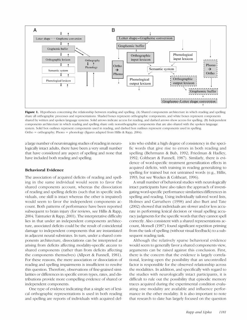
Reading and Spelling: Independent orShared Processes?
Both spelling and reading require various long-term andworking memory mechanisms that operate over lettersand word spellings. These are responsible for the transla-tion between letters and words and their correspondingsounds, and for words, there is the additional mapping be-tweenword spellings andmeanings. It is generally assumedthat the knowledge of word meanings (lexical semantics)and word sounds (the phonological lexicon) is not specificto reading or spelling as they form an essential part of thespoken language system. The debate regarding the relation-ship between reading and spelling, therefore, concerns thestatus of the orthographic components. One possibility isthat all orthographic mechanisms are shared by spellingand reading (the shared components architecture; see Fig-ure 1A). Another is that none is shared and that spelling andreading are independent production and comprehensionsystems (the independent components architecture; see Fig-ure 1B). A third position is that some mechanisms (e.g.,orthographic working memory) are shared whereas othersare not.It might seem to be a relatively straightforward matter to
adjudicate between these hypotheses; however, this hasnot been the case despite the fact that both behavioraland neural evidence have been brought to bear on thesequestions. The majority of the evidence has come from in-dividuals with acquired dysgraphia/dyslexia as a result ofneural injury, with only a few studies involving neurologi-cally intact adults. Furthermore, although there have beenJohns Hopkins University
© 2011 Massachusetts Institute of Technology Journal of Cognitive Neuroscience 23:5, pp. 1180–1197
a large number of neuroimaging studies of reading in neuro-logically intact adults, there have been a very small numberthat have considered any aspect of spelling and none thathave included both reading and spelling.
Behavioral Evidence
The association of acquired deficits of reading and spell-ing in the same individual would seem to favor theshared components account, whereas the dissociationof reading and spelling deficits (such that in specific indi-viduals, one skill is intact whereas the other is impaired)would seem to favor the independent components ac-count. Both patterns of performance have been reportedsubsequent to brain injury (for reviews, see Hillis & Rapp,2004; Tainturier & Rapp, 2001). The interpretative difficultylies in that under an independent components architec-ture, associated deficits could be the result of coincidentaldamage to independent components that are instantiatedin adjacent neural substrates. In turn, under a shared com-ponents architecture, dissociations can be interpreted asarising from deficits affecting modality-specific access toshared components (rather than from deficits affectingthe components themselves) (Allport & Funnell, 1981).For these reasons, the mere association or dissociation ofreading and spelling impairments is insufficient to resolvethis question. Therefore, observations of fine-grained simi-larities or differences in specific errors types, rates, and dis-tributions provide more compelling evidence of shared orindependent components.One type of evidence indicating that a single set of lexi-
cal orthographic representations is used in both readingand spelling are reports of individuals with acquired def-
icits who exhibit a high degree of consistency in the speci-fic words that give rise to errors in both reading andspelling (Behrmann & Bub, 1992; Friedman & Hadley,1992; Coltheart & Funnell, 1987). Similarly, there is evi-dence of word-specific treatment generalization effects inacquired deficits, with training in reading generalizing tospelling for trained but not untrained words (e.g., Hillis,1993; but see Weekes & Coltheart, 1996).
A small number of behavioral studies with neurologicallyintact participants have also taken the approach of investi-gating word-specific performance similarities/differences inspelling and reading. Using individually tailored word lists,Holmes and Carruthers (1998) and also Burt and Tate(2002) showed that individuals are slower and/or less accu-rate in performing lexical decision or visual spelling accu-racy judgments for the specific words that they cannot spellcorrectly. Also consistent with a shared representations ac-count, Monsell (1987) found significant repetition primingfrom the task of spelling (without visual feedback) to a sub-sequent reading task.
Although the relatively sparse behavioral evidencewould seem to generally favor a shared components view,arguments can be raised against this conclusion. First,there is the concern that the evidence is largely correla-tional, leaving open the possibility that an uncontrolledfactor is responsible for the observed relationship acrossthe modalities. In addition, and specifically with regard tothe studies with neurologically intact participants, it isdifficult to rule out the possibility that episodic memorytraces acquired during the experimental condition evalu-ating one modality are available and influence perfor-mance in the other modality. It is also important to notethat research to date has largely focused on the question
Figure 1. Hypotheses concerning the relationship between reading and spelling. (A) Shared components architecture in which reading and spellingshare all orthographic processes and representations. Shaded boxes represent orthographic components, and white boxes represent componentsshared by written and spoken language systems. Solid arrows indicate access for reading, and dashed arrows show access for spelling. (B) Independentcomponents architecture in which reading and spelling share only nonorthographic components that are also shared with the spoken languagesystem. Solid box outlines represent components used in reading, and dashed box outlines represent components used in spelling.Ortho = orthography; Phono = phonology (figures adapted from Hillis & Rapp, 2004).
Rapp and Lipka 1181
of a shared versus independent lexical orthographic com-ponent, largely ignoring the relationship between readingand spelling as concerns other orthographic components.With regard to the cognitive neuropsychological reportsthat have evaluated these other components, we find bothsome striking associations (e.g., Tainturier & Rapp, 2003;Caramazza, Capasso, & Miceli, 1996; Rapp & Caramazza,1989) as well as dissociations (Rapp & Caramazza, 1997;Beauvois & Dérouesné, 1981) between reading and spell-ing. Clearly, the evidence is not yet sufficiently clear orconsistent so as to support definitive conclusions.
Neural Substrates of Reading and Spelling
Given some of the difficulties of interpretation of the be-havioral evidence, data concerning the neural substratesthat support the specific processing components of read-ing and spelling may be especially useful.
Lesion-deficit Studies
Impairments in reading and/or spelling have been mostcommonly associated in both chronic and acute strokewith tissue dysfunction (lesion or hypoperfusion) in oneor more of the following left hemisphere areas: the angu-lar gyrus (Brodmannʼs area [BA] 39), the fusiform or infe-rior temporal gyrus (BA 37, 20), the supramarginal gyrus(BA 40), superior temporal gyrus (BA 22), and the inferiorfrontal gyrus (IFG; BA 44/45). In addition, reading but notspelling deficits have been associated with lesions in oc-cipital areas (BA 17, 18, 19), and spelling but not readingdeficits have been associated with lesions to premotorareas such as BA 6 (for reviews, see Philipose et al.,2007; Hillis & Rapp, 2004; Rapcsak & Beeson, 2002,2004; Friedman, Ween, & Albert, 1993; Roeltgen, 1993).Although some of the same neuroanatomical regionshave been identified in acquired deficits of both readingand spelling, there are two important issues that limit theprecision with which conclusions can be drawn. First,large lesions are usually involved, leaving open the possi-bility that different subregions within the same broadlydefined neural areas correspond to each modality. Sec-ond, very few studies have evaluated reading and spellingin the same individuals and as a result have not directlytested the hypothesis that specific lesions affect bothreading and spelling. With regard to the latter point, wereview the few recent studies that have considered bothtasks in the same individuals as these begin to allow for amore fine-grained analysis of the question.
Rapcsak and Beeson (2004) reported on eight individ-uals who, as a result of stroke, suffered damage to left hemi-sphere BA 37 and 20, corresponding to mid and anteriorfusiform regions (sparing the angular gyrus). All exhibitedboth reading and spelling impairments. Consistent withthese findings are the results of a study by Philipose et al.(2007), who examined reading and spelling in 69 cases
of acute stroke. They carried out analyses evaluating therelationship between presence/absence of behavioral im-pairments in reading and/or spelling and location of tissuedysfunction (lesion and/or hypoperfusion). They reporteda strong association between deficits in spelling and read-ing and tissue dysfunction in BA 40 and (superior) BA 37.In addition to these reports, the detailed examination of
individual patterns of behavioral impairment and lesioncan be particularly useful. Tsapkini and Rapp (2010) andGaillard et al. (2006) reported single case studies of readingand spelling in individuals with relatively circumscribedsurgical lesions of the left fusiform area. The Gaillard et al.case exhibited impairment in reading but not spelling, witha lesion in the posterior portion of the left fusiform gyrus.These authors argued for a disconnection of early readingareas in the posterior fusiform from the abstract ortho-graphic representations and processes localized in themid-fusiform region, which they refer to as the visual wordform area (VWFA). The Tsapkini and Rapp (2010) caseinvolved a more anterior mid-fusiform lesion that wasshown to specifically disrupt the mapping between ortho-graphic representations and word meanings in both read-ing and spelling while sparing the processing of visualobjects and auditorily presented words. The configurationof deficits and lesions in these two cases provides specificsupport for the conclusion that the left mid-fusiform is in-deed required both for reading and for spelling (for a dis-cussion, see Martin, 2006). Furthermore, it is encouragingthat this localization is generally consistent with the resultsof Philipose et al. (2007) and Rapcsak and Beeson (2004).
Functional Neuroimaging Studies
Although there have been a very large number of functionalneuroimaging studies of reading, only five studies haveprovided data concerning spelling in alphabetic systems1
(Norton, Kovelman, & Petitto, 2007; Beeson et al., 2003;Rapp & Hsieh, 2002; Menon & Desmond, 2001; Petrides,Alivisatos, & Evans, 1995). Evenmoreproblematic is the factthat there have been no functional neuroimaging studiesthat have considered both reading and spelling in the samesubjects.With regard to the neural substrates of reading, various
reviews and meta-analyses have reported a fair amount ofconvergence across studies (despite disagreement regard-ing the specific functions assigned to these areas) for theinvolvement in reading of the following left hemisphereareas: superior and middle temporal gyri (posterior andmiddle regions), supramarginal gyrus, fusiform gyrus, infe-rior temporal gyrus, extrastriate occipital cortex, lingualgyrus, and left IFG (triangularis and opercularis; for reviews,see Palmer, Brown, Petersen, & Schlaggar, 2004; Jobard,Crivello, & Tzourio-Mazoyer, 2003; Mechelli, Gorno-Tempini,& Price, 2003; Turkeltaub, Eden, Jones, & Zeffiro, 2002). Itis worth pointing out, however, that many of the studiesthat were reviewed involved oral reading or phonologicaljudgments on written forms so that it is not straightforward
1182 Journal of Cognitive Neuroscience Volume 23, Number 5
to specifically identify which of the implicated areas arespecifically involved in the orthographic aspects of readingand which with the planning and/or production of a spokenresponse.A challenge faced by the few paradigms that have used
the task of writing in the scanner is that of isolating thecentral components of the spelling process that might beshared by reading (see Figure 1A) from those compo-nents involved in the motor planning and execution as-pects of writing. Although Menon and Desmond (2001)and Petrides et al. (1995) do not address this problem,Beeson et al. (2003) carried out a study designed to sep-arate motor execution and planning from the more cen-tral aspects of spelling. Taking another approach, thestudies of Norton et al. (2007) and Rapp and Hsieh(2002) used tasks that required access to spelling knowl-edge but which did not involve writing in the scanner.However, the Norton et al. study used a complex spellingverification task that recruited both spelling and readingprocessing, making it impossible to determine if readingand spelling actually share substrates. If we focus on theresults of Beeson et al. (2003) and Rapp and Hsieh(2002), we find convergence in that they both identifyspelling-specific activation in the following left hemi-sphere areas: the fusiform gyrus, the precentral sulcus(BA 6), the posterior inferior/middle frontal gyrus, andthe supplementary motor cortex. Interestingly, althoughlesion studies since Dejerineʼs (1982) classic study haveprovided evidence that lesions affecting the angular gyrusare strongly associated with disruption to both reading(e.g., Black & Behrmann, 1994; Benson, 1979) and spell-ing (e.g., Rapcsak & Beeson, 2002; Hillis, Kane, Barker,Beauchamp, & Wityk, 2001; Roeltgen & Heilman, 1984),the neuroimaging evidence has not provided this samestrength of association. In their neuroimaging study ofspelling, Beeson et al. (2003) failed to find group level ef-fects, and in reading, Jobard et al. (2003) argued from theirmeta-analysis that at least some of the activations that havebeen reported for reading are more likely to be in posteriorpart of the middle temporal gyrus rather than the angulargyrus proper. In summary, if we evaluate the availableneuroimaging evidence, we find that reading and spellingboth recruit areas of the left fusiform gyrus and the IFG. Itis noteworthy that these areas are also included among theregions identified by the lesion-deficit analyses as givingrise to impairments of reading and spelling.
Orthographic Processing: Category andModality-specific Substrates?
Given the recency of literacy in human evolution, manyhave assumed that orthographic representation and pro-cessing are entirely parasitic on either visual object or spo-ken language processes and substrates. Contrary to theseexpectations, both neuroimaging and neuropsychologicalevidence have supported the claim that there are sub-
strates specifically necessary for orthographic processingwithin the posterior and inferior temporal/fusiform area.The claim is that these substrates are specialized for theprocessing of written stimuli and are not required for othervisual stimuli (category specificity) and also that they arenecessary for written but not spoken language stimuli (do-main specificity).
A number of functional neuroimaging studies havefound regions of the inferior temporal lobe, includingthe mid-fusiform region, to be reliably more activatedby written words relative to other categories of visual ob-jects, such as faces and houses (Baker et al., 2007; Ben-Shachar, Dougherty, Deutsch, & Wandell, 2007; Gaillardet al., 2006; Hasson, Levy, Behrmann, Hendler, & Malach,2002; Ishai, Ungerleider, Martin, Schouten, & Haxby,1999; Puce, Allison, Asgari, Gore, & McCarthy, 1996). Inaddition, there have been a handful of functional neuro-imaging studies that have specifically examined bothwritten and auditorily presented words and have foundactivation in this region only for written stimuli (Cohen &Dehaene, 2004; Cohen et al., 2004; Dehaene, Le ClecʼH,Poline, Le Bihan, & Cohen, 2002; Binder et al., 2000). Therehave also been several reports of individuals with selec-tive orthographic deficits subsequent to lesions affectingthis area. These include reports of selective alexia with-out prosopagnosia (Feinberg, Schindler, Ochoa, Kwan, &Farah, 1994) and vice versa (Farah,Wilson,Drain, & Tanaka,1998) and of selective visual object agnosia without alexiaor prosopagnosia (Humphreys & Rumiati, 1998; Rumiati& Humphreys, 1998) and vice versa (Buxbaum, Glosser,& Coslett, 1999; De Renzi & di Pellegrino, 1998). In ad-dition, the individuals, described by Tsapkini and Rapp(2010) andGaillard et al. (2006), suffered lesions to the fusi-form gyrus that resulted in either reading deficits alone orboth reading and spelling deficits while sparing object andface processing as well as spoken word comprehensionand production.
The proposal of modality- and category-specific ortho-graphic substrates has been vigorously challenged (Devlin,Jamison, Gonnerman, & Matthews, 2006; Hillis et al., 2005;Mechelli et al., 2005; Price & Mechelli, 2005; Price & Devlin,2003, 2004; Price et al., 2003). Generally speaking, the chal-lenges argue that neither the activation patterns nor thedeficits are as selective “as advertised.” With regard to theneuroimaging evidence, the argument is that althoughthere may be voxels that are more responsive to ortho-graphic than other visual stimuli, they are also responsiveto other visual categories, reflecting the fact that they areinvolved in certain types of complex visual analysis thatapply across a number of visual categories (Starrfelt &Gerlach, 2007; Martin & Chao, 2001; Moore & Price, 1999).Similarly, there are various reports of activation of the infe-rior temporal regions by spoken language stimuli and tasks(Vigneau, Jobard, Mazoyer, & Tzourio-Mazoyer, 2005; Price& Devlin, 2004; Jobard et al., 2003; Booth et al., 2002a,2002b; Thompson-Schill, Aguirre, DʼEsposito, & Farah,1999; Demonet, Price, Wise, & Frackowiak, 1994). With
Rapp and Lipka 1183
regard to the neuropsychological evidence, it has beenargued that in cases of selective impairments, other visualcategories and/or spoken language are indeed affectedand that the testing carried out was not sufficiently de-manding of these skills.
The work we report on here will not address the ques-tion of the possible modality specificity (written vs. spokenlanguage) of proposed written language substrates; how-ever, our use of face and house stimuli as functional local-izers will allow us to contribute to the ongoing debateregarding the proper characterization of the neural sub-strates that support written language processing and howthey are related to object processing more generally.
METHODS
Participants
Ten individuals participated in this investigation (4 men,6 women). They were all right-handed as determined bythe Edinburgh Handedness Inventory (Oldfield, 1971).They ranged in age from 18 to 42 years; all had a collegeeducation, were native speakers of English, and had noknown history of reading or spelling disability. Further-more, they all scored above 93% on a spelling pretest.All were paid for the participation in the research.
Tasks and Stimuli
Spelling, reading, and object processing (faces and houses)were evaluated in different tasks presented in differentruns, for a total of 10 runs administered in one scanningsession. The order of runs was varied across subjects. Alltasks were practiced before the scanning session. Readingand face and object processing were evaluated adoptingparadigms used in previous studies (reading: Cohen &Dehaene, 2004; object/face processing: Haxby et al., 2001).Using these fairly standard paradigms provided a solidbasis for comparison with the spelling results that involveda relatively novel paradigm (used before only in Hsieh &Rapp, 2004; Rapp & Hsieh, 2002). Tasks were presentedusing E-Prime software (Schneider, Eschman, &Zuccolotto,2002a, 2002b).
Silent reading was evaluated with three stimulus types:monomorphemic words, randomly generated consonantstrings, and a black and white checkerboard rectangle. Oneach trial, a central fixation cross appeared for 550 msec,followed by a stimulus presented in the center of the screenfor 200 msec. Participants were instructed to carefully at-tend to each stimulus. The words and consonant stringswere three to six letters in length. All alphabetic stimuli werepresented in lowercase in black font on a white back-ground; the checkerboard rectangle had an extension com-parable to that of a nine-letter word. Stimuli were presentedin blocks of 28 trials, and there were 6 blocks of each stimu-lus type for a total of 18 blocks per run; there were a total oftwo runs that were identical except that blocks were pre-
sented in different pseudorandom orders. No alphabeticstimulus was repeated in a run, and the checkerboard stim-ulus was always the same. The repetition time (TR) was1500, and 264 volumes were acquired in each run.Object processing was evaluated with four stimulus
types: faces, houses, and pixel-scrambled faces and houses,with 40 different stimuli of each type (from Haxby et al.,2001). Each stimulus was presented in the center of thescreen for 500 msec, and participants were instructed toattend carefully. Stimuli were presented in a blocked man-ner according to stimulus type with 40 stimuli per blockand 16 blocks per run (four of each stimulus type). Tworuns were presented; they were identical except for theorder of the blocks. The TR was 2000, and 169 volumeswere acquired in each run.Spelling was evaluated using two tasks: spelling probe
and case verification tasks that served, respectively, as theexperimental and sensory motor control tasks. The tasksinvolved identical sensory and motor components, varyingonly in the instructions given to subjects. In the spellingprobe task each trial was 6 sec long, with the followingsequence of events: (1) a centrally displayed task prompt(SPELLING?) for 1500 msec; (2) a central fixation crossfor 300 msec, (3) an auditory word (plus a variable periodof silence) for total duration of 1200 msec, (4) a single vi-sually displayed uppercase letter for 1000 msec, and (5) ablank response screen for 2000 msec. Participants wereinstructed to respond yes/no (right/left hand button press)whether with the visually presented letter was in the spell-ing of the heard word. The trials for the case verificationtask were identical to those of the spelling probe task, ex-cept that that there was a different task prompt (UPPER-CASE?) and the visually displayed letter could appear ineither upper- or lowercase. In this task, participants wereinstructed that the auditorily presented word was irrelevantand that they were simply to respond yes/no (button press)whether the visually presented letter was or was not inuppercase. Although with some limitations, the tasks weredesigned so that a comparison of the two tasks wouldidentify the central components of the spelling process,without requiring written responses in the scanner. Therationale was as follows: The two tasks involved listeningto a word, processing a visually presented letter, and mak-ing a yes/no decision. In addition, the spelling probe taskrequired searching long-term memory for the spellingof a word (and/or generating it via sublexical processes)and then engaging orthographic working memory whileverifying if the probe letter was contained in the spelling.Additional support for the appropriateness of the task forevaluating spelling comes from the Rapp and Kong (2002)report that individuals with acquired dysgraphia were im-paired in the spelling probe task.Across the two tasks, word stimuli were matched for
length, frequency, and grammatical category. In each task,half of the stimuli were high frequency (mean = 91.2) andhalf were low frequency (mean = 3.7) (Francis & Kucera,1982). Stimuli were either four or seven letters long. The
1184 Journal of Cognitive Neuroscience Volume 23, Number 5
majority of the words were nouns. In the spelling probetask, for four-letter words, all four-letter positions wereprobed, and for seven-letter words, Positions 1, 3, 5, 7were probed. In the case verification task, the letters pre-sented for case verification never occurred in the spellingof the heard word. Spelling probe and case verificationtrials were presented in a blocked manner, with six trialsper block, six blocks per run, for a total of six runs. High-and low-frequency word trials formed “mini-blocks” with-in these larger blocks that consisted of one to four trials.The TR was 1500 msec, and a total of 176 volumes wereacquired in each run.
Imaging Parameters
MRI data were acquired with a 3.0-T Phillips Intera Scannerat the F. M. Kirby Research Center for Functional Brain Im-aging at the Kennedy Krieger Institute (Baltimore, MD).Whole-brain T2-weighted gradient-echo, EPIs were acquiredwith an eight-channel SENSE (Invivio) parallel-imaginghead coil in 29 transverse slices (TR = 1500 msec, echotime = 30 msec, flip angle = 65°, field of view = 240 ×240 mm, matrix 128 × 128, slice thickness = 4 mm, gap =1mm). Structural images were acquired using an MP-RAGET1-weighted sequence that yielded images with a 1-mmisotropic voxel resolution (TR = 8.06 msec, echo time =3.8 msec, flip angle = 8°).
Data Analysis
Analysis of functional data was carried out using Brain-Voyager QX software (Maastricht, Netherlands) as well aswith programs developed in MatLab (MathWorks, Natick,MA) for specific analysis purposes. With regard to prepro-cessing, functional images were slice time and motion cor-rected and then temporal high-pass filtered to removecomponents occurring fewer than three cycles over thecourse of a run. The images were normalized to Talairachcoordinates, resampled to 3-mm isotropic voxels, and spa-tially smoothed (8-mm FWHM Gaussian kernel).The general linear model approach (Friston et al., 1995)
was used to estimate parameter values in a block design.Three general linear models separately modeled the spell,silent reading, and object processing tasks. Spelling wasmodeled with two regressors corresponding to the high-and low-frequency word blocks of the spelling probe task;all time points corresponding to the case verification trialswere used as the baseline condition. Silent reading wasmodeled with three regressors corresponding to stimulustype: words, consonant strings, and fixation; all time pointscorresponding to checkerboard trials served as the base-line. Finally, object processingwasmodeledwith two regres-sors for stimulus type: faces and houses; all time pointscorresponding to scrambled face and scrambled house trialsserved as the baseline. Regressors were created by convolv-ing a two-parameter gamma hemodynamic response func-tion with a boxcar function marking the temporal position
of each stimulus block type. In addition to the task-specificregressors, for all tasks and each run, six motion correctionregressors and an additional confound regressor (represent-ing run number) were included in the model. Group datawere subjected to random-effects analysis; for each contrast,cluster size thresholding was estimated using a plug-inimplemented in BrainVoyager. This plug-in implements aMonte Carlo randomization technique to estimate a mini-mum cluster size that, for a given uncorrected voxelwisep value, will achieve a corrected p value < .05 (Formanet al., 1995). On this basis, for all contrasts reported, weapplied a cluster size threshold corresponding to p valueof <.05. In addition, the default uncorrected voxelwisep value adopted was p< .005. However, for a small numberof contrasts, different uncorrected voxelwise values wereadopted (between p < .01 and p < .001). This was doneonly on those occasions on which the default value ob-scured a pattern that was reliable across a range of un-corrected voxelwise values. Cluster locations are reportedin terms of the location of the peak voxel of each clusterbecause the peak is more stable under threshold changesthan the geometric center. Talairach coordinate values(Talairach & Tournoux, 1988) are used in the text, butTable 1 provides both Talairach and MNI values. Clustersizes are reported as the number of 1-mm3 voxels; clusterssmaller than 50 voxels are not reported.
RESULTS
Analysis 1. Reading, Spelling, and Visual ObjectProcessing: Basic Neurotopography
Silent Reading
A brain-wide evaluation of words > checkerboards (voxel-wise threshold p< .005, cluster level p< .05) yielded threelarge clusters: left mid-fusiform (−39,−46,−5; 6,217 voxels),left IFG (−36, 20, 4; 17,242 voxels), and the right cingu-late gyrus (24, −16, 34; 8,677 voxels) (see Figure 2 andTable 1). The right cingulate activation was not limitedto the area of the peak in the anterior cingulate but ratherextended almost the length of the gyrus. Further analy-sis revealed that the large left IFG cluster was composedof three smaller clusters (with peaks at −39, 26, 19, IFGtriangularis; −36, 20, 4, IFG triangularis; and −39, 2, 28,IFG opercularis) that are reported in brackets in Table 1.The most posterior of the three was centered near an areaof the left hemisphere referred to as the inferior frontaljunction (IFJ) that is located at the junction of the inferiorfrontal sulcus and the inferior precentral sulcus. We willrefer to this region as the IFG/IFJ (see Figure 2B). Boththe fusiform and the IFG activations were highly left later-alized; in fact, right hemisphere fusiform activation (42,−52, −17) appeared only at an uncorrected p < .01 andright hemisphere IFG/IFJ activation did not appear at anymeaningful threshold. Activation in the region of the leftangular gyrus bordering on the intraparietal sulcus (IPS)was observed only at a more lenient voxelwise threshold
Rapp and Lipka 1185
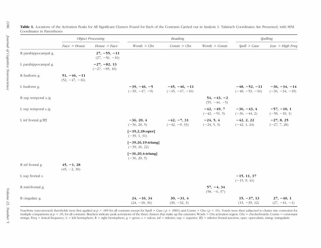
Table 1. Locations of the Activation Peaks for All Significant Clusters Found for Each of the Contrasts Carried out in Analysis 1: Talairach Coordinates Are Presented, with MNICoordinates in Parentheses
Object Processing Reading Spelling
Face > House House > Face Words > Chx Consts > Chx Words > Consts Spell > Case Low > High Freq
R parahippocampal g. 27, −55, −11(27, −56, −16)
L parahippocampal g. −27, −82, 13(−27, −85, 10)
R fusiform g. 51, −46, −11(52, −47, −16)
L fusiform g. −39, −46, −5(−39, −47, −9)
−45, −46, −11(−45, −47, −16)
−48, −52, −11(−48, −53, −16)
−36, −34, −14(−36, −34, −19)
R sup temporal s./g. 54, −43, −2(55, −44, −5)
L sup temporal s./g. −42, −49, 7(−42, −51, 5)
−36, −43, 4(−36, −44, 2)
−57, −10, 1(−58, −10, 1)
L inf frontal g/IFJ −36, 20, 4(−36, 20, 5)
−42, −7, 31(−42, −9, 33)
−24, 5, 4(−24, 5, 5)
−42, 2, 22(−42, 1, 24)
−27, 8, 25(−27, 7, 28)
[−39,2,28:oper](−39, 1, 31)
[−39,26,19:triang](−39, 26, 22)
[−36,20,4:triang](−36, 20, 5)
R inf frontal g 45, −1, 28(45, −2, 30)
L sup frontal s. −15, 11, 37(−15, 9, 41)
R mid-frontal g. 57, −4, 34(58, −6, 37)
R cingulate g. 24, −16, 34(24, −18, 36)
30, −31, 4(30, −32, 3)
15, −37, 13(15, −39, 12)
27, −40, 1(27, −41, −1)
Voxelwise (uncorrected) thresholds were first applied at p < .005 for all contrasts except for Spell > Case ( p < .0001) and Consts > Chx ( p < .01). Voxels were then subjected to cluster size correction formultiple comparisons at p < .05, for all contrasts. Brackets indicate peak activations of the three clusters that make up the extensive Words > Chx activation region. Chx = checkerboards; Consts = consonantstrings; Freq = lexical frequency; L = left hemisphere; R = right hemisphere; g = gyrus; s = sulcus; inf = inferior; sup = superior; IFJ = inferior frontal junction; oper: opercularis; triang: traingularis.
1186Jou
rnalof
Cogn
itiveNeu
roscience
Volume23,
Number
5

of p < 01, a level at which activations were more diffuseand less clearly differentiated into distinct clusters.A brain-wide evaluation of consonants > checkerboards
(voxelwise threshold p< .01, cluster level p< .05) yieldedthree clusters at the same left hemisphere locations asthose found for the words > checkerboards contrast: leftmid-fusiform (−45, −46, −11; 2,408 voxels), left IFG/IFJ(−42, −7, 31; 3,069 voxels), and right cingulate gyrus(30, −31, 4; 4,781 voxels).A comparison of the neural response for words versus
consonants (voxelwise threshold p < .005, cluster levelp< .05) revealed no voxels with significantly greater activa-tion for consonants than for words, although four clustersin which words > consonants were identified: bilateralposterior STS/middle temporal gyrus (left: 54, −43, −2;2,405 voxels; and right:−42,−49, 7; 6,888 voxels), right pos-terior middle frontal gyrus (57, −4, 34; 2,397 voxels), andleft IFG (−24, 5, 4; 5,955 voxels). In terms of the left fusiformspecifically, words > consonants activation was apparentonly at an uncorrected p< .003. We return to the compari-son of words and consonants strings in Analysis 3 in whichwe consider targeted small volume examinations.
Spelling
Whole-brain evaluation of spell > case (voxelwise thresh-old p < .0001, cluster level p < .05) revealed five clusters:left mid-fusiform (−48, −52, −11; 569 voxels), left IFG/IFJ(−42, 2, 22; 604 voxels), left superior frontal sulcus (−15,11, 37; 300 voxels), left posterior superior temporal gyrus/sulcus (−36, −43, 4; 261 voxels), and right posterior cin-gulate gyrus (15, −37, 13; 679 voxels) (see Figure 2 and
Table 1). Only at the more lenient voxelwise thresholds of0.01 or 0.05 was activation observed in the left intraparietalsulcus and supramarginal gyrus region; however, as wasthe case for the reading task, at these thresholds, activa-tions were poorly differentiated into clusters. Overall, acti-vation was highly left lateralized, such that the only righthemisphere activation was in the right posterior cingulate.This continued to be the case even as the threshold waslowered; in fact, at no reasonable threshold were signifi-cant spell > case activations observed in either the rightfusiform or the IFG/IFJ regions.
A brain-wide evaluation of case > spell yielded no sig-nificant voxels. Only at the very lenient uncorrected p <.01 did case > spell clusters appear in the right and lefthemisphere superior and middle temporal gyri (51, −58,19;−42,−61, 22; 57,−10,−14) and the left superior fron-tal gyrus (−18, 50, 34).
Using only the trials from the spelling probe task, weevaluated effects of lexical frequency by comparing low-frequency words directly to a baseline of high-frequencywords. Note that this direct contrast (without using thecase verification task as a baseline) identifies general lexicalfrequency effects and not orthographic effects specifically,that is, because the spelling probe task involves both listen-ing to a word and recovering its orthographic form fromlong-termmemory and the case verification baseline servesto isolate the orthographic components of the task by “re-moving” the auditory word processing aspects. In this waywe can evaluate frequency effects more broadly through-out the language system. We return to this point later.The results of the low-frequency > high-frequency con-trast (voxelwise threshold p < .005, cluster level p < .05)
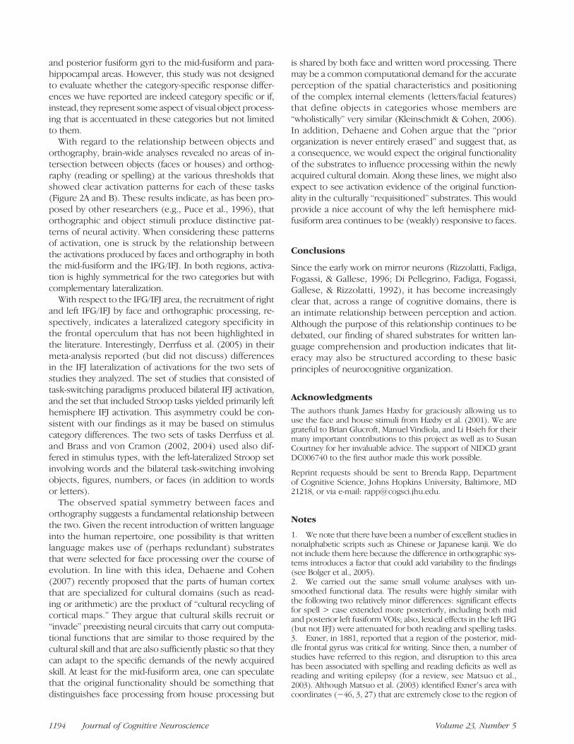
Figure 2. Neurotopographyof orthographic and objectprocessing. Significant clustersfrom Analysis 1 (vowelwisethreshold p < .005–.0001,cluster level p < .05) aredepicted: blue = words >checkerboards, green = spell >case, yellow = faces > houses,and pink = houses > faces.Both panels A and B depictoverlapping substrates forreading and spelling andsymmetrical activation fororthographic processing(left hemisphere) and faceprocessing (right hemisphere).(A) Horizontal image atTalairach z = −11 depictsclusters for silent reading(words > checkerboards:peak = −39, −46, −5), spelling(spell > case: peak = −48,−52, −11), passive viewing offaces (faces > houses: right hemisphere peak: 51, −46, −11), and passive viewing of houses (houses > faces: right hemisphere peak: 27, −55,−11; left hemisphere peak: −27, −82, 13). (B) Coronal image at Talairach = +4 includes the bilateral IFG and IFJ and depicts significantclusters for reading (peak = −36, 20, 4), spelling (peak = −42, 2, 22), and face processing (peak: 45, −1, 28).
Rapp and Lipka 1187

revealed a pattern of highly left-lateralized activation withfive clusters: left fusiform (−36, −34, −14; 5,257 voxels),left IFG/IFJ (−27, 8, 25; 3,219 voxels), left superior tem-poral gyrus (−57,−10, 1; 1,902 voxels), left posterior whitematter (−27, −55, 13; 2,503 voxels), and right posteriorcingulate (27, −40, 1; 2,310 voxels).
Objects
To identify brain areas that were especially responsive tofaces as compared with houses and vice versa, the neuralresponse to faces was contrasted with the response tohouses, relative to a baseline condition of scrambled faceand house images. Brain-wide analysis revealed two clus-ters for faces > houses and two clusters for houses > faces(voxelwise threshold p < .005, cluster level p < .05).
As can be seen in Figure 2, the two houses > faces clus-ters extended along the right and left parahippocampal/lingual gyri with peaks at 27, −55, −11 (cluster size =19,824) and −27, −82, 13 (cluster size = 14,604). Thetwo faces > houses clusters both appear in the right hemi-sphere. One is in the mid-fusiform gyrus (51, −46, −11;9116 voxels), and the other is in the IFG/IFJ (45, −1, 28;1561 voxels). At thresholds corrected for multiple com-parisons, we observed no left hemisphere activation forfaces > houses, and only at an uncorrected p < .005 didwe observe a left fusiform cluster (−36, −64, −17); in ad-dition, there was no left hemisphere IFG/IFJ activation forfaces > houses at any meaningful threshold.
Summary
The results we reported for object processing are consis-tent with those reported in the literature for both facesand houses. For example, the activation for faces > housesincludes the fusiform region most typically reportedas being especially responsive to faces and sometimesreferred to as the fusiform face area (39 ± 3, −40 ±7, −16 ± 5) (Grill-Spector, Knouf, & Kanwisher, 2004).Similarly, the activation for houses > faces included thelocation of what is sometimes referred to as the parahip-pocampal place area (21 ± 5, 54 ± 7, −9 ± 4) (Aguirre,Zarahn, & DʼEsposito, 1998).
The neurotopography of reading and spelling revealedby this analysis is also highly consistent with the lesion-deficit and neuroimaging research reviewed in the Intro-duction. Specifically, both tasks activated areas within theleft fusiform and the left IFG. The apparent sharing of sub-strates is investigated more thoroughly in Analysis 2.
Given the prominent role played by the angular gyrusin neurological theories of written language processing, itis worth briefly commenting specifically on the results re-garding this area. As indicated, we find activation in the in-traparietal sulcus and angular gyrus for both reading andspelling but only at thresholds more lenient than those thatclearly identified the fusiform and IFG/IFJ activations. One
possibility is that these relatively weaker activations as wellas the inconsistencies across imaging studies and discre-pancies between neuroimaging and deficit–lesion correla-tion studies (described in the Introduction) may be dueto greater variability in the individual recruitment of sub-strates within this region; certainly, this neuroanatomicalarea and these issues would benefit from more targetedinvestigation.
Analysis 2. Overlapping Substrates?
Reading and Spelling
To evaluate the relationship between the neural substratesof reading and spelling, we carried out two analyses. First,we identified areas of overlapping neural responsivity forreading and spelling, and second, we more carefully exam-ined the identified areas by comparing the cross-modalitydifferences in peak activations for reading and spelling tothe variability observed in repeated within-modality as-sessments. (Note that both analyses considered only datafrom word stimuli, given that the spelling task only in-volved words.)As depicted in Figure 3A, brain-wide evaluation of the
words > checkerboards and spell > case contrasts (usingthe same thresholds as in Analysis 1) identified only tworegions that were responsive to both modalities of ortho-graphic processing: one in the left fusiform gyrus (−42,−48, −13; 341 voxels) and another in the left IFG/IFJ(−42, 3, 24; 538 voxels). That is, spelling significantly ac-tivated voxels within two of the three regions that werefound to be significantly active for reading, and readingactivated voxels within two of the five regions that wereactive for spelling.Although this analysis indicates the existence of neural
tissue that is jointly sensitive to reading and spelling, it doesnot reveal the degree of similarity in the activation “topo-graphy” within these regions for the two orthographicmodalities. A key (and relatively stable) feature of the acti-vation topography is the location of the activation peaks.For example, one possibility to be considered is thatalthough reading and spelling coactivate voxels withinthe regions identified just above, their peak activationscould be distinct, suggesting a potentially critical distinc-tion between the two. In fact, the reading and spelling ac-tivation peaks in the fusiform, and the IFJ/IFG (opercularis)that were identified in Analysis 1, although geographicallyclose, are not identical (see Table 1). To evaluate the sig-nificance of these differences, we examined whether thepeak locations are reliably different across the modalities(reading and spelling) given the variability that could be ex-pected from repeatedmeasurements within eachmodality.To estimate this variability, for both the fusiform and theIFJ/IFG, we identified activation peaks for both readingand spelling on the basis of split-half samplings of the datain each modality. For spelling, we evaluated 20 samples,each consisting of three of the six run total, and for reading,
1188 Journal of Cognitive Neuroscience Volume 23, Number 5

we evaluated 20 samples each consisting of three of thesix block total (of words and checkerboards).The 20 values obtained for each of the x,y,z coordi-
nates for both the fusiform and the IFG/IFJ locations werecompared for reading versus spelling (see Table 2). Theresults of the six t test evaluations of these data sets re-vealed no significant differences ( p values ranging from.57 to .11) between reading and spelling activation peaks,except for the x-coordinates of the IFG/IFJ clusters ( p <.02). This indicates a potentially significant 2-mm differ-ence between the spelling (mean × value = −41) andthe reading (mean × value = −39) peaks. However, eventhis difference would not be significant if a correction for
the multiple comparisons (the six t tests) was applied tothis analysis.
Houses and Faces
The results of Analysis 1 revealed large clusters that weremore responsive to houses versus faces and vice versa.The finding of nonoverlapping areas for faces and housesdoes not mean, of course, that voxels within these clus-ters are not responsive to both faces and houses when eval-uated relative to some neutral baseline, such as scrambledimages. To address this question directly, we carried outa brain-wide conjunction analysis of faces > scrambledimages and houses > scrambled images. This analysisidentifies the areas of intersection in which both faces >scrambled images and houses > scrambled images aresignificant. The results revealed (voxelwise threshold p <.005, cluster level p< .05) bilateral, right lateralized regionswithin the fusiform and the middle occipital gyrus (righthemisphere: 27, −91, −2; left hemisphere: −33, −79,−14). Figure 3B depicts the regions identified in this con-junction analysis as well as the clusters identified in Analy-sis 1 in which houses > faces and faces > houses. Theresults indicate that (when the same correction for multi-ple comparisons is applied) there are areas that show“selective” responsivity to houses or faces that fall outsidethe areas that are jointly activated by houses and faces(relative to a low-level baseline).
Table 2. Mean and SD (in Parentheses) of the ActivationPeaks of Clusters in the Left Fusiform Gyrus and the LeftIFG/IFJ, Obtained from 20 Split-half Data Analyses of Reading(Words > Checkerboards) and Spelling (Spell > Case)
Coordinates
Left Mid-fusiform Left IFG/IFJ
Reading Spelling Reading Spelling
x −41.4 (1.6) −41.9 (3.3) −39.0 (2.6) −41.0 (2.4)
y −48.9 (4.5) −47.2 (6.2) 2.6 (2.5) 2.2 (2.1)
z −15.5 (5.0) −13.3 (3.6) 25.5 (4.9) 23.7 (2.3)
Only the x values for the IFG/IFJ clusters differ significantly ( p < .02),although not if a correction for multiple comparisons is applied. See textfor more details.
Figure 3. Shared substrates ofreading/spelling and faces/houses. (A) Lateral view ofactivations produced by reading(words > checkerboards) inblue and spelling (spell > case)in green. Indicated with redcircles are the regions ofoverlap between reading andspelling in the left mid-fusiform(−42, −48, −13; 341 voxels)and the left IFG/IFJ (−42, 3, 24;538 voxels). Correction formultiple comparisons: forreading = voxelwise p < .005,corrected p < .05, and forspelling = voxelwise p < .0001,corrected p < .05. (B) Orangedepicts shared voxels for faces >scrambled images and houses >scrambled images (righthemisphere: peak = 27, −91,−2; 18,405 voxels; lefthemisphere: peak = −33,−79, −14; 13,561 voxels).Included for comparisonpurposes are the regions(also depicted in Figure 2A)especially sensitive tofaces (yellow = faces >houses) and houses (pink =houses > faces).
Rapp and Lipka 1189

Objects and Orthography
No areas of intersection between objects (faces or houses)and orthography (reading or spelling) are seen when brain-wide activations specific to faces, houses, reading, andspelling (at a voxelwise threshold p < .005, cluster levelp < .05; or more stringent) are superimposed on oneanother. It is only when the voxelwise threshold is loweredbeyond this level and when activations become large and illdefined that we begin to see overlapping areas, and theseincrease in extent as the threshold is lowered. We addressthe question of the relationship between object and ortho-graphic substrates more systematically in Analysis 3.
Summary
With regard to the relationship between reading and spell-ing, the analyses clearly reveal that not only is there neuraltissue that is jointly sensitive to reading and spelling in theleft mid-fusiform and the IFG/IFJ but that the locations ofthe peak activity in the two modalities are statistically in-distinguishable. With regard to object processing and therelationship between object and orthographic processing,we find regions of cortex within the fusiform and occipitallobes that are differentially sensitive to the categories of
objects, faces, and written words. Although we cannot ruleout that these categories may also activate common sub-strates, it is quite clear that there are distinctive activationdistributions for stimulus processing in these categories.
Analysis 3. Small Volume Investigation of theFusiform, Occipital, and IFG/IFJ Regions
Targeted small volume analyses were carried out in thefusiform gyrus, the occipital lobe, and the IFG/IFJ region.Seven locations were selected bilaterally (for a total of four-teen 1-cm3 volumes), and t tests were used to examine theaverage responsiveness of the voxels within each volume(using the BrainVoyagerʼs VOI analysis procedure; seethe red squares in Figure 4A–C for VOI locations).A mid-fusiform volume was selected at coordinates com-
monly reported in the literature for the VWFA (±38, −44,−16), and then anterior (±38, −24, −16) and posterior(±38, −64, −10) volumes were selected to be equidistantfrom the mid-fusiform volume (while remaining within theneuroanatomical confines of the gyrus). Two occipital loca-tions were examined bilaterally, one in the middle occipi-tal gyrus, just posterior to the fusiform gyrus (±28, −84,−6), and another in the lingual gyrus (±10, −82, −3),
Figure 4. Results of bilateral VOI analyses. Fourteen 1-cm3 VOIs (depicted in red) were selected in the fusiform (A) and occipital gyri (A and B)and the IFJ (C). Each VOI was subjected to eight contrasts; significant contrasts are listed for each VOI (evaluated at Bonferroni-corrected p < .0063)and are depicted in the following colors: gray = checkerboards > words or consonant strings; pale yellow = faces > scrambled images; pale pink =houses > scrambled images; yellow: faces > houses; dark blue: words or consonants > checkerboards; light blue: words > consonants; dark green:spell > case; light green: low-frequency > high-frequency words.
1190 Journal of Cognitive Neuroscience Volume 23, Number 5

the latter permitting an evaluation of early visual processes.Two bilateral posterior frontal locations were selected: bi-lateral IFJ volumes were created centered on the averagecoordinates of IFJ activations reported in the recent lit-erature (±42, 3.5, 32; Brass & von Cramon, 2002, 2004;Derrfuss, Brass, & von Cramon, 2004) and located at thejunction between the inferior and middle frontal gyri andthe sulcus of the precentral gyrus; in addition, bilateral IFGvolumes were created so as to be fully contained within theposterior IFG (opercularis; ±48, 6, 20).At each volume, eight contrasts were examined: spell/
case, low/high frequency (spelling), words/checkerboards(reading), consonants/checkerboards (reading), words/consonants (reading), faces/houses, houses/scrambled,and faces/scrambled. To correct for the fact that each vol-ume was subjected to eight comparisons, a Bonferroni-corrected value of p < .0063 was applied to determinestatistical significance of the contrasts at each volume. Theresults are depicted in Figure 4A–C.With regard to the fusiform (see Figure 4A), the bilateral
posterior fusiform was responsive only to houses ( p <.00008) and faces ( p < .0003) relative to scrambled im-ages. The bilateral mid-fusiform was sensitive to the generalcontrast of face > scrambled (right: p < .0005 left: p <.0002). In addition, the right mid-fusiform was significantlysensitive to the specific contrast of faces > houses ( p <.002), and the left mid-fusiform showed a very strong trendtoward significance for this contrast ( p < .008). The mid-fusiform VOIs exhibited a markedly asymmetric respon-siveness to all of the orthographic contrasts, with only theleft mid-fusiform exhibiting significant sensitivity to the fol-lowing: in spelling, spell > case ( p< .0002) as well as low-frequency > high-frequency words ( p < .004); in reading,both words > checkerboards and consonants > checker-boards were significant (words: p < .002; consonants: p <.006) as was the comparison of words > consonants ( p <.003). In the anterior fusiform, the only significant effect wasin the left anterior fusiform, which exhibited sensitivity tolow-frequency > high-frequency words ( p < .002).In the occipital lobe (see Figure 4A and B), the middle
occipital gyrus exhibited bilateral effects of house > scram-bled images and face > scrambled images (houses: p <.00002; faces: p < .005), whereas the bilateral lingual VOIswere the only ones to show an effect of checkerboards >words ( p < .005) or consonant strings ( p < .003). In addi-tion, the right lingual gyrus exhibited an effect of scrambledimages > faces ( p < .004).The IFJ and the IFG volumes had highly similar patterns
of responsivity. They exhibited markedly asymmetric re-sponses (Figure 4C) such that in the left hemisphere, boththe IFJ and the IFGexhibited sensitivity only to orthographicconditions: spell > case ( p < .0003), words > checker-boards ( p < .00001), and consonants > checkerboards( p < .005); in the right hemisphere, the right IFJ exhib-ited significant effects only for faces, specifically faces >houses ( p < .004) and faces > scrambled images ( p <.005), and the right IFG exhibited similar sensitivity to
faces although the effect did not quite pass the Bonferronithreshold faces > houses ( p < .007).
Summary
The small volume analyses2 confirm the results of thebrain-wide analyses reported in Analyses 1 and 2 and reveala highly differentiated pattern of responsivity across theposterior brain and in the IFG/IFJ for both orthographicand object stimuli. The primary axes of this differentiationare posterior to anterior and right versus left hemispheres.We discuss these findings in more detail in the Generaldiscussion.
GENERAL DISCUSSION
In this article, we report on an fMRI investigation evaluatingthe brainʼs response to the tasks of silent reading, spelling,and the passive viewing of faces and houses. The objectiveswere to further our understanding of the relationship be-tween orthographic comprehension and production (read-ing and spelling) and, in turn, their relationship to visualobject processing. Whole-brain and small volume analysesconverge on the following empirical findings. (1) Neuraltissue in the left hemisphere mid-fusiform gyrus and theIFG (including the IFJ) are responsive to both reading andspelling. (2) The left mid-fusiform region, in addition to itsgeneral responsiveness to orthographic processing, exhibitssensitivity to lexical factors, namely, greater responsivity towords relative to consonant strings, and to low- relative tohigh-frequency words. (3) In contrast, the anterior portionof the left fusiform gyrus is responsive to differences in lexi-cal frequency but not to orthographic processing (eitherreading or spelling). (4) Within the inferior temporal lobes,we find bilateral regions that are responsive to both faces andhouses as well as additional regions that are more stronglyactivated by faces compared with houses or vice versa. (5)Substrates for objects (faces and houses) and orthographicprocessing (reading and spelling) are largely nonoverlap-ping, except for a strong trend in the left mid-fusiform forresponsivity to both faces and orthography. In fact, orthogra-phy (reading and spelling) and face processing activate gen-erally complementary homologous areas in the fusiform andIFG, with activations that are lateralized to the left and righthemispheres, respectively.
Reading and Spelling: Shared Substrates?
This study evaluated both reading and spelling in the sameindividuals, providing a strong test of the hypothesis ofshared components for reading and spelling. Both whole-brain and small volume analyses revealed highly reliableareas of overlapping activation for reading and spellingin both the left mid-fusiform gyrus and the left IFG/IFJ.Furthermore, the activation peaks for reading and spell-ing in these two areas are neuroanatomically close and
Rapp and Lipka 1191

statistically indistinguishable. In this way, the results pro-vide perhaps the strongest evidence to date that readingand spelling share at least some cognitive machinery (seeFigure 1A).
As would be expected, reading and spelling also showregions of nonoverlapping activation; for reading, this liesprimarily in the triangularis area of the IFG; for spelling, non-overlapping regions include the left superior temporal andsuperior frontal sulci. Thesemodality-specific activations arecertainly of interest and may indicate that some processesare not shared between reading and spelling. However,the absence of activation overlap in these regions presentsthe usual interpretative challenges. Namely, the landscapeof nonoverlapping regions is likely to change as thresholdsare relaxed and corrections for multiple comparisons are re-duced. Furthermore, the likely increased task demands—attentional, temporal, andotherwise—for the spelling probetask as compared with the silent reading task may result inmore and larger clusters reaching statistical significance inone task compared with the other. For these reasons, wefocus this discussionon thehighly reliable regions of overlapbetween the two tasks and discuss what these results revealabout the shared machinery of reading and spelling.
The Left Fusiform Gyrus
With regard to the left fusiform, the area of shared activa-tion for reading and spelling (Figure 3A) clearly coincideswith the region observed in a large number of neuroimagingstudies of reading and referred to as the VWFA (Cohen &Dehaene, 2004). It also falls within the region implicatedin lesion studies of spelling (Rapcsak & Beeson, 2004) andis consistent with the sparse neuroimaging data on spellingthat is available (Beeson et al., 2003; Rapp & Hsieh, 2002).The natural next question is which shared orthographicprocessing components (Figure 1A) make use of the leftmid-fusiform area? There are various aspects of the resultsthat are relevant in answering this question. First, the mid-fusiform area is responsive to both words and consonantstrings relative to checkerboards, indicating a role in ortho-graphic processing. Second, the area is more sensitive towords than consonant strings and more sensitive to low-than high-frequency words. This sensitivity to lexical factorsindicates that the region is not merely sensitive to orthogra-phy in general but to lexical orthography in particular. Third,we can rule out that the sensitivity to both orthographicand lexical factors is attributable to attentional factors thatcan be expected to affect the hemodynamic response morebroadly, because the pattern observed in mid-fusiformstands in clear contrast with that observed in the anteriorfusiform. This combination of findings leads to the conclu-sion that the mid-fusiform region is specifically involved inthe representation of (or access to/from) orthographic wordforms. In other words, it contributes to the retrieval or pro-cessing of the long-term memory representations of thespellings of words. This function, often referred to as theorthographic lexicon, mediates between letter forms and
meaning (see Figure 1A). This characterization of the func-tion of the left mid-fusiform is consistent with the proposalsof a number of researchers regarding reading (Glezer, Jiang,& Riesenhuber, 2009; Proverbio, Zani, & Adorni, 2008;Vinckier et al., 2007; Hillis et al., 2001; Samuelsson, 2000;but see Jobard et al., 2003; Mechelli et al., 2003; Howardet al., 1992) and also spelling (e.g., Tsapkini & Rapp, 2010;Rapcsak & Beeson, 2004). That is, the results support theview that reading and spelling share an orthographic lexi-con. Also noteworthy is the posterior–anterior transition insensitivity from both lexical and orthographic factors (in themid-fusiform) to only lexical frequency (in the anterior fusi-form). This finding is consistent with proposals in which theposterior–anterior axis of the left fusiform is characterized asinstantiating increasingly abstract, complex, and eventuallymodality-independent lexical processing (Vinckier et al.,2007; Binder, Medler, Westbury, Liebenthal, & Buchanan,2006; Dehaene, Cohen, Sigman, & Vinckier, 2005). Alongthese lines, a number of results have supported the claimthat anterior region of the left fusiform is an amodal lan-guage area representing the abstract word representationsthat mediate between orthographic, phonemic, and se-mantic information (among others, see Jobard, Vigneau,Mazoyer, & Tzourio-Mazoyer, 2006; Hillis et al., 2005; Cohenet al., 2004; Jobard et al., 2003; Damasio, 1989). Further-more, this claim is also consistent with the evidence fromsemantic dementia and other sources that underscoresthe role of the anterior temporal lobe in the representationand processing of word meaning (e.g., Mummery et al.,1999). Finally, the finding of substrates sensitive to non-orthographic lexical factors adjacent to those that appearto be specifically involved in orthographic processingis consistent with a number of reports reviewed in the In-troduction, indicating activation in the fusiform region forspoken language lexical retrieval (for reviews, see Price& Devlin, 2003, 2004). It should be evident that consider-able work is still required to understand the precise neuralunderpinnings of these various intimately related cognitiveoperations that apparently depend on left inferior temporalsubstrates.
The Left IFG/IFJ
The finding of shared substrates for reading and spellingwithin the left IFG /IFJ region is generally consistent with re-sults reported in the neuroimaging and neuropsychologicalliterature for reading (Bolger, Perfetti, & Schneider, 2005;Jobard et al., 2003; Mechelli et al., 2003; Turkeltaub et al.,2002; Paulesu et al., 2000; Price, 2000; Fiez & Petersen,1998). It is worth noting, however, that the precise locationof activations within the posterior IFG region for readingis quite variable across neuroimaging studies (x = −32 to−61; y = −5 to +20; x = 10 to 30), and the tasks used toevaluate reading are also quite disparate. For spelling, ithas been reported that dysgraphia can also result fromlesions to this general region (see Hillis et al., 2002 for areview).3
1192 Journal of Cognitive Neuroscience Volume 23, Number 5

The attribution of cognitive functions to the left pos-terior IFG/IFJ is not straightforward because, althoughactivation in the opercular IFG is often reported in neu-roimaging studies of reading, it has received markedlyless attention than has the fusiform. Furthermore, thecognitive functions that have been attributed to theposterior IFG are extremely diverse and include lexicalsemantics (Bolger et al., 2005), grapheme–phonologyconversion ( Jobard et al., 2003), lexical retrieval (Price,2000), phonological processing (Pugh et al., 1996), andthe orthographic lexicon (Hillis et al., 2002). The resultsof our VOI analyses do not strongly support any one ofthese proposals. The results reveal a highly similar pat-tern of responsiveness in both the left IFJ and the IFG,with both showing significant sensitivity to reading andspelling. In both areas, the effects of lexical factors (fre-quency, lexical status) did not meet the Bonferroni-corrected thresholds, although in all cases the p valuesfor the contrasts of low-frequency > high-frequencyand words > consonants were less than .05. In sum, interms of orthographic processing, the pattern of respon-sivity of the left IFG/IFJ region was quite similar to thatof the left mid-fusiform, except that sensitivity to lexicalfactors was weaker.In addition to possible language-specific functions of the
posterior IFG/IFJ mentioned just above, other types offunctions have been proposed. In recent articles, Derrfuss,Brass, Neumann, and von Cramon (2005) and Brass andvon Cramon (2002, 2004) have identified the IFJ as a func-tional area that is independent of themid-dorsolateral pFC,and they have proposed that it is involved in cognitivecontrol, with the specific function of updating task repre-sentations in situations where task and response demandsare changing. In a meta-analysis, Derrfuss et al. found bi-lateral IFJ activation for experimental paradigms that re-quired updating task representation (e.g., set switching,task switching, S-R reversal tasks) and also left IFJ activationfor various Stroop tasks. However, although the paradigmwe used for evaluating spelling certainly involved updatingtask representations (between the spell and case tasks),the silent reading task did not. The reading task was a pas-sive viewing task, and although the stimuli did switchbetween blocks of words, consonant strings, and checker-boards, the task remained constant throughout (to sim-ply attend carefully to the stimuli). Similarly, the right IFJsensitivity to faces > houses that we have reported was alsothe product of a passive viewing task that involved stimulusset switching (houses, faces, and scrambled images) butnot task changes. Given this, the function of updating rep-resentations does not provide a satisfactory candidate forthe processes that are shared by reading and spelling inthis area.Another direction for thinking about the functionality
of this region comes from the literature on mirror neu-rons. It has been argued that this system, in which thesame neurons are responsive to both seeing an actionperformed and performing the action, is likely to play a
key role in action imitation and/or in forming the basis ofaction understanding (for a review, see Rizzolatti &Craighero, 2004). In the monkey, premotor area F5 hasbeen identified as a key component of the motor neuronsystem. Intriguingly, the human homologue of area F5is considered to be the opercularis region of the IFG(Petrides & Pandya, 1997). fMRI studies in humans havesupported this localization, reporting responsiveness inthis area in a number of critical conditions (e.g., Buccinoet al., 2001; Iacoboni et al., 1999). However, it is notstraightforward to derive specific conclusions regardingthe functionality of this region for written language, espe-cially given the broader claim that spoken language mayhave its roots in gestural communication and the motorneuron system (Rizzolatti & Arbib, 1998). At this point, thisremains an avenue that merits additional scrutiny.
In sum, we find clear evidence of shared substrates forreading and spelling. The results specifically provide strongsupport for a shared lexical orthographic function in read-ing and spelling in the left mid-fusiform region. With regardto the IFG/IFJ, the results reveal a common recruitment ofthe posterior inferior frontal area by both reading and spell-ing, although the function of this area for written languageremains unclear. One concern that should be discussed isthe possibility that the spelling probe task recruits sharedsubstrates with reading, although these substrates wouldnot normally be recruited in spontaneous spelling or spell-ing to dictation. Although this cannot be ruled out, thereare good reasons to think that this is unlikely. First, as wehave noted, there is considerable lesion evidence indicat-ing that left fusiform and IFG lesions are associated withacquired dysgraphia. Second, previous neuroimaging stud-ies of spelling reported activation in these same regions.Thus, the neural substrates identified in this work are notunexpected, what the research does is (a) provide converg-ing evidence of spelling substrates from a different spellingtask and (b) allow for a strong test of the shared compo-nents hypothesis by examining both reading and spellingin the same individuals.
Objects and Orthography in the Literate Brain
With regard to object processing, we found bilateral regionsof the inferior temporal/occipital lobes that were jointlyresponsive to both faces and houses as well as regionsof special sensitivity to houses versus faces and vice versa(Figure 3B). These results support the claims of consider-able differentiation in the neural substrates that supportthe processing/representation of different object catego-ries (e.g., Haxby et al., 2001). Also consistent with the lit-erature is our report of left–right and anterior–posteriorasymmetries for object processing (e.g., Lerner, Hendler,Ben-Bashat, Harel, & Malach, 2001). Activation is rightlateralized for both faces and houses, and we see an in-creasing specificity of response along a posterior–anterioraxis from the lingual gyrus, through the middle occipital
Rapp and Lipka 1193
and posterior fusiform gyri to the mid-fusiform and para-hippocampal areas. However, this study was not designedto evaluate whether the category-specific response differ-ences we have reported are indeed category specific or if,instead, they represent some aspect of visual object process-ing that is accentuated in these categories but not limitedto them.
With regard to the relationship between objects andorthography, brain-wide analyses revealed no areas of in-tersection between objects (faces or houses) and orthog-raphy (reading or spelling) at the various thresholds thatshowed clear activation patterns for each of these tasks(Figure 2A and B). These results indicate, as has been pro-posed by other researchers (e.g., Puce et al., 1996), thatorthographic and object stimuli produce distinctive pat-terns of neural activity. When considering these patternsof activation, one is struck by the relationship betweenthe activations produced by faces and orthography in boththe mid-fusiform and the IFG/IFJ. In both regions, activa-tion is highly symmetrical for the two categories but withcomplementary lateralization.
With respect to the IFG/IFJ area, the recruitment of rightand left IFG/IFJ by face and orthographic processing, re-spectively, indicates a lateralized category specificity inthe frontal operculum that has not been highlighted inthe literature. Interestingly, Derrfuss et al. (2005) in theirmeta-analysis reported (but did not discuss) differencesin the IFJ lateralization of activations for the two sets ofstudies they analyzed. The set of studies that consisted oftask-switching paradigms produced bilateral IFJ activation,and the set that included Stroop tasks yielded primarily lefthemisphere IFJ activation. This asymmetry could be con-sistent with our findings as it may be based on stimuluscategory differences. The two sets of tasks Derrfuss et al.and Brass and von Cramon (2002, 2004) used also dif-fered in stimulus types, with the left-lateralized Stroop setinvolving words and the bilateral task-switching involvingobjects, figures, numbers, or faces (in addition to wordsor letters).
The observed spatial symmetry between faces andorthography suggests a fundamental relationship betweenthe two. Given the recent introduction of written languageinto the human repertoire, one possibility is that writtenlanguage makes use of (perhaps redundant) substratesthat were selected for face processing over the course ofevolution. In line with this idea, Dehaene and Cohen(2007) recently proposed that the parts of human cortexthat are specialized for cultural domains (such as read-ing or arithmetic) are the product of “cultural recycling ofcortical maps.” They argue that cultural skills recruit or“invade” preexisting neural circuits that carry out computa-tional functions that are similar to those required by thecultural skill and that are also sufficiently plastic so that theycan adapt to the specific demands of the newly acquiredskill. At least for the mid-fusiform area, one can speculatethat the original functionality should be something thatdistinguishes face processing from house processing but
is shared by both face and written word processing. Theremay be a common computational demand for the accurateperception of the spatial characteristics and positioningof the complex internal elements (letters/facial features)that define objects in categories whose members are“wholistically” very similar (Kleinschmidt & Cohen, 2006).In addition, Dehaene and Cohen argue that the “priororganization is never entirely erased” and suggest that, asa consequence, we would expect the original functionalityof the substrates to influence processing within the newlyacquired cultural domain. Along these lines, we might alsoexpect to see activation evidence of the original function-ality in the culturally “requisitioned” substrates. This wouldprovide a nice account of why the left hemisphere mid-fusiform area continues to be (weakly) responsive to faces.
Conclusions
Since the early work on mirror neurons (Rizzolatti, Fadiga,Fogassi, & Gallese, 1996; Di Pellegrino, Fadiga, Fogassi,Gallese, & Rizzolatti, 1992), it has become increasinglyclear that, across a range of cognitive domains, there isan intimate relationship between perception and action.Although the purpose of this relationship continues to bedebated, our finding of shared substrates for written lan-guage comprehension and production indicates that lit-eracy may also be structured according to these basicprinciples of neurocognitive organization.
Acknowledgments
The authors thank James Haxby for graciously allowing us touse the face and house stimuli from Haxby et al. (2001). We aregrateful to Brian Glucroft, Manuel Vindiola, and Li Hsieh for theirmany important contributions to this project as well as to SusanCourtney for her invaluable advice. The support of NIDCD grantDC006740 to the first author made this work possible.
Reprint requests should be sent to Brenda Rapp, Departmentof Cognitive Science, Johns Hopkins University, Baltimore, MD21218, or via e-mail: [email protected].
Notes
1. We note that there have been a number of excellent studies innonalphabetic scripts such as Chinese or Japanese kanji. We donot include them here because the difference in orthographic sys-tems introduces a factor that could add variability to the findings(see Bolger et al., 2005).2. We carried out the same small volume analyses with un-smoothed functional data. The results were highly similar withthe following two relatively minor differences: significant effectsfor spell > case extended more posteriorly, including both midand posterior left fusiform VOIs; also, lexical effects in the left IFG(but not IFJ) were attenuated for both reading and spelling tasks.3. Exner, in 1881, reported that a region of the posterior, mid-dle frontal gyrus was critical for writing. Since then, a number ofstudies have referred to this region, and disruption to this areahas been associated with spelling and reading deficits as well asreading and writing epilepsy (for a review, see Matsuo et al.,2003). Although Matsuo et al. (2003) identified Exnerʼs area withcoordinates (−46, 3, 27) that are extremely close to the region of
1194 Journal of Cognitive Neuroscience Volume 23, Number 5

shared activation for reading and spelling (−41, 3, 24), mostothers have identified Exnerʼs area with more superior premotorregions of the posterior middle and even superior frontal gyri(e.g., Roux, Dufor, Giussani, Draper, & Démonet, 2009).
REFERENCES
Aguirre, G. K., Zarahn, E., & DʼEsposito, M. (1998). An areawithin human ventral cortex sensitive to “building” stimuli:Evidence and implications. Neuron, 21, 373–383.
Allport, D. A., & Funnell, E. (1981). Components of themental lexicon. Philosophical Transactions of the RoyalSociety of London, Series B, Biological Sciences, 295,397–410.
Baker, C. I., Liu, J., Wald, L. L., Kwong, K. K., Benner, T.,& Kanwisher, N. (2007). Visual word processing andexperiential origins of functional selectivity in humanextrastriate cortex. Proceedings of the National Academyof Sciences, U.S.A., 104, 9087–9092.
Beauvois, M. F., & Dérouesné, J. (1981). Lexical or orthographicagraphia. Brain, 104, 21–49.
Beeson, P. M., Rapcsak, S. Z., Plante, E., Chargualaf, J.,Chung, A., Johnson, S., et al. (2003). The neural substratesof writing: A functional magnetic resonance imagingstudy. Aphasiology, 17, 647–665.
Behrmann, M., & Bub, D. (1992). Surface dyslexia anddysgraphia: Dual routes, single lexicon. CognitiveNeuropsychology, 9, 209–251.
Ben-Shachar, M., Dougherty, R. F., Deutsch, G. K., & Wandell,B. A. (2007). Differential sensitivity to words and shapesin ventral occipito-temporal cortex. Cerebral Cortex, 17,1604–1611.
Benson, D. F. (1979). Aphasia, alexia, and agraphia.New York: Churchill Livingstone.
Binder, J. R., Frost, J. A., Hammeke, T. A., Bellgowan, P. S.,Springer, J. A., Kaufman, J. N., et al. (2000). Humantemporal lobe activation by speech and nonspeechsounds. Cerebral Cortex, 10, 512–528.
Binder, J. R., Medler, D. A., Westbury, C. F., Liebenthal, E.,& Buchanan, L. (2006). Tuning of the human left fusiformgyrus to sublexical orthographic structure. Neuroimage,33, 739–748.
Black, S., & Behrmann, M. (1994). Localization in alexia.In A. Kertesz (Ed.), Localization and neuroimaging inneuropsychology. San Diego, CA: Academic Press.
Bolger, D. J., Perfetti, C. A., & Schneider, W. (2005).Cross-cultural effect on the brain revisited: Universalstructures plus writing system variation. Human BrainMapping, 25, 92–104.
Booth, J. R., Burman, D. D., Meyer, J. R., Gitelman, D. R.,Parrish, T. R., & Mesulam, M. M. (2002a). Functionalanatomy of intra- and cross-modal lexical tasks.Neuroimage, 16, 7–22.
Booth, J. R., Burman, D. D., Meyer, J. R., Gitelman, D. R.,Parrish, T. R., & Mesulam, M. M. (2002b). Modalityindependence of word comprehension. Human BrainMapping, 16, 251–261.
Brass, M., & von Cramon, D. Y. (2002). The role of thefrontal cortex in task preparation. Cerebral Cortex, 12,908–914.
Brass, M., & von Cramon, D. Y. (2004). Decomposingcomponents of task perception with functional magneticresonance imaging. Journal of Cognitive Neuroscience,16, 609–620.
Buccino, G., Binkofski, F., Fink, G. R., Fadiga, L., Fogassi, L.,Gallese, V., et al. (2001). Action observation activatespremotor and parietal areas in a somatotopic manner:
An fMRI study. European Journal of Neuroscience, 13,400–404.
Burt, J. S., & Tate, H. (2002). Does a reading lexicon provideorthographic representations for spelling? Journal ofMemory and Language, 46, 518–543.
Buxbaum, L. J., Glosser, G., & Coslett, H. B. (1999). Impairedface and word recognition without object agnosia.Neuropsychologia, 37, 41–50.
Caramazza, A., Capasso, R., & Miceli, G. (1996). The role of thegraphemic buffer in spelling. Cognitive Neuropsychology,13, 673–698.
Cohen, L., & Dehaene, S. (2004). Specialization within theventral stream: The case for the visual word form area.Neuroimage, 22, 466–476.
Cohen, L., Henry, C., Dehaene, S., Martinaud, O., Lehericy, S.,Lemer, C., et al. (2004). The pathophysiology of letter-by-letterreading. Neuropsychologia, 42, 1768–1780.
Coltheart, M., & Funnell, E. (1987). Reading and writing: Onelexicon or two? In A. Allport, D. G. MacKay, W. Prinz, &E. Sheerer (Eds.), Language perception and production:Shared mechanisms in listening, speaking, reading andwriting (pp. 313–339). London: Academic Press.
Damasio, A. R. (1989). Time-locked multiregionalretroactivation: A systems-level proposal for the neuralsubstrates of recall and recognition. Cognition, 33, 25–62.
De Renzi, E., & di Pellegrino, G. (1998). Prosopagnosia andalexia without object agnosia. Cortex, 34, 403–415.
Dehaene, S., & Cohen, L. (2007). Cultural recycling ofcortical maps. Neuron, 56, 384–398.
Dehaene, S., Cohen, L., Sigman, M., & Vinckier, F. (2005).The neural code for written words: A proposal. Trendsin Cognitive Sciences, 9, 335–341.
Dehaene, S., Le ClecʼH, G., Poline, J. B., Le Bihan, D., &Cohen, L. (2002). The visual word form area: A prelexicalrepresentation of visual words in the fusiform gyrus.NeuroReport, 13, 321–325.
Dejerine, J. (1982). Contribution a lʼetude anatomocliniqueet clinique des differentes varietes de cecite verbale.Memoires de la Societé de Biologié, 4, 61–90.
Demonet, J.-F., Price, C., Wise, R., & Frackowiak, R. S. J.(1994). A PET study of cognitive strategies in normalsubjects during language tasks: Influence of phoneticambiguity and sequence processing on phonememonitoring. Brain, 117, 671–682.
Derrfuss, J., Brass, M., Neumann, J., & von Cramon, D. Y.(2005). Involvement of the inferior frontal junction incognitive control: Meta-analyses of switching and Stroopstudies. Human Brain Mapping, 25, 22–34.
Derrfuss, J., Brass, M., & von Cramon, D. Y. (2004).Cognitive control in the posterior frontolateral cortex:Evidence from common activations in task coordination,interference control, and working memory. Neuroimage,23, 604–612.
Devlin, J. T., Jamison, H. L., Gonnerman, L. M., & Matthews,P. M. (2006). The role of the posterior fusiform gyrus inreading. Journal of Cognitive Neuroscience, 18, 911–922.
Di Pellegrino, G., Fadiga, L., Fogassi, L., Gallese, V., & Rizzolatti, G.(1992). Understanding motor events: A neurophysiologicalstudy. Experimental Brain Research, 91, 176–180.
Farah, M. J., Wilson, K. D., Drain, M., & Tanaka, J. N. (1998).What is “special” about face perception? PsychologicalReview, 105, 482–498.
Feinberg, T. E., Schindler, R. J., Ochoa, E., Kwan, P. C., &Farah, M. J. (1994). Associative visual agnosia and alexiawithout prosopagnosia. Cortex, 30, 395–411.
Fiez, J. A., & Petersen, S. E. (1998). Neuroimaging studiesof word reading. Proceedings of the National Academyof Sciences, U.S.A., 95, 914–921.
Rapp and Lipka 1195
Forman, S. D., Cohen, J. D., Fitzgerald, M., Eddy, W. F.,Mintun, M. A., & Noll, D. C. (1995). Improved assessmentof significant activation in functional magnetic resonanceimaging (fMRI): Use of a cluster-size threshold. MagneticResonance in Medicine, 33, 636–647.
Francis, W. N., & Kucera, H. (1982). Frequency analysis ofEnglish usage. Boston, MA: Houghton Mifflin Company.
Friedman, R. B., & Hadley, J. A. (1992). Letter-by-lettersurface alexia. Cognitive Neuropsychology, 9, 185–208.
Friedman, R. F., Ween, J. E., & Albert, M. L. (1993). Alexia.In K. M. Heilman & E. Valenstein (Eds.), Clinicalneuropsychology (3rd ed., pp. 37–62). New York:Oxford University Press.
Friston, K. J., Holmes, A. P., Worsley, K. J., Poline, J. B., Frith,C. D., & Frackowiak, R. S. J. (1995). Statistical parametricmaps in functional imaging: A general linear approach.Human Brain Mapping, 2, 189–210.
Gaillard, R., Naccache, L., Pinel, P., Clémenceau, S., Volle, E.,Hasboun, D., et al. (2006). Direct intracranial, fMRI, andlesion evidence for the causal role of left inferotemporalcortex in reading. Neuron, 50, 191–204.
Glezer, L. S., Jiang, X., & Riesenhuber, M. (2009). Evidencefor highly selective neuronal tuning to whole words in the“visual word form area.” Neuron, 62, 199–204.
Grill-Spector, K., Knouf, N., & Kanwisher, N. (2004). Thefusiform face area subserves face perception, not genericwithin-category identification. Nature Neuroscience, 7,555–562.
Hasson, U., Levy, I., Behrmann, M., Hendler, T., & Malach, R.(2002). Eccentricity bias as an organizing principle forhuman high-order object areas. Neuron, 34, 479–490.
Haxby, J. V., Gobbini, M. I., Furey, M. L., Ishai, A., Schouten,J. L., & Pietrini, P. (2001). Distributed and overlappingrepresentations of faces and objects in ventral temporalcortex. Science, 293, 2425–2430.
Hillis, A., & Rapp, B. (2004). Cognitive and neural substratesof written language comprehension and production. InM. Gazzaniga (Ed.), The new cognitive neurosciences(3rd ed., pp. 775–788). Cambridge, MA: MIT Press.
Hillis, A. E. (1993). The role of models of language processingin rehabilitation of language impairments. Aphasiology,7, 5–26.
Hillis, A. E., Kane, A., Barker, P., Beauchamp, N. J., & Wityk,R. J. (2001). Neural substrates of the cognitive processesunderlying reading: Evidence from magnetic resonanceperfusion imaging in hyperacute stroke. Aphasiology, 15,919–931.
Hillis, A. E., Kane, A., Tuffiash, E., Beauchamp, N. J., Barker,P. B., Jacobs, M. A., et al. (2002). Neural substrates ofthe cognitive processes underlying spelling: Evidencefrom MR diffusion and perfusion imaging. Aphasiology,16, 425–438.
Hillis, A. E., Newhart, M., Heidler, J., Barker, P., Herskovits, E.,& Degaonkar, M. (2005). The roles of the “visual wordform area” in reading. Neuroimage, 24, 548–559.
Holmes, V. M., & Carruthers, J. (1998). The relation betweenreading and spelling in skilled adult readers. Journal ofMemory and Language, 39, 264–289.
Howard, D., Patterson, K., Wise, R., Brown, W. D., Friston, K.,Weiller, C., et al. (1992). The cortical localization of thelexicons. Positron emission tomography evidence. Brain,115, 1769–1782.
Hsieh, L., & Rapp, B. (2004). Functional magnetic resonanceimaging of the cognitive components of the spellingprocess. Brain and Language, 91, 40–41.
Humphreys, G. W., & Rumiati, R. I. (1998). Agnosia withoutprosopagnosia or alexia: Evidence for stored visual memoriesspecific to objects. Cognitive Neuropsychology, 15, 243–277.
Iacoboni, M., Woods, R. P., Brass, M., Bekkering, H., Mazziotta,J. C., & Rizzollati, G. (1999). Cortical mechanisms ofhuman imitation. Science, 286, 2526–2528.
Ishai, A., Ungerleider, L. G., Martin, A., Schouten, J. L., &Haxby, J. V. (1999). Distributed representation of objectsin the human ventral visual pathway. Proceedings of theNational Academy of Sciences, U.S.A., 96, 9379–9384.
Jobard, G., Crivello, F., & Tzourio-Mazoyer, N. (2003).Evaluation of the dual route theory of reading: A metanalysisof 35 neuroimaging studies. Neuroimage, 20, 693–712.
Jobard, G., Vigneau, M., Mazoyer, B., & Tzourio-Mazoyer, N.(2006). Impact of modality and linguistic complexity duringreading and listening tasks. Neuroimage, 34, 784–800.
Kleinschmidt, A., & Cohen, L. (2006). The neural bases ofprosopagnosia and pure alexia: Recent insights fromfunctional neuroimaging. Current Opinion in Neurology,19, 386–391.
Lerner, Y., Hendler, T., Ben-Bashat, D., Harel, M., & Malach, R.(2001). A hierarchical axis of object processing stages inthe human visual cortex. Cerebral Cortex, 11, 287–297.
Martin, A. (2006). Shades of Déjerine–Forging a causal linkbetween the visual word form area and reading. Neuron,50, 173–175.
Martin, A., & Chao, L. L. (2001). Semantic memory and thebrain: Structure and processes. Current Opinion inNeurobiology, 11, 194–201.
Matsuo, K., Kato, C., Sumiyoshi, C., Toma, K., Duy Thuy, D. H.,Moriya, T., et al. (2003). Discrimination of Exnerʼs areaand the frontal eye field in humans—Functional magneticresonance imaging during language and saccade tasks.Neuroscience Letters, 340, 13–16.
Mechelli, A., Crinion, J. T., Long, S., Friston, K. J., Lambon-Ralph,M. A., Patterson, K., et al. (2005). Dissociating readingprocesses on the basis of neuronal interactions. Journalof Cognitive Neuroscience, 17, 1753–1765.
Mechelli, A., Gorno-Tempini, M. L., & Price, C. J. (2003).Neuroimaging studies of word and pseudoword reading:A comparison of PET and fMRI. Journal of CognitiveNeuroscience, 15, 260–271.
Menon, V., & Desmond, J. E. (2001). Left superior parietalcortex involvement in writing: Integrating fMRI with lesionevidence. Cognitive Brain Research, 12, 337–340.
Monsell, S. (1987). Nonvisual orthographic processing andthe orthographic input lexicon. In M. Coltheart (Ed.),Attention and performance XII (pp. 299–323). Hillsdale,NJ: Lawrence Erlbaum.
Moore, C. J., & Price, C. J. (1999). Three distinct ventraloccipitotemporal regions for reading and object naming.Neuroimage, 10, 181–192.
Mummery, C. J., Patterson, K., Wise, R. J., Vandenberghe, R.,Price, C. J., & Hodges, J. R. (1999). Disrupted temporallobe connections in semantic dementia. Brain, 122,61–73.
Norton, E. S., Kovelman, I., & Petitto, L. (2007). Are thereseparate neural systems for spelling? New insights intothe role of rules and memory in spelling from functionalmagnetic resonance imaging. Mind, Brain and Education,1, 48–59.
Oldfield, R. C. (1971). The assessment and analysis ofhandedness: The Edinburgh inventory. Neuropsychologia,23, 223–232.
Palmer, E. D., Brown, T. T., Petersen, S. E., & Schlaggar, B. L.(2004). Investigation of the functional neuroanatomy ofsingle word reading and its development. Scientific Studiesof Reading, 8, 203–223.
Paulesu, E., McCrory, E., Fazio, F., Menoncello, L., Brunswick, N.,Cappa, S. F., et al. (2000). A cultural effect on brain function.Nature Neuroscience, 3, 91–96.
1196 Journal of Cognitive Neuroscience Volume 23, Number 5

Petrides, M., Alivisatos, B., & Evans, A. C. (1995). Functionalactivation of the human ventrolateral frontal cortex duringmnemonic retrieval of verbal information. Proceedings ofthe National Academy of Sciences, U.S.A., 92, 5803–5807.
Petrides, M., & Pandya, D. N. (1997). Comparative architectonicanalysis of the human and the macaque frontal cortex.In F. Boller & J. Grafman (Eds.), Handbook ofneuropsychology (pp. 17–58). New York: Elsevier.
Philipose, L. E., Gottesman, R. F., Newhart, M., Kleinman,J. T., Herskovits, E. H., Pawlak, M. A., et al. (2007). Neuralregions essential for reading and spelling of words andpseudowords. Annals of Neurology, 62, 481–492.
Price, C. J. (2000). The anatomy of language: Contributionsfrom functional neuroimaging. Journal of Anatomy, 197,335–359.
Price, C. J., & Devlin, J. T. (2003). The myth of the visualword for area. Neuroimage, 19, 473–481.
Price, C. J., & Devlin, J. T. (2004). The pro and cons of labellinga left occipitotemporal region: “The visual word formarea.” Neuroimage, 22, 477–479.
Price, C. J., Gorno-Tempini, M. L., Graham, K. S., Biggio, N.,Mechelli, A., Patterson, K., et al. (2003). Normal andpathological reading: Converging data from lesion andimaging studies. Neuroimage, 20, 30–41.
Price, C. J., & Mechelli, A. (2005). Reading and readingdisturbance. Current Opinion in Neurobiology, 15,231–238.
Proverbio, A. M., Zani, A., & Adorni, R. (2008). The leftfusiform area is affected by written frequency of words.Neuropsychologia, 46, 2292–2299.
Puce, A., Allison, T., Asgari, M., Gore, J. C., & McCarthy, G.(1996). Differential sensitivity of human visual cortex tofaces, letterstrings, and textures: A functional magneticresonance imaging study. Journal of Neuroscience, 16,5205–5215.
Pugh, K. R., Shaywitz, G. A., Shaywitz, S. E., Constable, R. T.,Skudlarski, P., Fulbright, R. K., et al. (1996). Cerebralorganization of component processes in reading. Brain,119, 1221–1238.
Rapcsak, S. Z., & Beeson, P. M. (2002). Neuroanatomicalcorrelates of spelling and writing. In Neuropsychology,neurology, and rehabilitation (pp. 71–99). Philadelphia:Psychology Press.
Rapcsak, S. Z., & Beeson, P. M. (2004). The role of leftposterior inferior temporal cortex in spelling. Neurology,62, 2221–2229.
Rapp, B., & Caramazza, A. (1989). Letter processing in readingand spelling: Some dissociations. Reading and Writing, 1,13–33.
Rapp, B., & Caramazza, A. (1997). From graphemes toabstract letter shapes: Levels of representation in writtenspelling. Journal of Experimental Psychology: HumanPerception and Performance, 23, 1130–1152.
Rapp, B., & Hsieh, L. (2002). Functional magneticresonance imaging of the cognitive components ofthe spelling process. Poster presented at the CognitiveNeuroscience Society Meeting, San Francisco, CA.
Rapp, B., & Kong, D. (2002). Revealing the component functionsof the graphemic buffer. Brain and Language, 83, 112–114.
Rizzolatti, G., & Arbib, M. A. (1998). Language within ourgrasp. Trends in Neurosciences, 21, 188–194.
Rizzolatti, G., & Craighero, L. (2004). The mirror-neuronsystem. Annual Review of Neuroscience, 27, 169–192.
Rizzolatti, G., Fadiga, L., Fogassi, L., & Gallese, V. (1996).Premotor cortex and the recognition of motor actions.Cognitive Brain Research, 3, 131–141.
Roeltgen, D. P. (1993). Agraphia. In K. M. Heilman &E. Valenstein (Eds.), Clinical neuropsychology (pp. 63–89).New York: Oxford University Press.
Roeltgen, D. P., & Heilman, K. M. (1984). Lexical agraphiafurther support for the two-system hypothesis of linguisticagraphia. Brain, 107, 811–827.
Roux, F.-E., Dufor, O., Giussani, C., Draper, L., & Démonet,J.-F. (2009). The graphemic/motor frontal area (GMFA):Exnerʼs area revisited. Annals of Neurology, 66, 537–545.
Rumiati, R. I., & Humphreys, G. W. (1998). Recognition byaction: Dissociating visual and semantic routes to actionin normal observers. Journal of Experimental Psychology:Human Perception and Performance, 24, 631–647.
Samuelsson, S. (2000). Converging evidence for the roleof occipital regions in orthographic processing: A caseof developmental surface dyslexia. Neuropsychologia,38, 351–362.
Schneider, W., Eschman, A., & Zuccolotto, A. (2002a). E-Primereference guide. Pittsburgh: Psychology Software Tools, Inc.
Schneider, W., Eschman, A., & Zuccolotto, A. (2002b). E-Primeuserʼs guide. Pittsburgh: Psychology Software Tools, Inc.
Starrfelt, R., & Gerlach, C. (2007). The visual what for area:Words and pictures in the left fusiform gyrus. Neuroimage,35, 334–342.
Tainturier, M. J., & Rapp, B. (2001). The spelling process.In B. Rapp (Ed.), What deficits reveal about the humanmind/brain: A handbook of cognitive neuropsychology.Philadelphia: Psychology Press.
Tainturier, M. J., & Rapp, B. (2003). Is a single graphemic bufferused in reading and spelling? Aphasiology, 17, 537–562.
Talairach, J., & Tournoux, P. (1988). Co-planar stereotaxicatlas of the human brain. New York: Thieme.
Thompson-Schill, S. L., Aguirre, G. K., DʼEsposito, M., & Farah,M. J. (1999). A neural basis for category and modality specificof semantic knowledge. Neuropsychologia, 37, 671–676.
Tsapkini, K., & Rapp, B. (2010). The orthography-specificfunctions of the left fusiform gyrus: Evidence of modalityand category specificity. Cortex, 46, 185–205.
Turkeltaub, P. E., Eden, G. F., Jones, K. M., & Zeffiro, T. A.(2002). Meta-analysis of the functional neuroanatomy ofsingle-word reading: Method and variation. Neuroimage,16, 765–780.
Vigneau, M., Jobard, G., Mazoyer, B., & Tzourio-Mazoyer, N.(2005). Word and non-word reading: What role for thevisual word form area? Neuroimage, 27, 694–705.
Vinckier, F., Dehaene, S., Jobert, A., Dubus, J. P., Sigman, M.,& Cohen, L. (2007). Hierarchical coding of letter stringsin the ventral stream: Dissecting the inner organization ofthe visual word-form system. Neuron, 55, 143–156.
Weekes, B., & Coltheart, M. (1996). Surface dyslexia andsurface dysgraphia: Treatment studies and their theoreticalimplications. Cognitive Neuropsychology, 13, 277–315.
Rapp and Lipka 1197