Embed Size (px)
Citation preview

1
The Language of Fractures and Dislocations:
How to Describe an X-ray to an Orthopedic Surgeon Over the Phone
Joshua A. Tuck, D.O., M.S. (Med Ed)LECOM Orthopedic and Sports MedicinePeek’n Peak Primary Care Conference Winter 2016
Goals and ObjectivesGoal 1: Improve participant understanding of and ability to read basic fractures and dislocations on plain film x-rays.
Objectives: At the end of this lecture, participants should able able to:
-Determine and accurately name the fractured bone and / or dislocated joint
-Identify the specific location of the fracture and / or dislocation.
-Describe the basic characteristics of the fracture and / or dislocation.
Goal 2: Augment participant’s communication with orthopedic colleagues regarding radiographic findings, to enhance diagnostic accuracy and improve overall patient outcomes.
Objectives: At the end of this lecture, participants should able able to:
- Succinctly describe several radiographic examples of basic fractures and / or dislocations.
- Correctly answer 2-3 questions pertaining to the description of fractures and/or dislocation(s) as noted on plain radiographs.

2
Disclosures
The presenter has no relevant financial relationships to be discussed, directly or indirectly, referred to or illustrated with or without recognition within this presentation.
Relevance
Important to know how to describe fractures for:
Documentation
Communication with other physicians○ Colleagues
○ Specialists
“Ortho-speak”

3
Pre-reading Musculoskeletal Radiographs
1: Name, date, old films forcomparison
2: Identify type of view(s) 3: Identify bone(s) & joint(s)
demonstrated 4: Skeletal maturity
(physis: growth plate) 5: Soft tissue reactions/swelling 6: Bone & joint injury
(fractures & dislocations)
What is a (bony) fracture?
Disruption of a bone’s normal structure or continuity
Crack, break, or rupture in a bone
There are many how’s and why’s to bony fractures Terms used to describe each are related

4
Appropriate Imaging “One view is no view”. Need orthogonal
imaging (at least 2) to appropriately read & interpret x-rays. These views may differ per joint / bone being imaged.
-Shoulder: (AP, true AP, scapular Y, axillary)
-Knee: (AP, lat, oblique, merchant)
-Ankle (AP, lat, mortise)
-Wrist (AP, lat, oblique, carpal tunnel, scaphoid)
-Elbow (AP, lat, oblique, radial head / Greenspan)
Image joint above and below injury.
Classification In 1958 Swiss surgeons founded the AO
(Arbeitsgemeinschaft für Osteosynthesefragen/ Association for the Study of Internal Fixation) in order to the care for musculoskeletal injuries.
Müller AO Classification of fracture published in 1987 by the AO Foundation. Classifies fractures by location, type, and provides relative
prognosis of severity.
Very complicated and cumbersome
General rule is to describe what you see utilizing common verbiage and terminology.
5
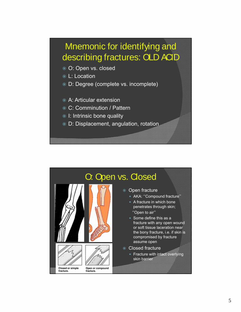
Mnemonic for identifying and describing fractures: OLD ACID O: Open vs. closed
L: Location
D: Degree (complete vs. incomplete)
A: Articular extension
C: Comminution / Pattern
I: Intrinsic bone quality
D: Displacement, angulation, rotation
O: Open vs. Closed Open fracture
AKA: “Compound fracture”
A fracture in which bone penetrates through skin;
“Open to air”
Some define this as a fracture with any open wound or soft tissue laceration near the bony fracture, i.e. if skin is compromised by fracture assume open
Closed fracture Fracture with intact overlying
skin barrier

6
L: Location Which bone?
Break into thirds (long bones) Proximal, middle, distal third
Anatomic orientation E.g. proximal, distal, medial,
lateral, anterior, posterior
Anatomic landmarks E.g. head, neck, body /
shaft, base, condyle
Segment (long bones) Epiphysis, physis,
metaphysis, diaphysis
Epiphysis
Metaphysis
Diaphysis
(Shaft)
Physis
Articular Surface
D: Degree of Fracture
Complete Complete cortical
circumference involved
Fragments are completely separated
Incomplete Cortex is not completely
compromised
“Only one cortex” involved
e.g “Greenstick fracture”

7
A: Articular Extension / Involvement
Intra-articular fractures
“Involves the articular surface”
Dislocation Loss of joint surface / articular congruity
Fracture-dislocation
C: Comminution / Pattern Transverse (Simple)
Oblique (Simple)
Spiral (Simple)
Linear / longitudinal
Segmental
Comminuted
Compression / impacted “Buckle / Torus”
Distraction / avulsion

8
Fracture Patterns
Atypical Fractures
• Greenstick
• Impacted
• Pathologic
• Stress
• Hairline
• Torus (buckle)

9
C: Comminution / Pattern Transverse (Simple)
C: Comminution / Pattern Oblique (Simple) Spiral (Simple)
Oblique in 2+ views

10
C: Comminution / Pattern Linear / longitudinal / split
C: Comminution / Pattern Segmental
Bone broken in 2+ separate places; Fx lines do not connect
11
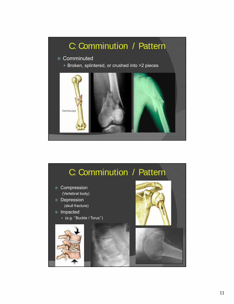
C: Comminution / Pattern Comminuted
Broken, splintered, or crushed into >2 pieces
C: Comminution / Pattern
Compression(Vertebral body)
Depression(skull fracture)
Impacted (e.g. “Buckle / Torus”)
12
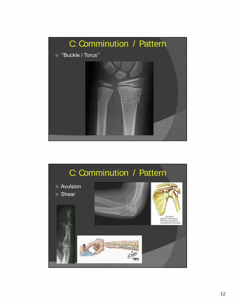
C: Comminution / Pattern “Buckle / Torus”
C: Comminution / Pattern Avulsion
Shear

13
I: Intrinsic Bone QualityOsteopenia– Decreased density
Normal
I: Intrinsic Bone Quality
Normal Osteopetrosis– Increased density

14
I: Intrinsic Bone Quality Osteopoikilosis
Focal areas of increased density
Normal
D: Displacement, Angulation, Rotation
Displacement
– Extent to which Fxfragments are not axially aligned
– Fragments shifted in various directions relative to each other
– Convention: describe displacement of distal fragment relative to proximal.
Complete, oblique tibial shaft fracture between distal & middle thirds; laterally displaced

15
D: Displacement, Angulation, Rotation
Angulation
– Extent to which fracture fragments are not anatomically aligned
In an angular fashion
– Convention: describe angulation as the direction the apex is pointing relative to anatomical long axis of the bone (e.g. apex medial, apex valgus), or direction of distal segment.
R tibial shaft fracture between proximal & middle thirds, angulated apex lateral (varus angulated)
D: Displacement, Angulation, Rotation
Angulation
Varus angulatedApex lateral
Valgus angulatedApex medial
ParallelNo angulation

16
D: Displacement, Angulation, Rotation
Rotation– Extent to which fracture
fragments are rotated relative to each other
– Convention: describe which direction the distalfragment is rotated relative to the proximal portion of the bone
ex: internal (towards midline) vs external (away from midline) rotation
D: Displacement, Angulation, RotationRotation
Normal AP view of hip– Greater trochanter in
profile
AP view of externally rotated hip Fx– Greater trochanter
perpendicular to film

17
Alternative Mnemonic: BLT LARD
B: Identify Bone
L: Location on bone
T: Type of fracture
L: Length changes
A: Angulation
R: Rotation
D: Displacement
Salter-Harris FracturesPediatric fracture involving physis (growth plate)

18
Salter-Harris II Fracture of Distal Femur
Salter-Harris IIIfracture distal tibia
Salter-Harris IV fracture distal tibia
19
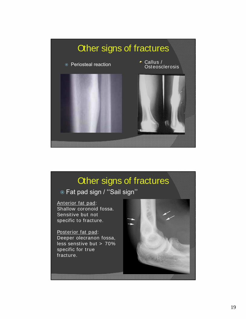
Other signs of fractures
Periosteal reaction Callus / Osteosclerosis
Other signs of fractures Fat pad sign / “Sail sign”
Anterior fat pad: Shallow coronoid fossa. Sensitive but not specific to fracture.
Posterior fat pad: Deeper olecranon fossa, less senstive but > 70% specific for true fracture.
20
Common Fracture Names and Eponyms
Jones’ Maisonneuve
Barton’s Monteggia
Bankart Segond
Bennet Pellegrini-stieda
Rolando Smith’s
Boxer’s Tillaux
Colles’ Lisfranc
Galleazzi Jefferson
Essex-Lopresti Chance
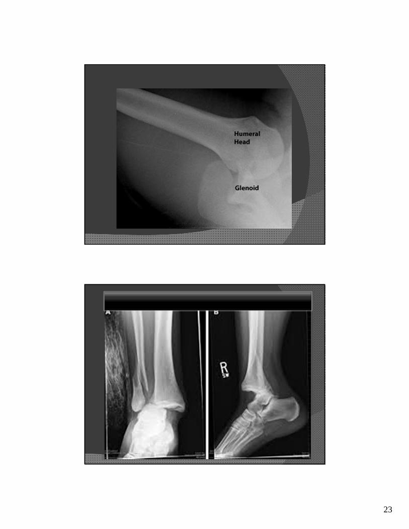
Joint Dislocations
Dislocation: Abnormal separation / discontinuity in a joint.
Subluxation: A partial / incomplete separation of a joint.
Same rules apply: Identify joint(s) involved in dislocation, determine direction of dislocation, and any associated fractures.

21
Description of Dislocations
Described by position of distal bone in relation to the proximal bone.
-Anterior (volar)
-Posterior (dorsal)
-Medial
-Lateral
-Any combination
Dorsal PIP Dislocation

22
23
24
Summary Systematically read X-rays
Bone, location, pattern, soft tissue
AO Classification complicated
Just describe what you see
Communicate and share with your consultants
Pre-reading
Succinct & accurate description of fractures
Interdisciplinary medical teams improve patient care
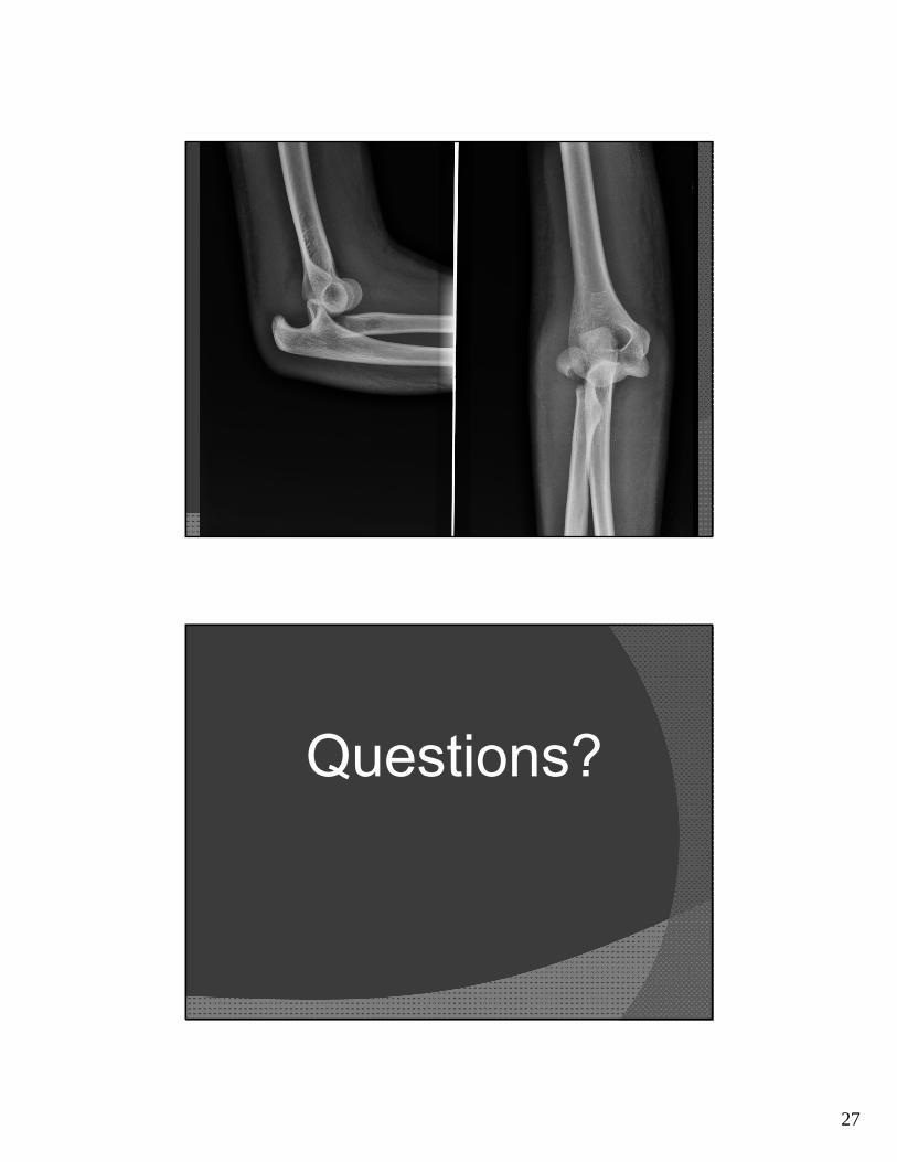
Examples
Let’s try a few examples…

25

26
27
Questions?

28
LECOM Health / Orthopedic & Sports Medicine 5401 Peach Street, Suite 3300, Erie, PA 16509
Ph: 814.868.7840 • Fax: 814.868.2139 [email protected]
Joshua A. Tuck, D.O., M.S. Orthopedic Surgeon
Specializing in Orthopedic Sports Medicine and Arthroscopy
Thank You!