Embed Size (px)
Citation preview

by Johan le Roux, Kim Laubscher & Michael Held
Introduction Fractures of the acetabulum and proximal femur are considered hip fractures. These injuries, and hip dislocations, are debilitating and must be treated urgently to allow the patient to regain mobilisation and avoid the morbidity and mortality associated with being bedridden.
Assessment Clinical Most patients with hip injuries have groin pain. In some patients with undisplaced or impacted fractures, mobilisation is still possible. Patients with displaced fractures or hip dislocations are usually not able to bear weight or raise a straight leg. The pull of the hip muscles will leave the leg in a particular position. With fractures, the leg is held in external rotation, abduction. With posterior dislocation (90% of dislocations) the leg held in flexion, adduction, internal rotation. Most commonly, the leg is shorter than the contralateral side.
Imaging AP and lateral X-ray of the hip and a full-length femur film are needed to exclude associated injuries. Disruption of Shenton’s line, loss of joint congruence, and a difference in femur head shape are indirect signs of an injury. In non-displaced cases, the fracture may be occult and not visible on X-rays. MRI is the gold standard for occult fractures. If this cannot be arranged within 24 hours, a CT scan is the next best choice. In hip dislocations, CT scans help visualise associated fractures of the acetabulum, femur head or neck, and exclude loose bodies in the joint.
X-ray pelvis (AP): Left hip fracture with disruption of the Shenton’s line (yellow)
Hip fractures and dislocations
Learning objectives 1. Low-energy hip fractures are often associated with significant osteoporosis. 2. Patients with hip fractures need a comprehensive medical investigation and
care. 3. Timely surgery and early mobilisation reduce complications and mortality in hip fractures.
Relevant anatomy The hip capsule originates from the acetabular margin and attaches on the greater and lesser trochanters and the intertrochanteric line. The femur neck is intracapsular, surrounded by synovial fluid, and not covered in periosteum, making the healing potential of these femoral neck fractures very poor. Another problem is the disruption of the medial circumflex femoral artery (the main blood supply to the femoral head), leading to avascular necrosis of the femoral head. The fractures can be grouped into displaced or undisplaced fractures, which guide surgical treatment. Peritrochanteric fractures occur between the lesser and greater trochanter. Therefore, they are extra-articular and do not cause medial circumflex femoral artery disruption and have a low risk of avascular necrosis.
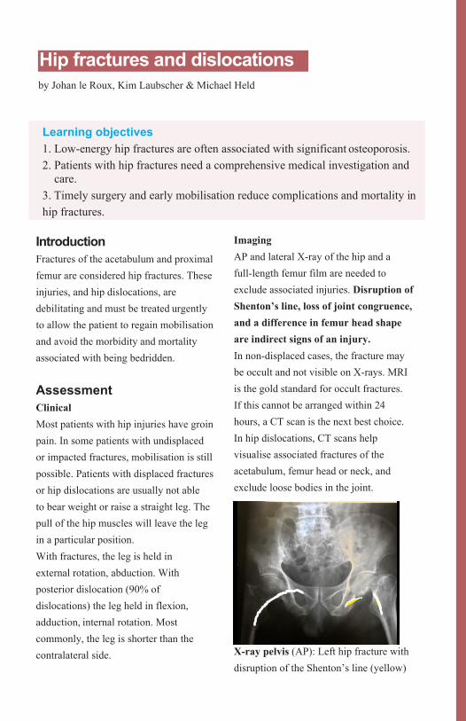
AP right hip: Peritrochanteric fracture with an intact femur neck.
Hip dislocation treatment These
are usually high-energy injuries with other associated injuries. A structured ATLS approach should be used during examination and initial treatment. Urgent closed reduction in line with the deformity under adequate analgesia and sedation are crucial. Neurovascular examination and documentation before and after reduction are mandatory. If unable to achieve closed reduction, closed reduction under general anaesthesia and muscle relaxant in theatre is the next step. If still unsuccessful, open reduction must be performed.
Femoral head fracture These are rare fractures and are usually associated with hip dislocations. The hip dislocation must be reduced initially. The fracture can be treated conservatively if it is in a non-weight bearing area, non-displaced and not associated with other injuries. Otherwise, fixation is required.
AP X-ray pelvis: Left hip dislocation. The Shenton’s line is disrupted. The hip is internally rotated (lesser trochanter is not visible), and the femoral head appears smaller than the contralateral side (closer to the X-ray film).
Hip fracture treatment After excluding other injuries (especially distal radius fractures and head injuries) and after neuromuscular structures are assessed, the patient should be made comfortable and evaluated for a general anaesthetic (see table). The race against time starts now as early mobilisation leads to improved outcomes if patients are treated within 48 hours of injury, thus preventing life-threatening complications such as DVT/PE, urinary tract infections and pneumonia. This is considered a surgical emergency and most centres plan the operation on the next available surgical list. A continued, coordinated orthogeriatric and multidisciplinary management of these patients with investigations for osteoporosis and fall prevention remains the gold standard, which few receive.
Operative treatment Non-operative treatment is only considered in patients who are unfit for anaesthesia and surgery. For neck of femur fractures, open reduction and internal fixation are indicated for most physiologically young patients. Undisplaced fractures are treated with cannulated screws or a sliding hip screw. Arthroplasty is indicated for elderly patients with displaced fractures.
For peri-trochanteric fractures, a dynamic hip screw is done in stable fracture patterns. In unstable fractures, a cephalomedullary nail is indicated.
(oblique fractures which would displace with dynamic compression of the hip screw, fractures with subtrochanteric extension or medial comminution)
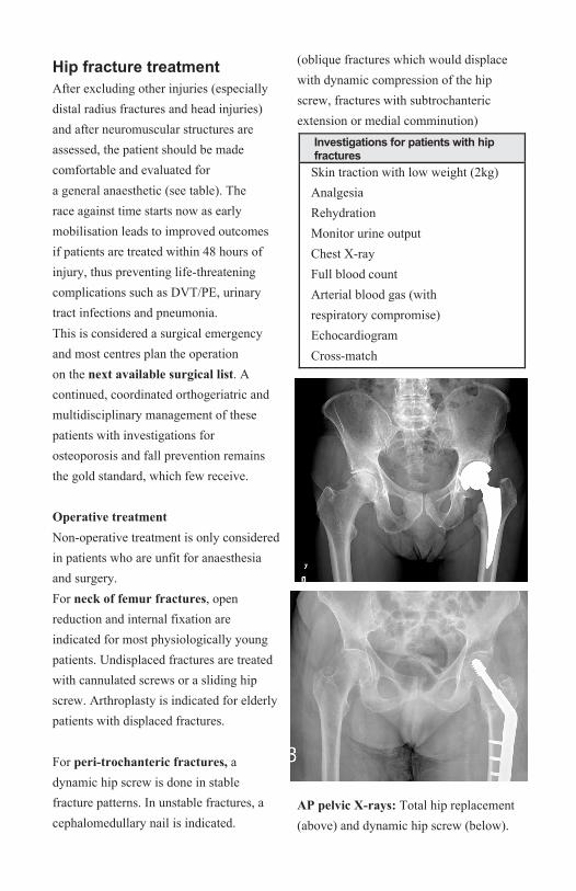
Investigations for patients with hip fractures Skin traction with low weight (2kg) Analgesia Rehydration Monitor urine output Chest X-ray Full blood count Arterial blood gas (with respiratory compromise) Echocardiogram Cross-match
AP pelvic X-rays: Total hip replacement (above) and dynamic hip screw (below).
![TREATMENT FRACTURES AND DISLOCATIONS, · [From theMedicaland Surgical ReporterofOctober 26, 1861.] A. NEW METHOD FOR THE TREATMENT OF FRACTURES AND DISLOCATIONS, WITHFRACTURESIN ANDNEARTHEELBOW^OSSf;](https://img.dokumen.tips/doc/110x75/6016d5bee3a1eb7ab135d0e2/treatment-fractures-and-dislocations-from-themedicaland-surgical-reporterofoctober.jpg)