Embed Size (px)
Citation preview

Three-dimensional Structure of Myelin Basic ProteinII. MOLECULAR MODELING AND CONSIDERATIONS OF PREDICTED STRUCTURES INMULTIPLE SCLEROSIS*
(Received for publication, September 19, 1996, and in revised form, December 2, 1996)
Ross A. Ridsdale‡, Daniel R. Beniac‡, Thomas A. Tompkins‡§¶, Mario A. Moscarello§, andGeorge Harauz‡i
From the ‡Department of Molecular Biology and Genetics, University of Guelph, Guelph, Ontario N1G 2W1,Canada and §Department of Biochemistry Research, Hospital for Sick Children, 555 University Avenue,Toronto, Ontario M5G 1X8, Canada
A computational model of myelin basic protein (MBP)has been constructed based on the premise of a phylo-genetically conserved b-sheet backbone and on electronmicroscopical three-dimensional reconstructions. Manyresidues subject to post-translational modification(phosphorylation, methylation, or conversion of argin-ines to citrullines) were located in loop regions and thusaccessible to modifying enzymes. The triproline seg-ment (residues 99–101) is fully exposed on the back sur-face of the protein in a long crossover connection be-tween two parallel b-strands. The proximity of thisregion to the underlying b-sheet suggests that post-translational modifications here might have potentialsynergistic effects on the entire structure. Post-transla-tional modifications that lead to a reduced surfacecharge could result first in a weakened attachment tothe myelin membrane rather than in a gross conforma-tional change of the protein itself. Such mechanismscould be operative in demyelinating diseases such asmultiple sclerosis.
Myelin basic protein (MBP)1 is one of the most importantproteins of the myelin sheath (1–6). Its significance is demon-strated in the shiverermutant of mouse, which has only a smallamount of structurally unstable myelin because the gene forMBP is mostly deleted (7, 8). This trait is recessive and inher-ited in a Mendelian manner, indicating that MBP is coded forby a single gene. In mammals, the gene consists of seven exons,and differential splicing of the primary MBP mRNA leads todifferent isoforms of MBP, i.e. forms of differing molecularweights (9–11). Alternative splicing of mRNA transcripts is acommon mechanism for generating protein diversity. The MBPgene is thus similar to the genes for SV40 T and t antigens,fibrinogen, lens a-A crystallin, and troponin T, in all of whichprimary transcripts with identical termini are alternativelyspliced to yield different mature mRNAs (10). The shark MBPgene has also been cloned and has revealed a similar exon
structure, indicating that this protein issued early in verte-brate evolution (11).In mammals, the 18.5- and 14-kDa isoforms of MBP are the
most common, although the relative proportions vary duringdevelopment and among species. Henceforth, unless otherwisespecified, we shall be using “MBP” to refer to the 18.5-kDaform. Each isoform of MBP can exist as one of many possiblecharge isomers, due to various post-translational modifications(3, 4). These charge isomers are denoted C1, C2, C3, C4, C5, C6,and C8, according to their elution profile on a cation exchangecolumn at pH 10.5 (12). Component C1 is the least modifiedand most basic component, while successive components differsequentially by the loss of a positive charge. Component C8 isan isoform of MBP that does not bind to the resin, and it is themost modified, containing several citrullinyl residues (13). Thepost-translational modifications include phosphorylation,ADP-ribosylation, and conversion of arginines to citrulline (3,4, 13). The latter change is relevant to multiple sclerosis; oftenfour or five arginines are so converted (14). In a recent case offulminating multiple sclerosis known as Marburg’s Disease, ayoung (26-year-old) woman presented with the disease anddied within 6 weeks. In the MBP extracted from the autopsiedbrain, 18 of 19 arginine residues had been citrullinated (15).The sequences of most forms of MBP from numerous species
are known, with the human and bovine forms having beensequenced first (16, 17). We shall henceforth be referring to thehuman sequence. The single Trp116 serves as a focus for im-munological properties of MBP and is proximal to a triproline(Pro99-Pro100-Pro101) segment (2). Residues around this tripro-line segment are often modified. Myelin basic protein has anumber of sequence and other similarities with other proteinfamilies (18, 19). Properties such as charge density, post-trans-lational modification by addition of fatty acids, and overallhydrophobicity are similar in MBP and proteolipid proteinfrom the central nervous system and in the pulmonary surfac-tant proteins SP-B and SP-C (20). Other sequence similaritieswith viral proteins have stoked conjectures on viral involve-ment in multiple sclerosis (21–24).Because of the reluctance of MBP to form crystals (25), its
detailed tertiary structure is not known. The main structuralmodels of this protein that exist are from the 1980s and repre-sent abstract syntheses of biochemical data and secondarystructure prediction algorithms (Refs. 26–29; see also Refs. 30,31). The structure of a small subsegment (five residues) hasrecently been solved by nuclear magnetic resonance (32). Themost sophisticated structural models of the whole protein re-main those of Stoner (27) and Martenson (28), which werebased on extensive biochemical and secondary structural data.These structures were represented at the time schematically,
* This work was supported by the Multiple Sclerosis Society of Can-ada (operating grant to G. H.). The molecular modeling workstationwas purchased with the assistance of an equipment grant to Dr. R.Yada (Food Science, University of Guelph) from the Natural Sciencesand Engineering Research Council of Canada. The costs of publicationof this article were defrayed in part by the payment of page charges.This article must therefore be hereby marked “advertisement” in ac-cordance with 18 U.S.C. Section 1734 solely to indicate this fact.¶ Present address: Lallemand Inc., Laboratoire R & D, 6100 Royal-
mount, Montreal, Quebec H4P 2R2, Canada.i To whom correspondence should be addressed: Tel.: 519-824-4120
(ext. 2535); Fax: 519-837-2075; E-mail: [email protected] The abbreviations used are: MBP, myelin basic protein; PDB,
Brookhaven Protein Data Bank.
THE JOURNAL OF BIOLOGICAL CHEMISTRY Vol. 272, No. 7, Issue of February 14, pp. 4269–4275, 1997© 1997 by The American Society for Biochemistry and Molecular Biology, Inc. Printed in U.S.A.
This paper is available on line at http://www-jbc.stanford.edu/jbc/ 4269
by guest on September 7, 2018
http://ww
w.jbc.org/
Dow
nloaded from

with parts thereof as plastic Corey-Pauling-Koltun space-fill-ing models. Our own recent work has comprised electron mi-croscopical investigations of the tertiary structure of bovineMBP, which is almost identical to human MBP (see accompa-nying paper (33)). In the process of recreating computationalrepresentations of both the Stoner and Martenson models forcomparison with our experimental results, we designed a re-vised structure, incorporating our new electron microscopicalreconstructions as tertiary structural constraints. This newmodel of human MBP is presented here.
MATERIALS AND METHODS
The main tool used in this aspect of our studies was the INSIGHT IImolecular modeling software package (Biosym Corp., Parsippany, NJ)running on an IBM RISC/6000 Powerstation 3AT (International Busi-ness Machines, Markham, Ontario, Canada). This commercial programcontains utilities for building polymers of nucleic acids and proteins,multiple sequence alignment and homology modeling, structure refine-ment by rotamer rotation, energy function minimization, and graphicdisplay of specified sites. There are no crystallographic or NMR struc-tures of any proteins with significant overall sequence similarity toMBP in the Brookhaven Protein Data Bank (PDB). We thus could notconstruct MBP by homology modeling but used instead a piecemealstrategy. We relied strongly on Stoner’s (27, 29) and Martenson’s (26,28) definitions of the residues forming a b-sheet secondary structure.For Stoner’s model (27), we required a flat antiparallel b-sheet (Fig.
1), which we derived initially from the structure of excitation energytransfer bacteriochlorophyll A protein (PDB accession code 3bcl). TheINSIGHT program was used to make the putative a-helical regionsdirectly using specified values of (f,c). These a-helices were placedapproximately where Stoner indicated them to lie. Loops then joined allthe secondary structure elements, and the entire structure was refinedby rotamer selection and energy function minimization. Rotamer selec-tion means choosing the best of a series of known possible amino acidside chain conformations to minimize steric overlap with any otheratoms in the structure. The energy function comprises electrostatic(including hydrogen bonding) and van der Waals forces among theatoms of the model. Physically implausible models (e.g. with overlap-ping atoms) yield divergent results at this point. This energy is not thefree energy of folding of the protein but serves only as an internal guideto refining the structure.For Martenson’s model (28), we required two layered b-sheets. The
relevant domain of the bacteriochlorophyll A protein was again thestarting point, although this time two bacteriochlorophyll A sheets wereplaced over one another and then spliced together to form the twist asindicated byMartenson in a schematic diagram (see Fig. 1). Loops againjoined the b-strands. There were no a-helices defined here.Finally, our model was based on the previous work by Stoner and
Martenson as well as on our new electron microscopical data (33). Forthis model, we first used the flat antiparallel b-sheet from the structureof bacteriochlorophyll A protein and later, for the results presentedhere, from the structure of severin (PDB accession code 1svr). Whereexperimental data on the structure were available, such as the NMRstructure of a tetradecapeptide repeat (32), they were incorporated intoour model. Amino acids 98–102 in human MBP (Thr-Pro-Pro-Pro-Ser)showed a 100% identity with a pentapeptide segment in endo-b-N-acetylglucosaminidase F1 (PDB accession code 2ebn), and the lattercoordinates were used directly to model the former region.To begin, the relevant regions of the human MBP sequence were
modeled as b-strands using calculated (f,c) angles and overlaid ontothe b-sheet of severin, and the b-strand coordinates of the knownstructure were used to form the nascent human MBP model. The 2ebnpentapeptide comprising the triproline segment was moved to fit intothe right-handed crossover region between adjacent parallel b-strandsb3 and b4. The (f,c) angles of the tetradecapeptide solved recently byNMR were used to model this segment, move it into a correct positionwith respect to the b-sheet, and assign coordinates. Finally, thesesegments were joined by loops using the electron microscopical recon-structions to constrain the loops’ positions. As before, structure refine-ment comprised rotamer definition and energy function minimization.Post-translational modifications were constructed interactively us-
ing the utilities of the INSIGHT II program. To perform a computa-tional phosphorylation, for example, a phosphate group was selectedfrom an internal library and then bonded onto the rest of the moleculeusing a built-in function. The bond type (e.g. single) is chosen by theuser, as well as the two hydrogen atoms to be removed by the bonding
reaction. A computational citrullinlization was performed essentiallythe same way after deleting one of the terminal NH2 moieties of theguanidino group of arginine and replacing it with an oxygen atom.
RESULTS AND DISCUSSION
Although the sequences of MBP from many species areknown, there is no accurate way to calculate a tertiary struc-ture from them alone. If the atomic structure of a protein witha significantly similar amino acid sequence were known, thenone could align the MBP sequence with this homologous se-quence and assign atomic coordinates to the amino acids ofMBP based on the known structure. Refinement of the pre-dicted structure then involves rotating amino acid side chainsto prevent steric overlap, filling in gaps in the structure byloops, and finally modifying bond lengths and atomic coordi-nates to minimize an energy function as defined above. Unfor-tunately, this homology modeling approach per se is not viablehere because no atomic structures are known of proteins sim-ilar enough to MBP. Nonetheless, we have created here threequantitative models of MBP starting from different premisesand essentially constructed interactively residue by residue.Previous Structural Models of Human MBP—In the 1980s,
three-dimensional models of MBP were proposed independ-
FIG. 1. The topologies of arrangement of b-strands in theStoner (27) (a) and Martenson (28) (b) models of myelin basicprotein.
Molecular Model of Myelin Basic Protein4270
by guest on September 7, 2018
http://ww
w.jbc.org/
Dow
nloaded from
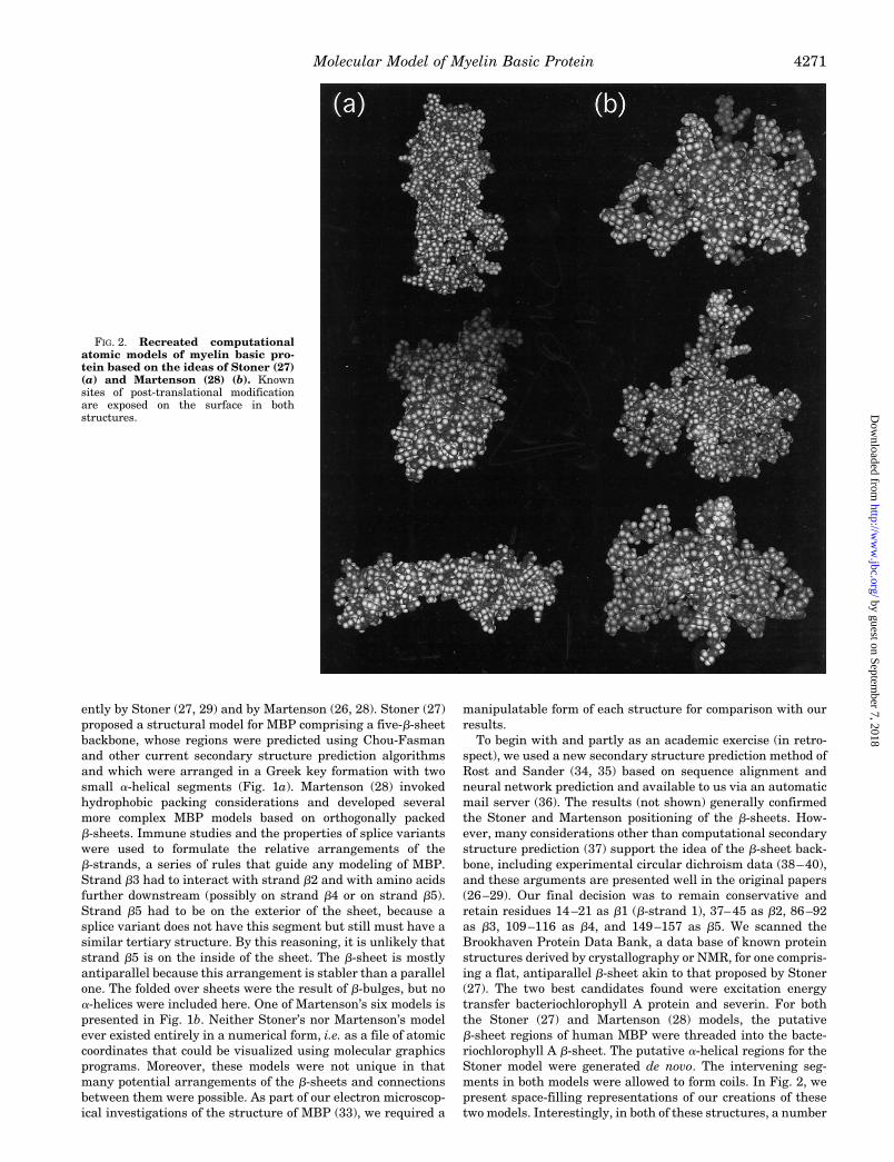
ently by Stoner (27, 29) and by Martenson (26, 28). Stoner (27)proposed a structural model for MBP comprising a five-b-sheetbackbone, whose regions were predicted using Chou-Fasmanand other current secondary structure prediction algorithmsand which were arranged in a Greek key formation with twosmall a-helical segments (Fig. 1a). Martenson (28) invokedhydrophobic packing considerations and developed severalmore complex MBP models based on orthogonally packedb-sheets. Immune studies and the properties of splice variantswere used to formulate the relative arrangements of theb-strands, a series of rules that guide any modeling of MBP.Strand b3 had to interact with strand b2 and with amino acidsfurther downstream (possibly on strand b4 or on strand b5).Strand b5 had to be on the exterior of the sheet, because asplice variant does not have this segment but still must have asimilar tertiary structure. By this reasoning, it is unlikely thatstrand b5 is on the inside of the sheet. The b-sheet is mostlyantiparallel because this arrangement is stabler than a parallelone. The folded over sheets were the result of b-bulges, but noa-helices were included here. One of Martenson’s six models ispresented in Fig. 1b. Neither Stoner’s nor Martenson’s modelever existed entirely in a numerical form, i.e. as a file of atomiccoordinates that could be visualized using molecular graphicsprograms. Moreover, these models were not unique in thatmany potential arrangements of the b-sheets and connectionsbetween them were possible. As part of our electron microscop-ical investigations of the structure of MBP (33), we required a
manipulatable form of each structure for comparison with ourresults.To begin with and partly as an academic exercise (in retro-
spect), we used a new secondary structure prediction method ofRost and Sander (34, 35) based on sequence alignment andneural network prediction and available to us via an automaticmail server (36). The results (not shown) generally confirmedthe Stoner and Martenson positioning of the b-sheets. How-ever, many considerations other than computational secondarystructure prediction (37) support the idea of the b-sheet back-bone, including experimental circular dichroism data (38–40),and these arguments are presented well in the original papers(26–29). Our final decision was to remain conservative andretain residues 14–21 as b1 (b-strand 1), 37–45 as b2, 86–92as b3, 109–116 as b4, and 149–157 as b5. We scanned theBrookhaven Protein Data Bank, a data base of known proteinstructures derived by crystallography or NMR, for one compris-ing a flat, antiparallel b-sheet akin to that proposed by Stoner(27). The two best candidates found were excitation energytransfer bacteriochlorophyll A protein and severin. For boththe Stoner (27) and Martenson (28) models, the putativeb-sheet regions of human MBP were threaded into the bacte-riochlorophyll A b-sheet. The putative a-helical regions for theStoner model were generated de novo. The intervening seg-ments in both models were allowed to form coils. In Fig. 2, wepresent space-filling representations of our creations of thesetwo models. Interestingly, in both of these structures, a number
FIG. 2. Recreated computationalatomic models of myelin basic pro-tein based on the ideas of Stoner (27)(a) and Martenson (28) (b). Knownsites of post-translational modificationare exposed on the surface in bothstructures.
Molecular Model of Myelin Basic Protein 4271
by guest on September 7, 2018
http://ww
w.jbc.org/
Dow
nloaded from

of the more interesting sites of post-translational modification,such as Ser7, Thr98, and the various arginines are all exposedon the surface.A New Structural Model of Human MBP—The Stoner and
Martenson models of MBP represent thoughtful syntheses ofbiochemical data available on this protein. Both structures areplausible within the limitations of a rarefied computationalrepresentation. However, the shapes of these models cannoteasily be reconciled with our derivation of the appearance ofMBP from electron microscopical data (33). Our three-dimen-sional reconstruction has an outer circumference of approxi-mately 15 nm and a thickness of 2.5 nm (roughly the differencebetween the outer and inner radii). The Stoner and Martensonmodels both have the two longest loop regions (betweenb-sheets b2 and b3 and sheets b4 and b5) on the same side ofthe molecule. As a result, the maximum length of these modelsis about 10 nm. Also, the Martenson model is too thick to fitinto the experimentally determined volume. As a result, weconstructed a new model to fit our electron microscopicalreconstruction.To begin our new model, we retained the idea of a b-sheet
backbone envisaged by Stoner and Martenson but did not re-tain their strand order. The b-sheet coordinates were derivedfrom the structure of severin (Fig. 3c) (41). We further reasonedon the basis of total length (circumference) considerations thatthe two long loops must be on opposite sides of the b-sheet. Theb-sheet was placed in the center of the electron microscopicalvolume to allow the long loops to fit into it. Martenson’s rules(based on immunological and other data and described above)are still consistent with approximately 60 arrangements of theb-sheets. Fortunately, the dimensions of the EM reconstructionconstrained the number of potential arrangements of theb-sheets to two (Fig. 3, a and b). The first topology has twocrossovers and an uncommon antiparallel b-sheet arrange-ment. We chose the second topology with only one right-handed(more common than left-handed in biological systems) cross-over connection. The first topology with two crossovers wouldhave made the molecule too thick, notwithstanding that onecrossover would have been too short to traverse the neededdistance. The b-sheet was oriented so that the right-handedcrossover connection was on the outer surface. Surface “bumps”on the exterior fit the crossover better, and modifiable aminoacids became accessible. Amino acids 5–11 were linked using(f,c) angles derived from a recent NMR study (32).In Fig. 4, we show the correspondence between our model
and the electron microscopical reconstruction of bovineMBP/C1 in low salt buffer (33). This structure was an open “C”shape. A second reconstruction in higher salt buffer yielded amore compact form of the protein, but a form that could be seento represent a closing in of the “C”. The human MBP modelcould be fit into this new volume simply by cutting bonds to theloop regions, reorienting them, and resplicing. In Fig. 5, a andb, more detailed space-filling representations show the generalshape and especially the modifiable sites of both of these newhuman MBP models. As noted above, the backbone of thestructure is a b-sheet modeled after that of severin. Both sev-erin and MBP are actin- and lipid-binding proteins (41–44).The triproline segment is fully exposed on the back surface ofthe protein, in a crossover loop between adjacent parallelstrands. There is a positive congruence in that this region inthe bacterial endo-b-N-acetylglucosamine F1 (2ebn) is also lo-cated in a crossover between parallel b-strands. In our humanMBP model, many sites of post-translational modification areclustered around the triproline segment. It is tempting to spec-ulate that the clustering of modifiable amino acids at such aposition connecting two b-strands has structural importance.
We can suggest with somewhat more certainty that the re-duced surface charge density upon citrullinization of arginines(as occurs in multiple sclerosis) will reduce the interaction withlipids in the myelin membrane, accounting for a certainamount of destabilization.Given the limited resolution of the first electron microscop-
ical reconstructions (33), the correspondence that could beachieved between the experimental data and our model isremarkable. Although we do not wish to encourage the practiceof formulating atomic models of proteins based solely on suchelectron microscopical structures, this strategy appears to havebeen fruitful here for human MBP. This is a small protein forwhich no direct structural information has hitherto been avail-able, yet for which a wealth of biochemical data could be ex-ploited to initiate and guide the model building process. Theenvelope of the three-dimensional reconstruction within whichwe had to fit the atomic model was a valuable constraint, whichreduced the number of possible topologies of b-strand and looparrangement to only two. The extended “C” shape of the proteinis credible given the peripheral membrane association of thisprotein, and its conformational flexibility in different condi-tions is also likely (38–40). The recent literature has examplesof other proteins that have been described as “holy” (sic., mean-ing “holey”, i.e. with a distinct nonglobular fold) (45–48). There
FIG. 3. An electron microscopical reconstruction on bovineMBP (33) suggested a total circumference of approximately 15nm for the “C”-shaped molecule. To fit inside this volume, it wasnecessary to have the putative loops of MBP on opposite sides of thecentral b-sheet. The number of possible topologies was thus reduced tothe two shown here in a and b. Both topologies are consistent withMartenson’s (28) rules. c, a ribbon diagram representation of severin,an actin-binding protein, whose backbone is a flat, antiparallel b-sheet(Brookhaven Protein Data Bank accession number 1svr) (41).
Molecular Model of Myelin Basic Protein4272
by guest on September 7, 2018
http://ww
w.jbc.org/
Dow
nloaded from

is also a family of pleckstrin homology domains (49) identifiedin various membrane-associated and signal transduction pro-teins that resembles our MBP model and might have a signif-icant relationship.Conclusions and Future Directions—Multiple sclerosis is a
human demyelinating disease. An autoimmune response to oneor more of its protein components is thought to be part of thepathogenesis. It has been postulated that MBP is the agent ofautoimmunity and that post-translational modifications ofMBP play a key role in the demyelinating process at the mo-lecular level. Knowledge of the tertiary structures of the MBPisoforms and charge isomers is essential to understanding theorganization of the myelin membrane and the mechanisms ofdevelopment of autoimmunity in multiple sclerosis. However,MBP has not been crystallized, and may never be crystallized,for high resolution x-ray diffractometry.
A three-dimensional model of MBP structure can now only beformulated with the assistance of structure prediction algo-rithms and on the basis of extensive biochemical and biophys-ical data that are available. In the accompanying paper (33), wehave described our structural studies of bovine MBP chargeisomer C1 associated with lipid monolayers. By electron micro-scopical angular reconstitution, we found bovine MBP/C1 toposses an overall “C” shape. In this paper, we used molecularmodeling software to create quantititive atomic coordinatemodels of human MBP and to localize certain post-transla-tional modifications relevant to multiple sclerosis. The three-dimensional electron microscopical reconstruction served as anenvelope within which an atomic model comprising fiveb-sheets and a large proportion of irregular coil could beuniquely packaged. In this model, the most important modifi-able amino acids are accessible to enzymes such as kinases. To
FIG. 4. Fitting a molecular model ofMBP to the electron microscopicalreconstruction. The reconstructed vol-ume is represented here by the light bluemesh contour, and a Corey-Pauling-Kol-tun representation of a model of MBP isrepresented in purple. a–d show the mol-ecule rotating at 90° intervals around ahorizontal axis lying in the plane of thepage. e shows the molecule rotated 90°from a around a vertical axis lying in theplane of the page. f is an oblique perspec-tive of the structure.
Molecular Model of Myelin Basic Protein 4273
by guest on September 7, 2018
http://ww
w.jbc.org/
Dow
nloaded from

this extent, the model is plausible.The model for human MBP that we present here is the first
of its kind for this very important protein. It was inspired bymodel-building exercises of the last decade, and we anticipatethat details will change as new data are incorporated into it. Itsvalue shall lie in its utility as a workbench for incorporatingexperimental evidence and for formulating experimentallytestable hypotheses. One idea suggested here is that deimina-tion of argininyl residues, with the accompanying loss of posi-tive charge, does not destabilize MBP’s tertiary structure per sebut rather its interactions with negatively charged lipids.
Acknowledgment—We are especially grateful to Bob Creedy (Com-puting and Communication Services, University of Guelph), for effortsin maintaining the molecular modeling workstation.
REFERENCES
1. Deber, C. M. & Reynolds, S. J. (1991) Clin. Biochem. 24, 113–1342. Lees, M. B. & Brostoff, S. W. (1984) in Myelin (Morell, P., ed) 2nd Ed., pp.
197–224, Plenum Press, New York3. Moscarello, M. A. (1996) in Cell Biology and Pathology of Myelin: Evolving
Biological Concepts and Therapeutic Approaches (Devon, R. M., Doucette,R., Juurlink, B. H. J., Nazarali, A. J., Schreyer, D. J. & Verge, V. M. K.)Plenum Publishing Corp., New York
4. Smith, R. (1992) J. Neurochem. 59, 1589–16085. Staugaitis, S. M., Colman, D. R. & Pedraza, L. (1996) BioEssays 18, 13–186. Wood, D. D. & Moscarello, M. A. (1996) in The Molecular Biology of Multiple
Sclerosis (Russell, W., ed.) John Wiley & Sons, Inc., New York7. Roach, A., Takahashi, N., Pravtcheva, D., Ruddle, F. & Hood, L. (1985) Cell 42,
149–1558. Mateu, L., Luzzati, V., Vonasek, E., Borgo, M. & Lachapelle, F. (1996) J. Mol.
Biol. 256, 319–3299. Campagnoni, A. T. (1988) J. Neurochem. 51, 1–1410. de Ferra, F., Engh, H., Hudson, L., Kamholz, J., Puckett, C., Molineaux, S. &
Lazzarini, R. A. (1985) Cell 43, 721–72711. Saavedra, R. A., Fors, L., Aebersold, R. H., Arden, B., Horvath, S., Sanders, J.
FIG. 5. Ribbon and van der Waalsrepresentations of our model of hu-man MBP viewed from different an-gles. a, a model to fit the three-dimen-sional electron microscopical reconstruc-tion (an open “C” shape) from low saltbuffer. b, a model to fit the three-dimen-sional electron microscopical reconstruc-tion (a more compact form) from high saltbuffer. Arginines subject to citrulliniza-tion are presented in red. The triprolinesegment forms part of the crossover con-nection, part of which is accentuated inlight blue. Serines that are colored greenare subject to phosphorylation. A threo-nine residue (Thr98) that is phosphoryl-ated by a mitogen-activated protein ki-nase is in purple. Hydrogen atoms arecoloredwhite, and other atoms are coloredyellow.
Molecular Model of Myelin Basic Protein4274
by guest on September 7, 2018
http://ww
w.jbc.org/
Dow
nloaded from

& Hood, L. (1989) J. Mol. Evol. 29, 149–15612. Chou, F. C.-H., Chou, C.-H. J., Shapira, R. & Kibler, R. F. (1976) J. Biol. Chem.
251, 2671–267913. Wood, D. D. & Moscarello, M. A. (1989) J. Biol. Chem. 264, 5121–512714. Moscarello, M. A., Wood, D. D., Ackerley, C. & Boulias, C. (1994) J. Clin.
Invest. 94, 146–15415. Wood, D. D., Bilbao, J. M., O’Connors, P. & Moscarello, M. A. (1996) Ann.
Neurol. 40, 18–2416. Carnegie, P. R. (1971) Biochem. J. 123, 57–6717. Eylar, E. H., Brostoff, S., Hashim, G., Caccam, J. & Burnett, P. (1971) J. Biol.
Chem. 246, 5770–578418. Karthigasan, J., Inouye, H. & Kirschner, D. A. (1995) Med. Hypotheses 45,
245–24019. Kirschner, D. A., Inouye, H., Ganser, A. L. & Mann, V. (1989) J. Neurochem.
53, 1599–160920. Perez-Gil, J. & Keough, K. M. W. (1994) J. Theor. Biol. 169, 221–22921. Fujinami, R. S. & Oldstone, M. B. A. (1985) Science 230, 1043–104522. Martenson, R. E., Deibler, G. E. & Kies, M. W. (1971) Nat. New Biol. 234,
87–8923. Shaw, S.-Y., Laursen, R. A. & Lees, M. B. (1986) FEBS Lett. 207, 266–27024. Steinman, L. (1996) Cell 85, 299–30225. Sedzik, J. & Kirschner, D. A. (1992) Neurochem. Res. 17, 157–16626. Martenson, R. E. (1981) J. Neurochem. 36, 1543–156027. Stoner, G. L. (1984) J. Neurochem. 43, 433–44728. Martenson, R. E. (1986) J. Neurochem. 46, 1612–162229. Stoner, G. L. (1990) J. Neurochem. 55, 1404–141130. Golubovich, V. P., Kirnaskh, L. I. & Galaktionov, S. G. (1989) Biophysics 34,
395–39931. Inouye, H. & Kirschner, D. A. (1991) J. Neurosci. Res. 28, 1–17
32. Mendz, G. L., Barden, J. A. & Martenson, R. E. (1995) Eur. J. Biochem. 231,659–666
33. Beniac, D. R., Luckevich, M. D., Czarnota, G. J., Tompkins, T. A., Ridsdale, R.A., Ottensmeyer, F. P., Moscarello, M. A. & Harauz, G. (1997) J. Biol. Chem.272, 4261–4268
34. Rost, B. & Sander, C. (1993) Proc. Natl. Acad. Sci. U. S. A. 90, 7558–756235. Rost, B. & Sander, C. (1994) Proteins 19, 55–7236. Rost, B., Sander, C. & Schneider, R. (1994) Comput. Applic. Biosci. 10, 53–6037. Lesk, A. M. (1988) in Computational Molecular Biology (Lesk, A. M., ed) pp.
192–197, Oxford University Press, Oxford38. Ramwani, J. J., Epand, R. M. & Moscarello, M. A. (1989) Biochemistry 28,
6538–654339. Stuart, B. H. (1996) Biochem. Mol. Biol. Int. 38, 839–84540. Surewicz, W. K., Moscarello, M. A. & Mantsch, H. H. (1987) Biochemistry 26,
3881–388641. Eichinger, L. & Schleicher, M. (1992) Biochemistry 31, 4779–478742. Barylyko, B. & Dobrowolski, Z. (1984) Eur. J. Cell Biol. 35, 327–33543. Dobrowolski, Z., Osinska, H., Mossakowska, M. & Barylyko, B. (1986) Eur. J.
Cell Biol. 42, 17–2644. Roth, G. A., Gonzalez, M. D., Monferran, C. G., DeSantis, M. L. & Cumar, F.
A. (1993) Neurochem. Int. 23, 459–46545. Hofsteenge, J. (1994) Curr. Opin. Struct. Biol. 4, 807–80946. Dijkstra, B. W. & Thunnissen, A.-M. W. H. (1994) Curr. Opin. Struct. Biol. 4,
810–81347. Neil, K. J., Ridsdale, R. A., Rutherford, B. L., Taylor, L., Larson, D. E., Glibetic,
M., Rothblum, L. I. & Harauz, G. (1996) Nucl. Acids Res. 24, 1472–148048. Scott, J. E. (1996) Biochemistry 35, 8795–879949. Lemmon, M. A., Ferguson, K. M. & Schlessinger, J. (1996) Cell 85, 621–624
Molecular Model of Myelin Basic Protein 4275
by guest on September 7, 2018
http://ww
w.jbc.org/
Dow
nloaded from

George HarauzRoss A. Ridsdale, Daniel R. Beniac, Thomas A. Tompkins, Mario A. Moscarello and
MULTIPLE SCLEROSISMODELING AND CONSIDERATIONS OF PREDICTED STRUCTURES IN
Three-dimensional Structure of Myelin Basic Protein: II. MOLECULAR
doi: 10.1074/jbc.272.7.42691997, 272:4269-4275.J. Biol. Chem.
http://www.jbc.org/content/272/7/4269Access the most updated version of this article at
Alerts:
When a correction for this article is posted•
When this article is cited•
to choose from all of JBC's e-mail alertsClick here
http://www.jbc.org/content/272/7/4269.full.html#ref-list-1
This article cites 45 references, 7 of which can be accessed free at
by guest on September 7, 2018
http://ww
w.jbc.org/
Dow
nloaded from