Embed Size (px)
Citation preview

Page 1 of 15
The inguinal and femoral canals: a practical step-by-stepapproach to accurate sonographic assessment.
Poster No.: C-1243
Congress: ECR 2014
Type: Educational Exhibit
Authors: P. Yoong1, C. A. Johnson1, L. A. Fowkes2, S. Duffy3, T. Marshall3;1Oxford/UK, 2London/UK, 3Norwich/UK
Keywords: Abdomen, Ultrasound, Diagnostic procedure, Education andtraining
DOI: 10.1594/ecr2014/C-1243
Any information contained in this pdf file is automatically generated from digital materialsubmitted to EPOS by third parties in the form of scientific presentations. Referencesto any names, marks, products, or services of third parties or hypertext links to third-party sites or information are provided solely as a convenience to you and do not inany way constitute or imply ECR's endorsement, sponsorship or recommendation of thethird party, information, product or service. ECR is not responsible for the content ofthese pages and does not make any representations regarding the content or accuracyof material in this file.As per copyright regulations, any unauthorised use of the material or parts thereof aswell as commercial reproduction or multiple distribution by any traditional or electronicallybased reproduction/publication method ist strictly prohibited.You agree to defend, indemnify, and hold ECR harmless from and against any and allclaims, damages, costs, and expenses, including attorneys' fees, arising from or relatedto your use of these pages.Please note: Links to movies, ppt slideshows and any other multimedia files are notavailable in the pdf version of presentations.www.myESR.org

Page 2 of 15
Learning objectives
To illustrate the relevant anatomy of the groin with reference to inguinal and femoralhernias, and to demonstrate a reproducible sonographic technique for identifying andassessing the integrity of the inguinal and femoral canals.
Background
A hernia is defined as a protrusion of a structure through a wall that normally containsit. There are several types of abdominal wall hernias, which are most commonly inguinal(1). These are associated with symptoms of discomfort and pain, as well as thesurgical complications of incarceration, bowel obstruction and strangulation (2). Clinicalexamination is the gold standard for diagnosing a groin hernia, and where there areclear clinical features, no further investigation is required. In equivocal cases, ultrasoundis increasingly used as a confirmatory modality, with high sensitivity and specificity indiagnosing groin hernias (3). It is a dynamic, non-invasive tool, which does not useionising radiation, unlike herniography and CT scanning. However, ultrasound has thelimitation of operator dependence and training in groin ultrasound scanning is anecdotallynon-uniform.
The three dimensional anatomy of the inguinal canal is conceptually difficult tounderstand; the femoral canal less so. (4). A detailed knowledge of the variouscomponents of the walls of the inguinal canal, conjoint tendon and the neurovascularcomponents of the spermatic cord is of interest, but not essential for identifying inguinalor femoral hernias on ultrasound. However, a basic awareness of the anatomy of theseregions is essential for competent diagnostic ultrasound scanning of the groin, especiallyto confirm structural integrity of the anterior abdominal wall and inguinal canal. In this briefreview, we aim to provide the necessary anatomical detail relevant to scanning the groinfor hernias, and relate this to an easily reproducible sonographic approach to examiningthe inguinal and femoral canals.
Anatomy
From superficial to deep, the anterior abdominal wall is composed of skin, subcutaneousfat, muscle/aponeuroses, fascia and peritoneum. Lying either side of the midline are therectus abdominis muscles, separated by the linea alba. More laterally, there are threelayers of flat muscles, the external oblique, internal oblique and transversus abdominis. Attheir medial aspect, these form flat broad tendons (aponeuroses), which run towards and

Page 3 of 15
then superficial or deep to the rectus abdominis. The inguinal ligament is the thickened,rolled up inferior edge of the external oblique aponeurosis, running from the anteriorsuperior iliac spine to the pubic tubercle.
The inguinal canal lies just superior to the medial half of the inguinal ligament. It is a shortoblique tunnel running through the anterior abdominal wall (Figure 1). Consequently, thewalls are primarily formed from the aponeuroses of the anterior abdominal wall muscles.The inguinal canal transmits the spermatic cord and ilioinguinal nerve in males, and theround ligament of the uterus and ilioinguinal nerve in females. There are two openings:the deep and superficial inguinal rings. The superficial ring is an opening in the externaloblique aponeurosis, lying above and medial to the pubic tubercle. The deep ring isa defect in the transversalis fascia, lying above the midpoint of the inguinal ligament.Indirect inguinal hernias pass through the deep inguinal ring, down through the canaltowards the superficial ring. Direct hernias are acquired and pass through a weakenedabdominal wall medial and inferior to the deep ring.
The inferior epigastric vessels are a critical landmark for sonographic assessment ofthe groin. They originate from the external iliac artery and vein immediately above theinguinal ligament. There are usually three vessels running together, two veins and anartery. They pass antero-medially, piercing the back of the anterior abdominal wall, thenrunning upwards on the posterior aspect of the rectus abdominis, which it eventuallypierces and supplies (Figure 1). This level is known as the arcuate line; below this levelthe posterior rectus sheath is deficient. During the course of its ascent towards the rectusmuscle, the inferior epigastric vessels pass behind the posterior wall of the inguinal canal,at the medial boundary of the deep inguinal ring. This facilitates the differentiation of directand indirect inguinal hernias on ultrasound (Figure 2) (2).
The anatomy of the femoral canal is conceptually easier to understand than the inguinalregion. The femoral canal lies just below the inguinal ligament and lateral to the pubictubercle. Consequently, a femoral hernia will pass below and lateral to the pubic tubercle,whereas an inguinal hernia will be seen above and medial to it. The key landmark for thefemoral canal is the femoral vein. This lies immediately lateral to the femoral canal, withthe femoral artery lateral to the vein. It normally contains only a lymph node, connectivetissue and fat. The saphenofemoral junction lies at the inferior aspect of the femoralcanal and is a useful sonographic landmark. When present, a femoral hernia enters thecanal through the femoral ring superiorly, compressing the medial aspect of the femoralvein, reducing its calibre. This is seen more reliably with an increase in intra-abdominalpressure, where the femoral vein distends in the absence of a hernia.
Images for this section:
Page 4 of 15
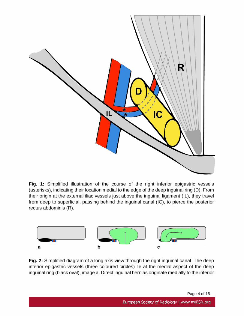
Fig. 1: Simplified illustration of the course of the right inferior epigastric vessels(asterisks), indicating their location medial to the edge of the deep inguinal ring (D). Fromtheir origin at the external iliac vessels just above the inguinal ligament (IL), they travelfrom deep to superficial, passing behind the inguinal canal (IC), to pierce the posteriorrectus abdominis (R).
Fig. 2: Simplified diagram of a long axis view through the right inguinal canal. The deepinferior epigastric vessels (three coloured circles) lie at the medial aspect of the deepinguinal ring (black oval), image a. Direct inguinal hernias originate medially to the inferior

Page 5 of 15
epigastric vessels (b) and indirect inguinal hernias pass through the deep ring laterallyand then over the inferior epigastric vessels (c).

Page 6 of 15
Findings and procedure details
Our approach is centred on the inferior epigastric vessels, which are followed in atransverse plane from within the rectus abdominis down the lower abdomen, towardstheir origin in the external iliac artery. The inguinal and femoral canals are superficial,non-rigid and small in calibre. We would therefore recommend a high frequency 12 MHzlinear transducer, light probe pressure and slow, controlled movements of the transducerduring an examination to maintain anatomical perspective. The normal inguinal canalbecomes difficult to visualise with excessive compression. The use of controlled Valsalvamanoeuvres, with spilt/dual screen before and after comparison images are essential.We favour using controlled instructions such as "blow out your cheeks with your mouthclosed", "tense up your stomach" or "raise your head from the pillow" rather thancoughing, which is less controlled. We would also advocate repeating the examinationwith the patient standing if initial findings are negative.
We summarise the stages in our technique below:
1. The patient is asked to point to the location of their symptoms and is brieflyexamined for a palpable mass.
2. The probe is orientated in a transverse plane over the rectus abdominis, justbelow the umbilicus in order to locate the deep inferior epigastric vesselswithin the rectus abdominis (Figure 3).
3. As the probe is moved inferiorly, these pass from medial to lateral andsuperficial to deep towards their origin at the external iliac vessels. Keepingthe image centred on the inferior epigastric vessels, a surrounding softtissue bulge gradually becomes apparent deep and lateral to the posterioraspect of the rectus abdominis (Figure 4). This is the superior aspectof the inguinal canal. This is easier to visualise in males, in whom theheterogenous tubular structures of the spermatic cord and surroundingechogenic fat are prominent. These are of course absent in the female.
4. The probe is now orientated slightly obliquely along the long axis of theinguinal ligament, which has a similar orientation to the inguinal canal. Thisenables the demonstration of a long axis view of the inguinal canal.
5. As the probe is moved slightly more inferiorly, the deep inguinal ring is seenas a hypoechoic defect in the posterior wall of the inguinal canal, just lateralto the inferior epigastric vessels (Figure 5). If this is difficult to localise, smallcraniocaudal movements of the probe over this region will demonstrate thecontents of the cord entering the deep ring.
6. This is the perfect location to assess for the presence of an indirect inguinalhernia. A controlled Valsalva manoeuvre is performed, keeping the deepring within view and the inferior epigastric vessels at the centre of the image.An indirect hernia is seen as a bulge emerging from the deep ring, passing

Page 7 of 15
over the inferior epigastric vessels, and moving down the inguinal canaltowards the superficial ring (Figure 6)
7. To assess for the presence of a direct hernia, the probe is moved slightlyinferior and medial to the deep ring but above the pubic tubercle. AnotherValsalva manoeuvre is performed. A direct hernia is seen as a "ballooning"of the posterior wall of the inguinal canal (Figure 7).
8. Moving below the inguinal ligament, the femoral artery and vein are locatedin a transverse plane. The sapheno-femoral junction is visualised and thislandmark used to assess for a femoral hernia (Figure 8). The probe ismoved slightly superior to this level, as a femoral hernia will not be seenbelow the junction. A controlled Valsalva manoeuvre is performed, normallyresulting in distension of the femoral vein with increased intra-abdominalpressure. In contrast, a femoral hernia descends from above, causing a softtissue bulge medial to the femoral vein, which is compressed (Figure 9). Forclarification, a further longitudinal section centred just medial to the femoralvein allows good visualisation of the normal peritoneal reflection (Figure 10)superior to the femoral canal. This will be seen to descend into the femoralcanal with a hernia.
9. Examine the soft tissues of the groin for evidence of soft tissue or vascularabnormality such as lymphadenopathy, abscess or aneurysm.
10. In the absence of any hernia, repeat the examination with the patientstanding.
11. In a radiological report, always note the reducibility of the hernial sac and itscontents (omental fat, bowel).
It is accepted practice to examine the inguinal region by identifying the pubic tubercleor inferior epigastric vessels at their origin inferiorly, or by obtaining a short axis view ofthe inguinal canal, medial to the femoral vessels in a longitudinal plane (5). However,the technique we have described is relatively straightforward to learn and reproducibleas a first-line assessment by radiologists in training, and less dependent on patient bodyhabitus, with the inferior epigastric vessels reliably located within the rectus abdominisand followed inferiorly.
Images for this section:

Page 8 of 15
Fig. 3: Transverse ultrasound image over the right rectus abdominis just below theumbilicus showing the inferior epigastric vessels (arrow) within the substance of theposterior muscle. Colour Doppler may used to locate the vessels if not immediatelyapparent.

Page 9 of 15
Fig. 4: Transverse ultrasound image. The inferior epigastric vessels have emerged fromthe right rectus abdominis and are travelling posterolaterally towards their origin. A softtissue bulge (arrowheads) is seen deep to the rectus, representing the superior aspectof the inguinal canal.

Page 10 of 15
Fig. 5: This is a long axis view through the right inguinal canal, inferior to Figures 3 and4, orientated parallel to the inguinal ligament. The deep inguinal ring (arrowheads) is ahypoechoic defect in the posterior wall of the inguinal canal immediately lateral to theinferior epigastric vessels.

Page 11 of 15
Fig. 6: Long axis ultrasound image through the right inguinal canal. The indirect inguinalhernia (arrows) emerges lateral to the inferior epigastric vessels (IEV) through the deepring (delineated by crosses), passing over the vessels and down the canal towards thesuperficial ring.

Page 12 of 15
Fig. 7: Transverse ultrasound image of a right direct inguinal hernia (arrows) arisingmedial to the inferior epigastric vessels (IEV) and deep ring.

Page 13 of 15
Fig. 8: Transverse ultrasound image of the right saphenofemoral junction, before (a) andduring a Valsalva manoeuvre (b) The femoral vein (FV) distends when there is increasedintra-abdominal pressure and no femoral hernia. This is the inferior margin of the femoralcanal. FA = Femoral artery; GSV = great saphenous vein.
Fig. 9: Transverse ultrasound image demonstrating the typical right femoral hernia(arrows) emerging medial to a compressed femoral vein (FV) with increased intra-abdominal pressure. Image a is taken at rest; image b during Valsalva.

Page 14 of 15
Fig. 10: Longitudinal ultrasound image through the femoral canal, above thesaphenofemoral junction and slightly medial to the centre of the femoral vein (FV). Inimage a, the normal peritoneal reflection (arrowheads) is seen superior to the femoralcanal, limited by the femoral ring. In the absence of a hernia, this distorts but the inferiormargin remains in a similar location (image b).

Page 15 of 15
Conclusion
We have described a reliable and reproducible technique for the sonographic assessmentof groin hernias. In our experience, following the inferior epigastric vessels inferiorly ina transverse plane, as it emerges from the posterior rectus sheath, allows for reliableidentification of the inguinal canal and deep inguinal ring in both males and females.The femoral vein at the saphenofemoral junction is the critical landmark for the femoralcanal. The ability to confidently demonstrate normal inguinal and femoral anatomy onultrasound is a valuable and clinically relevant skill. We hope that this description is auseful aid, especially for radiologists in training.
Personal information
References
1. Frequency of abdominal wall hernias: is classical teaching out of dates? Dabbas N,Adams K, Pearson K, Royle G. JRSM Short Rep. 2011 Jan 19;2(1):5.
2. A study of the risk of strangulation and obstruction in groin hernias. Rai S, ChandraSS, Smile SR.Aust N Z J Surg. 1998 Sep;68(9):650-4.
3. The groin hernia - an ultrasound diagnosis? Bradley M, Morgan D, Pentlow B, Roe A.Ann R Coll Surg Engl. 2003 May;85(3):178-180.
4. Sinnatamby CS. Last's anatomy: regional and applied. 12th ed. Edinburgh, New York:Churchill Livingstone/Elsevier; 2011.
5. Inguinofemoral hernia: accuracy of sonography in patients with indeterminate clinicalfeatures. Robinson P, Hensor E, Lansdown MJ, Ambrose NS, Chapman AH.AJR Am JRoentgenol. 2006 Nov;187(5):1168-78.