Embed Size (px)
Citation preview

Oral Oncology xxx (2014) xxx–xxx
Contents lists available at ScienceDirect
Oral Oncology
journal homepage: www.elsevier .com/locate /ora loncology
Review
The infrahyoid flap: A comprehensive review of an often overlookedreconstructive method
http://dx.doi.org/10.1016/j.oraloncology.2014.04.0111368-8375/� 2014 Elsevier Ltd. All rights reserved.
⇑ Corresponding author. Address: Clinic of Otolaryngology – Head and NeckSurgery, University of Florence Largo Brambilla 13 - 50134 Firenze. Tel.: +390557947054; fax: +39 055435649.
E-mail address: [email protected] (A. Deganello).
Please cite this article in press as: Deganello A, Leemans CR. The infrahyoid flap: A comprehensive review of an often overlooked reconstructive mOral Oncol (2014), http://dx.doi.org/10.1016/j.oraloncology.2014.04.011
Alberto Deganello a,⇑, C René Leemans a,b
a Academic Clinic of Otolaryngology – Head and Neck Surgery, Department of Translational Medicine and Surgery, University of Florence, Florence, Italyb Department of Otolaryngology – Head and Neck Surgery, VU Medical Center, Amsterdam, The Netherlands
a r t i c l e i n f o
Article history:Received 15 December 2013Received in revised form 22 April 2014Accepted 26 April 2014Available online xxxx
Keywords:Infrahyoid flapPedicled flapFree flapAlternative flapHead and neck reconstruction
a b s t r a c t
The infrahyoid flap is a myocutaneous pedicled flap mainly nourished by the superior thyroid vesselsthrough the perforators of the infrahyoid muscles. This thin and pliable flap provides a skin island ofabout 7 by 4 cm from the central part of the anterior neck. The flap can be transferred on its pedicle ofsuperior thyroid artery and vein to reconstruct medium sized head and neck defects created after cancerablation. We have successfully used this flap in a series of 40 cases with no total flap loss and with 1 caseof superficial skin necrosis. The aim of this review is to highlight the clinical usefulness of this pedicledflap even in the microvascular free flap era. A comprehensive review of the available literature reportingon the infrahyoid flap has been carried out using a web search. The history of the infrahyoid flap, the sur-gical technique with technical innovations, the clinical utility and limitations of this flap, are reported anddiscussed. Among the 7 larger series (cohort larger than 50 cases) a total of 956 flaps were performed, andthe global success rate was 91.7%, with failures being mainly related to partial skin necrosis, as the rate oftotal (skin and muscle) flap necrosis was only 1%.
This flap is reliable, easy to harvest during neck dissection, oncologically safe, it does carry a negligibledonor site morbidity. This paper highlights how the infrahyoid flap can represent an excellent reconstruc-tive solution in selected patients and head and neck sites.
� 2014 Elsevier Ltd. All rights reserved.
Introduction
The introduction of microvascular reconstructions has providedthe head and neck surgeon with the possibility of choosing amonga broad variety of free flaps. This reconstructive method representsa major evolution in the management of head and neck cancerwith a consequent reduction of pedicled flap reconstructions. TheIHF is a thin and pliable pedicled flap that has been developed ina free flap era, hence it is important to assess its usefulness in thismodern scenario.
History
The first report of using the infrahyoid system of muscles as apedicled flap with reconstructive intents came from Clairmontand Conley in 1977 [1]. In their report they described the transpo-sition of the infrahyoid muscles to repair anterior floor of mouth
defects arising from pull through composite resections with enblock neck dissection. In the report it was clearly specified thatonly the infrahyoid muscles were transposed upwards, and theAuthors recommended to make any effort to preserve the superiorthyroid artery and the innervation by the ansa hypoglossi in orderto ensure the viability of this newly designed flap.
In 1984 Eliachar et al. included the overlying skin to a transpo-sition of the infrahyoid muscles for the reconstruction of laryngo-tracheal defects. In their technique this myocutaneous flap wasused as a rotary-door flap with a double blood supply from thesuperior and inferior thyroid arteries [2,3]. Having thus substantiallimitation on the arch of rotation (coming from the need ofmaintaining both cranial and caudal pedicles), this rotary-door flapwas therefore recommended only for laryngotracheal defects.
In 1985 Rabson et al. pointed out how the inferior cervical skinapproaching the midline receives blood supply from perforatorvessels coming from the superior thyroid artery piercing the infra-hyoid muscles [4].
The most important and decisive step was taken by Wang et al.when in 1986 they first reported in the English literature thesurgical technique and the results of 112 head and neck recon-structions in 108 patients, describing the infrahyoid myocutaneous
ethod.

2 A. Deganello, C René Leemans / Oral Oncology xxx (2014) xxx–xxx
flap as we know it today [5]. The flap was mainly transposed toreplace intraoral defects, the blood supply being clearly identifiedin the superior thyroid vessels. It is important to remark that sincethis first report Wang noticed how this easy and quick reconstruc-tive method was particularly convenient and useful in weakelderly patients. This series starts from May 1979, so, even ifundoubtedly Wang is the father of this flap, credit for the originalidea (the grandfathers) must be given to Clairmont and Conley [1].
Methods
A comprehensive review of the available literature reporting onthe IHF has been carried out using a web search in Pubmed/Med-line, Google Scholar, Isi Web of Knowledge and Scopus. Nowadays,this method is reported in 61 published papers, including less than1400 patients. Only 24 full text papers in the English languagewere published in 28 years since 1986, with only 10 papersappearing in US Journals [5–14], the remaining 37 papers werepublished in other languages (Chinese, French, German, Polishand Japanese).
This review is intended to highlight the many advantages thatthis flap can offer to the head and neck surgeon even in a free flapera.
Surgical technique
In its original description by Wang et al. the IHF is harvested asa myocutaneous flap [5] after ipsilateral modified radical or selec-tive neck dissection is completed. Technically, the harvest of theIHF does not interfere with the extent of the neck dissection, sincethis flap lies in the central compartment of the neck, medial to thecarotid artery at neck level VI. When a therapeutic modified radicalneck dissection is indicated, this is performed according to thestandard technique, with the only mandatory requirement beingthe preservation of the superior thyroid vein and the caudal stumpof the internal jugular vein.
The infrahyoid muscles included in this flap are the sternohyoidmuscle, the superior belly of the omohyoid muscle [6] and thesternothyroid muscle. Usually the flap is unilateral and the sideis determined by the location of the defect, therefore the skin pad-dle and cervical incision for neck dissection are outlined in thesame neck side of the tumor resection. The shape of the flap is rect-angular or oval in a vertical position, and the skin paddle must befitted and included in the incision for unilateral or bilateral neckdissection. In 2005 Dolivet et al. [15] introduced a modificationfor the neck incision proposing an S instead of the original T shapedincision, and this modification was acquired in further reports(Fig. 1) [8,11,16–20].
Fig. 1. Neck incision: vertically oriented infrahyoid flap at the left
Please cite this article in press as: Deganello A, Leemans CR. The infrahyoid flaOral Oncol (2014), http://dx.doi.org/10.1016/j.oraloncology.2014.04.011
The medial edge of the IHF lies at the midline, the upper edge atthe level of the hyoid bone and the lower edge at the suprasternalnotch, the lateral edge lies three to five cm from the midline. Whena tracheotomy is required this is usually performed first and it isimportant to prevent tracheotomy site contamination to thewound bed. We recommend to place the caudal edge of the skinpaddle at list 1 cm above the incision for the tracheotomy, and toopen the trachea under the thyroid isthmus; the harvest of the flapwill eventually create a communication with the tracheotomy atthe side where the infrahyoid muscles are harvested, later on, toensure a tight separation, the thyroid isthmus and the sternal edgeof the sternocleidomastoid muscle are sutured to the subcutaneoustissue above the tracheotomy opening.
The skin and platysma all around the skin paddle are incised toallow prompt choke perforator vessels opening [8]; the skin flapsare elevated and, before starting with the intended modified radi-cal or selective neck dissection, the superficial cervical fascia alongthe anterior border of the sternocleidomastoid muscle, from thesternal insertion to the level of the hyoid bone, is incised and thedissection of the fascia proceeds until the omohyoid muscle isidentified at its intersection with the internal jugular vein.The intermediate tendon is divided and the fascia, together withthe anterior belly of the omohyoid muscle is elevated towardsthe lateral edge of the skin paddle and sutured to it Fig. 2.
Neck dissection and primary tumor resection are nowcompleted.
The elevation of the flap starts by dividing the anterior jugularvein and sectioning the sterno-hyoid and sterno-thyroid musclesdistally at the level of the suprasternal notch. The skin paddle isstitched to the underlying muscles and then the IHF is raised overthe avascular plane of the proper capsule of the thyroid gland,Fig. 3. When the dissection reaches the upper pole of the thyroidgland, the crico-thyroid artery (at the midline of the neck) andthe posterior branch of the superior thyroid artery (at its entrancein the upper pole of the gland) are cut, ligated and kept with theflap. The sterno-thyroid muscle is detached from the thyroid carti-lage, Fig. 4. Fascial connections between the superficial and mediancervical fascia are maintained in proximity of the neurovascularpedicle; these fascial connections are important to directly providemicrovascular venous return towards the median cervical fasciaand to protect the superior thyroid vein from twisting or kneeing[8,11].
Special care must be taken in preserving the external branch ofthe superior laryngeal nerve, and therefore the thyrohyoid muscleis usually spared and left in place. Finally, the hyoid insertions ofthe sternohyoid and omohyoid muscles are severed, the entire flapremains attached only by the neurovascular pedicle formed by thesuperior thyroid artery and vein, and nerves from the ansa cervica-lis, and is then ready to be transferred to reconstruct the defect,
side: neck incision for unilateral and bilateral neck dissection.
p: A comprehensive review of an often overlooked reconstructive method.
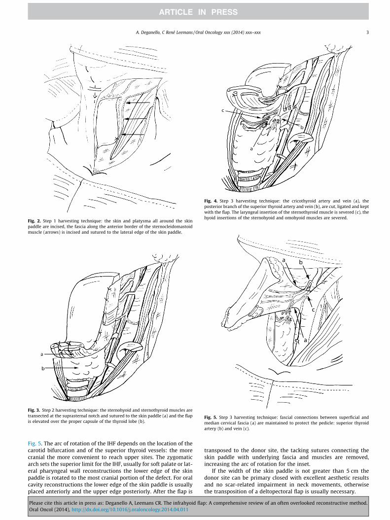
Fig. 2. Step 1 harvesting technique: the skin and platysma all around the skinpaddle are incised, the fascia along the anterior border of the sternocleidomastoidmuscle (arrows) is incised and sutured to the lateral edge of the skin paddle.
Fig. 3. Step 2 harvesting technique: the sternohyoid and sternothyroid muscles aretransected at the suprasternal notch and sutured to the skin paddle (a) and the flapis elevated over the proper capsule of the thyroid lobe (b).
Fig. 4. Step 3 harvesting technique: the cricothyroid artery and vein (a), theposterior branch of the superior thyroid artery and vein (b), are cut, ligated and keptwith the flap. The laryngeal insertion of the sternothyroid muscle is severed (c), thehyoid insertions of the sternohyoid and omohyoid muscles are severed.
Fig. 5. Step 3 harvesting technique: fascial connections between superficial andmedian cervical fascia (a) are maintained to protect the pedicle: superior thyroidartery (b) and vein (c).
A. Deganello, C René Leemans / Oral Oncology xxx (2014) xxx–xxx 3
Fig. 5. The arc of rotation of the IHF depends on the location of thecarotid bifurcation and of the superior thyroid vessels: the morecranial the more convenient to reach upper sites. The zygomaticarch sets the superior limit for the IHF, usually for soft palate or lat-eral pharyngeal wall reconstructions the lower edge of the skinpaddle is rotated to the most cranial portion of the defect. For oralcavity reconstructions the lower edge of the skin paddle is usuallyplaced anteriorly and the upper edge posteriorly. After the flap is
Please cite this article in press as: Deganello A, Leemans CR. The infrahyoid flaOral Oncol (2014), http://dx.doi.org/10.1016/j.oraloncology.2014.04.011
transposed to the donor site, the tacking sutures connecting theskin paddle with underlying fascia and muscles are removed,increasing the arc of rotation for the inset.
If the width of the skin paddle is not greater than 5 cm thedonor site can be primary closed with excellent aesthetic resultsand no scar-related impairment in neck movements, otherwisethe transposition of a deltopectoral flap is usually necessary.
p: A comprehensive review of an often overlooked reconstructive method.

Fig. 7. Anterior jugular vein: infrahyoid flap with venous drainage ensured by theanterior jugular vein (a) and draining with retrograde flow to the external jugularvein (b). The flap is ready to reconstruct a lateral floor of mouth and alveolar ridgedefect resulting from lateral pull through resection with marginal mandibulectomyand en block selective neck dissection I–III.
4 A. Deganello, C René Leemans / Oral Oncology xxx (2014) xxx–xxx
Technical modifications
The venous drainage is anatomically ensured by both theexternal and internal jugular systems, and the preservation ofone systems is crucial: the superior thyroid vein provides drainageto the internal jugular vein (Fig. 6); the cranial portion of theanterior jugular vein drains, with retrograde flow, into the externaljugular vein (Fig. 7). Some put particular emphasis on the preserva-tion of the cranial portion of the anterior jugular vein [12,13,21,22],which is perfectly feasible and reliable, nevertheless preservationof the external jugular system makes ipsilateral neck dissectiontechnically more demanding.
We described a new technique for tongue base reconstruction[11]: the neurovascular IHF is transposed without detaching itfrom the hyoid bone that acts as rotational pivot. During degluti-tion, the hyoid bone elevates and pushes the flap backwards, thushelping with bolus propulsion. For defects limited to the tonguebase, IHF is perfectly suited to the resected area having the desiredthickness Fig. 8.
As originally suggested by Wang et al. [5], it is wise to preservethe motor innervations of the infrahyoid muscles (provided by theansa cervicalis) in all cases of tongue reconstruction, to preventsubsequent atrophy. Conversely, for other sites, we recommendedto resect all motor innervations since denervation atrophy of theunderlying muscles will increase flap’s pliability with better func-tional results Fig. 9 [11].
Majoufre-Lefebvre et al. [23] introduced the horizontal infrahy-oid flap claiming less cosmetic sequelae at the donor site; this
Fig. 6. Superior thyroid vein: infrahyoid flap with venous drainage ensured by thesuperior thyroid vein. The picture shows how the flap easily reaches the oral cavity;after the flap is transposed (medial to the mandible) to the donor site, the tackingsutures connecting the skin paddle with underlying fascia and muscles are removedto increase the arc of rotation.
Fig. 8. Tongue base reconstruction: schematic representation of tongue basereconstruction maintaining the muscular insertions at the hyoid bone.
Please cite this article in press as: Deganello A, Leemans CR. The infrahyoid flaOral Oncol (2014), http://dx.doi.org/10.1016/j.oraloncology.2014.04.011
technique was then implemented in a large series of 276 casesfrom the same group [24], and the authors also stated that no addi-tional scars in the neck were required. This is certainly true when aselective neck dissection I-III is planned (as it happened for the 275squamous cell carcinoma patients in this series), nevertheless onlythe neck incision for a vertically oriented flap allows a comprehen-sive neck dissection without further incisions. Furthermore, a ver-tically oriented flap has a superior arc of rotation as compared toan horizontal flap, allowing for upper reconstructions that reachthe soft palate [19].
Clinical series and results
The web search identified 7 series with a study cohort largerthan 50 flaps (Table 1) [5,13,15,24–27], and 16 series with 10 to
p: A comprehensive review of an often overlooked reconstructive method.

Fig. 9. Oral cavity reconstruction: Postoperative result after reconstruction of thewhole retromolar trigone, the flap ensures good pliability (informed consent forpublication was obtained).
A. Deganello, C René Leemans / Oral Oncology xxx (2014) xxx–xxx 5
50 cases [6–8,10–12,18–22,28–32]. Among the 7 larger series atotal of 956 IHF were performed, and the global success rate was91.7%, with failures being mainly related to partial skin necrosis,as the rate of total (skin and muscle) flap necrosis was only 1%.
In 1991 Wang reported his global experience with the IHF ana-lyzing 260 cases [25]; this series came from the 112 flaps describedin 1986 with further 148 flaps in the following 3 years. In the firstreport, Wang stated how the rate of failure was 38% (7 of 18 cases),versus 4% (4 of 94 cases) when the internal and external jugularveins were both removed or not both removed respectively. Thesuccess rate reported by Wang in the further 148 flaps was 97%[25]. Wang indicated several technical points to increase the suc-cess rate, but the attention to venous drainage is the crucial step.Another aspect to be highlighted is that no total flap necrosiswas reported in this large series and the necrosis of the skin paddlenever lead to further surgery.
In a series of 276 cases, the horizontal IHF was used for oral cav-ity reconstructions in 95.6% of the cases, insufficient venous returnwas recorded in 22 cases (8%), leading to partial skin paddle necro-sis in 20 patients, and total sin paddle necrosis in the remaining 2patients [24]. Also in this series no total flap loss was recorded andno further surgery was required for the management of the super-ficial skin paddle necrosis. All these patients received a selectiveneck dissection of levels I–III; data on clinical and pathologicalneck involvement were not reported.
Table 1Overview of the 7 largest series.
Author No. of flaps Site Previous neck RT
Wang 1986 112 OC:101 Parotid:7 NRWang 1991 148 / /Faucher 1997 62 OC:19 OP:32 PL:9 Skin:2 1Zao 2001 53 OC:53 NRVerhulst 2004 153 OC:54 OP:99 1Dolivet 2005 152 OC:78 OP:47 PL:27 19Ricard 2009 276 OC: 264 OP:12 None
OC: oral cavity, NR: not reported, OP: oropharynx, PL: pharyngo–larynx.
Please cite this article in press as: Deganello A, Leemans CR. The infrahyoid flaOral Oncol (2014), http://dx.doi.org/10.1016/j.oraloncology.2014.04.011
Among the 16 series with 10–50 IHFs a total of 328 flaps werereported, and the overall success rate was 85.5%, with a large rangefrom 54% [7] to 100% [8,28,31]; also in these series the rate of total(skin and muscle) flap necrosis was low (2.7%). Among the 80 flapsused as myofascial transposition [10,28,29,32], 2 partial muscularnecrosis and 3 total were recorded (92.7% success rate).
Unfortunately, data regarding the oncologic appropriateness ofharvesting the IHF in N + necks are lacking.
Wang demonstrated how this flap was oncologically sound inN1 necks [25], but in the majority of other large series patientswere submitted to selective neck dissections I–III, and this wouldindicate a preponderance of cN0 cases. Only other 7 series in liter-ature report IHF in N + necks [8,11,19–21,28,29]; among the 153IHF harvested in these series, 88 flaps were harvested in N + necks:35 N1 out of 88 N+(39.7%), 51 N2 (58%), 2N3 (2.27%).
Therapeutic neck dissection is not a contraindication for IHF aslong as the oncologic radicality doesn’t require the resection of theinternal jugular vein, jeopardizing venous drainage.
Clinical utility
In head and neck reconstructions, especially for oral cavity andoropharyngeal defects, the pliability of the flap should allow for agood motility of the preserved structures all around the resectedarea. The majority of pedicled myocutaneous flaps for head andneck reconstruction (e.g., pectoralis major, trapezius, latissimusdorsi) are quite bulky, and this intrinsic characteristic carries a dis-advantage in terms of functional results; conversely the IHF is thinand pliable competing with fascio-cutaneous free flaps in the man-agement of medium sized defects of the floor of mouth, alveolarridge, and base of tongue. In our experience, for these sites, theresults are particularly high-quality, because the pliable skin pad-dle is placed and sutured all around the mucosal defect and theinfrahyoid muscles fill the deep tissue loss coming from resectionscarried en block with neck dissection. In case of marginal mandib-ulectomy, the flap’s muscles cover the denuded mandibular bonysurface, moreover the oval/rectangular shape of the IHF perfectlymatches the usual shape of the resections in these cases, Fig. 7(informed consent for publication was obtained). Excellent func-tional results are also obtained for base of tongue reconstructions[10,28], especially if the flap is not detached from the hyoid bone[11].
In a series of 32 consecutive oral cavity and oropharyngealreconstructions from our group, functional results of 18 patientsin poor general conditions unfit for a microvascular procedureand therefore receiving IHF reconstruction, were as good as thoseof the 16 patients in good general conditions receiving microvascu-lar free radial forearm flap transposition [11], furthermore, com-paring the medical costs, IHF reconstruction produced a savingsin this fragile cohort of patients [33]. We also used the IHF forintraoral reconstruction together with free fibula osseous mandib-ular reconstruction, whenever skin perforators for a fibular osteo-cutaneous harvest were not found or reliable [8].
Skin necrosis Flap necrosis Patients requiring further surgery
11 0 05Partial: 2 2 NRPartial: 2 Total: 2 1 0Partial: 17 4 48 2 NRPartial: 22 Total: 2 0 None
p: A comprehensive review of an often overlooked reconstructive method.

6 A. Deganello, C René Leemans / Oral Oncology xxx (2014) xxx–xxx
In literature this flap has been successfully used for defects ofthe oral cavity and oropharynx [5,8,11,19,25,27,30,31], the parotidregion [5], the pharyngolaryngeal tract [8,15,26,27] and the cervi-cal trachea [9]. As a myofascial transposition, it has been used toclose iatrogenic pharyngeal [34] and esophageal [35] fistulas fol-lowing anterior cervical spine surgery, or to prevent fistula forma-tion after total laryngectomy [36].
Limitations
This flap does carry dimensional limitations, which make itunsuitable for large sized and complex defects. The maximal lengthof the flap is usually around an average of 10 cm, depending on thelength of the patient’s neck. If the width of the flap exceeds 5 cm, afurther flap (usually a deltopectoral flap) is required to close thedonor site, and this would decrease all the intrinsic convenienceof the IHF; in most series the average dimensions of the flap is7 � 4 cm. It could be consequently argued that small or mediumsized defects within the oral cavity and oropharynx can also be pri-marily closed or reconstructed using local flaps and skin grafts,without requiring a pedicled flap or a free flap transposition. Thiscan be true when the defect comes from a transoral resection,but if the resection put in communication the oral cavity/orophar-ynx with neck spaces, as a result of tumour resection with en blockneck dissection, then primary closure usually leads to fixation ofmobile structures; furthermore in this situation local flaps or skingrafts are less able to ensure a tight separation between differentcompartments to prevent the occurrence of a salivary fistula withall its negative impacts.
Whenever the defect is large or encompasses more subsites,then a reconstruction with an more pliable fascio-cutaneous freeflap ensures better results as compared to IHF transposition,because microvascular flaps can better follow the contour of theoriginal anatomy, and can also be double folded in complexreconstructions.
Previous (chemo)radiotherapy is not an absolute contraindica-tion for IHF [5,11,15], but preoperative careful evaluation of theintended skin paddle is recommended: if lack of pliability, radia-tion induced fibrosis or teleangiectasias are encountered in the cer-vical skin, then a decrease in blood supply to the skin through theperforator vessels is probably occurring and the flap is contra-indi-cated. However if none of these features is present and the appear-ance of the skin is normal, then the flap can be considered [11,15].
Contraindications
Disadvantages of IHF mainly coincide with its contraindica-tions: previous thyroid surgery or neck dissection, N3 neck metas-tasis, and positive lymphnodes at level III–IV. All thesecontraindications pose consistent limitations to the use of thisreconstructive option. The IHF must always be planned in advanceand cannot represent a back-up solution in case of other flap fail-ure, since it cannot be used in a previously operated neck. In factprobable damages to the superior thyroid artery and/or vein and/or possible elevation of the skin overlying the strap muscles pre-vent the possibility to rely on this myocutaneous flap.
Conclusions
The infrahyoid flap is a quick, easy, and reliable reconstructivemethod, when specific contraindications are respected and whenused with knowledge of its clinical utility and limitations, the func-tional results are excellent with great patient’s satisfaction.
Please cite this article in press as: Deganello A, Leemans CR. The infrahyoid flaOral Oncol (2014), http://dx.doi.org/10.1016/j.oraloncology.2014.04.011
Conflict of interest statement
This work had no founding, furthermore there are no financialdisclosures from the authors.
Acknowledgment
The Authors thank Sabino Russo, MD, for surgical illustrations.
References
[1] Clairmont AA, Conley JJ. Surgical technique – the strap muscle flap. JOtolaryngol 1977;6:200–2.
[2] Eliachar I, Marcovich A, Shai YH. Rotary-door flap in laryngotrachealreconstruction. Arch Otolaryngol 1984;110:585–90.
[3] Eliachar I, Marcovich A, Har Shai Y, Lindenbaum E. Arterial blood supply to theinfrahyoid muscles: an anatomical study. Head Neck Surg 1984;7:8–14.
[4] Rabson JA, Hurwitz DJ, Futrell JW. The cutaneous blood supply of the neck:relevance to incision planning and surgical reconstruction. Br J Plast Surg1985;38:208–19.
[5] Wang HS, Shen JW, Ma DB, Wang JD, Tian AL. The infrahyoid myocutaneousflap for reconstruction after resection of head and neck cancer. Cancer1986;57:663–8.
[6] Rojananin S, Suphaphongs N, Ballantyne AJ. The infrahyoid musculocutaneousflap in head and neck reconstruction. Am J Surg 1991;162:400–3.
[7] Magrin J, Kowalski LP, Santo GE, Waksmann G, DiPaula RA. Infrahyoidmyocutaneous flap in head and neck reconstruction. Head Neck1993;15:522–5.
[8] Deganello A, Manciocco V, Dolivet G, Leemans CR, Spriano G. Infrahyoid fascio-myocutaneous flap as an alternative to free radial forearm flap in head andneck reconstruction. Head Neck 2007;29:285–91.
[9] Masuda M, Kamizono K, Ejima M, Fujimura A, Uryu H, Kadota H. Trachealreconstruction with a modified infrahyoid myocutaneous flap. Laryngoscope2012;122:992–6.
[10] Remmert SM, Sommer KD, Majocco AM, Weerda HG. The neurovascularinfrahyoid muscle flap: a new method for tongue reconstruction. PlastReconstr Surg 1997;99:613–8.
[11] Deganello A, Gitti G, Parrinello G, et al. Infrahyoid flap reconstruction of oralcavity and oropharyngeal defects in elderly patients with severe generalcomorbidities. Head Neck 2012;34:1299–305.
[12] Peng H, Wang SJ, Yang X, Guo H, Liu M. Infrahyoid myocutaneous flap formedium-sized head and neck defects: surgical outcome and techniquemodification. Otolaryngol Head Neck Surg 2013;148:47–53.
[13] Zhao YF, Zhang WF, Zhao JH. Reconstruction of intraoral defects after cancersurgery using cervical pedicle flaps. J Oral Maxillofac Surg 2001;59:1142–6.
[14] Perrenot C, Berengère P, Mastronicola R, Gangloff P, Dolivet G. Infrahyoidmyocutaneous flap for reconstruction after robotic transoral surgery fororopharyngeal tumors. Plast Reconstr Surg. 2014 Feb;133:236e–7e.
[15] Dolivet G, Gangloff P, Sarini J, et al. Modification of the infra hyoid musculo-cutaneous flap. Eur J Surg Oncol 2005;31:294–8.
[16] Deganello A, De Bree R, Dolivet G, Leemans CR. Infrahyoid myocutaneous flapreconstruction after wide local excision of a Merkel cell carcinoma. ActaOtorhinolaryngol Ital 2005;25:50–3.
[17] Deganello A, Struijs B, De Bree R. The infrahyoid myocutaneous flap in headand neck reconstruction [abstract]. Clin Otolaryngol 2005;30:298.
[18] Ouyang D, Su X, Chen WC, Chen YF, Men QQ, Yang AK. Anatomical study andmodified incision of the infrahyoid myocutaneous flap. Eur ArchOtorhinolaryngol 2013;270:675–80.
[19] Gangloff P, Deganello A, Lacave ML, et al. Use of the infra hyoid musculo-cutaneous flap in soft palate reconstruction. Eur J Surg Oncol 2006;32:1165–9.
[20] Mirghani H, Meyer G, Hans S, et al. The musculocutaneous infrahyoid flap:surgical key points. Eur Arch Otorhinolaryngol 2012;269:1213–7.
[21] Lockhart R, Menard P, Chout P, Favre-Dauvergne E, Berard P, Bertrand JC.Infrahyoid myocutaneous flap in reconstructive maxillofacial cancer andtrauma surgery. Int J Oral Maxillofac Surg 1998;27:40–4.
[22] Tincani AJ, Del Negro A, Araújo PP, Akashi HK, Neves Fda S, Martins AS. Headand neck reconstruction using infrahyoid myocutaneous flaps. Sao Paulo Med J2006;124:271–4.
[23] Majoufre-Lefebvre C, Laurentjoye M, Faucher A, Zwetyenga N, Siberchicot F,Ricard AS. The horizontal infrahyoid myocutaneous flap: surgical technique.Rev Stomatol Chir Maxillofac 2008;109:106–9 [French].
[24] 24 Ricard AS, Laurentjoye M, Faucher A, Zwetyenga N, Siberchicot F, Majoufre-Lefebvre C. 276 cases of horizontal infrahyoid myocutaneous flap. RevStomatol Chir Maxillofac 2009;110:135–7 [French].
[25] Wang HS. Further experiences with the infrahyoid myocutaneous flap inreconstruction after radical surgery of cheek and palate cancer. Zhonghua KouQiang Ke Za Zhi 1985;20(100-2):127–8 [Chinese].
[26] Faucher A, Verhulst J, Majoufre C, de Bonfils C. Infrahyoid musculocutaneousflaps: anatomical bases and indications in cervicofacial oncologic surgery. RevLaryngol Otol Rhinol 1997;118:43–6 [French].
[27] 27 Verhulst J, Souza Leão R. The infrahyoid musculocutaneous flap: experienceof 153 cases in the reconstruction of the oropharynx and oral cavity aftertumoral excision. Rev Laryngol Otol Rhinol 2004;125:49–53 [French].
p: A comprehensive review of an often overlooked reconstructive method.

A. Deganello, C René Leemans / Oral Oncology xxx (2014) xxx–xxx 7
[28] Windfuhr JP, Remmert S. Infrahyoid myofascial flap for tongue reconstruction.Eur Arch Otorhinolaryngol 2006;263:1013–22.
[29] Hell B, Heissler E, Gath H, Menneking H, Langford A. The infrahyoid flap. Atechnique for defect closure in the floor of the mouth, the tongue, the buccalmucosa, and the lateral pharyngeal wall. Int J Oral Maxillofac Surg1997;26:35–41.
[30] Li B, Li CH, Guo H, Chen J, Wang SX. Analysis of 27 cases of defect restorationusing infrahyoid myocutaneous flap after intraoral cancer surgery. ZhonghuaEr Bi Yan Hou Tou Jing Wai Ke Za Zhi 2008;43:826–9 [Chinese].
[31] Minni A, Mascelli A, Suriano M. The infrahyoid myocutaneous flap in intra-oralreconstruction as an alternative to free flaps. Acta Otolaryngol2010;130:733–8.
[32] Zhuang MA. Clinical study of infrahyoid muscle flap for repairing oral oncologypostoperative defect. Hebei Medicine 2011;5:595–8 [Chinese].
Please cite this article in press as: Deganello A, Leemans CR. The infrahyoid flaOral Oncol (2014), http://dx.doi.org/10.1016/j.oraloncology.2014.04.011
[33] Deganello A, Gitti G, Parrinello G, Muratori E, Larotonda G, Gallo O. Costsanalysis in oral cavity and oropharyngeal reconstructions withmicrovascular and pedicled flaps. Acta Otorhinolaryngol Ital 2013 [Epubahead of print].
[34] Seidl RO, Niedeggen A, Todt I, Westhofen M, Ernst A. Infrahyoid muscle flap forpharyngeal fistulae after cervical spine surgery: a novel approach – report ofsix cases. Eur Spine J 2007;16:501–5.
[35] 35 Wierzbicka M, Pabiszczak M, Smuszkiewicz P, Szyfter W. Wide esophagealwall rupture as iatrogenic complication of anterior cervical spine surgery.Otolaryngol Pol 2005;59:887–92 [Polish].
[36] Kadota H, Fukushima J, Kamizono K, et al. A minimally invasive method toprevent postlaryngectomy major pharyngocutaneous fistula using infrahyoidmyofascial flap. J Plast Reconstr Aesthet Surg. 2013 July;66(7):906–11.
p: A comprehensive review of an often overlooked reconstructive method.