Embed Size (px)
Citation preview
Faculdade de Farmácia, Universidade de Lisboa
Fakulteta za farmacijo, Univerza V Ljubljani
The influence of different nanocarriers on
skin’s biophysical parameters
Sara Catarina Henriques Andrade
Master of Science (MSc) in Pharmaceutical Sciences
Erasmus+ Programme
2017

1
2
Faculdade de Farmácia, Universidade de Lisboa
Fakulteta za farmacijo, Univerza V Ljubljani
The influence of different nanocarriers on
skin’s biophysical parameters
Sara Catarina Henrique Andrade
Supervisor: Assist. Prof. Dr. Pegi Ahlin Grabnar
Supervisor: Prof. Dr. Andreia Ascenso
Master of Science (MSc) in Pharmaceutical Sciences
Erasmus+ Programme
2017

i
Abstract
Introduction & Aims: Nanotechnology is a new trend in cosmetology and lipid nanoparticles
have shown higher degree of biocompatibility and versatility in this field compared to other
systems. The aim of this research project was to evaluate the influence of three different
systems containing lipid nanoparticles previously well characterized on skin hydration and
transepidermal water loss (TEWL).
Methods: Several formulations of lipid nanoparticles based systems (solid lipid nanoparticles
(SLN), nanostructured lipid carriers (NLC) with Dynasan® 114 (D114) or Glycerol
monostearate (GMS) and nanoemulsions (NE)) were fully characterized (particle size,
polydispersity index and zeta potential), and then incorporated into hydrogel form to study
different skin parameters such as skin hydration and TEWL on forearms of six human
volunteers.
Results: The capacitance basal values of human volunteers before application of any
hydrogel containing lipid nanoparticles were 31,48 (Control), 31,73 (SLN), 30,98 (NE), 30,42
(NLCD114
) and 32,07 a.u. (NLCGMS
). The TEWL basal values were 7,70 (Control), 7,55
(SLN), 7,72 (NE), 7,50 (NLCD114
) and 7,67 g/h/m2 (NLC
GMS). One hour after hydrogels`
application, the capacitance values measured were 40,07 (Control), 43,20 (SLN), 41,22 (NE),
41,15 (NLCD114
) and 44,15 a.u. (NLCGMS
). At the same time, the TEWL values obtained were
6,58 (Control), 4,67 (SLN), 4,13 (NE), 3,90 (NLCD114
) and 4,22 g/h/m2 (NLC
GMS).
Conclusion: The present study showed an increase in skin hydration after exposure to
different systems of lipid nanoparticles (even not statistically significant). On the other hand,
there was a statistically significant decrease in TEWL after that exposure, compared to the
control. In this way, nanolipid systems, i.e. solid lipid nanoparticles, nanostructured lipid
carriers and nanoemulsions, are promising systems to improve skin’s biophysical parameters
in cosmetodermatology.
Keywords: solid lipid nanoparticles, nanostructured lipid carriers, nanoemulsions,
transepidermal water loss, skin hydration
ii
Resumo
Introdução & Objetivos: A nanotecnologia é uma nova tendência no ramo da cosmetologia
e as nanopartículas lipídicas têm demonstrado um maior grau de biocompatibilidade e
versatilidade nesta área, comparando com outros sistemas já bem conhecidos. Este projeto de
investigação teve como objetivo principal a avaliação da influência de três sistemas diferentes
de nanopartículas lipídicas previamente bem caracterizados ao nível de parâmetros biofísicos
da pele, nomeadamente hidratação da pele e perda transepidérmica de água.
Métodos: Diversas formulações de sistemas baseados em nanopartículas lipídicas
(nanopartículas lípidicas sólidas (SLN), vetores lipídicos nanoestruturados (NLC) formulados
com Dynasan® (D114) ou Monoestearato de glicerol (GMS) e nanoemulsões (NE)) foram
rigorosamente caracterizadas (nomeadamente o tamanho das partículas, índice de
polidispersão e potencial zeta), sendo depois incorporadas na forma de hidrogel para ser
avaliada a influência destes nanosistemas lipídicos em diferentes parâmetros biofísicos da
pele, como a hidratação da pele e a perda transepidérmica de água nos antebraços de seis
voluntários.
Resultados: Os valores basais de capacitância nos voluntários antes da aplicação de qualquer
hidrogel contendo nanopartículas foram 31,48 (controlo), 31,73 (SLN), 30,98 (NE), 30,42
(NLCD114
) e 32,07 a.u. (NLCGMS
). Os valores basais da perda transepidérmica de água foram
7,70 (controlo), 7,55 (SLN), 7,72 (NE), 7,50 (NLCD114
) e 7,67 g/h/m2 (NLC
GMS). Os valores
da capacitância, uma hora após a aplicação dos hidrogeles, foram 40,07 (controlo), 43,2
(SLN), 41,22 (NE), 41,15 (NLCD114
) e 44,15 a.u. (NLCGMS
) e os valores de perda
transepidérmica de água medidos após o mesmo período de tempo foram 6,58 (controlo), 4,67
(SLN), 4,13 (NE), 3,90 (NLCD114
) e 4,22 g/h/m2 (NLC
GMS).
Conclusão: O presente estudo demonstrou um aumento na hidratação da pele após exposição
a diferentes sistemas de nanopartículas lipídicas, mesmo não tendo sido estatisticamente
significativo. Por outro lado, houve uma diminuição estatisticamente significativa na perda
transepidérmica de água após essa mesma exposição, comparativamente ao valor registado no
hidrogel controlo. Desta forma, os nanosistemas lipídicos (nanopartículas lipídicas sólidas,
vetores lipídicos nanoestruturados e nanoemulsões) são sistemas promissores ao nível da
melhoria dos parâmetros biofísicos da pele em Cosmética e Dermatologia.
Palavras-chave: nanopartículas lipídicas sólidas, vetores lipídicos nanoestruturados,
nanoemulsões, perda transepidérmica de água, hidratação da pele
iii
Acknowledgments
At the end of a journey that began five years ago, this thesis represents the final step
and simultaneously reflects the work developed in my last adventure as a student and one of
the best challenges I faced in my life: Erasmus+ Programme.
First of all, I want to thank, with all of my heart, my family for their support, their
patience and, mainly, for their love. They always helped me, in all good and bad moments. I
have to include also a special person that became as a second mother for me when I came to
Lisbon to study, my ‘aunt’ Nicole. Mother, father, brother and Nicole, thank you for keeping
me balanced and for making me the person I am today, you made this possible.
Secondly, a deepest thanks to my main supervisor from Portugal Prof. Andreia
Ascenso for her advices and recommendations, I am very grateful to have her as my
supervisor. Due to her extraordinary guidance and exemplary availability, she was crucial for
the development of this work. I wish you all the best.
Thirdly, the most sincere recognition to my supervisor in Slovenia, Prof. Pegi Ahlin
Grabnar, for her assistance and for the continuous support of my work. Without her guidance
it would not be possible to conduct this work since she helped me to develop this entire
research project, always in a critical way. I would like to express my gratitude and
appreciation to my co-supervisor in Slovenia, Maja Bjelosevic, for his friendly personal
support. She was incredibly available to help me, from working with instruments and
equipment to understanding results from my experiments. Thank you both, I hope to visit you
sooner than later.
I have to thank all the volunteers, who agreed to participate in this research project and
the University of Ljubljana for providing me access to its laboratories, as well as all the
materials that were necessary for the development of this study.
Remarkable thanks to 2Logical for the opportunity to join their team and to start my
professional career, facing very interesting projects in the area of health and medicine in
Mozambique. I have to highlight thanks to Pedro Rebelo, for all the flexibility, all the help
and important advices, definitely essential to be present in the professional field.
Lastly, I want to thank my friends who have always been a major source of support
when things would get a bit discouraging and with whom I have shared moments of deep
anxiety but also great memories from these five years. Regarding my Erasmus, I’m really glad
for my laboratory colleague, Joana Brito, with whom I shared several hours in the laboratory
and great moments in Ljubljana. A final and special thanks to Tiago Vieira, the one who has
supported me in all circumstances and who has the ability to surprise me always.
I couldn’t feel luckier, considering all the people who have crossed my path so far.
Life begins at the end of your comfort zone – N.D.W.
iv
Table of contents
Abstract .............................................................................................................................. i
Resumo……………………………….............................................................................. ii
Acknowledgements ......................................................................................................... iii
List of abbreviations ......................................................................................................... vi
List of figures ................................................................................................................. vii
List of tables .................................................................................................................. viii
1 Introduction ................................................................................................................ 1
1.1 Skin – A physiological barrier ............................................................................ 1
1.2 Nanotechnology in cosmetics – an overview ..................................................... 2
1.3 Lipid nanoparticles ............................................................................................. 3
1.3.1 Nanoemulsions (NE) ................................................................................... 3
1.3.2 Solid lipid nanoparticles (SLN)................................................................... 5
1.3.3 Nanostructured lipid carriers (NLC) ........................................................... 5
1.4 Production and incorporation of lipid nanoparticles into hydrogel .................... 6
1.4.1 High-energy mechanism ............................................................................. 6
1.4.2 Particle size, polydispersity index (PdI) and zeta potential analysis ........... 8
1.4.3 Nanolipidgel ................................................................................................ 9
1.5 Effectiveness testing - skin’s biophysical parameters measurement .................. 9
1.5.1 Skin hydration measurement ..................................................................... 10
1.5.2 Transepidermal water loss measurement .................................................. 10
2 Aim of the work ....................................................................................................... 11
3 Materials and Methods ............................................................................................. 12
3.1 Materials ........................................................................................................... 12
3.2 Equipments ....................................................................................................... 12
3.3 Preparation of lipid nanoparticles ..................................................................... 13
3.3.1 SLN ........................................................................................................... 13
3.3.2 NLC ........................................................................................................... 13
3.3.3 NE .............................................................................................................. 14
3.4 Evaluation of the physical parameters of nanosytems ...................................... 14
3.4.1 Particle size and polydispersity index ...................................................... 14
v
3.4.2 Zeta potential ............................................................................................. 15
3.5 Preparation of hydrogels ................................................................................... 15
3.6 Design of the clinical study .............................................................................. 16
3.6.1 Subjects ..................................................................................................... 16
3.6.2 Measurement conditions ........................................................................... 17
3.6.3 Application of the hydrogel ....................................................................... 17
3.6.4 Skin hydration measurement ..................................................................... 18
3.6.5 TEWL measurement ................................................................................. 19
3.6.6 Statistical analysis ..................................................................................... 19
4 Results and discussion ............................................................................................. 20
4.1. Pre-experimental work: the influence of different production parameters on
the particle size, polydispersity index and zeta potential ........................................... 20
i. Type of lipid and co-surfactant (SLN formulations) ................................. 21
ii. Homogenization time and shear rate (SLN formulations) ........................ 22
iii. Percentage of solid lipid and steric stabilizer (SLN formulations) ........... 23
iv. Percentage of solid lipid (NLC formulations) ........................................... 25
v. Percentage of liquid lipid (NE formulations) ............................................ 26
4.2 Final lipid nanoformulations ............................................................................ 27
i. Mean particle size measurement ............................................................... 27
ii. Evaluation of the physical stability ........................................................... 28
4.3 The influence of SLN, NLC and NE based hydrogels on skin hydration and
transepidermal water loss ........................................................................................... .29
i. Skin hydration measurement ..................................................................... 29
ii. TEWL measurement ................................................................................. 30
iii. Factors behind skin hydration and TEWL measurements ........................ 32
5 Conclusion ............................................................................................................... 33
References ....................................................................................................................... 34
Annexes ........................................................................................................................... 42
A1. Informed consent form ..................................................................................... 42
A2. Results of skin hydration in six volunteers ....................................................... 43
A3. Results of TEWL in six volunteers................................................................... 45
vi
List of abbreviations
SLN Solid lipid nanoparticles
NLC Nanostructured lipid carriers
NLCD114
Nanostructured lipid carriers formulated with Dynasan® 114
NLCGMS
Nanostructured lipid carriers formulated with Glycerol monostearate
NE Nanoemulsions
D114 Dynasan® 114
GMS Glycerol monostearate
PdI Polydispersity index
ZP Zeta potential
TEWL Transepidermal water loss
O/W Oil/Water
vii
List of figures
Figure 1 – Microemulsion vs Nanoemulsion (20) ..................................................................... 4
Figure 2 – Different type of NLC. From left to right: Highly imperfect type (I); multiple type
(II); amorphous type (III) (28) ................................................................................................... 6
Figure 3 – Homogenization of the mixture .............................................................................. 13
Figure 4 – Hydrogels. From left to right: hydrogel-control, SLN-based hydrogel, NLCD114
-
based hydrogel, NLCGMS
-based hydrogel, NE-based hydrogel ............................................... 15
Figure 5 – Tested area: 1) Hydrogel-control; 2) SLN-based hydrogel; 3) NE-based hydrogel;
4) NLCD114
-based hydrogel; 5) NLCGMS
-based hydrogel ....................................................... 17
Figure 6 – Influence of different nanolipid based hydrogels on skin hydration ...................... 29
Figure 7 – Influence of different nanolipid based hydrogels on TEWL .................................. 30
viii
List of tables
Table 1 – Composition (expressed in percentage) of each nanolipid system .......................... 14
Table 2 – Composition (expressed in percentage) of each hydrogel ....................................... 15
Table 3 – Interpretation of measured values of skin hydration with Corneometer ® CM 825 . 18
Table 4 – Interpretation of measured values of transepidermal water loss with Tewameter ®
TM 300 ..................................................................................................................................... 19
Table 5 – composition and parameters of SLN formulated with different lipids and steric
stabilizers and prepared at same conditions (results expressed as mean standard deviation,
n= 3) ......................................................................................................................................... 21
Table 6 – Composition and parameters of two SLN formulations prepared at different
homogenization conditions (results expressed as mean standard deviation, n= 3) ............... 22
Table 7 – Composition and parameters of SLN formulated with different solid lipid and steric
stabilizer concentrations and prepared at same conditions (results expressed as mean
standard deviation, n= 3) .......................................................................................................... 24
Table 8 – Composition and parameters of NLC formulated with different solid lipid type and
concentration and prepared at same conditions (results expressed as mean standard
deviation, n= 3). ....................................................................................................................... 25
Table 9 – Composition and parameters of NE formulated with different liquid lipid
concentration and prepared at same conditions (results expressed as mean standard
deviation, n= 3) ........................................................................................................................ 26
Table 10 – Size parameters of lipid formulations (SLN, NLC and NE) .................................. 27
Table 11 – Physical stability of final lipid nanoformulations .................................................. 28
1
1. Introduction
1.1. Skin – A physiological barrier
Skin is a viscoelastic organ and the largest organ of the human body constituted by
two mutually dependent layers, the epidermis, formed by keratinocytes in different stages of
differentiation and divided by different layers - stratum corneum, stratum lucidum, stratum
granulosum, stratum spinosum, stratum germinativum - and dermis (as well as the
subcutaneous fat tissue). This organ has several essential functions for human survival, such
as defensive (as a physical barrier), immunologic, thermoregulatory, metabolic and sensorial
(1).
Stratum corneum (SC) is the most external layer of the epidermis with corneocytes
molding a cornified envelope embedded in lipid bilayers, responsible for mechanical
resistance and involved in water permeability and exchange of substances with the external
environment, and thus contributing for skin barrier function. The last step of keratinocyte
differentiation is characterized by the constant replacement of corneocytes leading to a
renewable skin process (2). This outermost layer of the skin is also able to prevent invasion of
microbes and penetration of chemicals/radiation as well as to protect the body from excessive
water loss, sustaining the homeostasis. However, epidermis is not totally impermeable to
substances applied on its surface (3).
Some substances can pass through skin surface, more specifically compounds with
low molecular mass (approx. 600 Da), lipophilic and uncharged. Taking into account these
particularities, it is possible to obtain formulations with controlled drug release, allowing
specific pharmacologic effects and avoiding toxicological side effects (4). In fact, dermal
delivery has been highlighted among different routes of drug delivery for local and systemic
action, and it has been studied for delivery of several types of carriers, including
nanoparticles. Noteworthy, skin should be in good conditions regarding topical parameters
such as skin hydration, transepidermal water loss (TEWL) and transepidermal flux of carbon
dioxide, oxygen and ions (5).
2
1.2. Nanotechnology in cosmetics – an overview
Nanotechnology is an innovative science involved in the design, synthesis,
characterization, and application of structures, materials, devices and systems at the
nanometer scale, in the size range from 1 to 1000 nanometers (6). In this field, it is possible to
control the macroscopic chemical and physical properties of individual molecules and
interacting groups. In the last years, it has been observed an increasing interest in
nanoscience and nanotechnology in several fields, including cosmetic and pharmaceutical
products (7).
Therapy with nanocarriers has been developed since this drug delivery system allows
controlling the drug release, and thus leading to an improvement of pharmacokinetic
properties of drugs. For example, lipid nanoparticles have been studied for cancer therapy,
bacterial infections and dermatological disorders, due to their own characteristics as resistance
to chemical degradation, easy penetration through biological barriers and co-delivery of
different active substances among other advantages (8).
Nanotechnology is also a new trend in cosmetology, since nanoparticles present
several advantages compared to other systems, including a higher degree of biocompatibility
and versatility. Regarding their safety, no adverse effects of lipid nanoparticles on human skin
have been described so far (9). In this way, many cosmetic manufacturers already use
nanoscale products to provide an improvement on their effectiveness, such as higher UV
protection, deeper skin penetration and, consequently, long-lasting effects (10).
The potential that nanocosmetics brings is multifaceted as already referred, leading to
an improvement on the current production and characterization techniques with different
safety assessments (9). Concerns over the safety of nanoparticles are raised since their
properties (smaller size, chemical composition, surface structure, solubility, shape and
aggregation) may cause different risks to human life (direct or occupational risk) and also to
the environment. In the last years, a large number of in vitro and in vivo studies using
nanoparticles has been performed to prove their safety taking into account relevant
toxicological endpoints, like penetration through physiological barriers, cellular uptake and
translocation, cell damage or cytotoxicity, induction of cellular stress and mutagenicity/
genotoxicity (10). These studies have been conducted through different routes of
3
administration (inhalation, oral and dermal absorption), with special attention concerning the
entrance through skin, since there is less information about skin permeation (11).
In order to ensure the safety and efficacy of such nanocosmetics, FDA issued a report
prepared by its Nanotechnology Task Force (12). The Task Force report, formed in August
2006, presents an assessment of scientific and regulatory deliberations concerning the security
and effectiveness of FDA-regulated products that use nanotechnology materials. In the
meantime, this report submits recommendations regarding these considerations and
encourages the development of innovative, safe, and effective FDA-regulated nanoproducts
(13). European Comission has also a guidance on the safety assessment of nanomaterials in
cosmetics (14).
1.3. Lipid nanoparticles
Lipid nanoparticles have been investigated for different pharmaceutical applications
(parenteral, peroral, dermal, ocular and pulmonary). Since the last decade, a huge interest in
lipid nanoparticles for dermal use (in pharmaceutical and cosmetic products) has been clearly
emerged (15). Overall, these studies showed that lipid nanoparticles can be useful for topical
delivery of drugs and active compounds. Simultaneously, they offer several advantages in
this field, such as the enhancement of chemical stability of actives sensitive to light oxidation
and hydrolysis as well as the ability to increase the occlusion skin effect (decreasing
transepidermal water loss) and improve skin hydration (16).
These lipid-based nanodelivery systems are innovative carriers since they cover all the
advantages of other nanometric carriers (like emulsions, liposomes and polymeric
nanoparticles) (17). Different types of lipid nanoformulations have been studied so far, as
nanoemulsions, solid lipid nanoparticles and, the newest, nanostructured lipid carriers.
1.3.1. Nanoemulsions (NE)
An emulsion is composed by two immiscible phases (water and oil phases) and a
surfactant (steric stabilizer) on the interface between them. This surfactant decreases surface
tension, stabilizing the emulsion during the emulsification process, and its nature determines
the external phase of the emulsion (18). Several types of surfactants, like ionic surfactants and
non-ionic surfactants, can be used to stabilize the oil-in-water (O/W) emulsions.
4
When the surfactant cannot reduce surface tension, the addition of a co-surfactant can
play an important role, decreasing the percentage of the surfactant needed to stabilize
emulsions. This additive effect of surfactant and co-surfactant is important to avoid potential
toxic risks associated with the use of a high amount of the individual surfactant. Although
studies have already been developed in this area, there is still limited information regarding
the influence of a mixture of both emulsifiers on the reduction of the amount of the surfactant
needed (19).
In the last decades, progresses in nanotechnology have led to the development of
nanoemulsions, metastable dispersions of droplets (with a diameter from 50 to 1000 nm) of
one liquid within another (20). These emulsions can be O/W or W/O, and sometimes they are
confused with microemulsions. Microemulsions can be formed by a spontaneous process with
low energy since they are thermodynamically stable. However, these systems do not present
kinetic stability, and thus a huge concentration of surfactant in formulation would be needed
compared to emulsions, which may increase their irritation potential. Nevertheless,
nanoemulsions are produced by mechanical shear (high-energy methods) with less
concentration of surfactants. Nanoemulsions are also metastable instead of
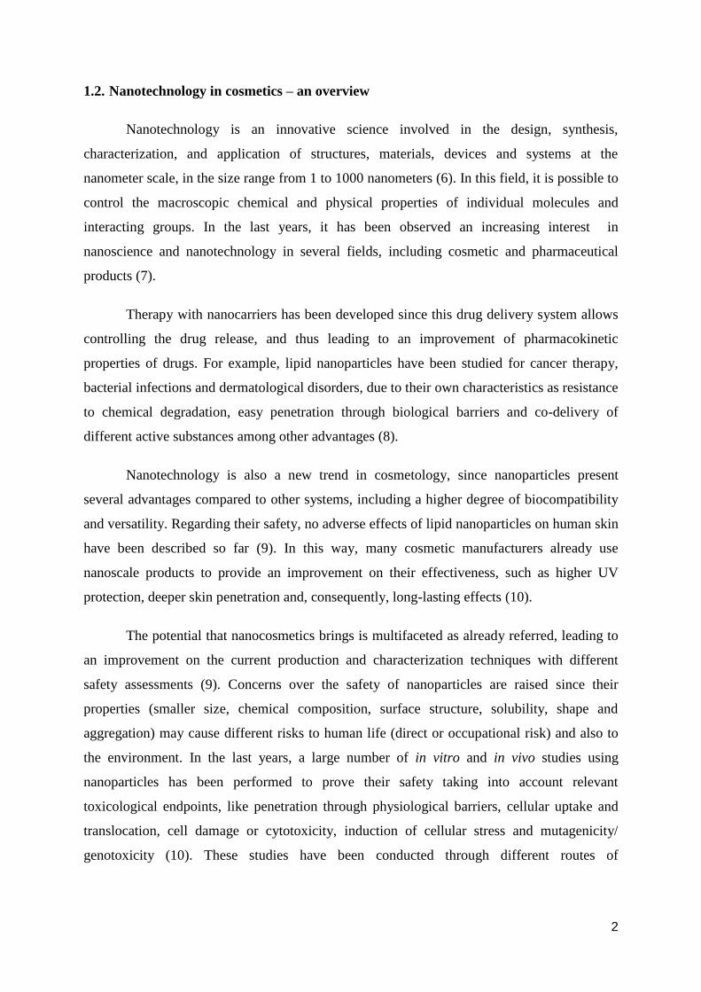
thermodynamically stable (21).
Although microparticles (10-6
) are
bigger than nanoparticles (10-9
), in this case is
different as droplets in microemulsions are
smaller than in nanoemulsions due to historical
development of these formulations (i.e. the term
‘microemulsion’ was firstly published, and just
after three decades the term ‘nanoemulsion’
appeared) (Figure 1) (22).
In this work, nanoemulsions were formulated with liquid lipid Miglyol® 812, a
mixture of medium-chain triglycerides (Caprylic/Capric Triglyceride) with excellent
emolliency and good user properties (23). This compound is able to form reservoir-type drug
delivery systems in the liquid oil core, where some drugs insoluble in water can be dissolved
with an increased payload (24).
Figure 1 – Microemulsion vs Nanoemulsion (20)
5
1.3.2. Solid lipid nanoparticles (SLN)
Solid lipid nanoparticles (SLN) were developed to combine the advantages of three
other systems: emulsions, liposomes and solid particles. SLN are produced by replacing the
liquid lipid (oil) of an O/W emulsion by a solid lipid or a mixture of solid lipids at 37⁰C (15).
These systems are produced with excipients generally recognized as safe (GRAS)
status for oral and dermal delivery, an important advantage concerning toxicity related with
previous formulations among other several benefits (25).
Incorporation of active substances in SLN can protect them from degradation,
increasing drug stability (26), and consequently, allowing a target strategy (27). In addition,
SLN allow a controlled drug release, since biphasic release profiles were observed in several
studies (an initial burst drug release followed by a prolonged release) (28). SLN also
incorporate lipophilic and hydrophilic drugs with no need of organic solvents (29). Despite
these advantages, SLN have shown several limitations, as drug expulsion during storage,
reduced particle concentration and drug loading and high water content of SLN dispersions.
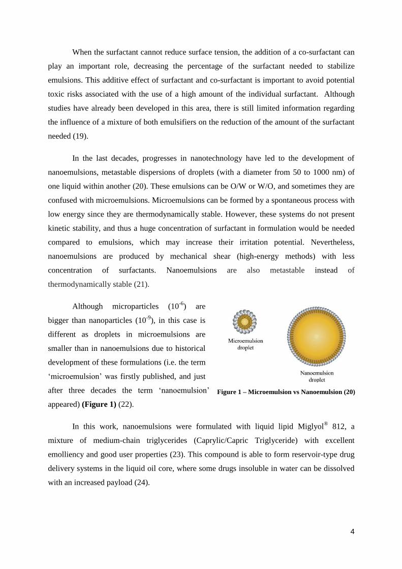
1.3.3. Nanostructured lipid carriers (NLC)
The disadvantages of SLN led to the development of a new generation of
nanosystems: nanostructured lipid carriers (NLC) (30). In this second generation, particles are
produced not only with solid lipids but also with liquid lipids (oils). To produce these blends
of lipids, solid lipids are mixed with liquid lipids (ratio from 70:30 up to 99.9:0.1) (26). There
is a melting point depression in NLC, compared with SLN, but the matrix is also solid at body
temperature (15). One problem verified with SLN is that during storage, a ‘perfect crystal’ is
formed (a matrix totally ordered in conditions of low energy that leads to drug expulsion). In
NLC preparation, the use of different molecules gives rise to an imperfect matrix that
accommodates the drug in its imperfections. To solve this question, there are three types of
NLC (26).
In type I, the highly imperfect type, exists a blend of low liquid lipid (oil) concentration
with solid lipid with the formation of a solid particle characterized by an extremely disordered
matrix. Type II, known as the multiple type, is processed with a higher liquid lipid
concentration, compared to type I, and this increase of lipid concentration leads to phase
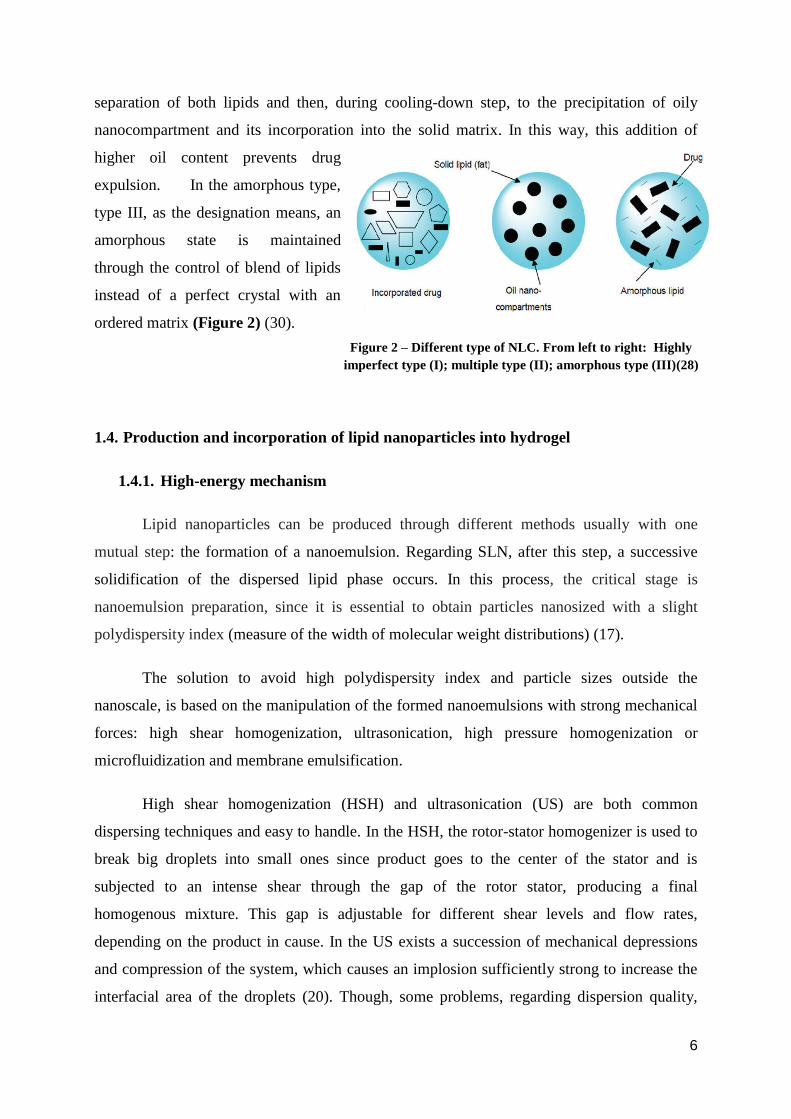
6
separation of both lipids and then, during cooling-down step, to the precipitation of oily
nanocompartment and its incorporation into the solid matrix. In this way, this addition of
higher oil content prevents drug
expulsion. In the amorphous type,
type III, as the designation means, an
amorphous state is maintained
through the control of blend of lipids
instead of a perfect crystal with an
ordered matrix (Figure 2) (30).
1.4. Production and incorporation of lipid nanoparticles into hydrogel
1.4.1. High-energy mechanism
Lipid nanoparticles can be produced through different methods usually with one
mutual step: the formation of a nanoemulsion. Regarding SLN, after this step, a successive
solidification of the dispersed lipid phase occurs. In this process, the critical stage is
nanoemulsion preparation, since it is essential to obtain particles nanosized with a slight
polydispersity index (measure of the width of molecular weight distributions) (17).
The solution to avoid high polydispersity index and particle sizes outside the
nanoscale, is based on the manipulation of the formed nanoemulsions with strong mechanical
forces: high shear homogenization, ultrasonication, high pressure homogenization or
microfluidization and membrane emulsification.
High shear homogenization (HSH) and ultrasonication (US) are both common
dispersing techniques and easy to handle. In the HSH, the rotor-stator homogenizer is used to
break big droplets into small ones since product goes to the center of the stator and is
subjected to an intense shear through the gap of the rotor stator, producing a final
homogenous mixture. This gap is adjustable for different shear levels and flow rates,
depending on the product in cause. In the US exists a succession of mechanical depressions
and compression of the system, which causes an implosion sufficiently strong to increase the
interfacial area of the droplets (20). Though, some problems, regarding dispersion quality,
Figure 2 – Different type of NLC. From left to right: Highly
imperfect type (I); multiple type (II); amorphous type (III)(28)
7
were observed when these techniques were used, such as the presence of microparticles and
metal contamination in case of HSH and US, respectively. An emulsifying agent (surfactant)
is a good solution when the problem is due to the average particle size, since the surfactant
allows the formation of nanoemulsion under simple high shear mixing. Another option could
be the simultaneous use of different methods to obtain better results, such as two short cycles
of HSH and US (to decrease processing intervals and obstacles associated to long preparation
times) (31) or different methods used in different steps (for example, one technique to get
macroemulsion as a pre-emulsion, and then another one to form nanoscale droplets) (20).
In the nineties, Muller applied a new technique, high pressure homogenization (HPH),
which is recognized nowadays as the main and most efficient technique to produce lipid
nanoparticles. This method is characterized by different steps in order to increase efficiency
and valuable results, and it is subdivided into hot and cold homogenization with dissolution,
solubilization or dispersion of the active in the melted lipid (26). Hot homogenization is the
most frequently applied technique since temperature sensitive compounds can be processed
by this way (exposure time is not so long). However, this technique is not able to incorporate
hydrophilic substances as well as extremely temperature sensitive compounds, and to solve
this limitation, cold homogenization was developed (32). Hot homogenization involves the
dissolution of the drug in the lipid and afterward this lipid should be melted at a specific
temperature (5-10ºC above its melting point) (33). After this optimization, this drug dissolved
in melted lipid is dispersed in an aqueous surfactant solution at the same temperature to form
a pre-emulsion. This pre-emulsion is homogenized with a piston-gap homogenizer and then
the lipid re-crystallizes when this O/W nanoemulsion is cooled down (to room temperature).
Overall, this procedure leads to lipid nanoparticles production at the end (31). Usually hot
homogenization is referred in association with high pressure homogenization since this type
of homogenization is on the cutting edge. However, to improve the effectiveness of
techniques as HSH and US, the principle of hot homogenization can be applied in these
techniques if a pre-emulsion is obtained as described before, and then the homogenization
process continues under hot temperature (26, 28). This association can be useful when is not
possible to use high pressure homogenization, for example, in the laboratory scale.
This work was performed following the hot homogenization principle since it was
produced a pre-emulsion subsequently subjected to high shear homogenization process to
obtain lipid nanoparticles.
8
1.4.2. Particle size, polydispersity index (PdI) and zeta potential analysis
Some properties of particles can play an important role regarding the efficacy of
dermal products based on nano-scale particles, such as particle size, polydispersity index
(PdI) and zeta potential.
Particle size influences skin penetration since smaller particles are more able to
pass through skin. Thus, nanoparticles have suitable topical and dermal delivery (10). In this
way, it is essential to measure particle size distributions to understand how this physical
property can affect the performance of products with nanoparticles. In addition, it is also
important to measure the width of molecular weight distributions (MWD), known as
polydispersity index (PdI), which should be at a narrow range (35). Photon correlation
spectroscopy is the method used to measure particle size and PdI by detecting the Brownian
motion of the particles subjected to dynamic light scattering (DLS). The oscillations intensity
of dispersed light arising from Brownian motion is analyzed (smaller particles have faster
diffusion than larger particles), and the particle size is obtained using the Stokes-Einstein
equation (16). To produce high quality data, it is important to dilute all samples (3) and
control the measurement temperature since different temperatures can influence the speed of
Brownian motion (36).
On another hand, it is important to measure zeta potential of the particles since this
property is also related with physical stability. This stability is due to an electric charge on the
particle surface that can repel other particles and avoid particle aggregation/flocculation. This
electric charge depends on the medium in which the particles are dispersed and some
alterations can decrease the stability. Therefore, it is important to control some formulation
parameters such as surfactants, pH and the type/concentration of ions. To keep particle
stability, zeta potential values usually should be more negative than -30 mV or more positive
than +30 mV (37). Zeta potential measurement is based on electrophoretic mobility of
particles, since particles with an electric charge will migrate to an electrode in the presence of
an electric field. There is a higher migration speed with stronger field and higher zeta
potential (in absolute values) (36).
9
1.4.3. Nanolipidgel
After the optimization and production of nanoparticles formulations, these dispersions
should be incorporated into a suitable dermal carrier with semi-solid consistency for topical
application. Due to adverse systemic effects of oral and parenteral formulations, topical
treatment has been extensively studied and used, as it offers many benefits: a) first-pass
metabolism is avoided; b) it is usually well accepted by patients since it is convenient, easy to
use and suitable for self-administration; c) its efficiency is achieved with a lower daily dose;
d) it prevents local fluctuations on the concentration of the active substance; e) active
substance is selectively delivered at the target site; f) it has fewer risks associated with oral or
intravenous administration, such as interactions or infections (38).
Among many topical formulations, the hydrogel is a good choice to incorporate
nanoparticles due to all advantages associated with this topical delivery system (39).
Hydrogels are constituted by a system of polymer chains with the ability to absorb huge
amounts of water due to their hydrophilic properties, with cross-linked compounds that
protect them from the dissolution. The water inside the hydrogel allows the free distribution
of some particles and the polymer works as a matrix to hold water. Gel is a system that is
considered neither liquid nor solid, exhibiting a semi-solid consistency (40).
When nanoparticles are embedded in a semi-solid form, using a gelling agent,
interactions between the constituents of the final formulation could lead to changes in the
physicochemical properties of nanolipid preparations. These modifications can be assessed
using rheological analysis, particle size and zeta potential measurement. Some studies were
performed to understand the influence of different gel-forming polymers used for hydrogel
preparation. According to the results, the performance of these systems is highly dependent on
the structure of these polymers and some polymers as Hydroxyethylcellulose 4000 (HEC) and
Carbopol® 934 preserve physical stability of nanoparticles (41).
1.5. Effectiveness testing – skin’s biophysical parameters measurement
In order to provide an adequate barrier effect, skin needs to be in good conditions as
was previously mentioned, including skin hydration conditions, TEWL and pH. Some dermal
products are designed to improve these skin’s biophysical parameters (42). Nowadays,
noninvasive techniques have been developed to measure these parameters with high
sensitivity and maintaining the skin barrier intact (5).
10
However, only two parameters (skin hydration and TEWL) will be discussed
according to the aim of this work. It is important to take into account that different factors
(age, sex, and anatomical site) and different environmental conditions (temperature, relative
humidity) can influence these values. Therefore, these measurements should be performed at
controlled conditions.
1.5.1. Skin hydration measurement
The water content of epidermis and dermis layers defines the skin hydration. Stratum
corneum (SC) is able to hold water, due to the presence of corneocytes (with hygroscopic
compounds inside mentioned as natural moisturizing factors, NMF) and intercellular lipids
bilayer matrix organized to prevent TEWL, and acting as a barrier. SC is characterized by the
presence of free water, involved in diffusion processes between skin and the environment, and
bound water, associated with NMF present in the epidermis. Lack of water in the skin leads to
defective hydration, responsible for dry and flaky skin surface (sometimes related with some
dermatological disorders). Moisturizers play an important role on skin hydration which is
fundamental to protect and maintain a healthy skin (43,44).
Skin hydration analysis is related with capacitance measurement of a dielectric
medium, detected by corneometry with high sensitivity. As the skin is a dielectric medium,
variations in hydration show up through changes in the dielectric constant. The corneometer
measures the change in the dielectric constant, changing the capacitance (defined as the
ability to store energy in an electric field) of a precision capacitor. In a final step, these
changes regarding water content are converted to arbitrary units of hydration (16,45).
1.5.2. Transepidermal water loss measurement
Transepidermal water loss (TEWL) is the loss of water from the stratum corneum,
affecting the level of epidermis moisture. During the normal skin metabolism, some water
evaporates from the skin continuously in a passive way depending on the relative humidity of
the environment, temperature, season and skin hydration. If the natural barrier function of the
skin is damaged, the water loss will increase, so TEWL is a sensitive indicator of the
defective skin barrier function (46). In this way, it is important to measure for all cosmetic
and dermatological products the influence of this parameter.
11
In order to measure TEWL under in vivo conditions, three techniques can be
performed: closed chamber, ventilated chamber and open chamber. In the closed chamber
method, a capsule is applied on the skin surface and then an electric hygrosensor measures the
water that evaporates and goes into the capsule. Ventilated chamber method is based on the
passage of a gas chamber (with gas and a pre-determined water content) along the skin and
the posterior measurement of the amount of water captured by the gas in the chamber. Then,
this collected water is analyzed by an incorporated hygrometer in the chamber. Despite being
a method that provides a continuous measurement of TEWL, incorrect results may occur in
case the gas inside the chamber becomes dehydrated, leading to an increase of water
evaporation from skin (5). Finally, the most approached method for determining TEWL is the
open chamber method based on the diffusion principle in an open chamber. There are several
instruments to measure TEWL by open chamber, but it will be discussed only the apparatus
used in this experiment: tewameter. This instrument has an open chamber that measures the
density gradient of the water evaporation from the skin by two pairs of sensors (temperature
and relative humidity) inside the cylinder (head of probe). This head of probe minimizes the
influence of air turbulence inside the probe. One pair is higher than the other one, and the
moisture at two different places is measured to determine the TEWL. During this
measurement, a microprocessor analyses the values expressing the evaporation rate in g/h/m2
(47).
2. Aim of the work
The aim of this research project was to evaluate the influence of three well
characterized different lipid nanosystems (solid lipid nanoparticles, nanostructured lipid
carriers and nanoemulsions) on skin hydration and transepidermal water loss on human
volunteers.
12
3. Materials and Methods
3.1. Materials
Lipids
➔ Dynasan® 114 (Sasol, Germany): Trimyristin, m.p. 55-58ºC (48)
➔ Glycerol monostearate 35-50 (Lex, Slovenia): Monoester of glycerin and stearic
acid, m.p. 56-59ºC (49)
➔ Miglyol® 812 (Sasol, Germany): Caprylic/Capric Triglyceride
Emulsifiers/Steric stabilizers
➔ Surfactant – Phospholipon® 80 (Phospholipid GmbH, Germany)
➔ Co-surfactant – Lutrol® F68: Poloxamer 188 (BASF, Germany)
➔ Co-surfactant – Tween® 80: Polysorbate 80 (Sigma-Aldrich Chemie GmbH,
France)
Ingredients of hydrogel
➔ Hydroxyethylcellulose 4000 (Merck, Germany)
➔ Glycerol 85% (Caesar&Loretz, Germany)
➔ Sodium methylparahydroxybenzoate (Lex, Slovenia)
Reagents/Solvents
➔ Purified water (Faculty of Pharmacy, University of Ljubljana)
3.2. Equipments
➔ Dual range Analytical Balance® AG245 (Mettler Toledo, Switzerland)
➔ Precision Balance® XP4002S (Mettler Toledo, Switzerland)
➔ Magnetic hotplate stirrer® RH basic 2 IKAMAG (IKA, Brasil)
➔ GFL multi-station water bath® TYP 1041 (GFL, Germany)
➔ Hotplate® EKP 3582 (Clatronic International GmbH, Germany)
➔ Ultra-Turrax® T25D rotor-stator homogenizer (IKA, Germany)
➔ PCS naprava Zetasizer Nano ZS® (Malvern Instruments, United Kingdom)
➔ Probe Heater® PR 100 (Courage & Khazaka GmbH, Germany)
➔ Tewameter® TM 300 (Courage & Khazaka GmbH, Germany)
➔ Corneometer® CM 825 (Courage & Khazaka GmbH, Germany)
13
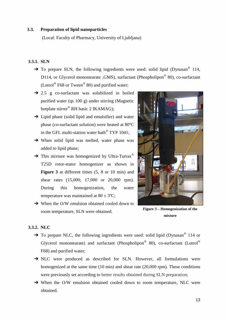
3.3. Preparation of lipid nanoparticles
(Local: Faculty of Pharmacy, University of Ljubljana)
3.3.1. SLN
➔ To prepare SLN, the following ingredients were used: solid lipid (Dynasan®
114,
D114, or Glycerol monostearate ,GMS), surfactant (Phospholipon® 80), co-surfactant
(Lutrol® F68 or Tween
® 80) and purified water;
➔ 2.5 g co-surfactant was solubilized in boiled
purified water (qs 100 g) under stirring (Magnetic
hotplate stirrer®
RH basic 2 IKAMAG);
➔ Lipid phase (solid lipid and emulsifier) and water
phase (co-surfactant solution) were heated at 80ºC
in the GFL multi-station water bath® TYP 1041;
➔ When solid lipid was melted, water phase was
added to lipid phase;
➔ This mixture was homogenized by Ultra-Turrax®
T25D rotor-stator homogenizer as shown in
Figure 3 at different times (5, 8 or 10 min) and
shear rates (15,000; 17,000 or 20,000 rpm).
During this homogenization, the water
temperature was maintained at 80 ± 3ºC;
➔ When the O/W emulsion obtained cooled down to
room temperature, SLN were obtained.
3.3.2. NLC
➔ To prepare NLC, the following ingredients were used: solid lipid (Dynasan® 114 or
Glycerol monostearate) and surfactant (Phospholipon® 80), co-surfactant (Lutrol
®
F68) and purified water;
➔ NLC were produced as described for SLN. However, all formulations were
homogenized at the same time (10 min) and shear rate (20,000 rpm). These conditions
were previously set according to better results obtained during SLN preparation;
➔ When the O/W emulsion obtained cooled down to room temperature, NLC were
obtained.
Figure 3 – Homogenization of the
mixture
14
3.3.3. NE
➔ To prepare NLC the following ingredients were used: liquid lipid (Miglyol® 812) and
surfactant (Phospholipon® 80), co-surfactant (Lutrol
® F68) and purified water;
➔ NLC were produced as described for SLN and NLC (homogenization at 20,000 rpm
for 10 min);
➔ When the O/W emulsion obtained cooled down to room temperature, NE were
produced.
The final composition of each nanolipid system is represented on Table 1.
Table 1 – Composition (expressed in percentage) of each nanolipid system
3.4. Evaluation of the physical parameters of nanosystems
After the production of these nanosystems, particle size, PdI and zeta potential of the
individual dispersions were measured on the Zetasizer Nano ZS®, to evaluate the main
physical parameters of the lipid formulations. The samples were previously diluted with
purified water.
3.4.1. Particle size and polydispersity index
These parameters were evaluated under the following conditions:
Dispersant: Water (Temperature: 25 ° C, Viscosity: 0.8872 mPa.s, RI: 1.330)
Temperature (T): 25 ° C
Equilibration time: 30 s
Cell type: DTS0012 - Disposable sizing cuvette
Measurement angle: 173 ° Backscatter (NIBS default)
SLN NLCD114 NLCGMS NE
Dynasan®
114 3.5 2.8 ----- -----
GMS ----- ----- 2.8 -----
Miglyol®
812 ----- 0.7 0.7 3.5
Lutrol® F68 1.5 1.5 1.5 1.5
Phospholipon® 80 1 1 1 1
Purified water qs 100 qs 100 qs 100 qs 100
15
3.4.2. Zeta potential
This parameter was evaluated under the following conditions:
Dispersant: Water (Temperature: 25 ° C, Viscosity: 0.8872 mPa.s, RI: 1.330,
Dielectric constant: 78.5)
F(Ka) selection: Model Smoluchowski
Temperature (T): 25 ° C
Equilibration time: 30 s
Cell type: DTS1060C – Clear disposable zeta cell
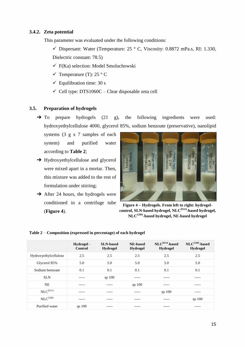
3.5. Preparation of hydrogels
➔ To prepare hydrogels (21 g), the following ingredients were used:
hydroxyethylcellulose 4000, glycerol 85%, sodium benzoate (preservative), nanolipid
systems (3 g x 7 samples of each
system) and purified water
according to Table 2;
➔ Hydroxyethylcellulose and glycerol
were mixed apart in a mortar. Then,
this mixture was added to the rest of
formulation under stirring;
➔ After 24 hours, the hydrogels were
conditioned in a centrifuge tube
(Figure 4).
Table 2 – Composition (expressed in percentage) of each hydrogel
Hydrogel -
Control
SLN-based
Hydrogel
NE-based
Hydrogel NLCD114-based
Hydrogel
NLCGMS-based
Hydrogel
Hydroxyethylcellulose 2.5 2.5 2.5 2.5 2.5
Glycerol 85% 5.0 5.0 5.0 5.0 5.0
Sodium benzoate 0.1 0.1 0.1 0.1 0.1
SLN ----- qs 100 ----- ----- -----
NE ----- ----- qs 100 ----- -----
NLCD114 ----- ----- ----- qs 100 -----
NLCGMS ----- ----- ----- ----- qs 100
Purified water qs 100 ----- ----- ----- -----
Figure 4 – Hydrogels. From left to right: hydrogel-
control, SLN-based hydrogel, NLCD114
-based hydrogel,
NLCGMS
-based hydrogel, NE-based hydrogel

16
3.6. Design of the clinical study
3.6.1. Subjects
The clinical study was performed in 6 healthy volunteers. These participants received
and signed an informed consent form, in order to prove that they accepted the test conditions
(Annex 1 (A1)). All procedures of this study were performed in accordance with the
principles of the Declaration of Helsinki and respective revisions.
The criteria for inclusion and exclusion in this study were previously established.
The inclusion criteria of volunteers were as follows: subjects aged between 21 and 27
years old; few or no hair on the volar forearms; absence of dermatological diseases, tattoos,
scars and/or history of frequent sunburns; to be present in the Faculty of Pharmacy of the
University of Ljubljana on the pre-set day to perform the measurements; to be cooperative,
discerning and able to follow instructions and comply with the study requirements.
On the other hand, the exclusion criteria taken into account were as follows: subjects
aged less than 18 years old; excessive hairiness on the forearms; presence of dermatological
diseases, tattoos, scars and/or history of frequent sunburns on the arms or forearms; the use of
other cosmetics products on the forearms that were not defined in the study protocol; known
history of allergy and/ or hypersensitivity to the ingredients contained in the composition of
the hydrogels tested; pregnant or lactating women; the presence of severe disease in the last 6
months prior to the beginning of the study; the presence of clinically relevant skin diseases or
any other physical disorder with cutaneous manifestations; the presence of mental/psychiatric
illness or systemic disease in the beginning of the study; the presence of any type of
immunological alterations, including autoimmune diseases; the presence of fever for more
than 24 hours, for less than 8 days before the beginning of the study; the participation in other
cosmetic or clinical studies for less than 2 weeks before the beginning of the study; recent
dermo-cosmetic or aesthetic treatments, 2 months prior to the beginning of the study; recent
(one month prior to the study) and intense exposure to ultraviolet radiation (sun/tanning beds);
the application of any topical medication in the forearms for less than 1 month prior the study
initiation; recent vaccinations, 2 weeks prior to the commencement of the study; taking any
systemic medication such as corticosteroids or antihistamines for less than 4 weeks, anti-
inflammatory or antibiotics for less than 2 weeks and retinoids for less than 3 months before
17
the beginning of the study; to have strong smoking habits (≥ 20 cigarettes per week over more
than 2 years); to be a consumer of drugs.
The participants were also asked not to apply any cream in the test area two days
before the measurement and not to smoke, exercise or drink coffee/tea or any other energy
drink in the measurement day.
3.6.2. Measurement conditions
The clinical study was carried out in a specific room in the Faculty of Pharmacy,
University of Ljubljana. It was attempted to control the temperature (T), to minimize sweat
production, and the relative humidity (RH) of the environment in order to perform all
measurements under certain room conditions (T: 20°C; RH: 40-60%) to obtain reproducible
results.
Accordingly, all volunteers were previously set in a comfortable position and prepared
removing the clothing from the arm area to be tested and letting the skin to acclimatize to
these environmental conditions for 20 minutes before the measurement.
During this clinical study, non-invasive biophysical measuring methods were used in
order to measure the influence of formulated hydrogels in skin hydration and TEWL. All
measurements were performed by one of the principal investigators.
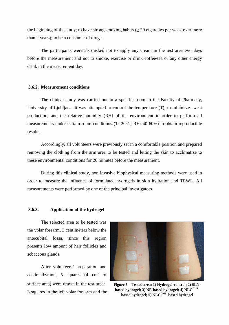
3.6.3. Application of the hydrogel
The selected area to be tested was
the volar forearm, 3 centimeters below the
antecubital fossa, since this region
presents low amount of hair follicles and
sebaceous glands.
After volunteers` preparation and
acclimatization, 5 squares (4 cm2
of
surface area) were drawn in the test area:
3 squares in the left volar forearm and the
Figure 5 – Tested area: 1) Hydrogel-control; 2) SLN-
based hydrogel; 3) NE-based hydrogel; 4) NLCD114
-
based hydrogel; 5) NLCGMS
-based hydrogel
18
others in the right volar forearm. Hydrogels with and without nanolipid systems (0.5 ml)
where applied as shown in Figure 5.
The study was designed as single blind. Therefore, the hydrogels were assigned by a
number, so that the volunteers were unaware of the type of system under test in each square.
Two measurements were performed in each square for both parameters (skin hydration
and TEWL). A first measurement was performed before hydrogels` application, to obtain the
basal values. Then, hydrogels were applied on the corresponding areas. After 30 minutes the
remnants of hydrogels were removed, and second measurements were performed after 60
minutes, measuring TEWL followed by skin hydration. Measurements were carried out in the
counterclockwise direction.
3.6.4. Skin hydration measurement
Skin hydration was measured using a Corneometer® CM 825. The probe was placed
perpendicularly to the skin area to be measured. The spring inside the probe head
ensures constant pressure (1.0 N ± 10 %) on the skin enabling exact and reproducible
measurement (uncertainty degree: ± 3%) (45). This spring covers an area of 49 mm2
and
assesses the epidermal water content from 20 to 30 µm (50).
Measurements were performed three times in each area. The display shows
immediately the measured values in arbitrary units (arbitrary Corneometer® units), and it is
necessary to wait about 5 seconds between each measurement. Between different squares, the
probe head was cleaned softly with dry paper.
To evaluate the level of skin hydration, the table below (Table 3) was used as
reference (this table was described in the data sheet of the corneometer used in this
experiment, Corneometer ® CM 825) (51).
Table 3 – Interpretation of measured values of skin hydration with Corneometer ®
CM 825
Skin hydration – arbitrary units
Interpretation
<30 Very dry
30-40 Dry
>40 Sufficiently moisturized
19
3.6.5. TEWL measurement
TEWL measurements were carried out with Tewameter® TM 300. The water
evaporated from the skin is measured indirectly by two pairs of sensors (T and RH), and it is
analyzed by a microprocessor. The results obtained are expressed in g/h/m2.
The probe was sited tightly on the skin surface, for approximately 90 seconds. During
this time, the volunteer could not move the arm to ensure reliable results. Between each
measurement, the probe was cleaned softly with wet paper (with deionized water).
The sensors in the probe usually have room temperature, however they should reach
skin temperature (32ºC) since the amount of evaporating water measured with the probe is
particularly low. Thus, the probe head is constantly warmed up to around 32 ºC in Probe
Heater® PR 100 in order to get very quickly accurate and stable results before and between
different measurements.
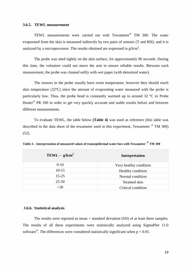
To evaluate TEWL, the table below (Table 4) was used as reference (this table was
described in the data sheet of the tewameter used in this experiment, Tewameter ® TM 300)
(52).
Table 4 – Interpretation of measured values of transepidermal water loss with Tewameter ®
TM 300
TEWL – g/h/m2
Interpretation
0-10 Very healthy condition
10-15 Healthy condition
15-25 Normal condition
25-30 Strained skin
>30 Critical condition
3.6.6. Statistical analysis
The results were reported as mean + standard deviation (SD) of at least three samples.
The results of all these experiments were statistically analyzed using SigmaPlot 11.0
software®. The differences were considered statistically significant when p < 0.05.
20
4. Results and Discussion
4.1. Pre-experimental work: the influence of different production parameters on the
particle size, polydispersity index and zeta potential
The first step of this experimental work was to study the influence of different
dynamic parameters (time and stirring rate of homogenization) as well as the effect of lipids
and co-surfactants (type and concentration) on the preparation of lipid nanoparticle systems.
In order to characterize the best formulations, the particle size (mean size), polydispersity
index and zeta potential were measured.
Regarding particle size, these particles should exhibit nanosize (from 1 to 1000 nm)
and be as small as possible, since an increase in the particle size will lead to lower physical
stability (16). It is important to obtain the optimal nanoparticle size range. In fact, several
studies, that have been developed in this field, showed the best clinical results with
nanoparticles at the size range of approximately 10–250 nm (53).
Measurement of PdI of nanoparticles is essential to obtain their size distribution. PdI is
dimensionless and scaled, so dispersity values range from 0 to 1. Samples are not suitable for
DLS technique if they have a very large size distribution, i.e. PdI values higher than 0.7 (54).
The higher the PdI value, the less monodispersed nanoparticle system is (55).
Nanoparticles should have zeta potential (ZP) more negative than -30 mV or more
positive than +30mV to be physically stable (16). Charged particles, with high zeta potential
modulus, repeal each other and prevent particle aggregation, allowing storage stability of
colloidal systems (56). In this case, the zeta potential of the developed systems (NE, SLN and
NLC) was negative due to the anionic nature of the surfactant components (Phospholipon® 80
has phosphatidylcholine with negatively charged phospholipids) (57).
These preliminary formulation studies were performed before the experimental design
in order to select the appropriate lipid, co-surfactant and homogenization conditions. These
conditions were firstly set during SLN preparation.
21
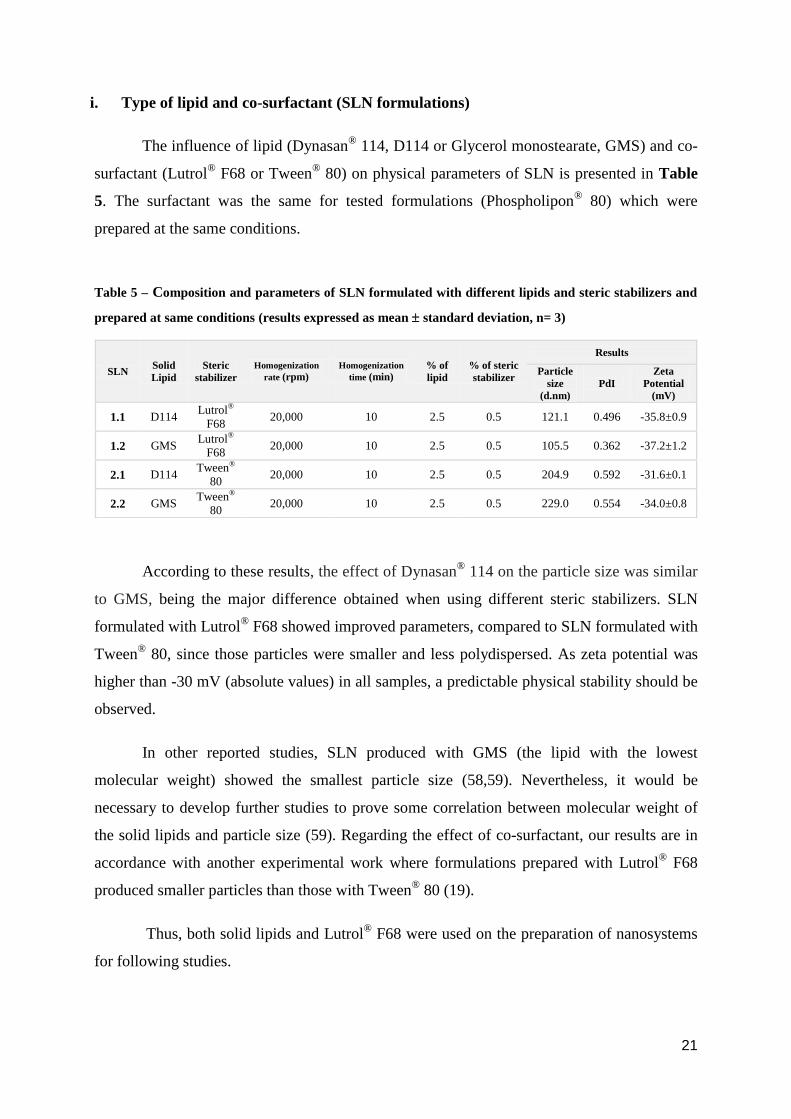
i. Type of lipid and co-surfactant (SLN formulations)
The influence of lipid (Dynasan®
114, D114 or Glycerol monostearate, GMS) and co-
surfactant (Lutrol® F68 or Tween
® 80) on physical parameters of SLN is presented in Table
5. The surfactant was the same for tested formulations (Phospholipon® 80) which were
prepared at the same conditions.
Table 5 – Composition and parameters of SLN formulated with different lipids and steric stabilizers and
prepared at same conditions (results expressed as mean standard deviation, n= 3)
According to these results, the effect of Dynasan®
114 on the particle size was similar
to GMS, being the major difference obtained when using different steric stabilizers. SLN
formulated with Lutrol® F68 showed improved parameters, compared to SLN formulated with
Tween® 80, since those particles were smaller and less polydispersed. As zeta potential was
higher than -30 mV (absolute values) in all samples, a predictable physical stability should be
observed.
In other reported studies, SLN produced with GMS (the lipid with the lowest
molecular weight) showed the smallest particle size (58,59). Nevertheless, it would be
necessary to develop further studies to prove some correlation between molecular weight of
the solid lipids and particle size (59). Regarding the effect of co-surfactant, our results are in
accordance with another experimental work where formulations prepared with Lutrol® F68
produced smaller particles than those with Tween®
80 (19).
Thus, both solid lipids and Lutrol® F68 were used on the preparation of nanosystems
for following studies.
SLN Solid
Lipid
Steric
stabilizer
Homogenization
rate (rpm) Homogenization
time (min) % of
lipid
% of steric
stabilizer
Results
Particle
size
(d.nm)
PdI
Zeta
Potential
(mV)
1.1 D114 Lutrol®
F68 20,000 10 2.5 0.5 121.1 0.496 -35.8±0.9
1.2 GMS Lutrol®
F68 20,000 10 2.5 0.5 105.5 0.362 -37.2±1.2
2.1 D114 Tween®
80 20,000 10 2.5 0.5 204.9 0.592 -31.6±0.1
2.2 GMS Tween®
80 20,000 10 2.5 0.5 229.0 0.554 -34.0±0.8
22
ii. Homogenization time and shear rate (SLN formulations)
It is crucial to optimize the homogenization conditions, i.e. shear rate, temperature and
time of homogenization to obtain a stable emulsion with nanoparticles, avoiding the formation
of microparticles.
Regarding temperature, the lipid is heated at approx. 5-10ºC above its melting point
for the hot pressure homogenization (HPH) process (33). Since high-shear homogenization
(HSH) was used in this experimental work, instead of HPH, a higher recommended
temperature, at least 10ºC above lipid melting point, was used (60). In that way, the lipids
were heated at 20ºC above their melting point to increase the effectiveness of this method. In
fact, higher temperature is beneficial for emulsification process due to reduction of surface
tension (61).
The influence of homogenization time and shear rate on physical parameters of SLN is
presented in Table 6.
Table 6 – Composition and parameters of two SLN formulations prepared at different homogenization
conditions (results expressed as mean standard deviation, n= 3)
SLN Solid
Lipid
Steric
stabilizer Homogenization
rate (rpm)
Homogenization
time (min)
% of
lipid
% of
steric
stabilizer
Results
Particle
size
(d.nm)
PdI
Zeta
Potential
(mV)
3.1 D114 Lutrol® F68 20,000 5 2.5 0.5 259.7 0.790 -30.4±1.2
3.2 D114 Lutrol® F68 20,000 8 2.5 0.5 208.4 0.582 -38.4±0.1
3.3 D114 Lutrol® F68 20,000 10 2.5 0.5 110.9 0.248 -34.0±0.8
3.4 GMS Lutrol® F68 20,000 5 2.5 0.5 220.5 0.575 -31.8±1.2
3.5 GMS Lutrol® F68 20,000 8 2.5 0.5 201.4 0.523 -34.8±0.9
3.6 GMS Lutrol® F68 20,000 10 2.5 0.5 178.5 0.345 -35.0±1.0
4.1 D114 Lutrol® F68 15,000 10 2.5 0.5 218.8 0.667 -36.5±1.0
4.2 D114 Lutrol® F68 17,000 10 2.5 0.5 163.5 0.491 -35.1±0.3
4.3 D114 Lutrol® F68 20,000 10 2.5 0.5 110.9 0.359 -39.4±1.1
4.4 GMS Lutrol® F68 15,000 10 2.5 0.5 233.4 0.452 -30.7±0.7
4.5 GMS Lutrol® F68 17,000 10 2.5 0.5 215.8 0.416 -31.4±1.0
4.6 GMS Lutrol® F68 20,000 10 2.5 0.5 101.0 0.286 -30.2±1.8
23
According to this table, the average SLN diameter and PdI were reduced with
increasing homogenization time (from 5 to 10 min) and shear rate (from 15,000 to 20,000
rpm), as theoretically expected. In addition, all formulations presented acceptable zeta
potential values.
It was previously observed that smaller particles were obtained by increasing the shear
rate. However, above 20,000 rpm (25,000 rpm) the average particle diameter did not
significantly changed. On the other hand, the shear rates below 15,000 rpm were not sufficient
for the formation of suitable SLNs, since large particles were visible in the dispersion. If
homogenization extends for more than 10 minutes, particles may become unstable due to high
energy input, leading to the formation of microparticles (60).
Therefore, better results were obtained for 10 minutes and 20,000 rpm, which were the
values selected to produce all lipid formulations.
iii. Percentage of solid lipid and steric stabilizer (SLN formulations)
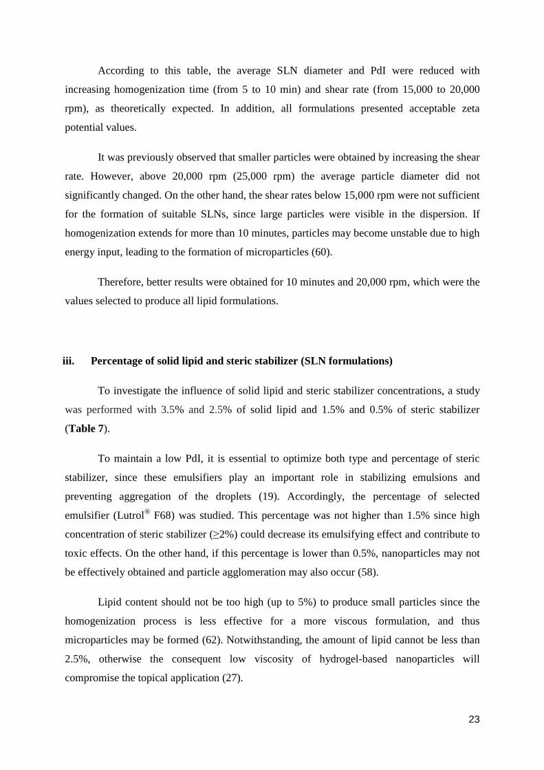
To investigate the influence of solid lipid and steric stabilizer concentrations, a study
was performed with 3.5% and 2.5% of solid lipid and 1.5% and 0.5% of steric stabilizer
(Table 7).
To maintain a low PdI, it is essential to optimize both type and percentage of steric
stabilizer, since these emulsifiers play an important role in stabilizing emulsions and
preventing aggregation of the droplets (19). Accordingly, the percentage of selected
emulsifier (Lutrol® F68) was studied. This percentage was not higher than 1.5% since high
concentration of steric stabilizer (≥2%) could decrease its emulsifying effect and contribute to
toxic effects. On the other hand, if this percentage is lower than 0.5%, nanoparticles may not
be effectively obtained and particle agglomeration may also occur (58).
Lipid content should not be too high (up to 5%) to produce small particles since the
homogenization process is less effective for a more viscous formulation, and thus
microparticles may be formed (62). Notwithstanding, the amount of lipid cannot be less than
2.5%, otherwise the consequent low viscosity of hydrogel-based nanoparticles will
compromise the topical application (27).
24
Accordingly, it is quite perceptible that it is essential to balance all possible
phenomena to optimize the composition of both steric stabilizer and lipid in formulation
study. This study was performed with two different percentages of each parameter since the
main goal was to understand whether a proportional correlation would be observed between
them.
Table 7 – Composition and parameters of SLN formulated with different solid lipid and steric stabilizer
concentrations and prepared at same conditions (results expressed as mean standard deviation, n= 3)
Table 7 shows that best results (regarding particle size and PdI) were obtained with
the formulations 5.1 (3.5% of D114 with 1.5% of Lutrol® F68), 5.4 (2.5% of D114 with 0.5%
of Lutrol® F68), 5.5 (3.5% of GMS with 1.5% of Lutrol
® F68), and 5.8 (2.5% of GMS with
0.5% of Lutrol® F68). Zeta potential presented good values in all formulations.
These values suggest that to obtain smaller particles with narrower PdI, the amount of
solid lipid and steric stabilizer should be proportional, i.e. if a higher percentage of lipid is
used (3.5%), then a higher percentage of steric stabilizer should be used as well (1.5%), and
vice versa, a lower content of steric stabilizer (0.5%) should be used with a lower lipid
amount (2.5).
SLN Solid
Lipid
Steric
stabilizer Homogenization
rate (rpm)
Homogenization
time (min)
% of
lipid
% of
steric
stabilizer
Results
Particle size
(d.nm) PdI
Zeta
Potential
(mV)
5.1 D114 Lutrol®
F68 20,000 10 3.5 1.5 214.6 0.431 -31.0±1.1
5.2 D114 Lutrol®
F68 20,000 10 3.5 0.5 291.2 0.761 -31.2±0.6
5.3 D114 Lutrol®
F68 20,000 10 2.5 1.5 265.5 0.792 -33.6±0.7
5.4 D114 Lutrol®
F68 20,000 10 2.5 0.5 174.4 0.590 -34.1±1.0
5.5 GMS Lutrol®F6
8 20,000 10 3.5 1.5 190.7 0.401 -32.1±0.9
5.6 GMS Lutrol®
F68 20,000 10 3.5 0.5 289.9 0.762 -33.4±0.6
5.7 GMS Lutrol®
F68 20,000 10 2.5 1.5 280.7 0.753 -32.4±1.0
5.8 GMS Lutrol®
F68 20,000 10 2.5 0.5 210.4 0.421 -31.0±0.9
25
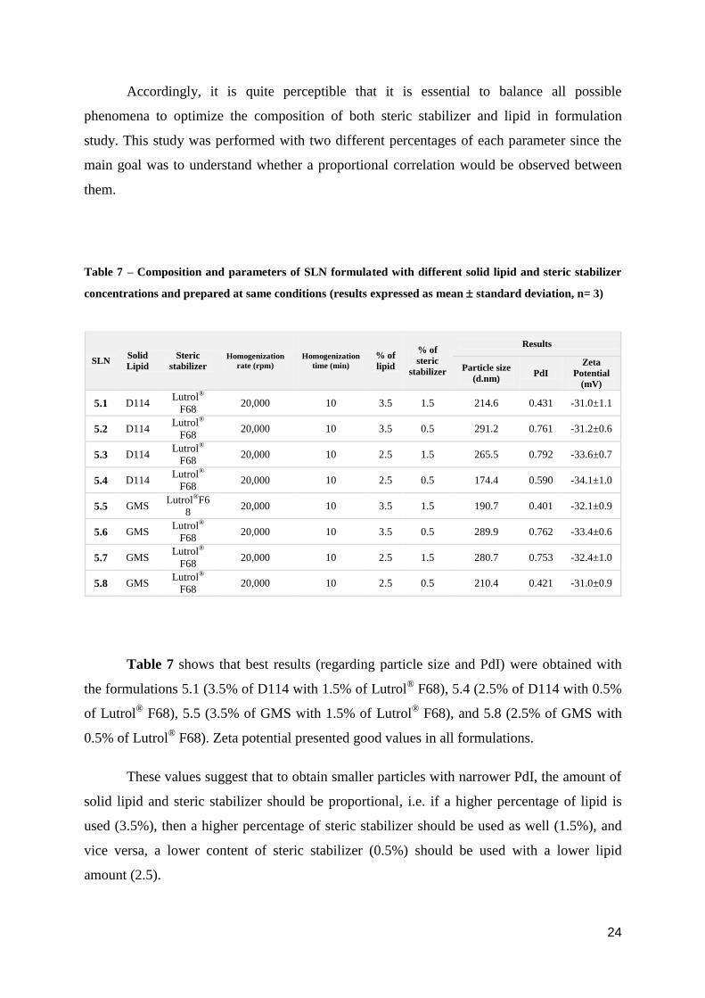
iv. Percentage of solid lipid (NLC formulations)
To study the influence of solid (s) lipid concentration in NLC, two solid lipids
(Dynasan® 114 and GMS) were used at different concentrations (2.8% and 2.1%,
respectively). On the contrary, the liquid (l) lipid, Miglyol® 812, was maintained at the same
concentration (0.7%) (Table 8).
Regarding steric stabilizer in this formulation, 1.5% was the selected amount (Table
8), taking into account the results from the last experiment (iii) (Table 7).
Since it was already studied the influence of liquid lipid percentage in NLC
formulations (using the same solid lipid content) reported in literature (63), here we proposed
to study the influence of only solid liquid percentage in NLC and how different total lipid
concentration and ratio solid/liquid lipid influences the particle size and zeta potential.
Table 8 – Composition and parameters of NLC formulated with different solid lipid type and
concentration and prepared at same conditions (results expressed as mean standard deviation, n= 3).
Smaller particle size and lower PdI values were obtained at higher solid lipid
concentration (samples 6.1 and 6.3), especially in NLC formulated with GMS. Some literature
reports suggest that the optimum solid / liquid lipid ratio to produce NLC varies from 70:30 to
99.9:0.1. Thus, samples 6.1 and 6.3 which presented a ratio of 70:30 are in accordance with
these reports (15).
NLC Solid
lipid
Liquid
lipid
Steric
stabilizer
Homogenization
rate (rpm)
Homogenization
time (min)
% of
lipid
(s/l)
% of
steric
stabilizer
Results
Particle
size
(d.nm)
PdI
Zeta
Potential
(mV)
6.1 D114 Miglyol®
812
Lutrol®
F68 20,000 10 2.8 / 0.7 1.5 271.3 0.601 -31.4±1.2
6.2 D114 Miglyol®
812 Lutrol®
F68 20,000 10 2.1 / 0.7 1.5 294.5 0.705 -36.2±0.7
6.3 GMS Miglyol®
812
Lutrol®
F68 20,000 10 2.8 / 0.7 1.5 128.1 0.448 -38.5±0.5
6.4 GMS Miglyol®
812
Lutrol®
F68 20,000 10 2.1 / 0.7 1.5 138.7 0.301 -37.6±1.3
26
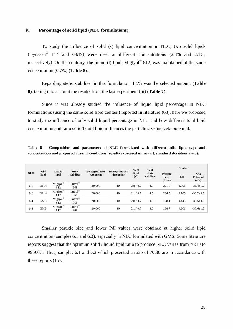
v. Percentage of liquid lipid (NE formulations)
To produce an emulsion, it is important to consider the optimum liquid lipid and steric
stabilizer ratio, which lead to a low surface tension and spontaneous droplet formation. It has
been reported that the surface tension between oil and water phases has a huge influence on
droplet size of the dispersed phase (even more than the oil viscosity). In this way, the
concentration of the liquid lipid and the liquid lipid/co-surfactant ratio are critical to optimize
droplet size through the influence of a lower interfacial tension (23,64).
Accordingly, the effect of the percentage of liquid lipid on nanoemulsions (NE)
formulations was also evaluated (Table 9), using the same percentage of steric stabilizer used
before in the design of NLC formulations (1.5%), and their ratio as well.
Table 9 – Composition and parameters of NE formulated with different liquid lipid concentration and
prepared at same conditions (results expressed as mean standard deviation, n= 3)
These results show that the use of both liquid lipid concentrations presented quite
similar and favorable results (nanoparticles with an acceptable PdI and zeta potential).
Regarding liquid lipid content, the percentages chosen were used to compare with the
results of another previous formulation with 3.5% of lipid and 1.5% of steric stabilizer (63).
Accordingly, 3.5% of liquid lipid was a good choice to produce a nanoemulsion with suitable
medium droplet size, being in accordance with the results here presented.
Finally, it is important to consider the liquid lipid/co-surfactant ratio. Several studies
proved that the use of Miglyol® 812/co-surfactant at 7:3 or 6:4 ratios presented similar results,
exhibiting an optimal droplet size (65). Since samples 7.1 and 7.2 with similar ratios showed
good results, it can be concluded that those ratios can decrease the interfacial tension, and
consequently, optimize the droplet size.
NE Liquid
lipid
Steric
stabilizer Homogenization
rate (rpm)
Homogenization
time (min)
% of
lipid
% of steric
stabilizer
Results
Droplet
size
(d.nm)
PdI
Zeta
Potential
(mV)
7.1 Miglyol®
812
Lutrol®
F68 20,000 10 3.5 1.5 181.0 0.341 -34.6±0.5
7.2 Miglyol®
812
Lutrol®
F68 20,000 10 2.5 1.5 185.0 0.391 -35.0±1.2

27
NLCGMS
Sample Size (nm)
10.1 141.5
10.2 162
10.3 157.9
10.4 131.2
10.5 178.7
10.6 151.2
10.7 141.9
Average 152.1
SD 15.8
RSD % 10.4
SLN
Sample Size (nm)
8.1 205.2
8.2 201.5
8.3 244.9
8.4 216.5
8.5 214.5
8.6 229.6
8.7 198.4
Average 215.8
SD 16.6
RSD % 7.7
NLCD114
Sample Size (nm)
9.1 174.8
9.2 183.6
9.3 194.3
9.4 202.5
9.5 209.4
9.6 173.4
9.7 211.2
Average 192.7
SD 15.8
RSD % 8.1
NE
Sample Size (nm)
11.1 225
11.2 214.2
11.3 216
11.4 196.5
11.5 188.7
11.6 180.2
11.7 201.4
Average 203.1
SD 16.1
RSD % 7.9
4.2. Final lipid nanoformulations
After these preliminary studies, the final formulation of each system was defined
according to Table 1, taking into account all parameters optimized in the pre-experimental
work.
i. Mean particle size measurement
To evaluate the mean particle size of each formulation, 7 samples (3 g) of each final
system were prepared and characterized (Table 10).
Overall, an average particle size of around 200 nm was obtained as expected. Among
all formulations developed, NLC presented significantly smaller particles compared to NE
and SLN (with the worst mean size). These outcomes confirm the results of other experiments
performed before in which particle size was optimized in different carriers (66–68).
Regarding NLC formulated with different solid lipids, NLC with GMS presented the
smallest particles, which coincides with the results from other experiments involving SLN
(58,59). As mentioned before (in topic 4.1.i.), this could be related with the lowest molecular
weight of GMS compared to D114.
Table 10 – Size parameters of lipid formulations (SLN, NLC and NE)
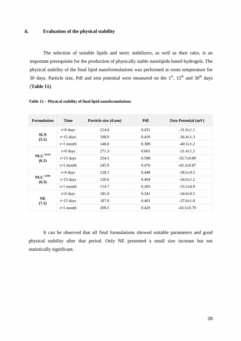
28
ii. Evaluation of the physical stability
The selection of suitable lipids and steric stabilizers, as well as their ratio, is an
important prerequisite for the production of physically stable nanolipids based hydrogels. The
physical stability of the final lipid nanoformulations was performed at room temperature for
30 days. Particle size, PdI and zeta potential were measured on the 1st, 15
th and 30
th days
(Table 11).
Table 11 – Physical stability of final lipid nanoformulations
It can be observed that all final formulations showed suitable parameters and good
physical stability after that period. Only NE presented a small size increase but not
statistically significant.
Formulation Time Particle size (d.nm) PdI Zeta Potential (mV)
SLN
(5.1)
t=0 days 214.6 0.431 -31.0±1.1
t=15 days 198.0 0.410 -36.4±1.3
t=1 month 148.0 0.389 -40.5±1.2
NLC D114
(6.1)
t=0 days 271.3 0.601 -31.4±1.2
t=15 days 254.5 0.598 -35.7±0.80
t=1 month 245.9 0.476 -43.3±0.87
NLC GMS
(6.3)
t=0 days 128.1 0.448 -38.5±0.5
t=15 days 120.6 0.404 -34.0±1.2
t=1 month 114.7 0.305 -33.2±0.9
NE
(7.1)
t=0 days 181.0 0.341 -34.6±0.5
t=15 days 187.6 0.401 -37.6±1.0
t=1 month 209.5 0.420 -43.5±0.79
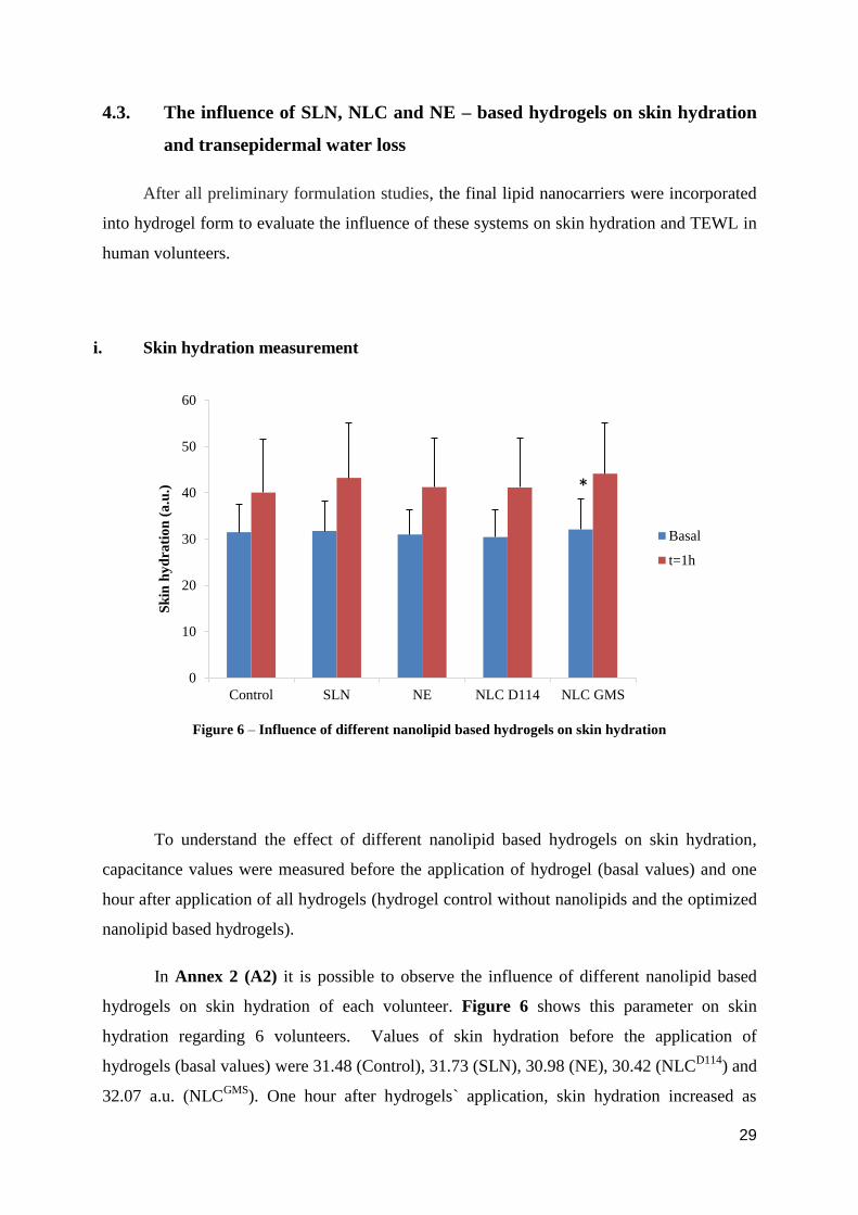
29
4.3. The influence of SLN, NLC and NE – based hydrogels on skin hydration
and transepidermal water loss
After all preliminary formulation studies, the final lipid nanocarriers were incorporated
into hydrogel form to evaluate the influence of these systems on skin hydration and TEWL in
human volunteers.
i. Skin hydration measurement
Figure 6 – Influence of different nanolipid based hydrogels on skin hydration
To understand the effect of different nanolipid based hydrogels on skin hydration,
capacitance values were measured before the application of hydrogel (basal values) and one
hour after application of all hydrogels (hydrogel control without nanolipids and the optimized
nanolipid based hydrogels).
In Annex 2 (A2) it is possible to observe the influence of different nanolipid based
hydrogels on skin hydration of each volunteer. Figure 6 shows this parameter on skin
hydration regarding 6 volunteers. Values of skin hydration before the application of
hydrogels (basal values) were 31.48 (Control), 31.73 (SLN), 30.98 (NE), 30.42 (NLCD114
) and
32.07 a.u. (NLCGMS
). One hour after hydrogels` application, skin hydration increased as
0
10
20
30
40
50
60
Control SLN NE NLC D114 NLC GMS
Sk
in h
yd
rati
on
(a
.u.)
Basal
t=1h
*
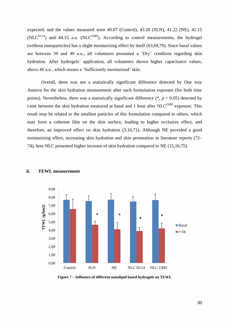
30
expected, and the values measured were 40.07 (Control), 43.20 (SLN), 41.22 (NE), 41.15
(NLCD114
) and 44.15 a.u. (NLCGMS
). According to control measurements, the hydrogel
(without nanoparticles) has a slight moisturizing effect by itself (63,69,70). Since basal values
are between 30 and 40 a.u., all volunteers presented a ‘Dry’ condition regarding skin
hydration. After hydrogels` application, all volunteers shown higher capacitance values,
above 40 a.u., which means a ‘Sufficiently moisturized’ skin.
Overall, there was not a statistically significant difference detected by One way
Annova for the skin hydration measurement after each formulation exposure (for both time
points). Nevertheless, there was a statistically significant difference (*, p < 0.05) detected by
t-test between the skin hydration measured at basal and 1 hour after NLCGMS
exposure. This
result may be related to the smallest particles of this formulation compared to others, which
may form a coherent film on the skin surface, leading to higher occlusive effect, and
therefore, an improved effect on skin hydration (3,16,71). Although NE provided a good
moisturizing effect, increasing skin hydration and skin permeation in literature reports (72–
74), here NLC presented higher increase of skin hydration compared to NE (15,16,75).
ii. TEWL measurement
Figure 7 – Influence of different nanolipid based hydrogels on TEWL
0,00
1,00
2,00
3,00
4,00
5,00
6,00
7,00
8,00
9,00
Control SLN NE NLC D114 NLC GMS
TE
WL
(g
/hm
2)
Basal
t=1h
* * *
*
31
The influence of different nanolipid based hydrogels on TEWL was evaluated
following the procedure described before.
According to Figure 7, basal values of TEWL were 7.70 (Control), 7.55 (SLN), 7.72
(NE), 7.50 (NLCD114
) and 7.67 g/h/m2 (NLC
GMS). One hour after hydrogels´ application, this
parameter decreased as expected, and the values measured were 6.58 (Control), 4.67 (SLN),
4.13 (NE), 3.90 (NLCD114
) and 4.22 g/h/m2 (NLC
GMS). These results are discriminated for
each volunteer in Annex 3 (A3). Since these values are between 0 and 10 g/h/m2, all
volunteers presented a ‘Very healthy condition’ regarding TEWL (even before hydrogels ´
application).
As shown in Figure 7, there was a statistically significant difference (*, p < 0.05)
detected by t-test between the TEWL measured at basal and 1 hour for all formulations,
except the control group. There was also a statistically significant difference (*, p < 0.05)
detected by One way Annova Pairwise Multiple Comparison Procedures (Holm-Sidak
method) between the TEWL measured 1 hour after each formulation exposure and the
control. However, no significant differences were obtained among nanolipid formulations for
this parameter. Therefore, it can be concluded that nanolipid systems incorporated into
hydrogels can lead to a decrease in TEWL, being in accordance with the preliminary findings
of some researchers (76).
Accordingly, a smaller particle size is associated with a decrease in TEWL since it
promotes a larger surface area of the particles reducing water evaporation from the skin
surface (3,16). Thus, it would be expected a higher reduction in TEWL after the application of
NLC-based hydrogel compared to SLN formulation. In addition, NLC usually decreases
TEWL more efficiently than NE due to a higher effect on skin hydration as well, leading to an
improved barrier function of the stratum corneum (16). However, no statistical differences
were detected among these formulations in this study, as mentioned before. In order to
understand these outcomes, it is important to approach the factors behind skin hydration and
TEWL measurements as described below.
32
iii. Factors behind skin hydration and TEWL measurements
Although the nanolipid systems were optimized before their incorporation in hydrogel
form, it should be noticed that some external conditions were not possible to control, which
might affect the final results.
Regarding environment conditions, measurements should be ideally obtained at 20°C
and 40 to 60% of RH, however, the room temperature and relative humidity varied from 23 to
26°C and 33% to 47% (respectively). This dry surrounding environment may have affected
the skin hydration and TEWL (77).
Concerning human error, the position and the pressure used between the probe and the
skin surface should be maintained through all measurements to obtain reproducible results.
However, it was difficult for the investigator to apply always the same exact pressure.
Moreover, the possible presence of remaining hydrogel should be considered as well since it
was difficult to select the optimal strength to remove all formulation of the skin of the
volunteers. If this strength was too high, it would affect skin properties, and on the other hand,
if it was too soft, the hydrogel would not be removed was supposed (50).
It is also important to consider the differences between volunteers, despite the
selection of a homogenous group following restrict inclusion and exclusion criteria. In fact,
some previous studies reported the influence of personal differences in skin’s biophysical
parameters (such as age, gender and race) but the results were controversial (78,79).
Regarding this work, the volunteers were Caucasian, 21-27 years old, four men and two
women. The gender is quite important mainly to the difference between the amount of hair on
the volunteers` forearms, which could affect the removal and drying of the hydrogel (79).
Finally, it is essential to focus the number of volunteers and the duration of this
clinical study. In future perspective, this study will be conducted on a larger number of
volunteers (at least 25 volunteers) (80) and under strict control conditions.
33
5. Conclusion
According to pre-experimental results, it can be concluded that particle parameters
such as particle size, PdI and zeta potential clearly affected the physical stability of these
nanosystems. Firstly, the optimal homogenization conditions were achieved at 20,000 rpm for
10 minutes. Besides homogenization parameters, different types of lipids have also influenced
the particle size in NLC production. The smallest nanoparticles were obtained with GMS
probably due to the lowest molecular weight of this lipid compared to D114. Regarding the
co-surfactants used, Lutrol® F68 produced smaller particles than Tween
® 80. Moreover, the
amount of solid lipid and steric stabilizer should be added at a proportional ratio. At least,
considering the liquid lipid/co-surfactant ratio, an optimal droplet size was obtained using
Miglyol® 812/ Lutrol
® F68 at 7:3 ratio. After formulation selection, all final formulations
showed suitable parameters and good physical stability for a month after its production.
Regarding skin hydration and TEWL measurements, the hydrogel-control had a slight
moisturizing effect by itself. In fact, none of skin hydration results presented statistical
significant differences for each formulation exposure, compared with the control.
Nevertheless, a statistically significant difference was observed for basal values 1 hour after
NLCGMS
exposure. Moreover, all volunteers presented dry skin regarding basal values and a
sufficiently moisturized skin after the hydrogels` application. Towards TEWL values, all
volunteers presented a very healthy condition. In addition, a statistically significant difference
was observed between the basal values and the TEWL values 1 hour after nanolipid-based
hydrogels` application for each formulation compared with the control. Accordingly,
nanolipid-based hydrogels could decrease TEWL values.
Notwithstanding, some factors might influence the measurement of skin’s biophysical
parameters such as environmental conditions, the human error and the skin differences
between participants. The number of participants and the duration of the study were quite
important as well. Thus, the standardization of all these conditions will be taken into account
in future studies.
34
References
1. Darlenski R, Kazandjieva J, Tsankov N. Skin barrier function: morphological basis and
regulatory mechanisms. J Clin Med. 2011;4(1):36–45.
2. Alberts B, Johnson A, Lewis J, Raff M, Robert K, Walter P. Molecular Biology of the
Cell. 4th ed. New York: Garland Science; 2002. 1616 p.
3. Estanqueiro M, Conceição J, Amaral MH, Sousa Lobo JM. Characterization, sensorial
evaluation and moisturizing efficacy of nanolipidgel formulations. Int J Cosmet Sci.
2014;36(2):159–66.
4. Patel D, Kumar V, Kesharwani R, Mazumdar B. Lipid nanoparticle a novel carrier for
cosmetics and topical preparation: a review. Inven Rapid Cosmeceuticals.
2015;2015(3):1–6.
5. Sotoodian B, Maibach HI. Noninvasive test methods for epidermal barrier function.
Clin Dermatol. 2012 May;30(3):301–10.
6. Mudshinge SR, Deore AB, Patil S, Bhalgat CM. Nanoparticles: Emerging carriers for
drug delivery. Saudi Pharm J [Internet]. 2011 Jul 1 [cited 2017 Mar 12];19(3):129–41.
Available from: http://www.sciencedirect.com/science/article/pii/S1319016411000302
7. Saini R, Saini S, Sharma S. Nanotechnology: the future medicine. J Cutan Aesthet
Surg. 2010;3(1):32–3.
8. Geszke-Moritz M, Moritz M. Solid lipid nanoparticles as attractive drug vehicles:
Composition, properties and therapeutic strategies. Mater Sci Eng C. 2016
Nov;68:982–94.
9. Ajazzuddin M, Jeswani G, Jha A. Nanocosmetics: Past, Present and Future Trends.
Recent Patents Nanomed. 2015;5(1):3–11.
10. Raj S, Jose S, Sumod US, Sabitha M. Nanotechnology in cosmetics: Opportunities and
challenges. J Pharm Bioallied Sci. 2012;4(3):186–93.
11. Crosera M, Bovenzi M, Maina G, Adami G, Zanette C, Florio C, et al. Nanoparticle
dermal absorption and toxicity: A review of the literature. Int Arch Occup Environ
Health. 2009;82(9):1043–55.
35
12. FDA. Guidance Documents - Guidance for Industry: Safety of Nanomaterials in
Cosmetic Products. 2012 [cited 2017 Jul 4];(June):1–16. Available from:
https://www.fda.gov/cosmetics/guidanceregulation/guidancedocuments/ucm300886.ht
m
13. USFDA. Nanotechnology: a Report of the Us Food and Drug administration
nanotechnology Task Force. 2007 [cited 2017 Jul 4];1–38. Available from:
https://www.fda.gov/downloads/ScienceResearch/SpecialTopics/Nanotechnology/ucm
110856.pdf
14. Scientific Committee on Consumer Safety. Guidance on the Safety Assessment of
Nanomaterials in Cosmetics. Eur Comm [Internet]. 2012 [cited 2017 Nov 12];0–136.
Available from:
http://ec.europa.eu/health/scientific_committees/consumer_safety/docs/sccs_o_073.pdf
15. Pardeike J, Hommoss A, Müller RH. Lipid nanoparticles (SLN, NLC) in cosmetic and
pharmaceutical dermal products. Int J Pharm. 2009 Jan 21;366(1–2):170–84.
16. Loo CH, Basri M, Ismail R, Lau HLN, Tejo BA, Kanthimathi MS, et al. Effect of
compositions in nanostructured lipid carriers (NLC) on skin hydration and occlusion.
Int J Nanomedicine. 2012;2013(8):13–22.
17. Puglia C, Bonina F. Lipid nanoparticles as novel delivery systems for cosmetics and
dermal pharmaceuticals. Expert Opin Drug Deliv. 2012;9(4):429–41.
18. Becher P, editor. Encyclopedia of Emulsion Technology: Volume 4 [Internet]. Marcel
Dekker, INC; 1996. p. 376. Available from:
https://books.google.pt/books?id=PfDaBQ-FoYgC&printsec=frontcover&hl=pt-
PT&source=gbs_ge_summary_r&cad=0#v=onepage&q&f=false
19. Sharma N, Madan P, Lin S. Effect of process and formulation variables on the
preparation of parenteral paclitaxel-loaded biodegradable polymeric nanoparticles: A
co-surfactant study. Asian J Pharm Sci. 2015;11(3):404–16.
20. Yukuyama MN, Ghisleni DDM, Pinto TJA, Bou-Chacra NA. Nanoemulsion: Process
selection and application in cosmetics - A review. Int J Cosmet Sci. 2016;38(1):13–24.
21. Montenegro L, Lai F, Offerta A, Sarpietro MG, Micicchè L, Maccioni AM, et al. From
nanoemulsions to nanostructured lipid carriers: A relevant development in dermal
delivery of drugs and cosmetics. J Drug Deliv Sci Technol. 2016;32:100–12.
36
22. McClements DJ. Nanoemulsions versus microemulsions: terminology, differences, and
similarities. Soft Matter. 2012;8(6):1719.
23. Jaworska M, Sikora E, Ogonowski J. The influence of glicerides oil phase on O/W
nanoemulsion formation by pic method. Period Polytech Chem Eng.
2014;58(SUPPL):43–8.
24. Dong X, Mattingly CA, Tseng M, Cho M, Adams VR, Mumper RJ. Development of
new lipid-based paclitaxel nanoparticles using sequential simplex optimization. Eur J
Pharm Biopharm. 2009 May;72(1):9–17.
25. Kakadia PG, Conway BR. Solid Lipid Nanoparticles: A Potential Approach for Dermal
Drug Delivery. Am J Pharmacol Sci. 2014;2(5A):1–7.
26. Müller RH, Radtke M, Wissing SA. Solid lipid nanoparticles (SLN) and nanostructured
lipid carriers (NLC) in cosmetic and dermatological preparations. Adv Drug Deliv Rev.
2002;54(SUPPL.):131–55.
27. Mäder K, Mehnert W. Solid lipid nanoparticles: production, characterization and
applications. Adv Drug Deliv Rev. 2001;47(2–3):165–96.
28. zur Mühlen A, Schwarz C, Mehnert W. Solid lipid nanoparticles (SLN) for controlled
drug delivery--drug release and release mechanism. Eur J Pharm Biopharm.
1998;45(2):149–55.
29. Khadka P, Ro J, Kim H, Kim I, Kim JT, Kim H, et al. Pharmaceutical particle
technologies: An approach to improve drug solubility, dissolution and bioavailability.
Asian J Pharm Sci. 2014;9(6):304–16.
30. Purohit DK, Nandgude TD, Poddar SS. Nano-lipid carriers for topical application:
Current scenario. Asian J Pharm. 2016;10(1):1–9.
31. Battaglia L, Gallarate M, Panciani PP, Ugazio E, Sapino S, Peira E, et al. Techniques
for the Preparation of Solid Lipid Nano and Microparticles. Appl Nanotechnol Drug
Deliv. 2014;51–75.
32. Müller RH, Lippacher A, Gohla S. Solid Lipid Nanoparticles (SLN) as a carrier system
for the controlled release of drugs. In: Wise DL, editor. Handbook of pharmaceutical
release technology. Marcel Dekker; 2000. p. 377–91.
33. Martins S, Sarmento B, Ferreira DC, Souto EB. Lipid-based colloidal carriers for
37
peptide and protein delivery - Liposomes versus lipid nanoparticles. Int J
Nanomedicine. 2007;2(4):595–607.
34. Chaturvedi SP, Kumar V. Production techniques of lipid nanoparticles: A review. Res J
Pharm Biol Chem Sci. 2012;3(3):525–41.
35. Rane SS, Choi P. Polydispersity Index: How Accurately Does It Measure the Breadth
of the Molecular Weight Distribution? Chem Mater. 2005 Feb;17(4):926–926.
36. Malvern. Zetasizer Nano Series. Malvern Instruments Ltd [Internet]. 2014 [cited 2017
Nov 5];20. Available from: https://www.malvern.com/en/assets/MRK1839_tcm22-
17228.pdf
37. Clogston JD, Patri AK. Zeta Potential Measurement. In: Clifton NJ, editor. Methods in
molecular biology. 2011. p. 63–70.
38. Chen YC, Liu DZ, Liu JJ, Chang TW, Ho HO, Sheu MT. Development of terbinafine
solid lipid nanoparticles as a topical delivery system. Int J Nanomedicine.
2012;7:4409–18.
39. Wavikar P, Vavia P. Nanolipidgel for enhanced skin deposition and improved
antifungal activity. AAPS PharmSciTech. 2013;14(1):222–33.
40. Okay O. General Properties of Hydrogels. In: Gerald G, Arndt K-F, editors. Hydrogel
Sensors and Actuators: Engineering and Technology - Volume 6 of Springer Series on
Chemical Sensors and Biosensors. New York: Springer Science & Business Media;
2009. p. 2–3.
41. Souto E., Wissing S., Barbosa C., Müller R. Evaluation of the physical stability of SLN
and NLC before and after incorporation into hydrogel formulations. Eur J Pharm
Biopharm. 2004 Jul;58(1):83–90.
42. Szczepanik MP, Wilkołek PM, Adamek ŁR, Pomorski ZJH. The examination of
biophysical parameters of skin (transepidermal water loss, skin hydration and pH
value) in different body regions of normal cats of both sexes. J Feline Med Surg. 2011
Apr;13(4):224–30.
43. Verdier-Sévrain S, Bonté F. Skin hydration: A review on its molecular mechanisms. J
Cosmet Dermatol. 2007 Jun;6(2):75–82.
44. Manfredini M, Giovanna M, Silvana C, Silvia S, Francesca F, Caterina L, et al. Does
38
skin hydration influence keratinocyte biology? In vivo evaluation of microscopic skin
changes induced by moisturizers by means of Reflectance Confocal Microscopy. Ski
Res Technol. 2014;19(3):299–307.
45. Corneometer® CM 825 - Courage + Khazaka Electronic, Köln [Internet]. [cited 2017
May 25]. Available from: http://www.courage-
khazaka.de/index.php/en/products/scientific/55-corneometer
46. Boer M, Duchnik E, Maleszka R, Marchlewicz M. Structural and biophysical
characteristics of human skin in maintaining proper epidermal barrier function. Postep
Dermatologii i Alergol. 2016 Feb;33(1):1–5.
47. Tewameter® TM 300 - Courage + Khazaka Electronic, Köln [Internet]. [cited 2017
Aug 12]. Available from: http://www.courage-
khazaka.de/index.php/en/products/scientific/139-tewameter#tm1
48. Ash M, Ash I. Trade name reference. In: Handbook of green chemicals. Synapse
Information Resources; 2004. p. 181.
49. Ash I, Ash M. Chemical Component CrossReference. In: Handbook of fillers,
extenders, and diluents. Synapse Information Resources; 2007. p. 273.
50. Gabard B, Clarys P, Barel AO. Comparison of Commercial Electrical Measurement
Instruments for Assessing the Hydration State of the Stratum Corneum. In: Serup J,
Jemec B, Grove G, editors. Handbook of non-invasive methods and the skin.
CRC/Taylor & Francis; 2006. p. 351–60.
51. Courage + Khazaka Electronic. Instructions - Corneometer® CM 825 Probe. Köln;
2016.
52. Courage + Khazaka Electronic. Instructions - Tewameter® TM 300 Probe. Köln; 2016.
53. Bhatia S. Nanoparticles Types, Classification, Characterization, Fabrication Methods
and Drug Delivery Applications. In: Natural Polymer Drug Delivery Systems. Cham:
Springer International Publishing; 2016. p. 33–93.
54. Malvern. Inform White Paper - Dynamic Light Scattering. 2011 [cited 2017 Aug
14];1–6. Available from: http://www.biophysics.bioc.cam.ac.uk/wp-
content/uploads/2011/02/DLS_Terms_defined_Malvern.pdf
55. Das S, Chaudhury A. Recent Advances in Lipid Nanoparticle Formulations with Solid
39
Matrix for Oral Drug Delivery. AAPS PharmSciTech. 2011 Mar;12(1):62–76.
56. Guimarães KL, Ré MI. Lipid Nanoparticles as Carriers for Cosmetic Ingredients: The
First (SLN) and the Second Generation (NLC). In: Beck R, Guterres S, Pohlmann A,
editors. Nanocosmetics and Nanomedicines: New Approaches for Skin Care. Berlim:
Springer Science & Business Media; 2011. p. 101–22.
57. American Lecithin Company. Technical Data - Phospholipon® 80 [Internet]. Oxford;
2017. Available from: http://www.americanlecithin.com/TDS/TDS_80.PDF
58. Gondrala UK, Dudhipala N, Kishan V. Preparation, Characterization and in vivo
Evaluation of Felodipine Solid-Lipid Nanoparticles for Improved Oral Bioavailability.
Int J Pharm Sci Nanotechnol. 2015;8(4):2995–3002.
59. Date A a, Vador N, Jagtap A, Nagarsenker MS. Lipid nanocarriers (GeluPearl)
containing amphiphilic lipid Gelucire 50/13 as a novel stabilizer: fabrication,
characterization and evaluation for oral drug delivery. Nanotechnology.
2011;22(27):275102.
60. Ahlin Grabnar P, Kristl J, Smid-Korbar J. Optimization of procedure parameters and
physical stability of solid lipid nanoparticles in dispersions. Acta Pharm.
1998;48(4):259–67.
61. Rosdi MRH, Ariffin A, Ishak ZAM. Optimizing homogenization parameters for
improving ethylene vinyl acetate emulsion stability in pour point depressant
application. J King Saud Univ - Eng Sci. 2016;11.
62. Khalil RM, El-bary AA, Kassem MA, Ghorab MM, Ahmed MB. Solid lipid
nanoparticles for topical delivery of Meloxicam: development and In Vitro
characterization. Eur Sci J. 2013;4(7):24–6.
63. Ńega K. Proučevanje vpliva vrste lipidov v nanosistemih na transepidermalno izgubo
vode in hidratacijo kože: [diplomska naloga]. 2013.
64. Mosqueira VCF, Legrand P, Pinto-Alphandary H, Puisieux F, Barratt G. Poly(D,L-
Lactide) nanocapsules prepared by a solvent displacement process: Influence of the
composition on physicochemical and structural properties. J Pharm Sci. 2000
May;89(5):614–26.
65. Hasan NMY. Role of medium-chain fatty acids in the emulsification mechanistics of
self-micro-emulsifying lipid formulations. Saudi Pharm J. 2014 Dec;22(6):580–90.
40
66. Aditya NP, Macedo AS, Doktorovova S, Souto EB, Kim S, Chang PS, et al.
Development and evaluation of lipid nanocarriers for quercetin delivery: A
comparative study of solid lipid nanoparticles (SLN), nanostructured lipid carriers
(NLC), and lipid nanoemulsions (LNE). LWT - Food Sci Technol. 2014;59(1):115–21.
67. Mitri K, Shegokar R, Gohla S, Anselmi C, Müller RH. Lipid nanocarriers for dermal
delivery of lutein: Preparation, characterization, stability and performance. Int J Pharm.
2011;414(1–2):267–75.
68. Gönüllü Ü, Üner M, Yener G, Karaman EF, Aydoʇmuş Z. Formulation and
characterization of solid lipid nanoparticles, nanostructured lipid carriers and
nanoemulsion of lornoxicam for transdermal delivery. Acta Pharm. 2015;65(1):1–13.
69. Hamidi M, Azadi A, Rafiei P. Hydrogel nanoparticles in drug delivery. Adv Drug
Deliv Rev. 2008;60(15):1638–49.
70. Sidley Chemical Co. L. Applications of CMC and HEC in Daily Chemical Products
[Internet]. 2014 [cited 2017 Aug 29]. Available from:
http://celluloseether.com/applications-cmc-hec-daily-chemical-products/
71. Müller RH, Petersen RD, Hommoss A, Pardeike J. Nanostructured lipid carriers (NLC)
in cosmetic dermal products. Adv Drug Deliv Rev. 2007;59(6):522–30.
72. Bajerski L, Michels LR, Colomé LM, Bender EA, Freddo RJ, Bruxel F, et al. The use
of Brazilian vegetable oils in nanoemulsions: An update on preparation and biological
applications. Brazilian J Pharm Sci. 2016;52(3):347–63.
73. Wu Y, Li Y-H, Gao X-H, Chen H-D. The application of nanoemulsion in dermatology:
an overview. J Drug Target. 2013 May 19;21(4):321–7.
74. Mordorski B, Landriscina A, Friedman A. An Overview of Nanomaterials in
Dermatology. In: Hamblin M, Avci P, Prow T, editors. Nanoscience in Dermatology.
2016. p. 31–46.
75. Guimarães KL, Ré MI. Lipid Nanoparticles as Carriers for Cosmetic Ingredients: The
First (SLN) and the Second Generation (NLC). In: Dragicevic N, Maibach HI, editors.
Percutaneous Penetration Enhancers Chemical Methods in Penetration Enhancement
Modification of the Stratum Corneum. 2016. p. 101–22.
76. López-García R, Ganem-Rondero A. Solid Lipid Nanoparticles (SLN) and
Nanostructured Lipid Carriers (NLC): Occlusive Effect and Penetration Enhancement
41
Ability. J Cosmet. 2015;5(June):62–72.
77. Sparr E, Millecamps D, Isoir M, Burnier V, Larsson Å, Cabane B. Controlling the
hydration of the skin though the application of occluding barrier creams. J R Soc
Interface. 2013 Mar 6;10(80):20120788.
78. van der Valk PG, Nater JP, Bleumink E. Skin irritancy of surfactants as assessed by
water vapor loss measurements. J Invest Dermatol. 1984;82(3):291–3.
79. Du Plessis J, Stefaniak A, Eloff F, John S, Agner T, Chou TC, et al. International
guidelines for the in vivo assessment of skin properties in non-clinical settings: Part 2.
transepidermal water loss and skin hydration. Ski Res Technol. 2013 Aug;19(3):265–
78.
80. Wesley NO, Maibach HI. Racial (Ethnic) Differences in Skin Properties: The Objective
Data. Am J Clin Dermatol. 2003;4(12):843–60.

42
Annexes
A1. Informed Consent Form
INFORMED CONSENT FORM
The influence of different nanocarriers on skin’s biophysical parameters
Principal Investigators: assist. prof. dr. Pegi Ahlin Grabnar and Sara Andrade
Organization: Faculty of Pharmacy – University of Ljubljana
This Informed Consent Form is for healthy subjects aged 21-27 years, who we are inviting to
participate in a research project in the framework of a Master's Thesis of Pharmaceutical Sciences.
The aim of our research project is to evaluate the influence of three well characterized
different lipid nanosystems (solid lipid nanoparticles, nanostructured lipid carriers and
nanoemulsions) on skin hydration and transepidermal water loss on human volunteers.
Nanotechnology is a new trend in cosmetology, since nanoparticles have shown higher degree of biocompatibility and versatility in this field compared to other systems. Several formulations of lipid nanoparticles were produced to control different parameters referred to dermal preparations such as TEWL and skin hydration. Because of their biocompatible chemical nature, no adverse effects of these compounds on human skin have been described so far.
This research will involve one application of four different nanolipid formulations (one with
SLN, two with NLC (different type of lipids) and one with NE) incorporated into hydrogel and one
control on your forearms. Therefore, you should come to the Faculty of Pharmacy on the scheduled
day. During this day, you will need to spend approximately two hours, twenty minutes to acclimatize
the skin and approximately one hour and a half to measure the biophysical parameters.
Your participation in this research is entirely voluntary and you may refuse or stop
participating at any time that you wish. If you have any questions you may ask us now or later.
After reading the foregoing information and explanations, I consent voluntarily to participate
as a participant in this research.
Signature of Participant: Date:
43
A2. Results of skin hydration in six volunteers
Control
SLN
NE
0
10
20
30
40
50
60
Basal t=1h
Sk
in h
yd
rati
on
(a
.u)
A.P.
A.D.
J.B.
M.A.
O.S.
V.Z.
0
10
20
30
40
50
60
70
Basal t=1h
Sk
in h
yd
rati
on
(a
.u)
A.P.
A.D.
J.B.
M.A.
O.S.
V.Z.
0
10
20
30
40
50
60
Basal t=1h
Sk
in h
yd
rati
on
(a
.u)
A.P.
A.D.
J.B.
M.A.
O.S.
V.Z.
44
NLCD114
NLCGMS
0
10
20
30
40
50
60
Basal t=1h
Sk
in h
yd
rati
on
(a
.u)
A.P.
A.D.
J.B.
M.A.
O.S.
V.Z.
0
10
20
30
40
50
60
70
Basal t=1h
Sk
in h
yd
rati
on
(a
.u)
A.P.
A.D.
J.B.
M.A.
O.S.
V.Z.
45
A3. Results of TEWL in six volunteers
Control
SLN
NE
0
2
4
6
8
10
Basal t=1h
TE
WL
(g
/hm
2) A.P.
A.D.
J.B.
M.A.
O.S.
V.Z.
0
2
4
6
8
10
Basal t=1h
TE
WL
(g
/hm
2) A.P.
A.D.
J.B.
M.A.
O.S.
V.Z.
0
2
4
6
8
10
Basal t=1h
TE
WL
(g
/hm
2) A.P.
A.D.
J.B.
M.A.
O.S.
V.Z.
46
NLCD114
NLCGMS
NOTE:
In a pre-experimental work, three different measurements were performed: 1st) before
application of the hydrogel; 2nd
) 30 min after the application of all hydrogels and 3rd
) 1 h after
the application of all hydrogels. After 30 min, it was possible to observe a great increase on
skin hydration that slightly decreased after 60 min. However, these results were discarded
since they might measure the hydration of the remaining gel that had not been yet absorbed
(whereas hydrogel was removed 30 min after hydrogels` application, immediately before the
second measurement).
0
2
4
6
8
10
Basal t=1h
TE
WL
(g
/hm
2) A.P.
A.D.
J.B.
M.A.
O.S.
V.Z.
0
2
4
6
8
10
Basal t=1h
TE
WL
(g
/hm
2) A.P.
A.D.
J.B.
M.A.
O.S.
V.Z.


![Review Stimuli-responsive nanocarriers for drug delivery ... · environment [45-47], such as CaCO 3 nanoparticles [39]. The ultrasound-responsive nanocarriers could be applied for](https://img.dokumen.tips/doc/110x75/5f3c0abafeae704f47101437/review-stimuli-responsive-nanocarriers-for-drug-delivery-environment-45-47.jpg)