Embed Size (px)
Citation preview

322 >
ABSTRACT The efficacy of root canal preparation is considerably en-hanced when an effort is made to provide an effective glide path. In addition the risk of undesirable fractures of instru-ments is reduced. The introduction of techniques and in-struments specifically directed at the preparation of a glide path have facilitated root canal shaping and provide a va-riety from which the clinician may choose, guided by the relevant design features and performance characteristics.
InTRoduCTIonThe instrumentation and preparation of the root canal sys-tem is regarded as being a most important stage of endo-dontic treatment for this has an influence on the efficacy of subsequent procedures in endodontic therapy.1,2
Root canal instrumentation was originally aimed at facilitating the placement of medicaments in the root canal and little attempt was made to clear the organic contents from the root canal system. The focus of instrumentation then shifted to preparing the root canal space to facilitate the placement of root canal fillings but the methods employed were mostly unrelated to the anatomy of the canal system or to the prop-erties of the obturation materials.1 In 1974, Schilder altered endodontic protocols forever with his innovative and revolu-tionary concepts that defined the design and biological ob-jectives for optimally shaping canal spaces and for debriding root canal systems.1 There were several primary objectives – shaping the root canal system to have a smooth taper from orifice to apex; keeping the apical foramen as small as was practical and in its original position; and ensuring that the preparation flowed with the original anatomy of the root canal system. Other objectives were to confine prepa-ration to the canal space, facilitate the removal of all tissue without forcing necrotic debris through the apical foramen, and ensuring that the final shape facilitated the placement of medicaments and exchange of irrigants. However, the
journey from orifice to apex can be perilous and proper root canal preparation remains one of the most difficult tasks in endodontic therapy.3
Canal scouting and preflaring are the first phases of canal instru-mentation and it has also been noted that during these phases the clinician might more frequently encounter procedural difficulties.4
Among such problems are ascertaining the location of the root canals, access cavity preparation, canal preparation without procedural errors, and establishment and mainte-nance of working length. Canal systems can have multiple geometric planes and curve significantly more than the roots that house them.1 Two-dimensional radiographs fail to reveal these morphological variations of canals in different spatial planes.1,5,6 Instrumentation of canals with multi-planar curva-tures and long, thin curved canals is fraught with possible procedural errors during either hand-file instrumentation5 or rotary nickel titanium (NiTi) instrumentation.7 These problems include instrument fracture, ledge formation, canal zipping or canal straightening, strip perforation, apical perforation, elbow formation and apical blockage. All of these errors can lead to incomplete debridement of the root canal system and con-tribute to decreased success rates of endodontic therapy.3
Technical protocols for shaping root canals have evolved to enable achievement of the objectives outlined by Schilder1 and to reduce the occurrence of procedural errors. Se-rial instrumentation was developed using multiple, curved hand files and reamers.1 The step-back technique involved preparation of the apical region of the root canal first, fol-lowed by coronal flaring to facilitate obturation.8 Crown-down techniques commenced with preparation using larger instruments at the canal orifice followed by progressively smaller files when proceeding down the root canal.9-11 The balanced-force technique enabled the shaping of curved canals to larger sizes using modified stainless steel files.12
Most of the procedural problems associated with achieving ideal shaping of curved canals were due to the stiffness of stainless steel instruments.13 The introduction of NiTi rotary instruments revolutionised endodontics as they have a lower modulus of elasticity than stainless steel instruments; and therefore ex-ert fewer lateral forces on the dentine walls in curved canals. Even though NiTi instruments are stronger and more flexible than their stainless steel counterparts14 fractures may still occur within their elastic limit. Instrument breakage can happen with-out evidence of previous permanent deformation15,16 and even
I Cassim: 1. BDS (Wits), PG Dip Dent (Endo)(Pret), Department of Odontology, School of Dentistry, University of Pretoria, South Africa.
PJ van der Vyver: 2. BChD(Pret), Dip Odont(Aesth.Dent.) Dip Odont( Endo) MSc(Endo) (Pret), Department of Odontology, School of Dentistry, University of Pretoria, South Africa.
Corresponding authorPJ van der Vyver: PO Box 2609, Cresta, 2118. Tel: 011 781 1020. Cell: 082 4104 293. Email: [email protected]
SADJ August 2013, Vol 68 no 7 p322 - p327
I Cassim1, PJ van der Vyver2
The importance of glide path preparation in endodontics: a consideration of instruments and literature
SCIENTIfIC

324 > SCIENTIfIC
without prior use.17-19 fracture is the most common procedural error that occurs during clinical use of rotary NiTi instruments,20 and the fear of such a mishap is the biggest deterrent to the adoption of the technology by clinicians.21- 23
fracture of rotary NiTi instruments may occur as a result of cyclic flexural fatigue (bending stress) or through torsion (shear stress).24,25 Pronounced canal curvature is considered to be the major risk factor in instrument fracture due to cyclic fatigue (bending stress).15,26 Torsional stress occurs when there is: (1) an extensive contact area between the cutting surface of
the instrument and the canal wall; (2) the canal cross section is much narrower than the cross
section of the tip of the instrument;27,28 and (3) when there is excessive axial pressure on the handpiece
during instrumentation.29 The instrumentation technique used and the preparation of a proper glide path therefore play a significant role in reducing torsional stress.23,30-33
SIgnIfICAnCe of glIde pATh pRepARATIonA glide path is defined as a smooth, though possibly nar-row, tunnel or passage from the coronal orifice of the canal to the radiographic terminus or electronically determined portal of exit.34 The maintenance of a glide path means having a smooth passage that is reproducible by files used succes-sively in the canal.35 All available NiTi rotary instruments have non-cutting tips36 and because of their extreme flexibility, these instruments are not designed for initial negotiation of the root canal.37 Bergmans et al. (2001) stated that during root canal preparation, no rotary instrument should be used where a hand instrument has not been placed before.22
Roland et al. (2002) showed that coronal pre-flaring can reduce the incidence of instrument fracture.38 The use of small hand files to confirm patency of the canal and to ensure sufficient space for rotary instruments to passively follow would greatly improve the safety of rotary NiTi instrument use.39 Teeth requiring endodontic therapy may have intra-canal calcifications (denticles) that have developed over time, especially in the aging population.35,40 The denticles may vary in size from 50 microns to several millimetres and may be present at any level along the canal walls.41 The passage of small hand files to the terminus of the canal beyond the pulp stones and denticles allows the clinician to establish full patency of the canal before commencement of mechanical preparation35,40 and reduces the risk of ledge formation,37 which is one of the major causes of a need for retreatment.42
Peters et al. (2003) in their study using extracted teeth, reported that no instrument fractures occurred during canal preparation when an appropriate glide path had been developed, even when high forces were used in constricted canals.27 It has been shown that the provision of an effective glide path also reduces torsional stress such that the average lifespan of a rotary instru-ment may be extended almost six-fold.30 In 2005, Patino et al. studied the influence of a manually prepared glide path on the separation rate of rotary NiTi instruments.43 These authors used three different file systems and tested them in root canals with a curvature larger than 30 degrees and found that separation was significantly reduced (12% with a glide path as opposed to 26% without a glide path). No difference existed between the types of file designs (K3, Profile and ProTaper).43 A study which ex-amined files after single clinical use, found that there was a high incidence of distortion and separation of rotary NiTi files when their use was not preceded by glide path preparation.19
The use of NiTi instruments in a reciprocating movement with unequal back and forth motion is another novel way in which the risk of file separation is reduced.44,45 Berutti et al. found in 2012 that fewer insertions of the WaveOne single file (Dent-sply, Maillefer, Ballaigues, Switzerland) were needed to reach working length when a glide path was prepared. These au-thors also found that preparation of a glide path resulted in less alteration to the original curvature of the canal.46
glIde pATh pRepARATIon meThodS(a) Hand stainless steel K-filesSeveral authors have recommended using stainless steel K-files by hand for preparing the glide path.30,31,34,40,47-49,53 The advantages of using stainless steel hand files and K-files compared with rotary NiTi files for creating the glide path are:
K-files provide better tactile sensation;• 48
less potential for separation;• 48
when a small size k-file is removed from the canal, the file of-•ten retains an impression of the canal, and in this way alerts the operator to the curvatures present in the canal;30,48,50,53
the stiffness of stainless steel hand files aids in path-finding •and in negotiating blockages and calcifications;37,48
lower cost;•no need for a dedicated hand piece.•
West (2006) recommends using stainless steel K-files in a verti-cal in and out motion with an initial amplitude of 1mm, gradually increasing as the dentine wall wears away and the file advances apically.34 In very narrow canals a “watch-winding” motion is rec-ommended to remove restrictive dentine, as well as to create an “envelope of motion”.40 West and Roane describe the “watch-winding” motion as a back and forth oscillation of a file (30 to 60 degrees) clockwise and counter-clockwise as the instrument is pushed downward into the canal. It is a definite inward progres-sion of the instrument in a filing motion. An “envelope of motion” occurs when a precurved file is advanced into the canal short of maximum resistance, then the file is removed while it is simul-taneously rotated in a clockwise direction.1,34,51 Schilder (1974) emphasises the need to use precurved hand instruments.1 The “envelope of motion” created by the rotation of the curved file as it is withdrawn from the canal scribes the side walls of the canal at random contact points, gradually widening and evolving the root canal shape to allow larger files to follow. This technique facilitates the suspension of debris in the irrigation solution.1,52 Both Schilder and West emphasise the importance of following the canal rather than forcing the file apically through any obstructions.1,40
Berutti et al. advocate that the diameter of the canal after glide path preparation should be at least one size larger than the tip of the first rotary file used to prepare the canal.30 West rec-ommends a minimum of a “super loose” size 10 K-file.40 This author also emphasises that if a glide path larger than size 10 K-file is required then it is advisable to use the “balanced force” motion described by Roane et al.12 for file sizes 15 and above in order to reduce the risk of ledge formation. This involves turning the handle of the file clockwise, and then turning it counter-clockwise using slight apical pressure so that the file does not “unscrew” its way out of the canal. During the clockwise mo-tion, the file blades cut into the dentine and during the apical counter-clockwise motion, the loose dentine is collected into the file’s flutes. This motion can be repeated several times as the file is advanced apically. After having carved a wider glide path the file is turned clockwise and removed.40
< 325www.sada.co.za / SADJ Vol 68 No.7 SCIENTIfIC
In order to confirm that a glide path is present, a size 15 or 20 K-file should slide easily to working length. The file is withdrawn 1mm without rotation and should slide to working length. Thereafter, the file is withdrawn 2mm without rotation and should slide to working length. When the file can be with-drawn 3mm to 5mm and slides to working length without the need for rotation a glide path is confirmed.53
Other hand files recommended for path finding and glide-path for-mation include the Antaeos Stiff “C” file (Schwed, Kew Gardens, NY), C file (Dentsply /Tulsa Dental Specialities, Oklahoma, USA), C file (Roydent, Hoboken, NJ), C+ file (Dentsply/Maillefer Ballaigues, Switzerland), D finder (Mani, Tochigi-ken, Japan), Hi-5 file (Miltex, York, PA), Pathfinder CS (SybronEndo, Glendora, CA), Pathfinder SS (SybronEndo, Orange, California, USA), S finder (JS Dental, Sendoline, Ridgefield, CT), Stiff K file (Brasseler, Savannah, GA), flexofile (Dentsply/Maillefer) and Senseus Profinder (Dentsply/Maillefer). The aforementioned instruments have varying tip dimen-sions, cross sections, tapers, pitch and flute design.54
The disadvantages of preparing a glide path with hand in-struments are:
operator fatigue.•hand fatigue. •time required in the preparation of the glide path.• 55
risk of the introduction of canal aberrations with larger file sizes.• 40, 55
greater change to original canal anatomy.• 55,56
increased apical extrusion of debris.• 57
(b) Hand files in reciprocating hand pieceThis technique involves using small size K-files mounted in a re-ciprocating hand piece in the preparation of the glide path.58,59 A small size K-file is used to negotiate the canal to length by hand before being attached to a reciprocating hand piece (figure 1a and 1 b). The hand piece is then moved vertically up and down, with an amplitude of 1mm to 3mm and bursts of reciprocation for approximately 15 to 30 seconds in each root canal. Sequen-tially larger size K-files (06 to 10) are inserted to just beyond the apical constriction to reduce the risk of blockage. Due to the relative stiffness of the file, Van der Vyver recommends placing a size 20 K-file one mm short of the apex during this method of glide path preparation to avoid apical transportation.53 The M4 reciprocating hand piece (SybronEndo) and Endo-Express reciprocating hand piece (Essential Dental Systems, NJ, USA) have a 30 degree equi-angle arc of reciprocation (five minutes on a clock face). The NSK Ti-Max Ti35L 10:1 reciprocating hand piece (NSK, Nakanishi, Japan) has a 90 degree angle of recip-rocation or 15 minutes on a clock face.
The advantages of using a stainless steel K-file in a recipro-cating hand piece for glide path preparation are:
reduced preparation time;•reduced operator fatigue;•
reduced hand fatigue, especially in canals with multi-planar curves;•reduced risk of instrument separation compared with ro-•tary NiTi methods.59
The disadvantages are:the need for a dedicated hand piece;•risk of apical transportation with files larger than a 15 K-file;• 53,59
risk of excess dentine removal as a result of the clinician •working the canal longer than necessary;60 risk of apical extrusion of debris if hand piece is inserted •apically with force;59 decreased tactile sensation.•
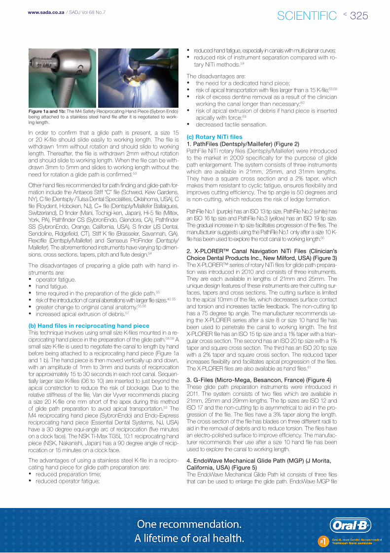
(c) Rotary NiTi files1. PathFiles (Dentsply/Maillefer) (Figure 2)Pathfile NiTi rotary files (Dentsply/Maillefer) were introduced to the market in 2009 specifically for the purpose of glide path enlargement. The system consists of three instruments which are available in 21mm, 25mm, and 31mm lengths. They have a square cross section and a 2% taper, which makes them resistant to cyclic fatigue, ensures flexibility and improves cutting efficiency. The tip angle is 50 degrees and is non-cutting, which reduces the risk of ledge formation.
Pathfile No.1 (purple) has an ISO 13 tip size, Pathfile No.2 (white) has an ISO 16 tip size and Pathfile No.3 (yellow) has an ISO 19 tip size. The gradual increase in tip size facilitates progression of the files. The manufacturer suggests using the Pathfile No.1 only after a size 10 K-file has been used to explore the root canal to working length.55
2. X-PLORER™ Canal Navigation NiTi Files (Clinician’s Choice Dental Products Inc., New Milford, USA) (Figure 3)The X-PLORER™ series of rotary NiTi files for glide path prepara-tion was introduced in 2010 and consists of three instruments. They are each available in lengths of 21mm and 25mm. The unique design features of these instruments are their cutting sur-faces, tapers and cross sections. The cutting surface is limited to the apical 10mm of the file, which decreases surface contact and torsion and increases tactile feedback. The non-cutting tip has a 75 degree tip angle. The manufacturer recommends us-ing the X-PLORER series after a size 8 or size 10 hand file has been used to penetrate the canal to working length. The first X-PLORER file has an ISO 15 tip size and a 1% taper with a trian-gular cross section. The second has an ISO 20 tip size with a 1% taper and square cross section. The third has an ISO 20 tip size with a 2% taper and square cross section. The reduced taper increases flexibility and facilitates apical progression of the files. The X-PLORER files are also available as hand files.61
3. G-Files (Micro-Mega, Besancon, France) (Figure 4)These glide path preparation instruments were introduced in 2011. The system consists of two files which are available in 21mm, 25mm and 29mm lengths. The tip sizes are ISO 12 and ISO 17 and the non-cutting tip is asymmetrical to aid in the pro-gression of the file. The files have a 3% taper along the length. The cross section of the file has blades on three different radii to aid in the removal of debris and to reduce torsion. The files have an electro-polished surface to improve efficiency. The manufac-turer recommends their use after a size 10 hand file has been used to explore the canal to working length.
4. EndoWave Mechanical Glide Path (MGP) (J Morita, California, USA) (Figure 5)The EndoWave Mechanical Glide Path kit consists of three files that can be used to enlarge the glide path. EndoWave MGP file
figure 1a and 1b: The M4 Safety Reciprocating Hand Piece (Sybron Endo) being attached to a stainless steel hand file after it is negotiated to work-ing length.
326 > SCIENTIfIC
No.1 (purple) has an ISO 10 tip size, file No.2 (white) has an ISO 15 tip size and file No.3 (yellow) has an ISO 20 tip size. All three instruments have a constant taper of 2% and can be rotated at 800 rpm at a torque of 30gcm or 0.3N/cm.
5. Scout-RaCe files (FKG Dentaire, La Chaux-de-Fonds, Switzerland) (Figure 6)Scout-RaCe files (fKG) are 2% tapered instruments which have been electro-polished to remove any irregularities formed during grinding and have a triangular cross section. The system consists of three instru-ments with a RaCe flute design (alternating cutting edges) and non-cutting tip. They are available in ISO tip size 10 (purple), 15 (white) and 20 (yellow) and should be used in a sequential manner (600 rpm) after initial canal exploration with a size 06 or 08 K-file to working length.
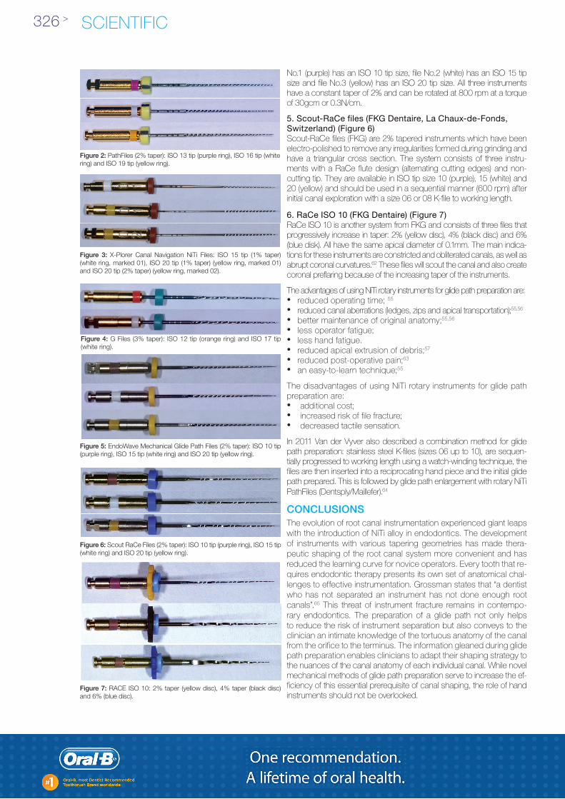
6. RaCe ISO 10 (FKG Dentaire) (Figure 7)RaCe ISO 10 is another system from fKG and consists of three files that progressively increase in taper: 2% (yellow disc), 4% (black disc) and 6% (blue disk). All have the same apical diameter of 0.1mm. The main indica-tions for these instruments are constricted and obliterated canals, as well as abrupt coronal curvatures.62 These files will scout the canal and also create coronal preflaring because of the increasing taper of the instruments.
The advantages of using NiTi rotary instruments for glide path preparation are:reduced operating time; • 55
reduced canal aberrations (ledges, zips and apical transportation);• 55,56 better maintenance of original anatomy;• 55,56
less operator fatigue;•less hand fatigue.•reduced apical extrusion of debris;• 57
reduced post-operative pain;• 63
an easy-to-learn technique;• 55
The disadvantages of using NiTi rotary instruments for glide path preparation are:
additional cost;•increased risk of file fracture;•decreased tactile sensation.•
In 2011 Van der Vyver also described a combination method for glide path preparation: stainless steel K-files (sizes 06 up to 10), are sequen-tially progressed to working length using a watch-winding technique, the files are then inserted into a reciprocating hand piece and the initial glide path prepared. This is followed by glide path enlargement with rotary NiTi Pathfiles (Dentsply/Maillefer).64
ConCluSIonSThe evolution of root canal instrumentation experienced giant leaps with the introduction of NiTi alloy in endodontics. The development of instruments with various tapering geometries has made thera-peutic shaping of the root canal system more convenient and has reduced the learning curve for novice operators. Every tooth that re-quires endodontic therapy presents its own set of anatomical chal-lenges to effective instrumentation. Grossman states that “a dentist who has not separated an instrument has not done enough root canals”.65 This threat of instrument fracture remains in contempo-rary endodontics. The preparation of a glide path not only helps to reduce the risk of instrument separation but also conveys to the clinician an intimate knowledge of the tortuous anatomy of the canal from the orifice to the terminus. The information gleaned during glide path preparation enables clinicians to adapt their shaping strategy to the nuances of the canal anatomy of each individual canal. While novel mechanical methods of glide path preparation serve to increase the ef-ficiency of this essential prerequisite of canal shaping, the role of hand instruments should not be overlooked.
figure 2: PathFiles (2% taper): ISO 13 tip (purple ring), ISO 16 tip (white ring) and ISO 19 tip (yellow ring).
figure 4: G Files (3% taper): ISO 12 tip (orange ring) and ISO 17 tip (white ring).
figure 5: EndoWave Mechanical Glide Path Files (2% taper): ISO 10 tip (purple ring), ISO 15 tip (white ring) and ISO 20 tip (yellow ring).
figure 6: Scout RaCe Files (2% taper): ISO 10 tip (purple ring), ISO 15 tip (white ring) and ISO 20 tip (yellow ring).
figure 3: X-Plorer Canal Navigation NiTi Files: ISO 15 tip (1% taper) (white ring, marked 01), ISO 20 tip (1% taper) (yellow ring, marked 01) and ISO 20 tip (2% taper) (yellow ring, marked 02).
figure 7: RACE ISO 10: 2% taper (yellow disc), 4% taper (black disc) and 6% (blue disc).

SCIENTIfICwww.sada.co.za / SADJ Vol 68 No. 7 < 327
declaration: No conflict of interest declared.
ReferencesSchilder H. Cleaning and shaping the root canal. Dent Clin North Am 1. 1974; 18: 269-96.Peters OA. Current challenges and concepts in the preparation of 2. root canal systems: a review. J Endod 2004; 30: 559-67.Hülsmann M, Peters OA, Dummer PMH. Mechanical preparation of 3. root canals: shaping goals, techniques and means. Endodontic Top-ics 2005; 10: 30-76.Jafarzadeh H, Abbott PV. Ledge formation: review of a great chal-4. lenge in endodontics. J Endod 2007; 33: 1155-62. Cunningham CJ, Senia ES. A three-dimensional study of canal curvatures 5. in the mesial roots of mandibular molars. J Endod 1992; 18: 294-300.Kartal N, Cimilli HK. The degrees and configurations of mesial canal 6. curvatures of mandibular molars. J Endod 1997; 23: 358-62.Haikel Y, Serfaty R, Bateman G, Senger B, Allemann C. Dynamic 7. and cyclic fatigue of engine-driven rotary nickel titanium endodontic instruments. J Endod 1999; 25: 434-40.Mullaney TP. Instrumentation of finely curved canals. Dent Clin 8. North Am 1979; 23: 575-92.Goerig AC, Michelich RJ, Schultz HH. Instrumentation of root canals in 9. molars using the step-down technique. J Endod 1982; 8: 550-4. fava LRG. The double-flared technique: an alternative for biome-10. chanical preparation. J Endod 1983; 9: 76-80.Morgan Lf, Montgomery S. An evaluation of the crown-down pres-11. sureless technique. J Endod 1984; 10: 491-8.R12. oane JB, Sabala CL, Duncanson MG. The “balanced force” concept for instrumentation of curved canals. J Endod 1985; 11: 203-11.Goldberg f, Araujo JA. Comparison of three instruments in the prepa-13. ration of curved root canals. Endod Dent Traumatol 1997; 13: 265-8.Walia H, Brantley WA, Gerstein H. An initial investigation of the bending and 14. torsional properties of Nitinol root canal files. J Endod 1988; 14: 346-51. Pruett JP, Clement DJ, Carnes DL. Cyclic fatigue testing of nickel-15. titanium endodontic instruments. J Endod 1997: 3: 77-85.Gambarini G. Cyclic fatigue of nickel-titanium rotary instruments after clinical use 16. with low- and high-torque endodontic motors. J Endod 2001; 27: 772-4.Arens fC, Hoen MM, Steiman HR, Dietz GC. Evaluation of single-use 17. rotary nickel-titanium instruments. J Endod 2003; 29: 664-6.Baumann MA. Nickel-titanium: options and challenges. Dent Clin N 18. Am 2004; 48: 55-67.Shen Y, Coil JM, Mclean AGR, Hemerling DL, Haapasalo M. Defects in 19. nickel-titanium instruments after clinical use. Part 5: Single use from endo-dontic specialty practices. J Endod 2009; 35: 1363-7.Spanaki-Voreadi AP, Kerezoudis NP, Zinelis S. failure mechanism 20. of ProTaper NiTi rotary instruments during clinical use: fractographic analysis. Int Endod J 2006; 39: 171-8.Kuhn G, Tavernier B, Jordan L. Influence of structure on nickel-titanium 21. endodontic instrument failure. J Endod 2001; 27: 516-20.Bergmans L, Van Cleyenbreugel J, Wevers M, Lambrechts P. Me-22. chanical root canal preparation with NiTi rotary instruments: ration-ale, performance and safety. Status report for the American Journal of Dentistry. Am J Dent 2001; 14: 324-33.Parashos P, Messer HH. Rotary NiTi instrument fracture and its con-23. sequences. J Endod 2006; 32: 1031-43.Serene TP, Adams JD, Saxena A. Nickel–titanium instruments. Appli-24. cations in endodontics. St. Louis: Ishiyaku EuroAmerica, 1995; 2: 92-4.Sattapan B, Nervo GJ, Palamara JE, Messer HH. Defects in rotary 25. nickel-titanium files after clinical use. J Endod 2000; 26: 161-5. Martín B, Zelada G, Varela P, Bahillo JG, Magán f, Ahn S, Rodríguez 26. C. factors influencing the fracture of nickel-titanium rotary instru-ments. Int Endod J 2003; 36: 262-6.Peters OA, Peters CI, Schönenberg K, Barbakow f. ProTaper rotary 27. root canal preparation: assessment of torque and force in relation to canal anatomy. Int Endod J 2003; 36: 93-9.Blum JY, Cohen P, Machtou P, Micallef JP. Analysis of forces devel-28. oped during mechanical preparation of extracted teeth using Profile Ni-Ti rotary instruments. Int Endod J 1999; 32: 24-31.Kobayashi C, Yoshioka T, Suda H. A new engine-driven canal preparation sys-29. tem with electronic canal measuring capabilities. J Endod 1997; 23: 751-4.Berutti E, Negro AR, Lendini M, Pasqualini D. Influence of manual 30. preflaring and torque on the failure rate of ProTaper instruments. J Endod 2004; 30: 228-30.Walsch H. The hybrid concept of nickel-titanium rotary instrumenta-31. tion. Dent Clin North Am 2004; 48: 183-202.Schrader C, Peters OA. Analysis of torque and force with differently ta-32. pered rotary endodontic instruments in vitro. J Endod 2005; 31: 120-3.Cheung GSP. Instrument fracture: mechanisms, removal of fragments, 33. and clinical outcomes. Endodontic Topics 2009; 16: 1-26. West J. Endodontic update 2006. J Esthet Restor Dent 2006; 18: 280-300.34. Khatavkar RA, Hegde VS. Importance of patency in endodontics. 35.
Endodontology 2010; 22: 85-91. Peters OA, Paqué f. Current developments in rotary root canal instrument 36. technology and clinical use: a review. Quintessence Int 2010; 41: 479-88.Young GR, Parashos P, Messer HH. The principles of techniques for 37. cleaning root canals. Aust Dental Journal 2007; 52 Suppl1: S52-S63. Roland DD, Andelin WE, Browning Df, Hsu GR, Torabinejad M. The 38. effect of pre-flaring on the rates of separation for 0.04 taper nickel-titanium rotary instruments. J Endod 2002; 28: 543-5.Blum JY, Machtou P, Ruddle CJ, Micallef JP. The analysis of me-39. chanical preparations in extracted teeth using ProTaper rotary instru-ments: value of the safety quotient. J Endod 2003; 29: 567-75.West J. The endodontic glide path: “Secret to rotary safety”. Dent 40. Today 2010; 29: 86-93.Goga R, Chandler NP, Oginni AO. Pulp stones: a review. Int Endod 41. J 2008; 41: 457–68.Castellucci A. Manual or Rotary Glide Path? Paper presented to the 42. tenth annual meeting of The Academy of Microscope Enhanced Dentistry, foundations and Expansion virtual meeting; 2011 Nov 10-12.Patino PV, Biedma BM, Liebana CR, Cantatore G, Bahillo JG. The 43. influence of a manual glide path on the separation rate of NiTi rotary instruments. J Endod 2005; 31: 114-6.You SY, Bae KS, Baek SH, Kum KY, Shon WJ, Lee W. Lifespan of 44. one nickel-titanium rotary file with reciprocating motion in curved root canals. J Endod 2010; 36:1991-4.Varela-Pati45. no P, Ibanez-Párraga A, Rivas-Mundina B, Cantatore G, Ot-ero XL, Martin-Biedma B. Alternating versus continuous rotation: a com-parative study of the effect on instrument life. J Endod 2010; 36: 157-9.Berutti E, Paolino DE, Chiandussi G, Alovisi M, Cantatore G, Castel-46. lucci A, Pasqualini D. Root canal anatomy preservation of WaveOne reciprocating files with or without glide path. J Endod 2012; 38:101-4.Gambarini G. The K3 rotary nickel titanium instrument system. En-47. dod Topics 2005; 10: 179-82.Mounce RE. Endodontic K-files: invaluable endangered species or 48. ready for the Smithsonian? Dent Today 2005; 24: 102-104. Ruddle CJ. The ProTaper technique. Endod Topics 2005; 10: 187-90. 49. Jerome CE, Hanlon RJ Jr. Identifying multiplanar root canal curva-50. tures using stainless-steel instruments. J Endod 2003; 29: 356-8.West JD, Roane JB. Cleaning and Shaping the Root Canal System. 51. In: Cohen S, Burns RC, editors. Pathways of the Pulp. 7th ed. Mosby Inc., St. Louis, Missouri; 1998.Ruddle CJ. Cleaning and shaping the root canal system. In: Cohen 52. S, Burns RC, editors. Pathways of the Pulp. 8th ed., St Louis, MO: Mosby Inc., St Louis, Missouri. 2002.Van der Vyver PJ. Creating a glide path for rotary NiTi instruments: 53. Part one. Endod Practice 2011 february: 40-3.Allen MJ, Glickman GN, Griggs JA. Comparative analysis of endo-54. dontic Pathfinders. J Endod 2007; 33: 723-6.Berutti E, Cantatore G, Castellucci A, Chiandussi G, Pera f, Migliaretti 55. G, Pasqualini D. Use of nickel-titanium rotary Pathfile to create the glide path: comparison with manual preflaring in simulated root ca-nals. J Endod 2009; 35: 408-12.Pasqualini D, Bianchi CC, Paolino DS, Mancini L, Cemenasco A, 56. Cantatore G, Castelucci A, Berutti E. Computed micro-tomograph-ic evaluation of glide path with nickel-titanium rotary Pathfile in maxillary first molar curved canals. J Endod 2012; 38: 389-93.Greco K, Carmignani E, Cantatore G. A comparative study between 57. manual and mechanic pre-flaring techniques. Paper presented to the fifteenth Biennial Congress of the European Society of Endod-ontology; 2011 Sept 14-17; Rome, Italy. Mounce RE. Blending reciprocation with the creation of larger apical 58. diameters. Dent News 2008; 15: 22-4.Kinsey B, Mounce RE. Safe and efficient use of the M4 safety hand-59. piece in endodontics. Roots 2008; 4: 36-40.Wagner MH, Barletta fB, Reis Mde S, Mello LL, ferreira R, fern-60. andes AL. NSK reciprocating handpiece: in vitro comparative analy-sis of dentin removal during root canal preparation by different opera-tors. Braz Dent J 2006; 17 (1):10-4.Nahmias Y, Cassim I, Glassman G. “Own the Canal”-The Importance 61. of a Reproducible Glide Path. Oral Health 2013; May: 74-82.Debelian G, Trope, M. Scouting the root canal with dedicated NiTi 62. files. Roots 2012, 2, 24-7.Pasqualini D, Mollo L, Scotti N, Cantatore G, Castellucci A, Migliaretti 63. G, Berutti E. Postoperative Pain after Manual and Mechanical Glide Path: A Randomized Clinical Trial. J Endod 2012; 38: 32-6.Van der Vyver PJ. Creating a glide path for rotary NiTi instruments: 64. Part two. Endod Practice 2011 May: 46 -53.Grossman LI. Guidelines for the prevention of fracture of root canal 65. instruments. Oral Surg Oral Med Oral Pathol 1969; 28: 746-52.
^ ^ ^