Embed Size (px)
Citation preview

The healing effects of Hyperium perforatum (St. John’s Wort) on experimental alkaline corrosive eosephageal and stomach burns
Erkan Güvenç, M.D.,1 Selahattin Kıyan, M.D.,2 Yiğit Uyanıkgil, M.D.,3 Emel Öykü Çetin, M.D.,4
Fatih Karabey, M.D.,5 Türker Çavuşoğlu, M.D.,3 Burak Gökçe, M.D.6
1Department of Emergency Medicine, Buca Seyfi Demirsoy State Hospital, İzmir-Turkey2Department of Emergency Medicine, Ege University Faculty of Medicine Hospital, İzmir-Turkey3Department of Histology and Embryology, Ege University Faculty of Medicine, İzmir-Turkey4Department of Pharmaceutical Technology, Ege University Faculty of Pharmacy, İzmir-Turkey5Department of Biotechnology, Ege University Graduate School of Natural and Applied Sciences, İzmir-Turkey6Department of Zoology, Ege University Faculty of Science, İzmir-Turkey
ABSTRACT
BACKGROUND: The most frequent etiologic cause is alkaline substances. We investigated the protective effects of the plant St. John ‘s Wort (Hypericum perforatum).
METHODS: We included 42 Wistar albino rats weighing between 200–300 grams and divided into six groups as Group 1: Con-trol, Group 2: Burn+Saline (BS), Group 3: Burn+St. John’s Wort (BSJW), Group 4: Burn+Plasebo (BP), Group 5: St. John’s Wort (SJW), Group 6: Placebo (P). After 15 days of treatment, esophagus, stomach and liver tissue samples were derived by dissection for histopathologic and biochemical markers. The cytotoxic effects of formulation on fibroblasts is evaluated in vitro on human dermoblast fibroblast line (HDFa, Gibco Invitrogen cell culture, C-013-5C).
RESULTS: The weight of the rats increased in Group 1, 3, 4, 6, decreased in Group 2 and did not change in Group 5. In the BSJW group, submucosal collagen accumulation, muscularis mucosa damage, tunica muscularis damage and collagen accumulation in esopha-gus were similar to the control group but lesser than BS and placebo group. In the stomach, mucosal damage, gastric gland dilatation, submucosal polymorphonuclear infiltration were similar to the control group and lesser than the BS group. The lethal concentration of SJW was 2.58 gr/mL.
CONCLUSION: SJW substrate is effective in protecting the esophagus and stomach in mild to moderate alcali corrosive burns in the subacute period. We should keep in mind the protective effects of STW substrate in alkaline corrosive burns of the gastrointestinal system.
Keywords: Alkaline; burn; corrosive; Hypericum Perforatum; Saint John ‘s Wort.
the prevention from the corrosive substances and ideal stor-age conditions and the limitations to the uncontrolled sale of some corrosive substances decrease the esophageal cor-rosive burns by %75.[1,2] The alkaline damages are frequently deep burns and cause liquefaction necrosis; the acidic dam-ages are frequently superficial burns and cause coagualation
EXPERIMENTAL STUDY
Ulus Travma Acil Cerrahi Derg, May 2020, Vol. 26, No. 3 373
INTRODUCTION
The corrosive burns in the gastrointestinal system arise from intentional or accidental oral ingestion of acidic or alkaline substances. They are frequently caused because of unsuitable packages. The printed and visual education programs about
Cite this article as: Güvenç E, Kıyan S, Uyanıkgil Y, Çetin EÖ, Karabey F, Çavuşoğlu T, et al. The healing effects of Hyperium Perforatum (St. John’s Wort-Kantaron) on experimental alkaline corrosive eosephageal and stomach burns. Ulus Travma Acil Cerrahi Derg 2020;26:373-383.
Address for correspondence: Erkan Güvenç, M.D.
Buca Seyfidemirsoy Devlet Hastanesi, Acil Servis Kliniği, İzmir, Turkey
Tel: +90 232 - 220 10 15 E-mail: [email protected]
Ulus Travma Acil Cerrahi Derg 2020;26(3):373-383 DOI: 10.14744/tjtes.2019.93428 Submitted: 08.03.2019 Accepted: 15.08.2019 Online: 15.05.2020Copyright 2020 Turkish Association of Trauma and Emergency Surgery

Güvenç et al. The healing effects of Hyperium perforatum on experimental alkaline corrosive eosephageal and stomach burns
necrosis except for hydrochloric acid.[3] The damage arises from acidic substances are located frequently in the pyloric region; if the stomach is congested, the acidic substances get mixed and cause disseminated tissue damage.[4,5] Although acidic substances cause the most damage in the stomach, in 6–10% of the cases, there are esophageal burns.[6]
St. John’s Wort (SJW) plant is also known as Hypericum per-foratum L. (Fig. 1) grows in Europe, Asia, North Africa and the United States for many years; it is used for many years as a medication for burns and poisonous animal bites.[7]
The distal flowering branches of SJW are gathered when fresh, put into olive oil without delay; it is kept in a closed glass jar under the sunshine for 41 days; its color turns to deep red, and the final extract is used for medical purposes.[8]
In the literature, there is not any clinical or experimental survey about the protective effects of the SJW extract on corrosive burns of esophagus or stomach. We investigated the possible protective effect of SJW on corrosive burns of esophagus and stomach.
MATERIALS AND METHODS
Experimental ProcedureThis experimental study is performed after the ethical ap-provement from Dokuz Eylul University Medicine Faculty, An-imal Ethics Committee with the protocol number of 65/2013; the survey is performed according to “The Guide for the Care and Use of Laboratory Animals-Institute of Laboratory Animal Resources Commission on Life Sciences National Re-search Council, USA”. The experimental part of the study is carried out in Dokuz Eylul University Multidisciplinary Ex-perimental Research and Animal Laboratory; histopatholog-ical analyses were carried out in Ege University, Faculty of Medicine Division of Histology and Embryology; biochemical analyses were carried out in Ege University Science Faculty, Department of Biology and AREL Laboratory. The study is supported by Ege University Scientific Research Project Fund as project 2013TIP-079.
The Preparation of the Extract and Analyses of SJWThe supranatant parts of the plant were collected and dried in the closed shade area. After drying, the material was ground homogenously by the mechanical grinder. The 6 gr of the powder was added to the glass balloon with 300 mL of
MeOH. We boiled the mixture firstly at 11ºC for 30 minutes then at 23ºC for 30 minutes with the ultrasonic bath. We covered the glass material with aluminium foil, providing a dark environment to protect the plant content.
After the sonification samples are centrifuged at 7 G for 10 minutes; the supernatant part was taken another glass bal-loon; 300 mL MeOH was added to the remaining material again and the extraction steps were repeated from the be-ginning for four times. The extract was obtained by collected supernatant parts. MeOH was removed by evaporation. The lyophilizer system (Christ®) was used to remove the solvent completely. The final dry extract was put placed into stability control media with dry-flask bottles.
The dry plant extract was dissolved with MeOH: Acetoni-trile (1:1) after weighted; than, the 500 ppm samples were prepared. We used vortex and ultrasonic bath in order to obtain a homogenous solution. The samples filtered through injector fiilter were taken in HPLC vials and made ready for analyses. The injection amount was 20 μl. The chromatogram of the hyperphosphoric standard was 270 nM RT:50.80. The chromatogram of the pseudohyperigenin standard was 590 nM RT:44.89. The chromatogram of the hypericin standard was 590 nM RT: 53.73. The chromatograms for analyzes of the extracted sample were determined as 270 and 590 nm. The analyses were performed by the HPLC system consist-ing of Shimadzu SCL-10VP, manual sampler, column oven and DAD detector elements. We used Hicrom® C18 column with particle size 5 μm, 250 mm length and 4.6 mm diameter; and Hicrom® C18 anterior column with particle size 5 μm, 10 mm length and 4.6 mm diameter. The column oven temperature was 30ºC, the solvent flow rate was 1 mL/min and the detec-tor wavelength 270 and 590 nm. The injection amount was 20 µl. The detailed SJW extract content is given in Table 1.
The Preparation of Oral Formulation, Including SJWA buffer solution is used, including 0.1 M acetic acid in a beaker. The beaker was placed on a calibrated pH-meter along with the mixer. The appropriate amount of 1M solution prepared with sodium hydroxide was added until the desired value was reached; then, the solution was taken into 100 mL balloon flask. The volume is completed by bidistillated water. The acetate buffer (pH=5) was prepared containing 12.5 mg/
Ulus Travma Acil Cerrahi Derg, May 2020, Vol. 26, No. 3374
Table 1. The chemical ingredents of St. John’s Wort extract
Sample RT Area Mean area SD %RSD
Pseudohypericin 44.917 56.8 58.60 2.5456 4.34
Hypericin 49.034 36.8 38.05 1.7678 4.65
Hyperforin 51.122 96.6 97.10 0.7071 0.73
SD: Standard deviation; RSD: Relative standard deviation; RT: Reaction time.
Figure 1. St. John’s Wort (Hypericum perforatum).
Division: SpermatophyaSubdivision: AngiospermaeClass: DicotyledoneaeSubclass: MagnolipsidaFamily: HypericaceaeGenus: HypericumSpecies: Hypericum Perforatum

Güvenç et al. The healing effects of Hyperium perforatum on experimental alkaline corrosive eosephageal and stomach burns
mL SJW extract and 2% hydroxypropyl cellulose (HPMC) as the viscosity enhancer. The extract was taken at regular in-tervals into storage containers to avoid the inappropriateness of storage conditions.
The Preparation of Placebo Gel ExtractThe acetate buffer (pH=5) was prepared containing 12.5 mg/mL SJW extract and 2% hydroxypropyl cellulose (HPMC) as the viscosity enhancer. The extract was taken at regular in-tervals into storage containers to avoid the inappropriateness of storage conditions.
The Subject Groups and Experimental MethodWe used 42 male and female Wistar albino rats weigh-ing between 200–300 gr. The rats were kept under normal day&night cyclus far from noise at 20–22ºC for adaptation. They were fed by tap water and standard artificial mouse feed ad libitum. The base of the cage consisted of wood dust. The alkaline corrosive material was 5% NaOH/0.2 cc consistent with Katrancioglu et al.’s[9] study. The groups and their speci-fications are explained in Table 2.
Five minutes before the corrosive application, intraperitoneal ketamine (Pfizer- Ketalar®; 50 mg/mL ketamine hydrochloride) 0.3–1.3 ml/kg was applied for sedation and analgesia without losing swallowing function. The corrosive material was given to the subjects in group 1, 2, 3 and 4 using mouth, which was ending at the level of the cervical esophagus. The admission time was nearly 20 minutes after oral ingestion of corrosive materials. Thus, we applied the placebo and SJW extract 20 minutes after the corrosive material application by feeding tube. The treatment and placebo applications were repeated every day for once for 14 days. To achieve analgesia, we applied acetaminophen 2 mg/mL (Bristol-Myers Squibb-Perfalgan®; 10 mg/mL acetaminophen). The subjects were sacrificed in the 15th day; tissue samples were derived from 3 cm proximal of the esophagogastric junction, stomach and liver.
Histopathologic EvaluationThe samples derived from the esophagus and stomach were
homogenised over ice in the homogenized buffer (50 mm phosphate buffer, pH:7.4) with a mechanic homogenizer (Hei-dolph Silent Crusher M). The samples were fixated by 4% formaldehyde; the 5 µm slices were stained by hematoxylin -eosin or Mallory Azan Stain. The histopathologic examina-tion was performed by a histologist under X10, X20, X40 magnification without knowing the groups.[10] The stenosis in-dex was calculated by determining the esophagus wall thick-ness and lumen diameter.[10–13] The submucosal cologen accu-mulation, muscularis mucosa damage, muscular layer damage and collagen accumulation in the muscular layer of esophagus were evaluated. The collagen accumulation level was deter-mined between 0–5 according to the histopathologic scoring system showed in Table 3.[10,12,13]
The gastric tissue samples were derived by dissection of the greater curvature of the stomach and getting the stained parts of the adjacent part of the corpus. The mucosal PMNL
Ulus Travma Acil Cerrahi Derg, May 2020, Vol. 26, No. 3 375
Table 2. Study groups
Group 1 – Control (n=7) We din’t do anything to this group
Group 2 – Burn (n=7) A corrosive burn was made by 5% NaOH/0.2 ml but any treatment didn’t applied
Group 3 – Burn + SJW (n=7) A corrosive burn was made by 5% NaOH/0.2 ml; and oral solution of SJW extract was given at
a dose of 50 mg/kg/day
Group 4 – Burn + Placebo (n=7) A corrosive burn was made by 5% NaOH/0.2 ml; and a placebo gel was given at a dose of
50 mg/kg/day
Group 5 – Control SJW (n=7) Oral solution of SJW extract was given at a dose of 50 mg/kg/day
Group 6 – Control Placebo (n=7) A placebo gel was given at a dose of 50 mg/kg/day
SJW: St. John’s Wort.
Table 3. Histopathologic scoring of the esophagus[10,12,13]
Score
Submucosal collogen accumulation
Absent 0
Mild (equal or less than 2 folds of the 1
muscularis mucosa)
Severe (more than 2 folds of the muscularis 2
mucosa)
Muscularis mucosa damage
Absent 0
Present 1
Tunica muscularis damage and collogen accumulation
Absent 0
Mild (collogen accumulation aroun muscle fibers) 1
Severe (mild damage and collogen accumulation 2
takes some of the fibers place)
Total score 0–5
infiltration, mucosal edema, gastric gland dilatation and sub-mucosal PMNL infiltration were performed.
Cytotoxicity AnalysesWe used the malonedialdehyde (MDA) level to determine the peroxidation level of the lipid inside the cell membrane, su-peroxide dismutase (SOD) level for free oxygen radicals and the level of glutathione peroxidase (GPX) and catalase (CTX) levels for their tissue-protective effects.[14]
The in-vitro cytotoxicity of the SJW was determined by the dermal fibroblast tissue line (HDFa, Gibco Invitrogen cell culture, C-013-5C). The frozen cells were cultured in RPMI (10% FBS, 100 U/mL penicillin and 100 μg/mL streptomycin) agar at 37°C, including 5% CO2. The number of the fifth passage fibroblasts were used for cytotoxicity determina-tion. The vitality of the cells was determined by MTT (Sigma In Vitro Toxicology Assay Kit, MTT based) vitality test and probit analyses was performed using the SPSS v.20 package program.
Statistical MethodSPSS 20.0 (IBM Corporation, Armonk, New York, United States) program was used to analyze the data. Continuous variables were expressed as mean±sd if they are normally distributed and expressed as median (min-max) if not. Normal distribution was determined by histogram and One-Sample Kolmogorov Smirnov test. To determine the difference of continuous variables between groups, non-parametric tests are used due to the low sample size; the Mann Whitney-U test for two groups and the Kruskal Wallis
test for more than two groups. The difference of the cathe-gorical variables between groups are determined using the Chi-square test and Fisher’s exact test. P<0.05 was accepted as significant.
RESULTS
Weight Analysis ResultsAll subjects in the Groups (n=42) were divided in a manner that each group contained seven subjects. Their weights were measured before the process. No statistically significant dif-ference in weight was found between the groups in everyday weight. In time, every group get weight significantly except Group 5 (p<0.05).
Esophageal Histopathologic FindingsThe microscopic specifications of the esophageal tissue mi-croscopy are shown in Figure 2.
Group 2 – Alkaline burn group: Diffused or focal mucosal erosion. There were moderate edema and sporadic mucosal neutrophil infiltration infiltration in the submucosa. Submu-cosal and mucosal collagen accumulation, inflammation and edema were detected. There were significant tissue damage and constriction in the lumen (Fig. 2b).
Group 3 – Alkaline burn + SJW group: Moderate in-flammation in all layers, mucosal and submucosal edema were detected. If we compare with Group 2, focal erosion findings, submucosal collagen accumulation, mucosal collagen accumu-lation, and stenosis index were lesser than Group 2 (Fig. 2c).
Ulus Travma Acil Cerrahi Derg, May 2020, Vol. 26, No. 3376
Güvenç et al. The healing effects of Hyperium perforatum on experimental alkaline corrosive eosephageal and stomach burns
(a)
(d)
(b)
(e)
(c)
(f)
Figure 2. The histopathologic appearence of the esophagus. H&E stain; X40 magnification (X40=125µm); the red arrows are stratum corneum. Blue arrows are stratum basale layer of epidermis; (a) Control group; (b) Burn Group; (c) Burn and SJW group; (d) Burn and placebo group; (e) SJW without burn group; (f) Placebo without burn group.

Group 4 – Alkaline burn + Placebo group: Similarly with Group 2, diffused or focal mucosal erosion; mild edema and sporadic mucosal neutrophil infiltration in submucosa; sub-mucosal and mucosal collagen accumulation, inflammation and edema were detected. There are significant tissue dam-age and constriction in the lumen (Fig. 2d).
Group 5 – Control + SJW group: There were mild edema and inflammation in mucosa, but not diffused erosion find-ings. Minimal inflammation and edema were present in the submucosa. The mucosal histopathologic examination was normal (Fig. 2e).
Group 6 – Control + Placebo group: The appearance was similar to Group 1 and 5. There are mild edema and in-flammation in the mucosa. Minimal inflammation and edema in the submucosa. The mucosal histopathologic examination was normal (Fig. 2f ).
The frequency of the histopathologic findings of all groups is expressed in Table 4 in detailed.
Submucosal collagen accumulation of esophagus in Group 3 was similar to Group 1 (p=0.559), significantly higher in Group
Ulus Travma Acil Cerrahi Derg, May 2020, Vol. 26, No. 3 377
Güvenç et al. The healing effects of Hyperium perforatum on experimental alkaline corrosive eosephageal and stomach burns
Table 4. The frequency of the histopathologic features of the groups
Experiment groups
Group 1 Group 2 Group 3 Group 4 Group 5 Group 6
n % n % n % n % n % n %
Submucosal collogenaccumulation of esophagus
Absent 6 85.7 – – 4 57.1 1 14.3 6 85.7 4 57.1 Mild 1 14.3 2 28.6 3 42.9 4 57.1 1 14.3 3 42.9 Severe – – 5 71.4 – – 2 28.6 – – – –
Muscularis mucosadamage of esophagus
Absent 6 85.7 2 28.6 6 85.7 2 28.6 4 57.1 6 85.7 Present 1 14.3 5 71.4 1 14.3 5 71.4 3 42.9 1 14.3
Tunica muscularis damage andcollogen accumulation in esophagus
Absent 7 100 – – 6 85.7 2 28.6 7 100 7 100 Mild – – 3 42.9 1 14.3 2 28.6 – – – – Severe – – 4 57.1 – – 3 42.9 – – – –
PMNL infiltration and mucosalin gastric mucosa
Absent 6 85.7 – – 1 14.3 – – 5 71.4 4 57.1 Mild 1 14.3 2 28.6 3 42.9 3 42.9 2 28.6 3 42.9 Moderate – – 2 28.6 2 28.6 2 28.6 – – – – Severe – – 3 42.9 1 14.3 2 28.6 – – – –
Gastric mucosal edema
Absent 6 85.7 – – 3 42.9 1 14.3 2 28.6 4 57.1 Mild 1 14.3 3 42.9 2 28.6 2 28.6 5 71.4 2 28.6 Moderate – – 3 42.9 2 28.6 4 57.1 – – 1 14.3 Severe – – 1 14.3 – – – – – – – –
Gastric gland dilatation
Absent 5 71.4 – – 1 14.3 – – 4 57.1 4 57.1 Mild 2 28.6 3 42.9 4 57.1 4 57.1 3 42.9 3 42.9 Severe – – 4 57.1 2 28.6 3 42.9 – – – –
Submucosal PMNLinfiltration in stomach
Absent 5 71.4 – – 2 28.6 1 14.3 5 71.4 5 71.4 Mild 2 28.6 2 28.6 3 42.9 2 28.6 2 28.6 2 28.6 Moderate – – 3 42.9 2 28.6 3 42.9 – – – – Severe – – 2 28.6 – – 1 14.3 – – – –
PMNL: Polymorphonuclear lymphocytes.

2 than Group 1 (p=0.005); higher in Group 4 than Group 3 but there was not any statistical significance (p=0.266). Consequently, SJW has a significant effect on degreasing the submucosal collagen accumulation of esophagus and also it is better than placebo.
Muscularis mucosa damage in esophagus in Group 2 was higher than Group 1 but lack of statistical significance (p=0.103); similar in Group 3 and 1 (p=1.000); higher in Group 2 than Group 3 but lack of statistical significance (p=0.103); similar in Group 4 and 2 (p=1.000). Conse-quently, SJW has a significant effect on degreasing the mus-cularis mucosa damage in esophagus also it is better than placebo.
Tunica muscularis damage and collagen accumulation in esophagus was significant in Group 2, which was higher than the control group (p=0.001), similar in Group 3 and 1 (p=1000); higher in Group 4 than Group 1 (p=0.021). Conse-quently, SJW has a significant effect on degreasing the tunica muscularis damage and collagen accumulation in esophagus. Also, it is better than placebo.
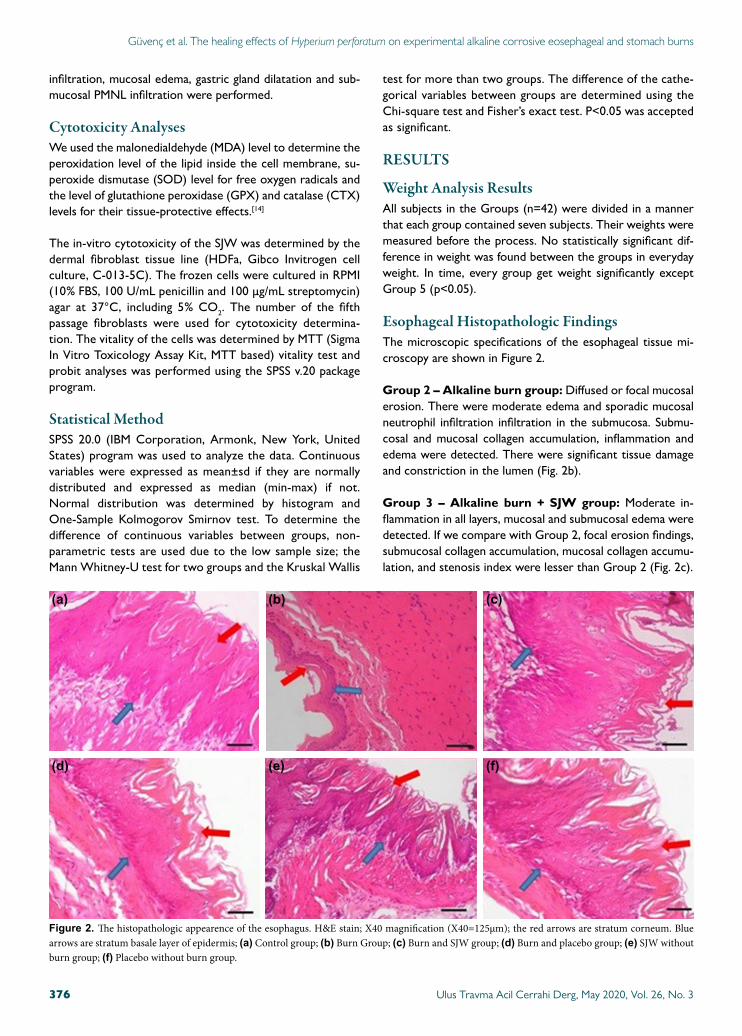
Gastric Histopathologic FindingsThe microscopic spesifications of the gastric tissue mi-croscopy are shown in Figure 3.
PMNL infiltration in gastric mucosa was higher in Group 2, 3 and 4 than Group 1 (p-values are 0.005, 0.029, 0.005, respec-tively); similar in Group 3 and 2 (p=1.000).
Edema in gastric mucosa was higher in Group 2 than Group 1 (p=0.005); similar in Group 3 and 1 (p=0.266); similar in Group 4 and 3 (p=0.559). Consequently, SJW has a significant effect on degreasing the edema in gastric mucosa and also similar with placebo.
Dilatation in gastric gland was higher in Group 2 than Group 1 (p=0.021); similar in Group 3 and 1 (p=0.103); similar in Group 4 and 3 (p=1.000). Consequently, SJW has a significant effect on degreasing the dilatation in the gastric gland and also similar to placebo.
PMNL infiltration in gastric submucosa was higher in Group 2 than Group 1 (p=0.021); similar in Group 3 and 1 (p=0.286); similar in Group 4 and 1 (p=0.103); similar in Group 4 and 3 (p=1.000). Consequently, SJW has a significant effect on degreasing the dilatation in the gastric gland and also similar with placebo. Consequently, SJW has a significant effect on degreasing the PMNL infiltration in gastric submucosa and also similar to placebo.
The mean stenosis index, epithelium thickness and compar-ison of them are given in Table 5. The stenosis index was
Ulus Travma Acil Cerrahi Derg, May 2020, Vol. 26, No. 3378
Güvenç et al. The healing effects of Hyperium perforatum on experimental alkaline corrosive eosephageal and stomach burns
(a)
(d)
(b)
(e)
(c)
(f)
Figure 3. The histopathological appearance of all groups. The blue arrows indicate superficial mucosal layer and mucus cells. The red arrows are foveola gastricea, the red stars are the dilatation of the gastric glands, yellow stars are the PMNL cells accumulation areas. (a) Control group; X40 magnification, essential cells; (b) Burn Group; X20 magnification, mild dilatation of gastric glands and capillary hemorhagia, submucosal PMNL infiltration, edema and congestion in vessels; (c) Burn and SJW group; X20 magnification, submucosal PMNL infiltration, edema and congestion in vessels; (d) Burn and placebo group; X20 magnification, moderate dilatation in gastric glands and capillary hemorhagia, submucosal PMNL infiltration, edema and congestion in vessels; (e) SJW without burn group; X40 magnification, partial mucosal damage and decrease in mucus accumulation; (f) Placebo without burn group; X40 mag-nification. (X20=250 µm, X40=125 µm).

higher in the SJW group (Group 3) than the control group but lower than the burn group (Group 2) and placebo (Group 4). Contrary, epithelium thickness was lower in the SJW group (Group 3) than the control group but higher than burn group (Group 2) and placebo (Group 4). Consequently, from the point of stenosis index, epithelium thiskness, SJW has a significant effect on degreasing the stenosis index and pro-tecting the decrease in epithelium thickness due to alkaline burn and better than placebo.
MTT cell vitality test is calculated: LC50 = 2580564,906 µg/ml = 2580.564 mg/ml = 2.580564 gr/ml.
The formulation of the medication is applied as 50 mg/kg. The amount of active ingredient in the oral formulation was 12.5 mg/mL, which was very low than the calculated LC50 value. Consequently, the SJW extract has no cytotoxicity on fibroblasts.
The Enzyme Levels in GroupsThe MDA, CAT, SOD and GPX levels in groups are expressed in Table 6. MDA and CAT levels were different in groups, but
SOD and GPX levels were similar in all groups. The box-blot graphs of all groups are given in Figure 4.
The MDA level was higher in Group 2 than Group 1 (p=0.048), but CAT levels were similar in Group 1 and 2 (p=0.701). Ad-ditionally, the MDA level was higher in Group 3 than Group 2 (p=0.001), but CAT levels were similar in Group 2 and 3 (p=0.200).
If we compare with placebo, the MDA level was similar in Group 2 and 4 (p=0.949), but CAT levels were lower in Group 4 than Group 3 (p=0.001). The MDA and CAT levels were higher in Group 3 than group 4 (p-values are 0.001 and 0.002, respectively). As a summary, we can say that the peroxidation level of the lipid inside the cell membrane, which is determined by MDA, was higher in the SJW group than the burn group and also placebo. Additionally, the tissue-protective effects which were determined by CTX was similar in SJW and burn group, but the SJW group was higher than placebo.
DISCUSSIONSJW extract has been used for many years.[15] Previous stud-
Ulus Travma Acil Cerrahi Derg, May 2020, Vol. 26, No. 3 379
Güvenç et al. The healing effects of Hyperium perforatum on experimental alkaline corrosive eosephageal and stomach burns
Table 5. Stenosis index and epithelium thickness of the groups
Stenosis index Epithelium thickness
Mean±SD (micrometer) Comparison (p) Mean±SD (micrometer) Comparison (p)
Group 2 Group 3 Group 2 Group 3
Group 1 0.12±0.01 0.002 0.002 160.49±3.10 0.002 0.002
Group 2 0.42±0.03 – 0.002 66.76±2.76 – 0.002
Group 3 0.22±0.02 0.002 – 127.70±3.82 0.002 –
Group 4 0.38±0.03 0.029 0.002 101.06±2.52 0.002 0.002
Group 5 0.23±0.03 0.002 0.698 157.67±2.72 0.002 0.002
Group 6 0.20±0.03 0.002 0.120 158.02±1.51 0.002 0.002
SD: Standard deviation.
Table 6. Mean enzyme levels in all of he groups
MDA (nmol/mg protein) CAT (U/mg protein) SOD (u/mg protein) GPX (U/mg protein)
Mean±SD Mean±SD Mean±SD Mean±SD
Group 1 1.41±0.13 71.43±3.95 86.29±3.45 12.34±0.57
Group 2 1.56±0.11 71.0±8.64 85.14±2.79 12.16±0.66
Group 3 1.89±0.12 65.43±6.95 86.29±3.82 12.41±0.58
Group 4 1.57±0.08 56.14±2.54 83.57±2.76 11.67±1.11
Group 5 1.70±0.10 66.29±6.10 86.00±3.11 12.66±0.93
Group 6 1.39±0.09 60.29±3.20 83.43±2.37 12.81±0.75
p <0.001 0.001 0.249 0.200
SD: Standard deviation; MDA: Malonedialdehyde; CAT: Catalase; SOD: Superoxide dismutase; GPX: Glutathione peroxidase.

ies have investigated the beneficial effects of this extract on thermal burns and traumatic injuries; its useful effects on the healing of linear and circular lacerations; it also shortens the re-epithelisation time.[16] Additionally, SJW extract protects epithelium thickness and reduces the degeneration of hair follicles in thermal burns.[15] In Burning Mouth Syndrom, to use SJW three times a day in 300 mg dose for 12 weeks re-duce the burning sensation, but it does not benefit the pain sensation.[17]
The pharmacologic studies with SJW are generally about the antidepressant effect using the ingredients of the plant called hypericin and hyperforin. The flower and branch part of the plant include 2–4.5% hyperforin and 0.2–1.8% adhiperforin which are a branch of phloroglucinol group; the flower and buds of the plant include 0.05–0.3% naftodiantron; the parts of the plant over the ground like leaves, stalks, flowers and buds include 2–4% flavonoids.[18] The plant extract is used as antidepressant, antitumor, antiviral, antimicrobial, an-tibacterial, analgesic, hepatoprotective and gastroprotective purposes.[19–28] It is argued that the extract could be used as antiviral in acquired immune deficiency syndrome (AIDS).[29]
According to some of the in-vitro studies, the ingredients of SJW inhibits some of the steps in inflammatory reactions.[26,30,31] The SJW extract inhibits the free radical production, myeloperoxidase, cyclo-oxygenase-1, 5-lipo-oxygenase and inducible cyclo-oxygenase and nitric oxide synthase.[32–34]
The first treatment method of the strictures due to corro-sive esophagus burn is the antegrade dilatation of the esoph-agus with oiled whalebone in the 17th century; the rubber dilatators are first used in 1837 for the same purpose; it was developed by contrast-enhanced radiologic studies after the discovery of bismuth in 1895; finally, a new era is started with the invention of distally illuminated esophagoscope by Cheva-lier Jackson in 1902.[35–37] The cases with esophageal stricture due to corrosive burns were reduced by the prophylactic use of the antibiotics in the 1940s, also using steroids and early-prophylactic dilatation in the 1950s.[35,36]
While the endoscopic examination of the corrosive esophageal burns, deep burns, massive hemorrhagia, ulcer-ation, focal necrosis and simple inflammation findings could be seen.[38] The protective effects of colchicine was reported
Ulus Travma Acil Cerrahi Derg, May 2020, Vol. 26, No. 3380
Güvenç et al. The healing effects of Hyperium perforatum on experimental alkaline corrosive eosephageal and stomach burns
Figure 4. The MDA, CAT, SOD and GPX levels in groups. MDA: Malonedialdehyde; CAT: Catalase; SOD: Superoxide dismutase; GPX: Glutathione peroxidase.
2.200
100 16.00
14.00
12.00
10.00
8.00
80
MD
A (n
mol
/mg
prot
ein)
SOD
(U/m
g pr
otei
n)
GPX
(U/m
g pr
otei
n)C
AT (U
/mg
prot
ein)
2.000
95
75
70
65
70
55
1.800
90
1.600
85
80
1.400
75
1.200
70
50
Group 1
Group 1 Group 1
Group 1Group 2
Group 2 Group 2
Group 2Group 3
Group 3 Group 3
Group 3Group 4
Group 4 Group 4
Group 4Group 5
Group 5 Group 5
Group 5Group 6
Group 6 Group 6
Group 6
24
3
31
38
8

in the animal experiment.[39] N-acetyl cysteine was used due to the intermolecular disulfide bond feature, but its clinical usage is precluded because of excessive bronchial secretion.[40] Indomethacin was tried for its anti-inflammatory effect, but it was not advised due to its inhibitory effect of throm-bocyte aggregation.[39] The experimental studies reported the protective effect of antioxidant substances like vitamin E and C in stricture formation by reducing collagen synthesis.[41] Epidermal growth factor (EGF) and interferon γ (IFN–γ) are significantly reduce the residual stenosis rate after corrosive esophageal burns.[42]
As we see, various experimental methods are tried to reduce the stenosis due to corrosive esophageal burns. However, the desired clinical target and clinical usage could not be achieved because the treatment method generally could not be applied in clinical practice and also there are not enough clinical hu-man studies.
To our knowledge, there is not any research into the effects of SJW in corrosive esophageal burns. Thus, we could not compare all of the data results obtained in previous studies. In our study, mucosal edema and collagen accumulation in the epithelium, diffuse edema, inflammation and collagen accumu-lation were detected in the burn model group. The benefits of SJW and the comparison with placebo were investigated in this study. When we gave the SJW extract to the burn model, submucosal and mucosal inflammation, edema, focal erosion and collagen accumulation were reduced and also it was superior to placebo. Also, the muscularis mucosa damage and collagen accumulation were lower than burn and placebo group. The collagen accumulation between muscle fibers and stenosis index was lower in the SJW treatment group, and also epithelium thickness was higher in the SJW group than placebo group. PMNL infiltration in gastric mucosa, gastric mucosal edema, gastric gland dilatation was lower in the SJW group than the burn group.
The anti-oxidant activity of the SJW, which is rich in flavonoids, is showed in-vitro.[43] With the use of the medium (75 mg/kg of body weight/day) or high (150 mg/kg of body weight/day) dose, SJW reduces the malondialdehyde levels in plasma or liver of rats fed by cholesterol reach diet.[43] Intraperitoneal single dose (50 mg/kg) SJW administration for 15 minutes to rats with hepatic ischemia reduces the liver enzymes and malondialdehyde and increases catalase activity.[44]
The medications that will be applied to the gastrointestinal system should be safe for the patient. While the application of SJW to chicken embryo cells, active fibroblast cells get into polygonal shape and fibroblasts cell production increases.[45] In our study, the lethal concentration of the extract is deter-mined as 2.580564 gr/ml; the amount of active ingredient in the oral formulation was 12.5 mg/mL, which was very low than the calculated LC50 value. Thus, our findings suggest that our extract is safe for using in the gastrointestinal system.
SJW has lower side effects in normal doses. The most fre-quently seen side effects are gastrointestinal symptoms, aller-gic reactions, vertigo, confusion, discomfort, apathy and xe-rostomia.[46,47] These effects are generally mild, moderate and transient.[48] The side effects are dose-dependent and also the medications that the patient had already using may affect the side effects.[46,47,49–52] SJW extracts have no genotoxic potential or mutagenic activity, according to in-vivo and in-vitro studies, but acute toxic neuropathy and mania are reported.[46,53]
In conclusion, SJW has protective beneficial effects on corro-sive esophageal burns in the early phase. Its lethal concentra-tion is very high, so it can be used safely in corrosive burns in esophagous. However, we should be careful while using the extract in humans. There is not any human model; only with further human studies can detect the benefits and harms of SJW on the gastrointestinal system.
Ethics Committee Approval: This experimental study is performed after the ethical approvement from Dokuz Eylul University Medicine Faculty, Animal Ethics Committee with the protocol number of 65/2013.
Peer-review: Internally peer-reviewed.
Authorship Contributions: Concept: E.G., S.K.; Design: E.G.; Supervision: S.K.; Fundings: E.G., Y.U., E.Ö.Ç.; Materials: E.G., Y.U., T.Ç., F.K.; Data: E.G., F.K., B.G.; Analysis: E.G., Y.U; Literature search: E.G., S.K., Y.U., T.Ç; Writing: E.G.; Critical revision: E.G.
Conflict of Interest: None declared.
Financial Disclosure: The authors declared that this study has received no financial support.
REFERENCES
1. Spitz L, Lakhoo K. Caustic ingestion. Arch Dis Child 1993;68:157–8.2. Christesen HB. Epidemiology and prevention of caustic ingestion in chil-
dren. Acta Paediatr 1994;83:212–5. [CrossRef ]
3. Hugh TB, Kelly MD. Corrosive ingestion and the surgeon. J Am Coll Surg 1999;189:508–22. [CrossRef ]
4. Moore WR. Caustic ingestions. Pathophysiology, diagnosis, and treat-ment. Clin Pediatr (Phila) 1986;25:192–6. [CrossRef ]
5. Hodgson JH. Corrosive stricture of the stomach; case report and review of literature. Br J Surg 1959;46:358–61. [CrossRef ]
6. Friedman EM. Caustic ingestions and foreign bodies in the aerodigestive tract of children. Pediatr Clin North Am 1989;36:1403–10. [CrossRef ]
7. Hışıl Y, Şahin F, Omay SB. Composition of Hypericum perforatum L. and Its Medical Importance. UHOD 2005;4:212−8.
8. Miller AL. St. John’s wort (Hypericum perforatum): clinical effects on depression and other conditions. Alternative medicine review: a journal of clinical therapeutic 1998;3:18−26.
9. Katrancioglu O, Nadir A, Şahin E, Arici S, Akkaş Y, Kaptanoğlu M. Acid and alkali burns of the esophagus: An experimental study. Scientific Re-search and Essays 2011;6:3959−63. [CrossRef ]
10. Türkyilmaz Z, Sönmez K, Demirtola A, Karabulut R, Poyraz A, Gülen S, et al. Mitomycin C prevents strictures in caustic esophageal burns in rats. J Surg Res 2005;123:182−7. [CrossRef ]
11. Gehanno P, Guedon C. Inhibition of experimental esophageal lye stric-
Ulus Travma Acil Cerrahi Derg, May 2020, Vol. 26, No. 3 381
Güvenç et al. The healing effects of Hyperium perforatum on experimental alkaline corrosive eosephageal and stomach burns

tures by penicillamine. Arch Otolaryngol 1981;107:145–7. [CrossRef ]
12. Millar AJW, Cywes S. Caustic strictures of esophagus, in Pediatric surgery. O’Neill JAO, Rowe MI, editors. Mosby: St Lous; 1998.p.969−79.
13. Koltuksuz U, Mutuş HM, Kutlu R, Özyurt H, Çetin S, Karaman A, et al. Effects of caffeic acid phenethyl ester and epidermal growth fac-tor on the development of caustic esophageal stricture in rats. J Ped Surg 2001;36:1504−9. [CrossRef ]
14. Loeb MP, Eisenstein AM. Caustic injury to the upper gastrointestinal tract, in Sleisenger and fordtran’s gastrointestinal and liver disease. 6th edition. WB Saunders Co; 1998.p.335−42.
15. Kıyan S, Uyanıkgil Y, Altuncı YA, Çavuşoğlu T, Çetin Uyanıkgil EÖ, Karabey F. Investigation of acute effects of Hypericum perforatum (St. John’s Wort-Kantaron) treatment in experimental thermal burns and comparison with silver sulfadiazine treatment. Ulus Travma Acil Cerrahi Derg 2015;21:323–36. [CrossRef ]
16. Prisăcaru AI, Andriţoiu CV, Andriescu C, Hăvârneanu EC, Popa M, Motoc AG, et al. Evaluation of the wound-healing effect of a novel Hypericum perforatum ointment in skin injury. Rom J Morphol Embryol 2013;54:1053−9.
17. Sardella A, Lodi G, Demarosi F, Tarozzi M, Canegallo L, Carrassi A. Hypericum perforatum extract in burning mouth syndrome: a random-ized placebo-controlled study. J Oral Pathol Med 2008;37:395–401.
18. Brolis M, Gabetta B, Fuzzati N, Pace R, Panzeri F, Peterlongo F. Identi-fication by high-performance liquid chromatography–diode array detec-tion–mass spectrometry and quantification by high-performance liquid chromatography–UV absorbance detection of active constituents of Hypericum perforatum. J Chromatography A 1998;825:9−16.
19. Szegedi A, Kohnen R, Dienel A, Kieser M. Acute treatment of moderate to severe depression with hypericum extract WS 5570 (St John’s wort): randomised controlled double blind non-inferiority trial versus paroxe-tine. BMJ 2005;330:503. [CrossRef ]
20. Gaster B, Holroyd J. St John’s wort for depression: a systematic review. Arch Intern Med 2000;160:152–6. [CrossRef ]
21. Linde K, Ramirez G, Mulrow CD, Pauls A, Weidenhammer W, Melchart D. St John’s wort for depression--an overview and meta-analysis of ran-domised clinical trials. BMJ 1996;313:253–8. [CrossRef ]
22. Colasanti A, Kisslinger A, Liuzzi R, Quarto M, Riccio P, Roberti G,et al. Hypericin photosensitization of tumor and metastatic cell lines of human prostate. J Photochem Photobiol B 2000;54:103−7. [CrossRef ]
23. Tang J, Colacino JM, Larsen SH, Spitzer W. Virucidal activity of hyper-icin against enveloped and non-enveloped DNA and RNA viruses. An-tiviral Res 1990;13:313–25. [CrossRef ]
24. Reichling J, Weseler A, Saller R. A current review of the antimicrobial ac-tivity of Hypericum perforatum L. Pharmacopsychiatry 2001;34:S116–8. [CrossRef ]
25. Saddiqe Z, Naeem I, Maimoona A. A review of the antibacterial activity of Hypericum perforatum L. J Ethnopharmacol 2010;131:511–21.
26. Kumar V, Singh PN, Bhattacharya SK. Anti-inflammatory and anal-gesic activity of Indian Hypericum perforatum L. Indian J Exp Biol 2001;39:339–43.
27. Herekman-Demir T, Ozturk N, Ozturk Y. Hepatoprotective effect of St John’s Wort. in Fundamental & Clinical Pharmacology. Wiley-Blackwell Commerce Place; MA:USA, 2001.
28. Cayci MK, Dayioglu H. Hypericum perforatum extracts healed gastric lesions induced by hypothermic restraint stress in Wistar rats. Saudi Med J 2009;30:750–4.
29. Lenard J, Rabson A, Vanderoef R. Photodynamic inactivation of infectiv-ity of human immunodeficiency virus and other enveloped viruses using hypericin and rose bengal: inhibition of fusion and syncytia formation. Proc Natl Acad Sci U S A 1993;90:158–62. [CrossRef ]
30. Raso GM, Pacilio M, Di Carlo G, Esposito E, Pinto L, Meli R. In-vivo and in-vitro anti-inflammatory effect of Echinacea purpurea and Hyper-icum perforatum. J Pharm Pharmacol 2002;54:1379–83.
31. Abdel-Salam OM. Anti-inflammatory, antinociceptive, and gastric effects of Hypericum perforatum in rats. ScientificWorldJournal 2005;5:586–95.
32. Pabuçcuoğlu A, Konyalioğlu S, Baş M, Meral GE. The in vitro effects of Hypericum species on human leukocyte myeloperoxidase activity. J Ethnopharmacol 2003;87:89–92. [CrossRef ]
33. Albert D, Zündorf I, Dingermann T, Müller WE, Steinhilber D, Werz O. Hyperforin is a dual inhibitor of cyclooxygenase-1 and 5-lipoxygenase. Biochem Pharmacol 2002;64:1767−75. [CrossRef ]
34. Tedeschi E, Menegazzi M, Margotto D, Suzuki H, Förstermann U, Kleinert H. Anti-inflammatory actions of St. John’s wort: inhibition of human inducible nitric-oxide synthase expression by down-regulating signal transducer and activator of transcription-1alpha (STAT-1alpha) activation. J Pharmacol Exp Ther 2003;307:254–61. [CrossRef ]
35. Tucker JA, Yarington CT Jr. The treatment of caustic ingestion. Oto-laryngol Clin North Am 1979;12:343–50.
36. Ulman I, Mutaf O. A critique of systemic steroids in the management of caustic esophageal burns in children. Eur J Pediatr Surg 1998;8:71–4.
37. Jackson C. Bronchoscopy: Past, present and future. J Med N Engl 1928;199:758−63. [CrossRef ]
38. Di Costanzo J, Noirclerc M, Jouglard J, Escoffier JM, Cano N, Martin J, et al. New therapeutic approach to corrosive burns of the upper gastroin-testinal tract. Gut 1980;21:370−5. [CrossRef ]
39. Thompson JN. Corrosive esophageal injuries. II. An investigation of treat-ment methods and histochemical analysis of esophageal strictures in a new animal model. Laryngoscope 1987;97:1191–202. [CrossRef ]
40. Liu AJ, Richardson MA. Effects of N-acetylcysteine on experimentally in-duced esophageal lye injury. Ann Otol Rhinol Laryngol 1985;94:477–82.
41. Günel E, Cağlayan F, Cağlayan O, Akillioğlu I. Reactive oxygen radical lev-els in caustic esophageal burns. J Pediatr Surg 1999;34:405–7. [CrossRef ]
42. Berthet B, di Costanzo J, Arnaud C, Choux R, Assadourian R. Influence of epidermal growth factor and interferon gamma on healing of oesophageal corrosive burns in the rat. Br J Surg 1994;81:395–8. [CrossRef ]
43. Zou Y, Lu Y, Wei D. Hypocholesterolemic effects of a flavonoid-rich ex-tract of Hypericum perforatum L. in rats fed a cholesterol-rich diet. J Agric Food Chem 2005;53:2462–6. [CrossRef ]
44. Bayramoglu G, Bayramoglu A, Engur S, Senturk H, Ozturk N, Colak S. The hepatoprotective effects of Hypericum perforatum L. on hepatic is-chemia/reperfusion injury in rats. Cytotechnology 2014;66:443–8.
45. Oztürk N, Korkmaz S, Oztürk Y. Wound-healing activity of St. John’s Wort (Hypericum perforatum L.) on chicken embryonic fibroblasts. J Ethnopharmacol 2007;111:33–9. [CrossRef ]
46. Barnes J, Anderson LA, Phillipson JD. St John’s wort (Hypericum perfo-ratum L.): a review of its chemistry, pharmacology and clinical properties. J Pharm Pharmacol 2001;53:583–600. [CrossRef ]
47. Greeson JM, Sanford B, Monti DA. St. John’s wort (Hypericum perfora-tum): a review of the current pharmacological, toxicological, and clinical literature. Psychopharmacology (Berl) 2001;153:402–14. [CrossRef ]
48. Ernst E, Rand JI, Barnes J, Stevinson C. Adverse effects profile of the herbal antidepressant St. John’s wort (Hypericum perforatum L.). Eur J Clin Pharmacol 1998;54:589–94. [CrossRef ]
49. Izzo AA. Drug interactions with St. John’s Wort (Hypericum perforatum): a review of the clinical evidence. Int J Clin Pharmacol Ther 2004;42:139–48. [CrossRef ]
50. Mannel M. Drug interactions with St John’s wort : mechanisms and clinical implications. Drug Saf 2004;27:773–97. [CrossRef ]
51. Di YM, Li CG, Xue CC, Zhou SF. Clinical drugs that interact with St. John’s wort and implication in drug development. Curr Pharm Des 2008;14:1723–42. [CrossRef ]
52. Zhou SF, Lai X. An update on clinical drug interactions with the herbal antidepressant St. John’s wort. Curr Drug Metab 2008;9:394–409.
53. Nierenberg AA, Burt T, Matthews J, Weiss AP. Mania associated with St. John’s wort. Biol Psychiatry 1999;46:1707–8. [CrossRef ]
Ulus Travma Acil Cerrahi Derg, May 2020, Vol. 26, No. 3382
Güvenç et al. The healing effects of Hyperium perforatum on experimental alkaline corrosive eosephageal and stomach burns

Ulus Travma Acil Cerrahi Derg, May 2020, Vol. 26, No. 3 383
Güvenç et al. The healing effects of Hyperium perforatum on experimental alkaline corrosive eosephageal and stomach burns
OLGU SUNUMU
Deneysel alkali koroziv ösefageal ve mide yanıklarındaHyperium perforatum’un (Sarı Kantaron) iyileştirici etkisiDr. Erkan Güvenç,1 Dr. Selahattin Kıyan,2 Dr. Yiğit Uyanıkgil,3 Dr. Emel Öykü Çetin,4
Dr. Fatih Karabey,5 Dr. Türker Çavuşoğlu,3 Dr. Burak Gökçe6
1Buca Seyfidemirsoy Devlet Hastanesi, Acil Servis Kliniği, İzmir2Ege Üniversitesi Tıp Fakültesi Hastanesi, Acil Tıp Anabilim Dalı, İzmir3Ege Üniversitesi Tıp Fakültesi, Histoloji ve Embriyoloji Anabilim Dalı, İzmir4Ege Üniversitesi Eczacılık Fakültesi, Farmasötik Teknoloji Anabilim Dalı, İzmir5Ege Üniversitesi Fen Bilimleri Enstitüsü, Biyoteknoloji Anabilim Dalı, İzmir6Ege Üniversitesi Fen Fakültesi, Biyoloji Anabilim Dalı, İzmir
AMAÇ: Koroziv yanıklarda en sık etiyolojik neden alkali maddelerdir. Hypericum Perforatum’un deneysel özofagus ve mide alkali koroziv yanık modelinde etkili olup olmadığı araştırıldı.GEREÇ VE YÖNTEM: Araştırmada 42 adet, 200–300 gram ağırlığında, Wistar Albino sıçanlar seçildi ve 6 grup oluşturuldu; Grup 1: Kontrol, Grup 2: Yanık+SF (YSF), Grup 3: Yanık+Kantaron (YK), Grup 4: Yanık+Plasebo (YP), Grup 5: Kantaron (K), Grup 6: Plasebo (P). Tedavi sonrasında 15. gün diseksiyon uygulanarak alınan özofagus, mide ve karaciğer doku örneklerinden, histopatolojik ve biyokimyasal belirteçlere (SOD, GPX, MDA, CAT) bakıldı. Uygulanan ilaç formulizasyonunun fibroblastlar üzerine sitotoksitesi invitro koşullarda erişkin insan dermal fibroblast hücre hattında değerlendirildi (HDFa, Gibco invitro hücre kültürü, C-013-5C).BULGULAR: Deneklerin ağırlık değerlerinin karşılaştırmasında Grup 1, 3, 4 ve 6 da ağırlık artış, Grup 2’de ağırlık kaybı saptandı, Grup 5’te ise anlamlı bir fark saptanmadı. YK grubunda özefagusta submukozal kollojen birikimi, muskularis mukoza hasarı, tunika muskularis hasarı ve kollojen akümülas-yonu kontrol grubu ile benzerdi, fakat YSF ve plasebodan daha azdı. Midede mukozal hasar, gastrik bez dilatasyonu, submukozal PMNL infiltrasyonu YK grubunda kontrol grubu ile benzer ve YSF grubundan daha az idi. Kantaronun letal kosantrasyonu 2.58 gr/mL idi.TARTIŞMA: Kantoron özefagus ve midenin orta derecede alkali koroziv yanıklarında subakut periyotta korumada etkilidir. Kantaronun gastrointes-tinal sistemin koroziv yanıklarında kullanılabileceği akılda tutulmalıdır.Anahtar sözcükler: Alkali; Hypericum Perforatum; koroziv; Saint John’s Wort; yanık.
Ulus Travma Acil Cerrahi Derg 2020;26(3):373-383 doi: 10.14744/tjtes.2019.93428
DENEYSEL ÇALIŞMA - ÖZET