Embed Size (px)
Citation preview
Bricd Journal of Plastic Surgery (1984) 37, 149- I59 ‘i, 1984 The Trustees of British Association of Plastic Surgeons
The free thigh flap: a new free flap concept based on the septocutaneous artery
Y. G. SONG, G. Z. CHEN, and Y. L. SONG
The Plastic Surgery Hospital, Beijing, People’s Republic of China
Summary-Based on the septocutaneous artery flap concept, the thigh, which is the commonest conventional donor site for split-skin grafts, can also become a donor area for skin flaps. The thigh flap, with its large and long neuro-vascular pedicle, can be used either as a free flap or as an island flap as an alternative to the lower abdominal flap, groin flap, tensor fasciae latae myocutaneous flap, sartorius myocutaneous flap or the gracilis myocutaneous flap. The anatomical basis, operative technique and characteristics of the thigh flap are discussed.
Free flaps have been widely used clinically for over 10 years. Although many types of free skin and myocutaneous flap are being used at present, surgeons are still looking for new flaps to suit the specific requirements of different recipient sites, to reduce the deformity at the donor site, to ease the management of the flap and to increase the success rate of these operations.
While studying the circulation structure of the forearm flap, Professor Ruyao Song (Song, 1979; Song et al., 1982a) found that although the cutaneous arteries were generally rather short and unsuitable for vascular anastomosis, they could be traced to the intermuscular septa where they originated as vessels of a larger diameter and by making use of these intermuscular septal vessels as the pedicle, a large free flap could be formed. Therefore, in addition to the conventional axial flap and myocutaneous flap, we have at our disposal a new type of septocutaneous arterial flap with a wide choice of suitable donor sites.
In 1980, after making studies of the circulation of the upper arm skin, we developed four types of upper arm flap based on Professor Song’s theory of the septocutaneous flap (R. Y. Song et al., 1982b). Towards the end of 1981, on the basis of this same theory we developed three thigh flaps on the widely used free skin graft donor site: one on the anterolateral surface, one on the anteromedial surface and one on the posterior surface. Clinically we found these thigh flaps to possess many advantages. Firstly, they are easily raised, have long vascular pedicles with a large lumen and also several cutaneous nerves that can be used for anastomosis. Secondly, the quality of the skin is good and these flaps are suitable for use on the face
and neck. Thirdly, flaps of an irregular shape and large size can be raised as required. Lastly, the donor site is hidden and therefore more acceptable to the patient.
Anatomical considerations
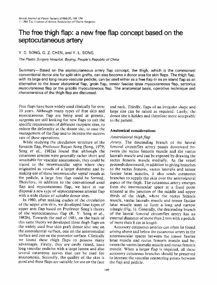
Anterolateral thigh flap Artery. The descending branch of the lateral femoral circumflex artery passes downward be- tween the rectus femoris muscle and the vastus lateralis muscle and can be exposed by drawing the rectus femoris muscle medially. As the vessel proceeds downward, in addition to giving branches to the rectus femoris, vastus lateralis and tensor fasciae latae muscles, it also sends cutaneous branches to supply the skin over the anterolateral aspect of the thigh. The cutaneous artery emerges from the intermuscular space at a fixed point situated at the junction of the middle and upper thirds of the thigh, where the rectus femoris muscle, vastus lateralis muscle and tensor fasciae latae muscle meet to form a long and narrow triangle (Fig. 1). Generally, the descending branch of the lateral femoral circumflex artery has an external diameter of more than 2 mm with a pedicle of more than 8 cm in length.
Accessory cutaneous arteries can often be found arising above and below the cutaneous artery in the intermuscular spaces between the tensor fasciae latae muscle and rectus femoris muscle and be- tween the vastus lateralis muscle and rectus femoris muscle. When a larger flap is required, all these accessory cutaneous branches should be preserved to increase the vascular connecting points between the flap and pedicle.
149
150 BRITISH JOURNAL OF PLASTIC SURGERY
Veins. There are one or two venae comitantes accompanying the descending branch of the lateral femoral circumflex artery. Their external
Cutaneous branch A
A
femoral cutaneous and the lateral femoral cutaneous nerves. These nerves generally have two or three branches running on the surface of the
Descending br. lat. circumflex fem. artery
Cut. br. -
of
B
Fig. 1
Figure l-The artery of the anterolateral thigh flap is the descending branch of the lateral femoral circumflex artery. As this vessel proceeds downwards, in addition to supplying branches to the rectus femoris, vastus lateralis and tensor fasciae latae muscles, it also gives cutaneous branches to supply the skin over the anterolateral aspect of the thigh. The cutaneous artery emerges from the intermuscular space at a fixed point situated at the junction of the middle and upper thirds of the thigh (A). It is a cutaneous branch of the descending branch of the circumflex lateral femoral artery (B).
diameters are larger than that of the artery and are therefore sufficient to provide good drainage to the thigh flap. The lateral femoral vein, which drains into the great saphenous vein, can be used to supplement the venae comitantes should it be necessary.
Nerves. The nerve supply to the anterolateral surface of the thigh is provided by the anterior
deep fascia and are about 1 mm in diameter which is adequate for micro-anastomosis.
Thickness and dimensions. The maximum size of this flap may extend from a horizontal line at the level of the greater trochanter down to a parallel line 3 cm above the patella which includes the anterior surface and the lateral surface of the thigh. In an adult male, the total area of this flap

THE FREE THIGH FLAP: A NEW FREE FLAP CONCEPT BASED ON THE SEPTOCUTANEOUS ARTERY 1.51
can exceed 800 cm?. The flap is thinnest (about 1 cm) over the iliotibial tract and thickest (exceeding 3 cm) near the gluteal region.
Anteromedial thigh flap Artery. There is often an innominate branch of the lateral circumflex femoral artery which arises near its base and descends medially on the medial aspect of the rectus femoris muscle and between the gartorius ‘and vastus medialis muscles. This in-
Cut. br. of innom. desc. br. of lat.
. circumflex fem. artery
Cut. br. artery to Y sartorius muscle
nominate descending artery can be exposed by drawing the sartorius muscle medially; it runs parallel and 2 to 3 cm in front and lateral to the femoral artery. Along its course, branches are distributed to the vastus medialis muscle, sartorius muscle and rectus femoris muscle; the distal end of this artery penetrates the deep fascia to become a cutaneous artery, supplying the skin on the antero- medial surface of the thigh. The point of exit of this cutaneous artery is generally in the mid-section
Innom. desc. br. of
lat. circumflex fem. artery
Fig. 2
Figure Z-The artery of the anteromedial thigh flap is often an innominate branch of the circumflex lateral femoral artery which arise, near its baw and descends medially on the medial aspect of the rectus femori5 muscle. between the sartoriu\ and vastus
medialis muscles. Along its course, branches are given to the vastw medialis muxle, tartoriuh muscle and rectum femori\ musclcr; the distal end of this artery penetrates the deep fascia to become a cutaneouh artery, wpplyinp the skin on the anteromedial surface
of the thigh. The point of exit of this cutaneous artery is generally in the mid+ection of the thieh. Should this innominate
descending artery be too slender or non-existent, a muscular branch supplying the \artoriu\ and gracifis muscles at the junction of the middle and lower thirds of the thigh can be found and used. (A) Note the cutaneous branch of the innominate descending branch
of the lateral femoral circumflex artery; and the cutaneous branch of the muwular artery to the \artoriu\ and pracili, muwlej. (B) Innominate dexending branch of the lateral femoral circumflex artery.
152 BRITISH JOURNAL OF PLASTIC SURGERY
of the thigh, in the narrow triangular inter- muscular space formed by the sartorius, rectus femoris and vastus medialis muscles (Fig. 2). By making use of this cutaneous branch and the innominate medial descending artery, an antero- medial thigh flap pedicle up to 12 cm in length and with an external diameter of more than 2 mm can be prepared.
Cutaneous branches:
1st perf. artery
2nd perf. artery +
3rd perf. artery
Should this innominate descending artery be too slender or non-existent, a muscular branch supplying the sartorius and gracilis muscles which arises directly from the femoral artery at the junction of the middle and lower thirds of the thigh, can be found. This muscular branch often has large cutaneous arteries which supply the skin on the anteromedial aspect of the thigh: therefore
Fig. 3
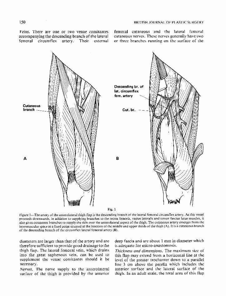
Figure 3-The artery of the posterior thigh flap is the third perforating artery of the profunda femoris artery. (A) The cutaneous branches of the first, second and third perforating arteries. (B) The first, second and third perforating arteries arising from the profunda femoris artery.

THE FREE THIGH FLAP: A NEW FREE FLAP CONCEPT BASED ON THE SEPTOCUTANEOUS ARTERY 153
the muscular branch and its cutaneous arteries may be used for the anteromedial thigh flap pedicle with a length of about 5 cm and an external diameter of upto2mm. Veins. The medial descending branch of the lateral circumflex femoral artery and the muscular vessels to the gracilis and sartorius muscles all have venae comitantes with external diameters slightly larger than that of the arteries and therefore suitable for anastomosis. The superficial tributary of the vastus medialis vein, which drains into the great saphenous vein, can also be used as a sup- plementary drainage vessel. Nerves. .The flap is innervated by the anterior cutaneous branch of the femoral nerve. The cutaneous nerve has a diameter of about 1 mm and can be used for micro-anastomosis. Thickness and dimensions. The maximum size of this flap can include the anterior and medial surface of the thigh. The flap is about 2 to 3 cm thick.
Posterior thigh flap Artery. The profunda femoris artery provides 4 perforating arteries aligned in a cephalad-caudal direction, to supply the muscles on the posterior and lateral aspect of the thigh. Each perforator terminates as a cutaneous branch to the postero- lateral aspect of the thigh (Fig. 3). Of the 4 perforators, the third is generally the largest and can be used as a pedicle for the posterior thigh flap. After the third perforator artery is given off by the profunda femoris artery, it circles the crista femoris and traverses between the vastus lateralis muscle and biceps femoris muscle, going upwards superficially and sending branches to the two muscles en route. After the artery passes through the biceps femoris muscle, it then perforates the deep fascia to supply the skin of the posterior thigh. The length of the third perforating artery which can be used is 5 cm, its external diameter exceeds 2 mm. Veins. The perforating artery has a vena comitans which is of similar calibre and can be used for anastomosis.
Nerves. The posterior femoral cutaneous nerve and lateral femoral cutaneous nerve are both within the limits of the posterior thigh flap and available for use. Thickness and dimensions. The upper margin of the posterior thigh flap may extend to the gluteo- femoral crease, the lower margin to the popliteal
fossa. The skin of the popliteal fossa and lateral aspect of the thigh is about 1 cm thick, whereas the skin at the mid-line of the posterior thigh is 2 to 3 cm thick and may exceed 3 cm near the gluteo- femoral crease.
Operative technique
Planning of the flap A transparent pattern of the recipient site is prepared on a piece of plastic film with the position of the recipient vessels marked out.
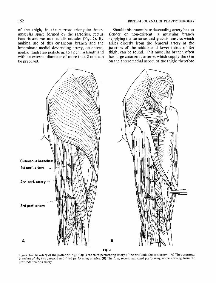
The position of the vascular pedicle is located with a Doppler flowmeter on the donor site and then marked with gentian violet. The vascular pedicles of all three thigh flaps are large and can be easily detected. The cutaneous branches of the perforating arteries can also be located with this instrument: the other cutaneous arteries are all comparatively small and cannot be detected with the Doppler flowmeter thus avoiding confusion.
The prepared pattern is placed on the donor thigh in accordance with the position of the selected vascular pedicle and the flap outlined with a marker. The pedicle can be at the centre of the flap or on one side: the flap may be large or small: it can have long narrow protruding angles with no restrictions in shape, but care must be taken to enlarge the flap according to the thickness of the skin at the donor site (Fig. 4).
We generally use the anterolateral thigh flap as it is the thinnest and easiest to raise. Positioning of thepatient. The patient is placed in the supine position for taking anterolateral and anteromedial thigh flaps. For posterior thigh flaps, the patient is placed in the lateral decubitus position. Locating the vascular pedicle. The skin on the thigh is incised near the vascular pedicle according to the flap design. The deep fascia is exposed and carefully dissected towards the pedicle. The flap is then lifted and the branches sent out by the cutaneous artery to the skin can be seen 2 to 3 cm away from where it perforates the fascia. The deep fascia is incised here and carefully dissected towards the intermuscular space where the cutaneous artery originates. The muscles are retraced to expose the vascular bundle and the length and diameter measured. If found suitable, the flap may then be completely raised from the donor site. Lastly, 1 cm of the intermuscular septal tissue on both sides of the vascular bundle should be removed with the pedicle. The motor nerves
154 BRITISH JOURNAL OF PLASTIC SURGERY
Desc. br. of lat. circumflex fem. artery
It-mom. desc. br. of lat. circumflex fem. artery
Cut. br. artery to sartorius muscle
A ,’
1 /’ ’ \ \ I I , : :
Fig. 4
Figure 4-Flap design. The position of the vascular pedicle is located with a Doppler flowmeter on the donor site and marked with gentian violet. A prepared pattern is placed on the thigh in accordance with the position of the selected vascular pedicle and the flap outline with a marker. The pedicle can be at the centre of the flap or on one side. (A) The descending branch of the lateral femoral circumflex artery of the anterolateral thigh flap. The innominate descending branch of the lateral femoral circumflex artery of the anteromedial thigh flap. (B) The first, second and third perforating arteries and the posterior thigh flap.
which accompany the vascular bundle should be pedicle should be dissected from this layer of meticulously dissected and left behind. muscle fibres. Dissection is not difficult, for there
At times, the vascular pedicle (especially the is often a thin layer of connective tissue separating perforating artery) of the three thigh flaps passes the vessels from the muscle and there are only 4 to 6 through a thin layer of muscle fibres before muscle branches which can be easily ligated. entering the skin. When this is encountered, the The cutaneous nerves of the thigh lie above the
THE FREE THIGH FLAP: A NEW FREE FLAP CONCEPT BASED ON THE SEPTOCIJTANEOUS ARTERY 157
Fig, 6
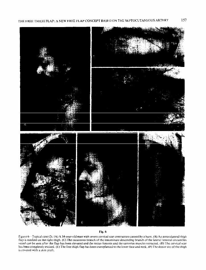
Figure 6-Typical case (2). (A) A 34.year-old man with severe cervical scar contracture caused by a burn. (B) An anterolateral thigh flap is marked on the right thigh. (C)The cutaneous branch of the innominate descending branch of the lateral femoral circumflex vessel can be seen after the flap has been elevated and the rectus femoris and the sartorius muscles retracted. (D) The cervical scar has been completely excised. (E) The free thigh flap has been transplanted to the lower face and neck. (F) The donor site of the thigh is covered with a skin graft.
158 BRITISH JOURNAL OF PLASTIC SURGERY
The cutaneous arteries and veins from the intermuscular vessels are concomitant from their origin to the two plexus forming a complete circulatory system. The superficial venous system formed by the great and small saphenous veins have a different direction from that of the cutaneous vessels. During operation, if only one vein can be anastomosed, the vena comitans should be used and not the superficial vein (though the latter may have a larger diameter), because the use of a superficial vein may result in partial or total venous congestion.
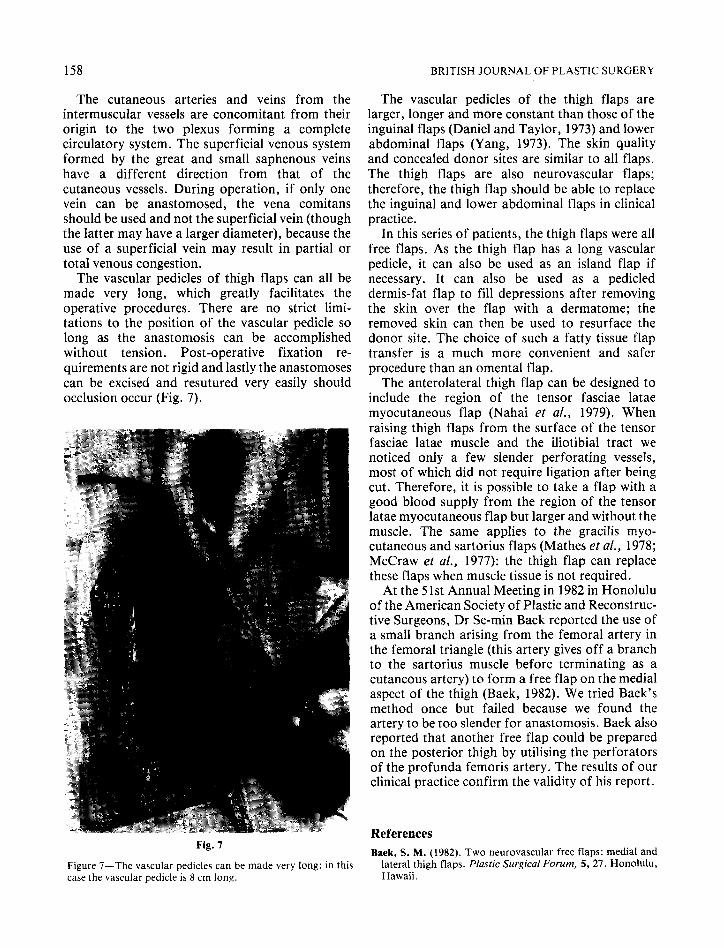
The vascular pedicles of thigh flaps can all be made very long, which greatly facilitates the operative procedures. There are no strict limi- tations to the position of the vascular pedicle so long as the anastomosis can be accomplished without tension. Post-operative fixation re- quirements are not rigid and lastly the anastomoses can be excised and resutured very easily should occlusion occur (Fig. 7).
Fig. 7
Figure ‘I-The vascular pedicles can be made very long: in this case the vascular pedicle is 8 cm long.
The vascular pedicles of the thigh flaps are larger, longer and more constant than those of the inguinal flaps (Daniel and Taylor, 1973) and lower abdominal flaps (Yang, 1973). The skin quality and concealed donor sites are similar to all flaps. The thigh flaps are also neurovascular flaps; therefore, the thigh flap should be able to replace the inguinal and lower abdominal flaps in clinical practice.
In this series of patients, the thigh flaps were all free flaps. As the thigh flap has a long vascular pedicle, it can also be used as an island flap if necessary. It can also be used as a pedicled dermis-fat flap to fill depressions after removing the skin over the flap with a dermatome; the removed skin can then be used to resurface the donor site. The choice of such a fatty tissue flap transfer is a much more convenient and safer procedure than an omental flap.
The anterolateral thigh flap can be designed to include the region of the tensor fasciae latae myocutaneous flap (Nahai et al., 1979). When raising thigh flaps from the surface of the tensor fasciae latae muscle and the iliotibial tract we noticed only a few slender perforating vessels, most of which did not require ligation after being cut. Therefore, it is possible to take a flap with a good blood supply from the region of the tensor latae myocutaneous flap but larger and without the muscle. The same applies to the gracilis myo- cutaneous and sartorius flaps (Mathes et al., 1978; McGraw et al., 1977): the thigh flap can replace these flaps when muscle tissue is not required.
At the 5 1 st Annual Meeting in 1982 in Honolulu of the American Society of Plastic and Reconstruc- tive Surgeons, Dr Se-min Baek reported the use of a small branch arising from the femoral artery in the femoral triangle (this artery gives off a branch to the sartorius muscle before terminating as a cutaneous artery) to form a free flap on the medial aspect of the thigh (Baek, 1982). We tried Baek’s method once but failed because we found the artery to be too slender for anastomosis. Baek also reported that another free flap could be prepared on the posterior thigh by utilising the perforators of the profunda femoris artery. The results of our clinical practice confirm the validity of his report.
References
Baek, S. M. (1982). Two neurovascular free flaps: medial and lateral thigh flaps. Plastic Surgical Forum, 5, 27. Honolulu, Hawaii.
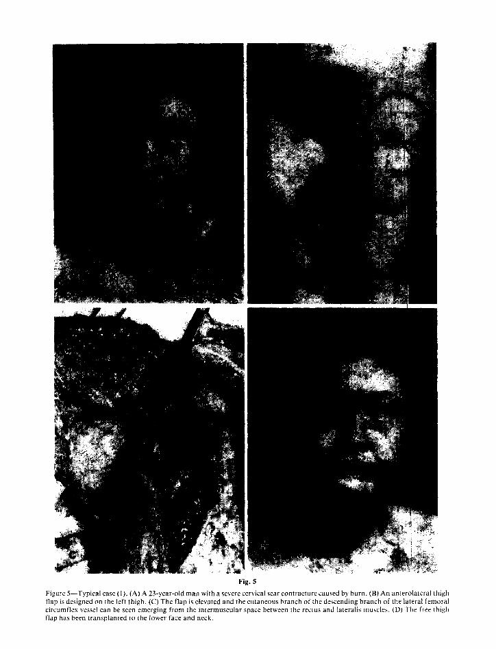
Fig. 5
Figure 5--Typical case (1). (A) A 23-year-old man with a severe cervical scar contracture caused by burn. (B) An anterolateral thigh flap is designed on the left thigh. (C)The flap is elevated and the cutaneous branch of the descending branch of the lateral femoral circumflex vessel can be seen emerging from the intermuscular space between the rectus and lateralis muscles. (D) The free thigh flap has been transplanted to the lower face and neck.

THE FREE THIGH FLAP: A NEW FREE FLAP CONCEPT BASED ON THE SEPTOCUTANEOUS ARTERY 155
deep fascia and are easily located. If necessary, the cutaneous nerve can be included in the flap and anastomosed to the recipient nerve.
If the flap is large and there are veins available for anastomosis at the recipient site, the proximal portion of the lateral femoral vein or the medial femoral vein may be dissected to form a pedicle together with the flap.
Preparation of the recipient site A second surgical team prepares the recipient site by removing the diseased tissues, dissecting out the recipient vessels and sensory nerves for anasto- mosis with the thigh flap while the thigh flap is being raised by the first team.
After making certain that the position and length of the vascular pedicles are correct, the vessels may be cut and the flap transferred to the recipient site. 10-O atraumatic sutures are used to anastomose blood vessels under the operating microscope. A vena comitans should be anasto- mosed before the artery. Twenty minutes should be allowed to elapse after the flap circulation has been re-established: if the anastomosis is patent and the flap circulation good, the flap may then be sutured to the recipient site. When possible, another vena comitans or superficial vein should be anasto- mosed.
If the flap is small, the donor site can be closed directly. In most cases, a split-skin graft is required and this can be taken from the same extremity.
Results
We have performed 15 free thigh flap transfers on 15 patients with late head and neck burn contrac- tures: the anterolateral thigh flap in 9 cases, the anteromedial thigh flap in 4 cases and the posterior thigh flap in 2 cases. All the flaps survived and the contractures were corrected, but all 15 flaps were too thick and secondary thinning procedures were required (Figs. 5 and 6).
Discussion
We differ from the general viewpoint expressed by Daniel and Williams (1973) and McGraw and Dibbell(1977) in that we believe that the cutaneous branches of the intermuscular septal artery, not the myocutaneous perforating arteries, provide the major blood supply to the skin of the extremities. Large free flaps can survive on pedicles utilising the cutaneous branches of the intermuscular septal arteries. The excellent blood supply provided by the cutaneous branches of the intermuscular septal
arteries is the anatomical and haemodynamical basis of the thigh flap, which we have named the septocutaneous artery flap.
The cutaneous branch of the descending branch of the lateral femoral circumflex artery, which is the nutrient vessel for the anterolateral thigh flap and the cutaneous branch of the innominate medial descending artery arising from the lateral artery, which is the nutrient vessel for the antero- medial thigh flap, often have a reciprocal growth relationship (i.e. should the former artery be large, the latter is often slender or absent and vice versa). Fortunately this condition can be detected pre- operatively with a Doppler probe, therefore the decision to use the anterolateral or the antero- medial thigh flap and the position of the vascular pedicle can be accurately determined before the operation.
The primary function of the intermuscular septal artery, which constitutes the vascular pedicle for thigh flaps, is to supply blood to the neighbour- ing musculature; therefore, the blood flow of the muscular branches exceeds that of the cutaneous branches. Once the flap is completely raised and the muscular branches all ligated, the blood flow to the flap is greatly increased; the skin becomes flushed, pulsation can be observed not only in the cutaneous artery but also in its 0.3 mm branches, the vessels being in a state of “hyperperfusion” due to the ligation of all the muscular branches. The circulation is excellent, the large calibre of the vessels allows easy anastomosis and so makes the thigh flap easy to master with a correspondingly high rate of success.
According to our previous anatomical studies (R. Y. Song et al., 1982b) the skin of the extremities is supplied by two vascular plexuses, the subdermal plexus and the plexus above the deep fascia. There are some vertical vessels passing through the subcutaneous fatty tissue to connect the two plexuses. After the cutaneous branch of the intermuscular septal artery enters the deep fascia, it does not present an axial distribution but divides into several branches which join the vascular plexus above the deep fascia. Through the sub- dermal plexus and the vascular plexus above the deep fascia, the cutaneous artery can provide blood to a large area of skin. As the thigh flap is supplied through two vascular plexuses and not through an axial system, its shape is not limited and can be designed as required: its vascular pedicle need not be centrally placed and can be situated at one side of the flap.

THE FREE THIGH FLAP: A NEW FREE FLAP CONCEPT BASED ON THE SEPTOCUTANEOUS ARTERY 159
Baek, S. M. (1983). Two new cutaneous free flaps: the medial and lateral thigh flap. Plastic and Reconstructive Surgery, 71, 354.
Daniel, R. K. and Williams, H. B. (1973). The free transfer of skin flaps by microvascular anastomosis. An experimental study and re-appraisal. Plastic and Reconstructive Surgery, 52, 16.
Daniel, R. K. and Taylor, G. I. (1973). Distant transfer of an island flap by microvascular anastomosis. Plastic and Reconstructive Surgery, 52, I 11.
Mathes, S. J., Nahai, F. and Vasconez, L. 0. (1978). Myocutaneous free flap transfer, anatomical and experi- mental considerations. Plastic and Reconstructive Surgery, 62, 162.
McGraw, J. B. and Dihbell, D. G. (1977). Experimental definition of independent myocutaneous vascular territories. Plastic and Reconstructive Surgery, 60, 212.
McGraw. J. B., Dibbell, D. G. and Carraway, J. H. (1977). Clinical definition of independent myocutaneous vascular territories. Plastic and Reconstructive Surgery, 60, 341.
Nahai, F., Hill, H. L. and Hester, T. R. (1979). Experience with the tensor fascia lata flap. Plastic and Reconstructive Surgery, 63. 788.
Song, R. Y. (1979). Personal communication. Song, R. Y., Gao, Y. Z., Song, Y. G., Yu, Y. S. and Song, Y. L.
(1982a). The forearm flap. Clinics in Plastic Surgery, 9, 21. Song, R. Y., Song, Y. G., Yu, Y. S. and Song, Y. L. (1982b).
The upper arm free flap. Clinics in Plastic Surgery, 9, 27. Yang, D. Y. (1973). Cited in editorial addendum. Plastic and
Reconstructive Surgery, 52, 116.
The Authors
Ye-guang Song, MD, Plastic Surgeon, Plastic Surgery Hospi- tal, Beijing.
Guo-zhang Chen, MD, Plastic Surgeon, Plastic Surgery Hospi- tal, Beijing.
Ye-liang Song, MD, Plastic Surgeon, Friendship Hospital, Beijing.
Requests for reprints to: Dr Ye-guang Song, MD, Plastic Surgery Hospital, Ba-Da-Chu, Beijing, People’s Republic of China.
Postscript
In early March 1983 when this paper was received in the editorial office and accepted for publication we had not yet received our copy of the March issue of Plastic and Reconstructive Surgery which contained a more recent paper by Baek (Two new cutaneous free flaps: the medial and lateral thigh flaps. Plastic and Reconstructive Surgery, 1983, 71, 354, with a short discussion by Dr Luis 0. Vasconez) in which he described two clinical cases performed after extensive anatomical dissections on cadavers. I wrote to Dr Ye-guang Song at the end of April 1983 drawing his attention to this article and he kindly sent a reply from which I have taken the following paragraphs:
“In early March 1983 a group of 38 plastic surgeons from Australia visited the Plastic Surgery Hospital in Ba-Da-Chu, Beijing and during their ward rounds noticed, and were very impressed by, three patients on whom free thigh flap transfers had been carried out . . .”
It was at their instigation that Professor Ruyao Song, the Director of the Institute, promptly sent the text and the illustrations of this paper to this Journal.


![HFM Free Flap Versus Fat Grafting[1]](https://img.dokumen.tips/doc/110x75/54f83fc94a7959fe478b459b/hfm-free-flap-versus-fat-grafting1.jpg)
