Embed Size (px)
Citation preview

CASE REPORT Open Access
The first report of molecular characterizedBRD4-NUT carcinoma in Brazil: a casereportLeandro J. C. Oliveira1*, Aline B. L. Gongora1, Marcela T. Latancia2, Felipe G. Barbosa3, João Vitor A. M. Gregorio1,4,Leonardo A. Testagrossa5, Mariane T. Amano2 and Olavo Feher1,4
Abstract
Background: NUT midline carcinoma is a rare and aggressive subset of squamous cell carcinoma, which is characterizedby the translocation of nuclear protein in testis gene that is mostly fused with bromodomain and extraterminal familyproteins. We describe here the first Brazilian case of NUT midline carcinoma with BRD4-NUT fusion detected in a next-generation sequencing panel and we present the clinical evolution of this patient.
Case presentation: A 42-year-old Caucasian man was diagnosed with poorly differentiated squamous cell carcinoma ofthe left maxillary sinus, with negative in situ hybridization for Epstein–Barr encoding region and human papillomavirusgenotyping. He received induction therapy, chemoradiotherapy with weekly systemic chemotherapy, and, concurrently,weekly intra-arterial chemotherapy. New imaging evaluation, 1 month after the end of the last treatment, revealed agood partial response in the primary lesion. However, positron emission tomography-computed tomography showedmultiple suspicious lesions in his bones and lungs, which were histologically confirmed. He died exactly 2 months aftermetastatic disease was diagnosed.
Conclusions: NUT midline carcinoma is usually very aggressive. Currently, there is no standard of care for treatment ofNUT midline carcinoma. The definitive diagnosis must be by demonstration of NUTM1 rearrangement. Immunohistochemicalstaining of greater than 50% of tumor nuclei on formalin-fixed paraffin-embedded tissue using the monoclonalrabbit antibody to NUT (clone C52B1), has a specificity of 100%, and sensitivity of 87% for the diagnosis of NUTmidline carcinoma. Our case is the first Brazilian case of NUT midline carcinoma with BRD4-NUT fusion.
Keywords: NUT midline carcinoma, Poorly differentiated squamous cell carcinoma, Molecular pathology,Targeted therapy
BackgroundNUT midline carcinoma (NMC) is a rare and aggressivesubset of squamous cell carcinoma, which is character-ized by the translocation of nuclear protein in testis(NUT) gene that is mostly fused with bromodomain andextraterminal (BET) family proteins, such as BRD3 andBRD4 [1], generating BRD3-NUT and BRD4-NUT,respectively. A non-BET member, nuclear receptorbinding SET domain 3 (NSD3), was also seen to be fusedwith NUT (NSD3-NUT) [2, 3], but this seems to be less
frequent than BRD3-NUT and BRD4-NUT. The major-ity of the cases occur in the head, neck, and mediasti-num, although there are reports involving the bladder,pancreas, adrenal gland, kidney, and salivary gland [4].NMC differs from other carcinomas, which usually havecomplex karyotypes, because it is characterized by a fewor a single translocation, most often the BRD4-NUT:t(15;19)(q14;p13.1) [4].Until 2011, NMC was considered an extremely rare
carcinoma, with only 28 reported cases worldwide [5].Since 2012, the number of diagnosed cases increasedenormously; this was identified in a review in theInternational NUT Midline Carcinoma Registry, whichdetected 48 cases of NMC from 2011 to 2014, of which
© The Author(s). 2019 Open Access This article is distributed under the terms of the Creative Commons Attribution 4.0International License (http://creativecommons.org/licenses/by/4.0/), which permits unrestricted use, distribution, andreproduction in any medium, provided you give appropriate credit to the original author(s) and the source, provide a link tothe Creative Commons license, and indicate if changes were made. The Creative Commons Public Domain Dedication waiver(http://creativecommons.org/publicdomain/zero/1.0/) applies to the data made available in this article, unless otherwise stated.
* Correspondence: [email protected] de Oncologia, Hospital Sírio Libanês, Rua Dona Adma Jafet, 91. 2ndfloor. Building A, São Paulo 01308-050, BrazilFull list of author information is available at the end of the article
Oliveira et al. Journal of Medical Case Reports (2019) 13:279 https://doi.org/10.1186/s13256-019-2213-6

86% were BRD4-NUT positive [6]. This review suggeststhat NMC is probably underdiagnosed.We describe here the first Brazilian case of NMC with
BRD4-NUT fusion detected in next-generation sequen-cing (NGS) panel and we present the clinical evolutionof this patient. Until recently, to the best of ourknowledge, no cases of NMC had been reported in LatinAmerica [4, 7]. Salles et al. described four patients withtypical NMC histopathology and NUT positivity in thenucleus [8]. However, the fusion was not molecularlydescribed. Since there is a clear failure in detectingNMC in patients, and further studies are required toprovide better chances for these patients, we understandthat there is a need to report and better characterizecases of NMC.
Case presentationA 42-year-old Caucasian man presented in December2016 with productive cough, facial pain, and rhinorrhea.He is an engineer, who does not smoke tobacco, and hehad no significant premorbid conditions. There was nohistory of prescription drug use, and no significant familyhistory. Neurological, cardiovascular, respiratory, and ab-dominal examinations were normal, except for tendernessof the face elicited by palpation. He was first diagnosed ashaving acute rhinosinusitis and treated with antibiotics(details were not available), without improvement. Due tosymptoms persistence, magnetic resonance imaging (MRI)of his face was ordered. This study revealed an expansiveirregular heterogeneous lesion (4.5 × 3.5 × 4.0 cm), with itscentral portion located on the interface between the leftmaxillary sinus and the pterygopalatine fossa. This lesion
invaded medially the nasal cavity and posterosuperiorlythe left orbit apex with an intracranial extension throughthe inferior orbital fissure (Fig. 1a–d). There was nolymphadenopathy and no perineural invasion. A biopsydemonstrated poorly differentiated squamous cell carci-noma with negative in situ hybridization for Epstein–Barrencoding region (EBER). Human papillomavirus (HPV)genotyping test was negative as well. Positron emissiontomography-computed tomography (PET-CT) wasnegative for nodal and systemic metastases. His totalleukocyte and platelet counts, as well as hemoglobinlevels, were all within normal limits. His biochemicalparameters, including serum electrolytes, renal functiontest, and liver function test, were also normal.He received, from January 2017 to April 2017, induc-
tion therapy with docetaxel 75 mg/m2 at day 1, cisplatin100 mg/m2 at day 1, and fluorouracil (5-FU) 1000mg/m2
per day at day 1 to day 4 (DCF) for six cycles every 3weeks, with clinical benefit and stable disease by MRIand PET-CT (Figs. 1 and 2). Chemoradiotherapy (radio-therapy 35 fractions – 70 Gy) with weekly systemicchemotherapy based on carboplatin 1.5 area under thecurve (AUC), paclitaxel 45 mg/m2, and cetuximab 400mg/m2 was administered for 7 weeks from May 2017 toJuly 2017. Concurrently, weekly intra-arterial chemo-therapy with cisplatin 150 mg/m2 was performed for 5weeks with grade 2 myelotoxicity and nausea.New imaging evaluation, 1 month after the end of the
last treatment, revealed a good partial response in theprimary lesion (Figs. 1e–h and 2). However, PET-CTshowed multiple suspicious lesions in his bones and lungs(Fig. 3), which were histologically confirmed (Fig. 4a, b).
A B C D
E F G H
Fig. 1 Local tumor response seen in head and neck magnetic resonance images. Magnetic resonance images from baseline before second-linechemotherapy (a–d) showing locally advanced left maxillary sinus mass (arrows), infiltrating adjacent sinuses and skull base foramina. Follow-up magneticresonance images (e–h) showing significant tumor shrinkage (arrows) with areas of necrosis, representing local morphological partial response
Oliveira et al. Journal of Medical Case Reports (2019) 13:279 Page 2 of 7

A
B
Fig. 2 Local complete metabolic response seen in 18F-fluorodeoxyglucose positron emission tomography-computed tomography.Fluorodeoxyglucose-positron emission tomography-computed tomography head and neck images from baseline before second-linechemotherapy (a) showing locally advanced left maxillary sinus enhanced mass (arrows) with fluorodeoxyglucose avidity. Positron emissiontomography-computed tomography of 1 month after end of treatment (b) confirming morphological shrinkage of the left maxillary sinus withextensive necrosis (arrows) without any fluorodeoxyglucose uptake, representing local complete metabolic response
A B
Fig. 3 Systemic disease progression evidenced in 18F-fluorodeoxyglucose positron emission tomography-computed tomography maximumintensity projection images. Maximum intensity projection fluorodeoxyglucose-positron emission tomography-computed tomography whole-body images from baseline before second-line chemotherapy (a) and 1 month after end of treatment (b). Image a shows no evidence ofsystemic disease. Image b shows disease progression with new bone and pulmonary fluorodeoxyglucose-avid metastasis (arrows)
Oliveira et al. Journal of Medical Case Reports (2019) 13:279 Page 3 of 7

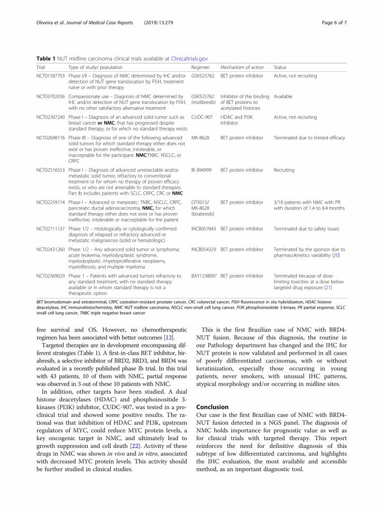
Programmed death-ligand 1 (PD-L1) expression by immu-nohistochemistry (IHC) was negative (SP263 Ventana). Inorder to look for other therapeutic possibilities, a formalin-fixed paraffin-embedded (FFPE) tumor biopsy was sent tothe Foundation Medicine with patient consent. Deoxyri-bonucleic acid (DNA) extracted from FFPE and hybridcapture-based NGS was applied to perform the Founda-tionOne™ test, which comprises a panel of 315 genesknown to carry somatic mutations in human solidtumors, as well as introns of 28 genes involved in rear-rangements. The test showed a rearrangement of BRD4-NUT characterizing NMC. No other mutations werefound; microsatellite stability and low mutationalburden were reported. The IHC for NUT protein wasperformed and stained positively in neoplastic nuclei(Fig. 5).Due to the lack of available clinical trials in the country
for this highly aggressive tumor, our patient received two
more cycles of chemotherapy, but, unfortunately, withoutany response. He died exactly 2 months after metastaticdisease was diagnosed.
DiscussionWe describe the first report of a molecular characterizedBRD4-NUT carcinoma in Brazil and Latin America. Ourpatient showed a significant partial response in theprimary lesion but progressed with multiple metastaticlesions in bone and lungs.Reports of NMC have been increasing since it was ori-
ginally described as a genetically defined entity in 2004[9]. It can affect individuals of all ages, despite the initialthought that it only affected children and young adults.The median age of diagnosis is 24 years. The mostcommon primary sites are thorax, in 50% of the patients,and head and neck, in 39% of patients. There are otherprimary sites (non-midline, non-thoracic, or head and
Fig. 4 a Bone metastasis of poorly differentiated carcinoma showing neoplastic epithelioid clusters with small cells (hematoxylin andeosin, × 200). b Higher magnification of the neoplastic clusters (hematoxylin and eosin, × 400)
Oliveira et al. Journal of Medical Case Reports (2019) 13:279 Page 4 of 7

neck) described in reports of NMCs, such as soft tissueor bone, which can account for 11% of all cases [10]. InBrazil, lung and head and neck cancers are among the tenmost frequent cancers, especially in men [11]. However,until now, there were no cases of NUT-BRD4 NMCconfirmed in Brazil, probably due to underdiagnosis.NMC is usually very aggressive. Not rarely, it is meta-
static at diagnosis and has poor survival rates. An analysisof a clinical database with 63 patients with NMC and out-come data of 54 patients showed a median overall survival(OS) of 6.7 months. The 2-year OS was 19% [12]. A sys-tematic review and individual patient data analysis of 119cases showed median OS of only 5 months. Radiotherapyand chemotherapy had a significant impact on OS, whilesurgery, BET inhibitors, and histone deacetylase inhibitors(HDACi) did not affect survival significantly [13].As previously discussed, the karyotypes of NMC are not
complex; they frequently have a clonal translocation of theNUTM1 gene, classically t(15;19)(q14;p13.1), which placesNUT in the frame with BRD4. This feature differs fromother carcinomas because the latter develop over years dueto an accumulation of mutations and genetic aberrations[14]. The BRD4-NUT fusion oncoproteins drive a series ofdisruptions of gene expression through chromatin remodel-ing [15], overexpression of MYC [16], and sequestration ofthe activated form of p53 [17]. Another possibility is the fu-sion of NUT with BRD3 or NSD3, which are functionallyrelated to BRD4. Therefore, recruitment of NUT to thechromatin through the BET family proteins is probablynecessary for the oncogenesis of NMC [17].On histological examination, NMC can have a
broad morphologic range, in many cases re-sembling squamous cell carcinoma, in others show-ing small undifferentiated cells with or withoutareas of abrupt keratinization. Therefore, diagnosis
should not be assumed just based on histologicalaspect [18].The definitive diagnosis must be by demonstration of
the NUTM1 rearrangement. There is no consensus onwhich pathology material of the patient should be submit-ted to molecular tests. Some authors suggested perform-ing IHC or molecular tests in all poorly differentiatednon-cutaneous carcinomas, with or without squamous dif-ferentiation, with a monomorphic appearance. Glandulartumors do not need to be tested. Viral etiology, such asHPV or Epstein–Barr virus (EBV), is not associated withNMC and can be used to rule this diagnosis out. However,NMC can be strongly positive for p16, despite its negati-vity for HPV; therefore, it should not be used forexcluding NMC [14].IHC staining of greater than 50% of tumor nuclei on
FFPE tissue using the monoclonal rabbit antibody toNUT (clone C52B1) has a specificity of 100%, and sensi-tivity of 87% for the diagnosis of NMC. Some germ cellstumors can also stain, but usually focally (< 10%) [19].Confirmatory molecular tests are not mandatory. How-ever, they can be used if the C52B1 antibody is notavailable or to identify the fusion partner to NUT, whichmay have clinical and prognostic relevance. For example,NSD3 or BRD3-NUT-positive NMC was associated witha better OS than those with BRD4-NUT, especially innon-thoracic NMC [14]. The assays that can be used forthis purpose are fluorescence in situ hybridization(FISH), reverse transcriptase-polymerase chain reaction(RT-PCR), cytogenetics, and NGS-based approaches.Molecular diagnostic methods have limitations, such asthe cost and access to quality services [14].Currently, there is no standard of care for treatment of
NMC. Incomplete or no surgical resection and no initialradiotherapy have been associated with shorter progression-
Fig. 5 NUT immunohistochemistry staining positively in neoplastic nuclei
Oliveira et al. Journal of Medical Case Reports (2019) 13:279 Page 5 of 7
free survival and OS. However, no chemotherapeuticregimen has been associated with better outcomes [12].Targeted therapies are in development encompassing dif-
ferent strategies (Table 1). A first-in-class BET inhibitor, bir-abresib, a selective inhibitor of BRD2, BRD3, and BRD4 wasevaluated in a recently published phase Ib trial. In this trialwith 43 patients, 10 of them with NMC, partial responsewas observed in 3 out of these 10 patients with NMC.In addition, other targets have been studied. A dual
histone deacetylases (HDAC) and phosphoinositide 3-kinases (PI3K) inhibitor, CUDC-907, was tested in a pre-clinical trial and showed some positive results. The ra-tional was that inhibition of HDAC and PI3K, upstreamregulators of MYC, could reduce MYC protein levels, akey oncogenic target in NMC, and ultimately lead togrowth suppression and cell death [22]. Activity of thesedrugs in NMC was shown in vivo and in vitro, associatedwith decreased MYC protein levels. This activity shouldbe further studied in clinical studies.
This is the first Brazilian case of NMC with BRD4-NUT fusion. Because of this diagnosis, the routine inour Pathology department has changed and the IHC forNUT protein is now validated and performed in all casesof poorly differentiated carcinomas, with or withoutkeratinization, especially those occurring in youngpatients, never smokers, with unusual IHC patterns,atypical morphology and/or occurring in midline sites.
ConclusionOur case is the first Brazilian case of NMC with BRD4-NUT fusion detected in a NGS panel. The diagnosis ofNMC holds importance for prognostic value as well asfor clinical trials with targeted therapy. This reportreinforces the need for definitive diagnosis of thissubtype of low differentiated carcinoma, and highlightsthe IHC evaluation, the most available and accessiblemethod, as an important diagnostic tool.
Table 1 NUT midline carcinoma clinical trials available at Clinicaltrials.gov
Trial Type of study/ population Regimen Mechanism of action Status
NCT01587703 Phase I/II – Diagnosis of NMC determined by IHC and/ordetection of NUT gene translocation by FISH, treatmentnaïve or with prior therapy
GSK525762 BET protein inhibitor Active, not recruiting
NCT03702036 Compassionate use – Diagnosis of NMC determined byIHC and/or detection of NUT gene translocation by FISH,with no other satisfactory alternative treatment
GSK525762(molibresib)
Inhibitor of the bindingof BET proteins toacetylated histones
Available
NCT02307240 Phase I – Diagnosis of an advanced solid tumor such asbreast cancer or NMC, that has progressed despitestandard therapy, or for which no standard therapy exists
CUDC-907 HDAC and PI3KInhibitor
Active, not recruiting
NCT02698176 Phase IB – Diagnosis of one of the following advancedsolid tumors for which standard therapy either does notexist or has proven ineffective, intolerable, orinacceptable for the participant: NMC;TNBC; NSCLC; orCRPC
MK-8628 BET protein inhibitor Terminated due to limited efficacy
NCT02516553 Phase I – Diagnosis of advanced unresectable and/ormetastatic solid tumor, refractory to conventionaltreatment or for whom no therapy of proven efficacyexists, or who are not amenable to standard therapies.Part Ib includes patients with SCLC, CRPC, CRC or NMC
BI 894999 BET protein inhibitor Recruiting
NCT02259114 Phase I – Advanced or metastatic: TNBC, NSCLC, CRPC,pancreatic ductal adenocarcinoma, NMC, for whichstandard therapy either does not exist or has provenineffective, intolerable or inacceptable for the patient
OTX015/MK-8628(birabresib)
BET protein inhibitor 3/10 patients with NMC with PRwith duration of 1.4 to 8.4 months
NCT02711137 Phase 1/2 – Histologically or cytologically confirmeddiagnosis of relapsed or refractory advanced ormetastatic malignancies (solid or hematologic)
INCB057643 BET protein inhibitor Terminated due to safety issues
NCT02431260 Phase 1/2 – Any advanced solid tumor or lymphoma;acute leukemia, myelodysplastic syndrome,myelodysplastic /myeloproliferative neoplasms,myelofibrosis, and multiple myeloma
INCB054329 BET protein inhibitor Terminated by the sponsor due topharmacokinetics variability [20]
NCT02369029 Phase 1 – Patients with advanced tumors refractory toany standard treatment, with no standard therapyavailable or in whom standard therapy is not atherapeutic option
BAY1238097 BET protein inhibitor Terminated because of dose-limiting toxicities at a dose belowtargeted drug exposure [21]
BET bromodomain and extraterminal, CRPC castration-resistant prostate cancer, CRC colorectal cancer, FISH fluorescence in situ hybridization, HDAC histonedeacetylase, IHC immunohistochemistry, NMC NUT midline carcinoma, NSCLC non-small cell lung cancer, PI3K phosphoinositide 3-kinase, PR partial response, SCLCsmall cell lung cancer, TNBC triple negative breast cancer
Oliveira et al. Journal of Medical Case Reports (2019) 13:279 Page 6 of 7

AcknowledgementsNo funding received.
Authors’ contributionsLJCO, ABLG, MTL, MTA, and LAT described the case and wrote the initialarticle. OF was the patient’s personal doctor and was a reviewer of thearticle, together with JVAMG. FGB analyzed, selected, and wrote the subtitlesfor the images. All authors read and approved the final manuscript.
FundingNot applicable.
Availability of data and materialsNot applicable.
Ethics approval and consent to participateNot applicable.
Consent for publicationWritten informed consent was obtained from the patient’s next of kin forpublication of this case report and any accompanying images. A copy of thewritten consent is available for review by the Editor-in-Chief of this journal.
Competing interestsThe authors declare that they have no competing interests.
Author details1Centro de Oncologia, Hospital Sírio Libanês, Rua Dona Adma Jafet, 91. 2ndfloor. Building A, São Paulo 01308-050, Brazil. 2Instituto de Ensino e Pesquisa,Hospital Sírio Libanês, São Paulo, Brazil. 3Serviço de Medicina Nuclear,Hospital Sírio Libanês, São Paulo, Brazil. 4Serviço de Oncologia, Instituto doCâncer do Estado de São Paulo, Universidade de São Paulo, São Paulo, Brazil.5Serviço de Anatomia Patológica, Hospital Sírio Libanês, São Paulo, Brazil.
Received: 26 March 2019 Accepted: 30 July 2019
References1. French C. NUT midline carcinoma. Nat Rev Cancer. 2014;14(3):149–50.2. French CA, et al. NSD3-NUT fusion oncoprotein in NUT midline carcinoma:
implications for a novel oncogenic mechanism. Cancer Discov. 2014;4(8):928–41.3. Suzuki S, et al. NSD3-NUT-expressing midline carcinoma of the lung: first
characterization of primary cancer tissue. Pathol Res Pract. 2015;211(5):404–8.4. French CA. Pathogenesis of NUT midline carcinoma. Annu Rev Pathol. 2012;
7:247–65.5. Stelow EB. A review of NUT midline carcinoma. Head Neck Pathol. 2011;5(1):31–5.6. Chau NG, et al. Intensive treatment and survival outcomes in NUT midline
carcinoma of the head and neck. Cancer. 2016;122(23):3632–40.7. French CA. The importance of diagnosing NUT midline carcinoma. Head
Neck Pathol. 2013;7(1):11–6.8. Salles PG, et al. Expression of P16 in NUT carcinomas with no association
with human papillomavirus (HPV). Appl Immunohistochem Mol Morphol.2014;22(4):262–5.
9. French CA, Kutok JL, Faquin WC, et al. Midline carcinoma of children andyoung adults with NUT rearrangement. J Clin Oncol. 2004;22:4135–9.
10. Chau NG, Ma C, Danga K, et al. A novel prognostic risk classification modelfor NUT midline carcinoma: A largest cohort analysis from the NMC registry.J Clin Oncol. 2018;36(suppl):6085.
11. Ministério da Saude, Instituto Nacional de Câncer José Alencar Gomes daSilva. Estimate/2018 – Cancer Incidence in Brazil (Rio de Janeiro), 2017. ISBN978–85–7318-361-0.
12. Bauer DE, et al. Clinicopathologic features and long-term outcomes of NUTmidline carcinoma. Clin Cancer Res. 2012;18(20):5773–9.
13. Giridhar P, Mallick S, Kashyap L, Rath GK. Patterns of care and impact ofprognostic factors in the outcome of NUT midline carcinoma: a systematicreview and individual patient data analysis of 119 cases. Eur ArchOtorhinolaryngol. 2018;275(3):815–21.
14. French CA. NUT Carcinoma: Clinicopathologic features, pathogenesis, andtreatment. Pathol Int. 2018;68(11):583–95.
15. Alekseyenko AA, Walsh EM, Wang X, et al. The oncogenic BRD4-NUTchromatin regulator drives aberrant transcription within large topologicaldomains. Genes Dev. 2015;29:1507–23.
16. Grayson AR, Walsh EM, Cameron MJ, et al. MYC, a downstream target ofBRD-NUT, is necessary and sufficient for the blockade of differentiation inNUT midline carcinoma. Oncogene. 2014;33:1736–42.
17. Reynoird N, Schwartz BE, Delvecchio M, et al. Oncogenesis by sequestrationof CBP/p300 in transcriptionally inactive hyperacetylated chromatindomains. EMBOJ. 2010;29:2943–52.
18. Evans AG, French CA, Cameron MJ, et al. Pathologic characteristics of NUTmidline carcinoma arising in the mediastinum. Am J Surg Pathol. 2012;36:1222–7.
19. Haack H, Johnson LA, Fry CJ, et al. Diagnosis of NUT midline carcinoma using aNUT-specific monoclonal antibody. Am J Surg Pathol. 2009;33:984–91.
20. Lewin J, Soria JC, Stathis A, et al. Phase Ib trial with birabresib, a smallmolecule inhibitor of bromodomain and extraterminal proteins, in patientswith selected advanced solid tumors. J Clin Oncol. 2018;36(30):3007–14.
21. Postel-Vinay S, Herbschleb K, Massard C, et al. First-in-human phase I doseescalation study of the Bromodomain and Extra-Terminal motif (BET)inhibitor BAY 1238097 in subjects with advanced malignancies. Eur JCancer. 2016;69:S7–8.
22. Sun K, Atoyan R, Borek MA, et al. Dual HDAC and PI3K inhibitor CUDC-907downregulates MYC and suppresses growth of MYC-dependent cancers.Mol Cancer Ther. 2017;16(2):285–99.
Publisher’s NoteSpringer Nature remains neutral with regard to jurisdictional claims inpublished maps and institutional affiliations.
Oliveira et al. Journal of Medical Case Reports (2019) 13:279 Page 7 of 7