Embed Size (px)
Citation preview

The Female Urogenital System of the lYIarsupialia with special reference to the Vaginal Complex
By
JOSEPH PEARSON
(Read 13th November, 1944)
PLATES X-XII
It is proposed to discuss the general disposition of the female urogenital organs in the Marsupialia and in particular to examine the variations to be found in the vaginal complex. Special reference will be made to the genera PotoToiis and Bettongia, partly because the urogenital system of the former ha~ not been described or figured hitherto and also because the two genera have been confused by previous writers on marsupial comparative anatomy and it is important that the matter should be clarified once and for all (see Pearson, 1944).
The foundation of our knowledge of the urogenital system of the Marsupialia was well and truly laid by Richard Owen, and after the lapse of a century students of comparative anatomy of the group find themselves returning to the admirable and, on the whole, accurate accounts of the urogenital system which the distin· guished comparative anatomist has placed on record.
A second stage may be said to have been reached when Lister and Fletcher (1881) and Fletcher (1882, 1883, 1884) made a careful investigation of the vaginal cul-de-sac of the MacTopodidae and were able to confirm and supplement some of the observations on marsupial parturition which had first been made by Home as far back as 1795 and later by Owen, Alix (1879), Brass (1880) and others.
Perhaps the most fruitful period is associated with the name of ,J. P. HiIl who since the concluding years of the last century has issued a series of monographs dealing with the compal'ative anatomy of the urogenital system and more particularly with the embryology of the Marsupialia. In the course of their anatomical researches Hill (11'99, etc.) and Hill and Fraser (1925) have rounded off the earlier work and have demonstrated that the marsupials have forsaken the primitive parturient route by way of the MUllerian ducts and have acquired secondarily an amazing method of parturition by a direct median passage.
The above names are those which come most readily to mind, though many other investigators have made valuable contributions to our knowledge of the morphology or the marsupial urogenital system.
The Prototypal Marsupial (Text fig, 1)
In attempting to reconstruct the urogenital system of the ancestral marsupial one is confronted by an intriguing and difficult problem. The evidence of the cemparative anatomy and embryology of recent forms requires careful handling, and those best qualified to judge are hardly in accord reg'arding the prototypal plan on which the marsupial urogenital system was laid down or in the interpretation of' the method of' evolution which followed.
There is complete agreement, however, that the arrangement of' this system in modern marsupials, though showing' considerable variation, is based upon a common
71

.1. PEARSON
Figs 1-5(1)
VIG. -I." ~-PrototYDal marsupial, dorsal view.
JTI<;. 2. Paramedial seetion of fig. 4 01' fig. 5. (In figs 2, 7, 11., 13, Hi, and 17 the right lateral vagina 18 outside the vlane of the H€ction but is ~hown by dotted lines.)
.,
.1,- -- Dasyuru..::;, d()1'~;al view x about 2.
Fw. ·1.---Didelphid, with the euIs-de-sac geJ),arate (modified, after Hill and Fraser, 1 fl25) x about .J.
FlG. 5.--Didelphid, with the culs-de-sae fused (modified, aHe}' Hill and Fl'USel'.
1925) x about :i.
(]) Figs 1-7 and 10-23 are diag-ramrnatic representat.ions of the female urogenital system. Reference letters of all text ngLlres and plates are given on p. ~)8.
73

74 UROGENITAL SYSTJ<jM OF THE MARSUPIALIA
antel'ior portion of the urogenital sinus, so that there is a definite gap filled with connective tissue of the genital strand which separates the culs-de-sac from the anterior end of the urogenital sinus. In some instances the septum separating the culs-dc-sac of right and left side,; becomes perforated, or in extreme cases may disappear altogether, thus linking up the vaginal eavities of the two sides (text fig. 5, m.e.). This latter condition is almost universal in the more highly specialized marsupials, at least after the first parturition. Hill and Fraser (1925), after a careful examination of the vaginal system in the Didelphidae, came to the conclusion that parturition in that family took place through a ' pseudo-vaginal passage' forming a direct median passage from the median culs-de-sac to the urogenital sinus. This method of parturition is similar to that described in PeY'O/IIwles by Hill (1899) in an earlier paper. A rent appears in the posterior wall of the vaginal cul-de-sac through which the embryos pass. They then make their way caudally through chinks in the connective tissue of the genital cord and find entrance to the urogenital sinus through a temporary break in its anterior wall. These authors expressed the view that, though parturition took place down the simple lateral Mullerian ducts in the primitive marsupials, the usual method of parturition in recent forms is by means of 'a direct median passage. Further reference to this question of parturition will be made in a later section of the present paper.
Immediately posterior to the uteri the main Mullerian ducts, or lateral vaginae (l.v.), sweep outward like the handles of 'a vase and in due course converge as they proceed caudally to open together dorso-laterally into the urogenital sinus, while the urinary bladder (bl.) is connected with the sinus by means of a short urethra at about the same level but on the ventral side. The urogenital sinus (u.g.s.) is a relatively long tube which carried the clitoris on its ventral wall near the posterior extremity. It is important to bear in mind the disposition of the urinary bladdf,r at the anterior end of the long urogenital sinus as this arrangement obtains not only in the Didelphidae but also in most marsupials, with the notable exceptions of the genera PotOI'OUS and Bettongia as well as in members of the Peramelidae.
In most marsupials, whether primitive or highly specialized, the bladder is connected with the urogenital sinus by means of a short urethra. There can be little doubt that such was the condition in the prototype of the group and when, as in Pemmele,s, Poto'l'oiis, and Bettongia, the 'attachment of the bladder has shifted forward so as to require a long urethral extension from the point of attachment to the urogenital sinus, it is reasonable to conclude that such a condition denotes a dE'parture from the simple and primitive plan.
It should b? noted that the urogenital sinus in the Didelphidae, the most primitive of recent marsupials, is long, and the same is true of most members of this group.
Dasyurus and Sarcophilus (Text fig. i3)
The species examined are the Tasmanian Native Cat, Dasyunts qllo11 (= Dasym'us 1)i,'e)'1'inus), and the Tasmanian Devil, Sal'cophilu8 hWI'l'isi,i.
On the whole, the female urogenital system of the Australian polypl'otocionts is more advanced than that of American forms, particularly as regards the develoJlment of the vaginal culs-de-sac. In the American polyprotodonts these are often small and usuaily there is a definite pseudo-vaginal gap between the culs-de-sac and the urog'enital sinus. On the other hand, in Australian forms which have achieved parity the pseudo-vaginal gap hardly exists, as the walls of the euls-de-sac

J. PEARS01\ 75
and the urogenital sinus 'are in apposition, though their cavities are separated by the intervening walls. In young females the pseudo-vaginal gap is present and gradually becomes reducpd as maturity approaches. HilI (1900, b) describe!l the femalp urogenital system of 'a young MYI'1necoliius in which a pseudo-vaginal gap was shown to exist, but there is no reason to doubt that in the parous adult the condition would be substantially the same as in adult specimens of Dasyunls and Sa1'cophilus. All three forms possess a long urogenital sinus and this probably holds good for all true polyprotodonts.
In all essential l'esppcts the female urogenital system is built upon the same common plan in Dnsyurus and SaTCophillls and the proportions of the various parts are 'almost identical (compare, for example, the measurements of Dasyurus, No. 23, and Snrcophilus, No. 50, given in the table which follows). Text fig. il shows the arrangements in Dasyu·rus, and for general purposes this may be taken to represent the condition in Sal'cophilus. Except where otherwise stated, the following description of the urogenital system is based upon an examination of' DasyuTu8.
The following measurements of the reproductive system of DasyuTlis and Sarcophilus were made in the course of the present investigation:-
(A) Total length of genital system from the anteriur end of the uteri to the posterior end. of the urogenital sinus
(B) Antero-postel'ior length of the .Iateral vaginae
(C) Antero-posterior distance between anterior end of vaginal culs-de-sac and anterior extremity of urogenital sinus
(D) Length of urogenital slnm,
Vterus
Das1Jurus f Sa1'fwphilus
Nu. 1'7
I ~------.~--------~~_I._. No. 27 No. 51)
42 mIT!. 65 mm. I 78 mm. 130 mill.
7 rnm. 6 nun. 91um. 12 fillTI.
3-5 mm. 3 mm. 4'5 mm. 6 mm.
29 rrnn. 45 nlm. 70 mm.
This is divided into an anterior uterine body and a posterior uterine neck. The body is fusiform in shape and is slightly less than one-third of the length of the entire uterus. The neck is a narrow tube which is clearly divided into two portions. The anterior half converges towards its fellow of the other side. When they reach the middle line they turn abruptly in a caudal direction in close contact with each other. Thus the two uterine necks are Y -shaped (text fig. 3, ut.n.). Specimens of SaTcophilus dissected in the course of the present investigation show substantially the same arrangement. Flynn (1910) figures the urogenital sinus of a female Sarcophilu8 but does not show the posterior half of the uterine necb running side by side as described above. However, in his description he states that' the two necks approximate and run side by side '. Mackenzie (1919) referring to Sal'co[Jhilus regards the posterior median portion of the' Y' as being vaginal rather than uterine. The uterine neck and the vagina are contiguous parts of the MUllerian duct and it might be regarded as an arbitrary matter to define where the one ends and the other begins. The uterine neck is considered to be

76 UROGENITAL SYSTEM OF THE MARSUPIALIA
that part of the Mullerian duct which lies between the body of the uterus and the vagina, and immediately anteriOl' to the level at which the vaginal cul-de-sac and the lateral vagina arise, Moreovel', the posterior end of the uterine neck uwally has its opening into the vagina, the os utori, on a papilla which projects into the cavity of the vaginal cuI-de-sac. Flynn (1910) l'efers rather obscurely to the os ?ltm'i in SaTcophilns which would appear to be at posterior extremity of the medial portion of the 'Y '.
It would appear that Mackenzie's view is untenable and that the whole of the Y-shaped portion of the Miillerian ducts should be reganled as utel'ine.
Vaginal system The vaginal system is very small relative to the size of the entire Ul'ogenital
~ystem, as may be seen from text fig. 3 and from the table of measurements given above. In both DasYIITl1s and Sw'co]ihiI1/s the distance C (see Table) is only about 9 per cent of D. For purposes of comparison approximate measurements have been taken of the photographs of the Didelphid urogenital system given by Hill and Fraser (1925) and it is found that in the different species C varies from 26 to 57 per cent of D with an average of 37 per cent. It may be said that in all cases where measurements have been recorded of the components of the polyprotodont urogenital system C is less than D and generally C/ D is considerably less than half. In diprotoclonts, on the other hand, C is almost invariably greater than D.
Vaginal culs-de-sac In his account of the urogenital system of Sa/'cophilus Mackenzie (1919)
states that the septum which sepal'ates the l'ight and left cuIs-de-sac may be absent. It would appear that his observations were based upon dissections alone. Flynn (HnO), on the other hand, found that an examination of serial sections through the culs-de-sac of an adult Sarr;ophilus revealed the presence of a welldeveloped septum. Further investigations are called for and it is hoped to pursue this matter as a sequel to the present work.
Urogenital sinus As indicated above, the u]'ogenital sinus of polyprotodonts is a relatively lal'ge
structure, which would appeal' to refute the contention of HilI (1899, 1900 b) that the short urogenital sinus of Perameles is a primitive chal'acter.
Urinary bladder and urethra The bladder is attached by means of a short urethra to the anterior end of
the urogenital sinus neal' the level at which the two lateral vaginae open into the sinus. This arrangement is typical of most marsupials, with the exception of Pemmeles, Potoroiis, and Bettol1,qiu,.
Perameles (Text figs 6 and 7)
Since Hill (1899) has given an adequate account of th(' female urogenital system of Perameles thel'e is no l'eason to traverse the g],ound already covered by h;m. For the purposes of the present paper it is sufficient to summarize the outstanding features of the urogenital system of this interesting form only in so far as they are relevant to a discussion on t.he general morphology of the female urogenital system of the Mm'supialia.
Vaginal cuI-dc-sac (m.e.) This is reminiscent of the condition found 111 some Didelphids. It is poorly
developed and the posterior prolongation found 111 most marsupials hardly exists

,T. PEARSON 77
and in consequence the cul-de-sac is separat(~d from the urogenital sinus by a Etretch of connective tissue, unequalled in length in any oth(~r marsupial. In young animals the right and left chambers are completely separated, but in parous females the median septum disappears (text fig. 6, u~.c.). However, the vaginal caeca (v.c.) which arise from the embryonic Mullerian ducts retain their double structure throughout life. The short cul-de-sac is, on the face of it, a primitive character, though the breaking down of the septum in parous females indicates some specialization. The question whether the small size of the cul-de-sac in Pemrneles ma~' not have been acquired secondarily is discussed below under Parturition.
Vaginal caeca (v. c.}
The presence of these large expansions, which lie anterior to the main vaginal complex, is perhaps the most characteristic feature of the female uro~enital system of Perameles. At first sight there seems to be a single bladder-like expansion about 30 mm. wide and approximately the same length, though its actual dual structure is betrayed to some extent by the bilobed character of the anterior wall. The double nature is clearly revealed, however, if an incision is made in the dorsal 01' ventral wall, when a thin median septum (text fig. G, sept.) is seen completely sep'arating the right chamber from the left. In the young non-parous female each caecal chamber is connected with the corresponding vaginal cul-de-sac, and although in the parous female,s, as has been seen, the two culs-de-sac become a single median chamber, the two vaginal caeca retain their integrity thl'Oughout life. Now these large anterior expansions of the vaginal system in the Peramelidae are a definite specialization and are only found in one other mal'sup'aI, the diprotodont genus Bettong'ia.
Lateral vaginae (1.1).}
These are long, narrow tubes about 45 111m. in length which arise from the postel'o-lateral corners of the right and left vaginal caeca. They pass caudally, almost in a straight line, to open into an extremely short urogenital sinus and throughout their entire course they are closely applied to the ,medially placed urethra and careful dissection is necessary to separate the component parts. It is probable that these long, straight vaginae are more primitive than the vasehandle shaped lateral vaginae so characteristic of the Didelphidae and indeed of most marsupials.
Urinary bladder (bl.) and urethra (uTeth.)
The bladder arises about the same level as the short vaginal cul-de-sac and is thus placed much farther forward than in most marsupials. In consequence the urethra is inordinately long (45 mm.) and occupies a medial position between the two lateral vaginae. This extreme anterior position of the bladder and the great length of the urethra are not found elsewhere in the Marsupialia, except in the dipl'otodont genera POtOTOUS and Bettongia, and this state of affairs must be regarded as being highly specialized. In addition, the Peramelid arrangement is unique in that the long urethra, as it passes caudally to the urogenital sinus. is unaccompanied by any medial portion of the vaginal system.
U rog'euital sinus (1I.g.s.)
This is extremely short, being only about 5 mm. in length, and Paamelcs diffel's from all other marsupials in this respect. The nearest approach is found in P%Toiis where, however, the two lateral vaginae lose their separate identity a considerable distance anterior to the urogenital sinus. whereas in PeTa-meZes they

78 UROGENITAL SYSTEM OF THE :cIARSUPIALIA
uys
_~ "_~ ____ ~_ ct"
Figs 6 and 7 PeTameles--Fig. d(1l':':ial view~ x 2; fig. 7~ parmnedial ~~eetion, x ,2.

J. PEARSON 79
remain separate until they reach the sinus. Hill (1899, 1900 b) claims that the short urogenital sinus in this form is primitive, but it is significant that all known primitive marsupials have a relatively long sinus.
Parturition
It was in this genus that Hill (1899) first discovered the amazing method of parturition by means of a ' pseudo-vaginal passage', which, later, Hill and Fraser (1925) found to be common to IrlOst, if not all, Didelphids as well as to many other marsupials. In PerU1neles Hill found that the embryos, instead of passing to the exterior by way of the lateral vaginae, as must undoubtedly have been the case in the earlier stock in which the viviparous method was established, take a short cut through the connective tissue lying between the culs-de-sac and the urogenital sinus. An adventitious channel, unlined by epithelium, is formed and the embryos find their way along this improvised route to the urogenital sinus, thus shortcircuiting the devious COUl'se followed by the lateral vaginae in most marsupials. Incidentally, it is noted that this median passage is comparable in position to that of the single median vagina of the Monodelphia.
Consideration of the method of parturition by means of this median passage in various members of the Marsupialia justifies the assumption that pseudo-vaginal parturition first appeared in forms in which the culs-de-sac were poorly developed and the pseudo-vaginal gap was, in consequence, relatively long. As this new method gradually established itself its efficienc:y would be more readily ensured if the culs-de-sac: were to extend in a caudal direction thus reducing the size of the gap which had to be traversed by the pioneer embryos in their efforts to reach the urogenital sinus by a direct median route. Ultimately, as has been proved to be the case in the more highly specialized marsupials, the cllls-de-sac would reach the urogenital sinus and in some few cases even fuse with it to form a true medial vagina.
Fl'om this it might be inferred that Perameles, by reason of its having a much longer pseudo-vaginal gap than that of any other marsupial, c represents an early phase in the evolution of the pseudo-vaginal apparatus. The weight of evidence, however, appears to be against such a view, as it would be hard to envisage this pseudo-vaginal experiment achieving success if in the first instance the pseudo-vaginal gap had been as gJ'eat as that found in recent Peramelids. PeToimeles is by no means a primitive marsupial and its urogenital system betrays specialization in at least two important features, viz., the presence of the vaginal caeca and the position of the urinary bladder. It is the view of the present writer that the inordinately long pseudo-vaginal gap in PeTameles, which at first sight appears to be primitive, is likely to have been acquired secondarily and is, in point of fact, a specialized condition, and it is suggested that the arrangement of this pseudo-vaginal apparatus in Perwneles has been derived from the Didelphid condition. Thus, the pseudo-vaginal gap in the Peramelids may have been gradually lengthened either by the shortening of the posterior prolongations of the culs-de-sac, 01' by the shifting and (:ontraction of the urogenital sinus, or by both. There seems to be no reason to doubt that once the pseudovaginal method of parturition had been well established in a form in which the pseudo-vaginal gap was of moderate si:r.e, adaptation to the gradual lengthening of the pseudo-vaginal gap in the Peramelid stock would have been possible.
Summary
It is considered, therefore, that on the whole the female urogenital system of Penondes is not so primitive as Hill claimed. At first sight the short undeveloped

80 UROGENITAL SYSTEM 0]<' THE MARSUPIALIA
vaginal cul-de-sac and the straight lateral vaginae would be regarded as primitive characteristics and considerable support is given to this view by the presence of the genital cord throughout life. On the other hand, the syndactylous Des in the Peramelids is highly specialized and in at least three charaderistics the female urogenital system shows a departure from the primitive eondition. These are the development of two large vaginal eaeca; the forward position of the urinary bladder and the consequent elongated urethra; and the great length of the pseudovaginal gap, which in the writer's opinion has been acquired secondarily.
Potorous (Plates X and XI; Text figs 8, 9, and 12)
So far as can be ascertained the urogenital system of PotvToiis has not been described or figured hitherto, though by a confusion in synonomy Owen misled his successors to believe that he had described the urogenital system of a female potm'oo when, in fact, he had dealt with a bettong (see Pearson, 1944).
Members of this genus are fast disappearing throughout Amltralia and for practiC'al purposes it may be said that the Tasmanian v~riant of Poto'l'oiis tl"idactylnsis the only form which can be obtained, and in a few year's time it may be too late to make an anatomical survey of this interesting marsupial. Apart from these considerations the striking condition of the urogenital organs in Potol'oils justifies a description in some detail.
The following outstanding features of the urogenital system of Potoroiis tl"iclactylns are based upon the dissection of five females.
Fallopian tubes and uteri
The two narrow Fallopian tubes nm in a mesial direction and are continuous with their respective uteri which lie in contact with each other in the medial line almost parallel to the longitudinal axis of the body. The uteri are fusiform structures each one narrowing at its posterior end into a well-defined neck.
Anterior vaginal expansion (a.'v.e.)
Immediately behil\d the uteri the vaginal apparatus begins as a fairly capacious thin-walled chamber to which it is proposed to apply the term' anterior vaginal expansion '. This may be regarded as an extension of the anterior portions of the vaginal cul-de-sac and the lateral vagina. By opening up the anterior vaginal expansion it is seen that each uterine neck terminates within this chamber in a well-developed papilla which is perforated at the apex by the os utwri. The two papillae lie medially side by side and project from the roof of the chamber into its cavity. The anterior vaginal expansion may be regarded as an incipient caecum such as is found in a much more advanced condition in PwrameZes and Bettongin and, as in those genera, probably functions as a receptaculum seminis. Though of double origin it is a single chamber without any sign of a median septum in the adult, and is about 11 111m. long in its antero-posterior axis and 17 111m. wide.
Vag'inal cul·de·sac (m.e.)
The anterior vaginal expansion is connected medially with the cuI-de-sac and laterally with the two lateral vaginae. The cul-de-sac is about 25 mm. long and 4 111111. wide and extends as far back as the posterior vaginal sinus with which it appears to fuse. A. close examination, however, shows that the cavities of the two structures remain separate. The original median septum is not present in

J, PEAI~SON 81
the adult though remnants of it may be seen in some specimens, The singlE" ehamber ends caudally in two small pockets separated by a median partition which is the last remnant of the original septum. This condition of the posterior region of the cul-de-sac is similar to that described by Lister and Fletcher (1881) in fJysi;01ymnus g[i'imnl'di which, in the opinion of the writer (Pearson, 1944), was wrongJy identified by them and was, in effect, a po to roo and not a bettong.
Lateral vaginae (l.'v.)
These are two almost straight tubes 25-;30 mm. in length which arise from the IJostero-lateral corners of the anteriol' vaginal expansion and run caudally parallel to the cul-de-sac and somewhat closely applied to it. At their caudal extremities they converge and open, not directly into the urogenital sinus as in the case in other marsupials, but into a median dorsal tube which may be designated the posteriu)' l'oginnl sinu.8. This sinus, which lies dorsal to the urethra, but completely separated from it, runs caudally for a distance of about 17 mm. before opening into the urogenital sinus along with the urethra. So far as is known a welldefined posterior vaginal sinus is not found elsewhere in other marsupials except in Bpttongio where, however, the length of the sinus is only about one-half of that in Potm·oii,s.
It has been clearly demonstrated (Lister and Fletcher, J 881; Buchanan and Fraser, 1918; Hill and Fraser, 1925; Baxter, 1935) that the lateral vagina of marsupials is not del'ived from the MUllerian duct alone, but is compounded of the MUllerian duct, which forms the anterior portion, and a much smaller posteriol' element. In some cases (Macropus, TTichosU'l'lls, PC'l'n1nelcs, Da,syu//,us) this posteriOl' section is the caudal part of the Wolffian duct which persists and retains its connexion with the urogenital sinus; while in others (Didelphidae, etc.) the non-MUllerian portion of the lateral vagina represents a fOl'ward solid epithelial proliferation (' sinus cord ') of the urogenital sinus which later acquires a lumen and links UIJ the MUllerian duct with the urogenital sinus. As pointed out by Baxter (1984), this lack of a uniform scheme of vaginal development is by no means confined to the Marsupialia and serves to bring them into line with the Monodelphia where, too, the development of the posterior part of the vagina seems to follow no fixed course. This dual origin of the lateral vagina in marsupials is often shown externally by a constriction marking the junction of the component parts (Lister and Fletcher, 1881), or may reveal itself internally by the posterior section having a smaller lumen or by having a different type of epithelial lining (Hill and Fraser, 1925) In the case of Potoyoiis there is no such stricture in the course of the lateral vagina and it is conceivable that the posterior vaginal sinus represents the fused posterior portions of the two lateral vaginae which are, of non-M'Llllerian origin. It is interesting to note that a somewhat comparable state of affairs exists in some rodents. In the rat, for example, the urethl'a and vagina remain separate throughout their entire length. According to Mijsbel'g the embryonic urogenital sinus divides into two canals, dorsal and ventral. 'rhe ventral canal becomes the urethra and the dorsal canal receives the fused vag'inae, This suggests the possibility that in the case of POtOT01:i,s the posterior vag'ina] sinus may be part of the embryonic urogenital sinus, and not, as suggested above, the fused posterior non-MUllerian elements of the lateral vaginae. However, in the absence of any precise knowledge regal'ding the development of the posterior portion of the Miillerian duct and its method of connexion with the urog'enital sinus in Potol'oiis it would be unprofitable to spe,culate further on the significance and origin of the posterior vaginal sinus.

82 UROGENITAL SYSTE:\'l OF THE MARSUPIALIA
r.ut. __
1: U/~. ~ ________ _
av·e __ _
8 ____ . 01 ____ _ 9
--""""";g.. _____ d.v. ______ _
'-+-!-,,+ _____ . ________ 7tJ. C ________ _
vreth
____ ~. ______ 1J.v.s __ . _________ _
--- .----- __ 'C< '15. _. __________ _ ct. __________ ~
-~4i__------------ v. C--______ f-.-'-'-11
_~ _"_~ r v.. r. _____ _
_ ______ 1". u.l. _________ _
++c-+------------'m c. ___ _
______ . __ r Iv.
_______________ u.gs ____ . __________ . _____ -\
v...-l----------- d. ---------------f'l
Figs 8-11 PotoToii.s-Fig. 8. dorsal vjew, x : fig, fl, pal"amediaI section, x l~,
BeUon!1ia.-Fig. 10. dorsa1 "\-"lew, x 1~; fig. 11. parmnedial section, xLi.

J. PEARSON 83
LTrinary bladder (bl.) and urethra (ureth.)
The bladder is attached to the ventral side of the cul-de-sac immediately posterior to the anterior vaginal expansion. A urethra of considerable length (about 40 mm.) runs caudally from this point of attachment, its anterior portion being immediately ventral to the cul-de-sac and closely applied to it, while its posterior section runs ventral to the posterior vaginal sinus as far as the urogenital £inus into which it opens. As stated above in the case of Pcrameles, this extreme anterior position of the bladder is a secondary adaptation.
Urogenital sinus (a.g.s.)
This is very short and has a length of about 10 mm. With the exception of the condition in PCTMneles, this i~, so fal' as is known, the shortest urogenital sinus in the Marsupialia.
Parturition
Flynn (1923) has placed on record a definite case, which came under his notice, of parturition through the lateral vagina of Potorous tTiclnctylus. A unique photograph taken by him has been lodged in the Tasmanian Museum and is here reproduced (pI. X) by courtesy of Professor Flynn. This photograph bears out the conclusions of Lister and Fletcher (1881) in the case of Hypsi]JJ"ymnu.s gnimal'di (probably a wrongly identified specimen of Poto1'(Jiis, see Pearson, 1944) that parturition in that species took place through the lateral vagina. These have been confirmed by observations made in the course of the present inquiry in which it was found that in two specimens of PotO'J'o'us tTiclnetylus which carried small pouch young, there was no conn ex ion between the vaginal cul-de-sac and the posterior vaginal canal. It seems probable, therefore, that in this g'enus parturition takes place through the lateral vaginae. This is to be regarded not as a primitive characteristic but as a secondary reversion to the prototypal arrangement.
Summary
The urogenital system of PotoJ'oiis is noteworthy in the following respects;-
1. Unlike most recent marsupials parturition takes places by way of the lateral vaginae.
2. The anterior portion of the vaginal complex forms a relatively capacious chamber (anterior vaginal expansion) which functions as a receptaculum seminis.
3. The vaginal cul-de-sac has lost all signs of its double origin in parous adults and ends blindly at its caudal end.
4. The lateral vaginae open into the posterior vaginal ~inus and not, as is usually the case, directly into the urogenital sinus.
5. The urinary bladder has taken up an anterior position and is connected with the urogenital sinus by means of a long urethra,
6. The urogenital sinus is shorter than in other marsupials, with the single known exception of the Peramelidae.
Bettongia (Plate XII; Text figs 10, 11, 12, and 13)
The female urogenital system of Betto1'l[fia has been described and illustrated on two previous occasions, first by Owen (1834) and again by Brass (1880). Both these writers figured the characteristic vaginal caecum which is present in this genus. Unfortunately, Owen confused the issue by changing his identification of

84 UROGENITAL SYSTEM OF THE MARSUPIALlA
the marsupial which he examined, and it is necessary to review briefly the circumstances which led to the perpetuation of this error. Owen (18:34) described and figured the female urogenital organs of the rat-kangaroo HY}JsipTymnlls 'whitei (= Bettangia gairnal'd1) and drew attention to the large anterior caecum which arises from the vaginae. Later the same author (Owen, 1841) refened again to this characteristic caecum in exactly the same words but changed his identifi·cation of the marsupial to HJjpsip-rynmusmuil"inus (= Patm'ous tridactJjllls). In a still later work Owen (1868) repeated this description in the same words and stood by his second identification. Brass (1880) did not clarify the position, as the mm'supial dissected by him was given as Hypsip1"Jjmnus sp. which might have been either a potoroo or a bettong. There can be no question, however, that Brass and Owen examined and described the same form, as the presence of the characteristic caecum in both specimens makes this clear beyond the shadow of a doubt. Lister and Fletcher (1881) stated that the caecum was not present in HYPHip/,yrnnu.s gai,mardi (= Bettongia grtirnrtrdi) and Hill (1899) and Hill and Fraser (1925) stated that the caecum was present in Potorous.
From this brief review of the history of the case one would be inclined to conclude that the vaginal caecum was present in PotoToiis and absent in Betf;ongiu. It has been demonstrated recently (Pearson, 1944) that the reverse is actually the case. Perhaps the confusion has been assisted by the failure of earlier systematists and morphologists to have a clear conception of the nomenclature of the rat kangaroos. The marsupial genus Poto1'01:is was established by Desmarest in 1804, though most biologists until recent years appear to have used, without justification, the name of flypsipryrnnus given by Illiger in 1811. The genus Bettongia was established by Gray in 1837 to emphasize the important differences between the potoroos ancl bettongs. Although the bushmen generally I ump these two forms under the general name of 'kangaroo rats' or 'rat kangaroos' they can be distinguished with ease.
A 8 Fig. 12
Rhinuria of (A) PotOTO'i[::;, (B) Hettoongia x L
There are at least three important external charadm's which serve as ready means of distinguishing members of the genus Bettongin from those the genus Potoroiis. These are as follows:-
l. The rhinarium is naked and tessellated in both genera but differs markedly in shape. This difference is clearly shown in text fig. 12 in which it is seen that the aboral boundary in Potoroiis is much 1110re pointed than in Bettongia.

J. PEARSON 85
2. In PotoroiJs the pes is shorter than the head. In Betton!lia the pes is longer than the head.
3. In Bettongia there is a well-developed digital process about 8 mm. long and 1'5 mm. in diameter arising from the median dorsal side of the cloacal opening (pl. XII, fl.p.). In Porotoiis this process is absent, but in the sa111e position there is a tuft of long hairs about 50 111m. long in the adult.
Bettongia, though once abundant, is now almost extinct on the mainland of Australia. It is still fairly common in Tasmania, though not easily procurable. In the circumstances no apology is needed f01' giving a somewhat detailed description of the urogenital system of the Tasmanian form (Bettongia cuniculus), particularly as certain features of the system are important in a general review such as is given in the present paper.
The following description is based upon an examination of three mature females.
Fallopian tubes and uteri
These are similar in general disposition and appearance to the same parts in Poto)'oiis and call for no special comment. The uterine necks terminate posteriorly in two well-marked papillae which lie side by side and project from the roof of the anterior portion of the median cul-de-sac into its cavity. Each papilla bears a clearly marked os nte)'i at its tip.
Vaginal cul-de-sac (m.e.)
In the adult the cul-de-sac shows no signs of its dual origin. It is a long, narrow chamber about 25 mm. long and about 5 mm. wide, which ends blindly at its caudal extremity in close contact with the posterior vaginal sinus. At its anterior extremity a narrow median passage runs forward from the floor of the chamber immediately below the uterine papillae to open into the large singlechambered caecum which lies ventral to the uteri and dorsal to the urinary bladder.
Vaginal caecum (v.c.)
The narrow median outlet from the cul-de-sac, already referred to, leads into the vaginal caecum which is a single-chambered sac occupying the same position as the varinal caeca of Pemmelos, that is, between the urinary bladder and the uteri. When fully developed its anterior boundary lies well beyond the uteri and Fallopian tubes. In the adult condition it shows no signs of its dual origin and in this respect differs from the similar structure in Pcramcles which throughout life is divided into two chambers by a thin median se.ptum. As pointed out by Hill (1899) and HIll and Fraser (1925) this large diverticulum is no doubt identical in function with the vaginal caecum of Perameles and serves as a receptaculum seminis. (J)
Lateral vaginae (l.v.)
These are straight, narrow tubes whieh arise from the postero-Iateral corners of the vaginal caecum, one on each side, and pass caudally parallel to the cul-de-sac and closely applied to it, in much the same manner as in PotoToiis. As in that genus the two lateral vaginae open posteriorly into a common dorsal chamber,
(1) These authors refcY'l'cd erroncGusly to P()toroi~8 which they ,had confused with Betton,qia (see Pearson, 194·1).

86 UROGENITAL SYSTEM OF' THE MARSUPIALIA
the posterior vaginal sinus, which, however, is much shorter in Bettongia. being only 9 mm. in length. This sinus lies dorsal to the urethra and both open into a connnon urogenital sinus.
Urinary bladder (bl.) and urethra (w·eth.)
As in the case of Per'ameles and Potoroiis the bladder has an anterior position and accordingly the urethra is considerably longer than in most marsupials. It runs caudally for a distance of about 30 mm. ventral to the cul-de-sac and posterior vaginal sinus and opens into the urogenital sinus.
Fig. 13 Paramedial section of the urogenital system in Bettongia x 1~.
Urogenital sinus (u.g.s.)
This is about 18 mm. long in a mature female and is therefore considerably longer than the urogenital sinus of Poto'/'oiis.
Parturition
There is no direct evidence to indicate how parturition takes place in this genus, though Lister and Fletcher (1881), after an examination of HypsiprY11Inus gairna.rdi (= Bettongin gairnardi) , concluded that the vaginal cul-de-sac does not acquire a connexion, temporary or otherwise, with the urogenital sinus and that the young pass down the lateral vaginae. But it has been shown (Pearson, 1944) that their identification was wrong and that their specimen was probably a member of the genus PotoT'o·iis, since the vaginal caecum was not present. As Bettong·ia possesses a posterior vaginal sinus similar to but much shorter than the comparable structure in Potor'oiis the reasons given in the case of the latter genus for parturition taking place through the lateral vaginae may also hold good in Bcttongia.
Summary
The main points in which the female urogenital system of B",ttongia differs from that in most marsupials may be summal'ized as follows :--
1. The presence of a large single-chambered vaginal caecum., 2, The extreme anterior attachment of the bladder and the consequent
unusual length of the urethra.

J. PEARSON 87
3. The presence of a short posterior vaginal sinus comparable to the highly developed structure in Potoroiis.
4. The possibility that, as in Potonliis, parturition may takE' place by of the lateral vaginae.
Vombatus (== Phasco]omys) (Text figs 14 and 15)
The following notes are based upon an examination of several specimens the Tasmanian wombat, Vombatus (:=:: PhasC'olO'lnlls) urs1:nus tas1naniensi.s.
Uteri
The two uteri lie with their posterior two-thirds in contact along the median line. In the fully developed condition each uterine body is almost globular, the antero-posterior diameter being about 21 mm. and the transverse diameter about 17 mm. '1'he neck of the uterus is much narrower and most of it is hidden by the walls of the vaginal culs-de-sac. Thepnsterior portion of each neck projects a considerable distance into the cavity of the corresponding cul-de-sac and is comparatively large so that it almost fills the entire anterior portion of the cul-de-sac. This portion of the neck is noteworthy in having an irregular papillose surface. The os uteri lies slightly anterior to the free tip.
Vaginal culs-de-sac These are robust structures and their cavities are completely separated from
one another by a thick septum which, so far as is known, remains intact throughout life. The posterior extremities of the culs-de-sac are in close contact with the converging posterior ends of the lateral vaginae, but do not open into them. The antero-posterior length of the culs-de-sac is about 32 mm.
Lateral vaginae These lie closely applied to the sides of the vaginal culs-de-sac. Externally
the two lateral vaginae appear to converge into a common median portion which lies immediately caudal to the culs-de-sac. Dissection reveals, however, that this common median portion is separated internally for a short distance by a median septum which seems to be variable in size in different specimens and which terminates in front of the urethral opening.
Bladder and urogenital sinus The bladder is connected with a short urethra which opens into the urogenital
sinus about 8 mm. behind the caudal extremity of the culs-de-sac. The urogenital sinus passes caudally for a distance of about 24 mm. There is a well-developed clitoris situated ventrally at the posterior end of the sinus. Internally the entire length of the sinus is marked by " series of strong longitudinal muscle bands.
Parturition According to Hill and Fraser (1925) parturition takes place in the koala by
means of a short pseudo-vaginal passage and it is not improbable that the same holds good in the wombat. The latter has a type of cul-de-sac which is reminiscent of the condition found in some Australian polypl'otodonts and in this respect differ from other diprotocionts. In polypl'otoclonts, however, the length of C (median vaginal. length) is less than the length of D (length of the urogenital sinus), whereas in the wombat C (32 111m.) is greater than D (24 mm.).

88 UROGENITAL SYSTEM THE "IARSUPIALIA
15
----- dv----------b
_____ hi
.-------- U g.5 --------______ .
16 17
Figs 14-17 Vumbatus-Fig. 14, dOl'sal view, x .~, fig. 15, panlmedial ;:;ection, x ~o
Trtchosu.rus~-Fig. 16, dcrsal view, x ~; fig. 17. tFall(l,hia. paramedial section (after Brass. 1880)

.T. PEARSON 89
Brass (1880) flgul'ed the urogenital system of a wombat, His drawing makes it clear that his specimen was an immature female as the vaginal culs-de-sac are shown as ending freely in front of the posterior junction of the two lateral vaginae. e nfortunately Wiedersheim in his Vel'yleichende A natomie del' W irb('ltiere took over Brass' illustration and this has since been repeated in the English edition of the same work and in all the editions of a well-known English text-book on zoology. The consequence has been that several generations of zoologists have been given an erroneous impression of the condition of the culs-de-sac in Vombatus.
The above account of the female urogenital system of the wombat also appJie;; substantially to the koala, Phu,scolal'ctos C'iner'eus (see Forbes, 1881). However, in the koala the relative proportions of the vaginal system and the urogenital sinus differ from those of the wombat. According to Forbes the proportion;,; in the koala are as follows:-
Total antero-postel'ior length of vagina
Length of cul-de-sac
Length of the urogenital sinus
So that, unlike the wombat, D is greater than C 111 the koala.
Trichosurus (Text fig. 16)
16 mm.
11 111m.
32 mm.
The condition of the female Ul'ogenital system in Trichosll1'us is probably typical of the diprotodont family Phalangeridae as a whole.
The right and left uterine necks are not in contact in the middle line but are separated at their posterior ends by a distance of about 6 111m, and the two ora uterorum, in consequence, are situated laterally.
The two vaginal euls-de-sac form a single chamber in pal'OUS females and in such specimens only slight traces of the septum are to be found on the dorsal and ventral walls of the chamber. As Hill (1900, c) has pointed out, the septum is intact prior to the first parturition. Here again, as in the case of the wombat, Brass (1880) figured an immature specimen. His figure shows a well-defined septum separating the two culs-de-sac thus giving the impression that this is the typical condition in Trichosurus. Unfortunately, as in the case of the wombat, this figure has found its way into some of the text-books. This single median chamber has an antero-posterior length of about 25 mm. in a fully grown female. Its posterior wall is in close apposition to the anterior wall of the urogenital sinus, or more conectly, to that short median chamber which lies immediately anterior to the true urogenital sinus and which is formed by the confluence of the two lateral vaginae. It should be emphasized, however, that the vaginal cul-de-sac ends blindly and does not open into the urogenital sinus.
The urogenital sinus has a totai length of about GO 111m. in a fully-grown ~pecimen, so that, unlike many diprotodonts, D is greater than C. This is also true of the ring-tailed phalanger, Pseudocheinls, and the flying phalanger, Petam'us bl'Cl,iceps, the honey phalanger, Ta'Y'sipes s)!enCCl'ae, and the pygmy glider, Ac)'oiJates Pllgnw.eus. It is not unlikely, therefore, that the possession of a long urogenital sinus is characteristic of all the Phalangeridae.
Hill (1900, c) has shown that parturition takes place in this genus by means of a pseudo-vaginal passage.

90 VIWGENITAL SYSTEM OF THE MARSlJPIALIA
Macropodinae (Text fig. 17)
In this sub-family the vaginal cuIs-de-sac reach their highest development ann, with perhaps a single exception, can no longer be regarded as blind median outgrowths of the vaginal system, except during their earlier stages. For the first time they become a true functional vagina, that is to say, they lose their median septum in the matuI'(~ condition and sooner or later this single median chamber acquires a permanent connexion with the urogenital sinus. (l)
There are three conditions:~
1. There appEars to be only one exception to the above general rule, viz., lViacro]Ju8 'major, in which the median cul-de-sac never acquires a permanent connexion with the urogenital sinus at any stage, and parturition probably takes places through a short pseudo-vaginal passage as in such forms as T l'ichoslI1'us.
2. The most common condition is that in which a permanent through passage is established at the time of the first parturition. Such is probably the case in Thylo,gale hWm'dim'ii (Desmarest) where a permanent connexion between the median vagina and the urogenital sinus exists in parous females. This was first recorded by Luca in 1867 and the presence of this connexion in a female carrying a pouch-young has been confirmed in the course of the present investigation.
3. In rare instances the median vagina establishes a permanent connexion with the urogenital sinus in non-parous females. This condition, which may be regarded as the highest development in the vaginal system of the Marsupialia, has been recorded by Brass (1880) in Bennett's wallaby, Wallach-in rufogrisea /I'utica (Ogilby) (text fig. 17), and by Lister and Fletcher (1881) in Wallnhia bicolO1' Desmarest (= Halmaturus ualabatus).
General Summary Though the female urogenital system of all marsupials is based upon a
common plan it is clear that there is considerable disparity between the relatively simple design of the vaginal apparatus in the American polyprotodonts at one end of the scale and the more complex condition found in the highly specialized Australian diprotodonts at the other end. This is mainly due to the elaboration of the vaginal culs-de-sac and their adaption to the novel mode of parturition by way of a median passage. The differences are also caused in some measure by such complications as the development of vaginal caeca in Perameles and Bettongin, the forward shifting of the urinary bladder and the consequent lengthening of the urethra in the Peramelidae and the Potoroinae, and by the variability in the length of the urogenital sinus. These matters are treated in some detail in the present paper and the review thus presented emphasizes the unimportant role played by the lateral vaginae in recent Marsupialia. Deposed from their original important status as parturient ducts they now serve only to ensure that the
(1) Many authors use the term' n1edian vagina' even in those cases in which the n1edian portion of the vaginal system ends blindly and never acquires a connexion with the urogenital sinus. In such instances the luore appropriate, if rather clumsy, term 'cul-de-sac' is used in the present paper. It seems more correct to limit the term 'median yagina' to those cases in which there ia a direct connexion \vith the urogenital sinus.

J. PEARSON 91
spermatozoa reach the uteri and even this service is probably no longer necessary in those Macropodinae in which a true permanent median vagina exists. The process of parturition has now been transferred to a new and more direct route by way of those secondary outgrowths of the Mullerian ducts, the vaginal culs-de-sac. The present paper attempts to trace the gradual elaboration of this new vaginal apparatus from the primitive arrangement of the culs-de-sac in some Didelphidae to the establishment in the highest marsupials of a complete median vagina lying between the ureters as in the Monodelphia. Except in the specialized Potoroinae, where there is a secondary return to the primitive condition, partm'ition probably takes place by means of a median passage in all recent marsupials. This method started as an amazing makeshift contrivance (pseudo-vaginal passage) which must have been attended by considerable risk, and has culminated in the establishment of a true median vagina in some of the Macropodinae.
In the following summary of the points dealt with in the present paper attention is focussed mainly upon the elaboration of the median vaginal apparatus, though other characteristics of the urogenital system of the Marsupialia are also given. In this summary the marsupials which are dealt with in the present paper are arranged in a logical sequence according to the degree of elaboration of the median vaginal apparatus, starting with the hypothetical prototypal marsupial and culminating in the specialized Macropodinae. The Pel'amelidae and Potoroinae (PotoToiis and Bettongia) cannot be fitted into such a series and consequently they have been placed by themselves at the end of the series, but not as part of it.
Prototypal marsupial (Text fig. 1)
No vaginal culs-de-sac. Parturition by way of the primitive Mullerian ducts.
Didelphidae (Text figs 2, 4, and 5)
Vaginal culs-de-sac small and never reach the urogenital sinus. Septum sometimes present throughout life but may break down in parous females.
Parturition by moderately long' pseudo-vaginal passage. Urinary bladder attached by shQrt urethra to anterior end of urogenital sinus. Vag'inal antero-posterior length shorter than urogenital sinus.
Dasyuridae (Text fig. 3)
Vaginal culs-de-sac reach the urogenital sinus but do not open into it. Septum usuaJly present throughout life.
Parturition by very short pseudo-vaginal passage. Urinary bladder attached by short urethra to anterior end of urogenital sinus. Vaginal antero-posterior Jength shorter than urogenital sinus.
Vcrnhatidae (Text figs 14 and 15)
Vaginal culs-de-sac reach urogenital sinus but do not open into it. Septum present throughout life.
Parturition by very short pseudo-vaginal passage. Urinary bladder attached by short urethra to anterior end of urogenital sinus. Vaginal antero-posterior length greater than length of urogenital sinus.
Phalangeridae (Text fig. Hi)
Vaginal culs-de-sac reach urogenital sinus but do not open into it. Septum usually not present in parous females.
92 UROGEl\TI'AL SYSTEM OF THE; MARSUPIALIA
Parturition by very short pseudo-vaginal passage. Urinary bladder attached by short urethra to anterior end of urogenital sinus. Vaginal antero-posteriol' length Jess than that of urogenital sinus.
Macropodinae (Text fig. 17)
Vvith exception of lvlarropus ?najoT, where the vaginal relationship to the urogenital sinus is similar to that in the Phalangerinae, a true median vagina is formed. The two culs-de-sac fuse into a single chamber, which acquires a permanent connexion with the urogenital sinus.
Parturiiion takes place through the median vagina. Urinary bladder attached by short urethra to anterior end of urogenital sinus. Vaginal antero-posterior length greater than that of urogenital sinus.
Peramelidae (Text figs 6 and 7)
Vaginal culs-de-sac feebly developed. No larger than in some primitive Didelphidae. Septum breaks down in parous females. Right and left vaginal caeca.
Parturition by extremely long pseudo-vaginal passage. Urinary bladder fixed at considerable distance anterior to urogenital sinus
with which it is connected by a long urethra. Very long lateral vaginae. Very short urogenital sinus.
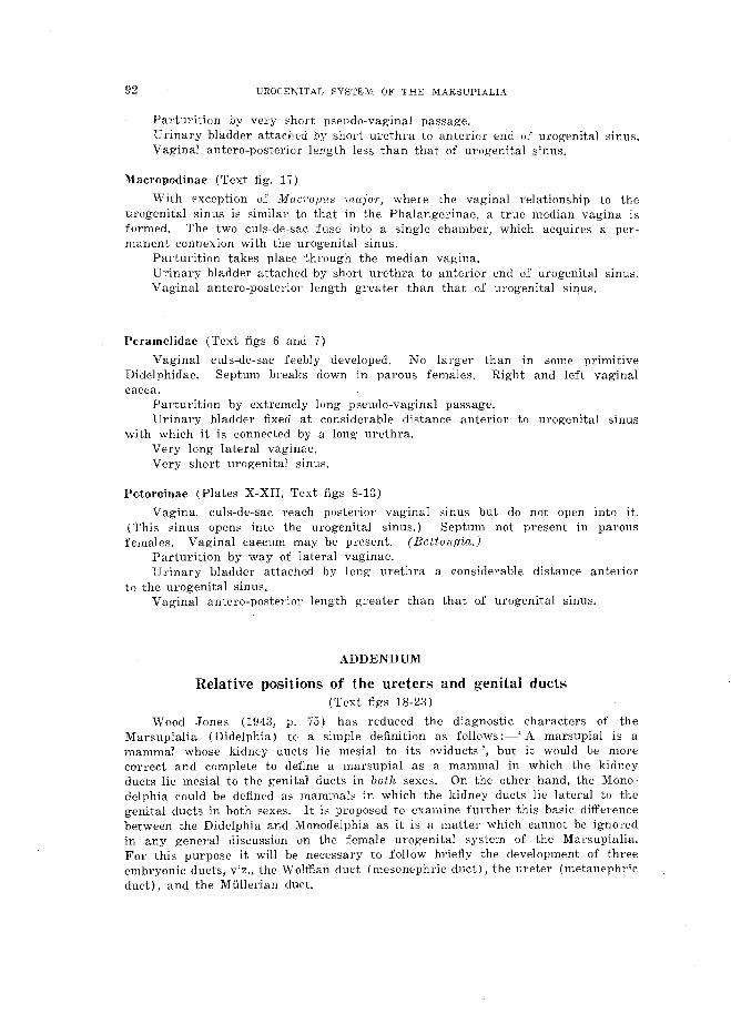
P{Jtoroinae (Plates X-XII, Text figs 8-13)
Vaginal culs-de-sac reach posterior vaginal sinus but do not open into it. I This sinus opens into the urogenital sinus.) Septum not present in parous females. Vaginal caecum may be present. (Bettongia.)
Parturition by way of lateral vaginae. Urinary bladder attached by long urethra a considerable distance anterior
to the urogenital sinus. Vaginal antel'o-posterior length greater than that of urogenital sinus.
ADDENDUM
Relative positions of the ureters and genital ducts (Text figs 18-23)
Wood Jones (1943, p. 75) has reduced the diagnostic characters of the Marsupialia (Didelphia) to a simple definition as follows :-' A marsupial is a mammal whose kidney ducts lie mesial to its oviducts', but it would be more correct and complete to define a marsupial as a mammal in which the kidney ducts lie mesial to the genital ducts in both sexes. On the other hand, the MOl1odelphia could be defined as mammals in which the kidney ducts lie lateral to the genital ducts in both sexes. It is proposed to examine further this basic difference between the Didelphia and Monodelphia as it is a matter which cannot be ignored in any general discussion on the female urogenital system of the Marsupialia. For this purpose it will be necessary to follow briefly the development of three embryonic ducts, viz., the Wolffian duct (mesonephric duct), the ureter (metanephric duct), and the Mullerian duct.

.T. PEARSON 93
Phylogenetically the Wolffian and Mullerian ducts probably arose by the longitudinal splitting of the archinephric duct. In ontogeny the Mullerian duct arises lateral to the Wolffian duct at each side of the body, but in the development of all known vertebrates and particularly in the Mammalia, there is a considerable time-lag in the first appearance of the Mullerian duct. For example, in the marsupial TTichosul'uS (Buchanan and Fraser, 1918) the Wolffian duct actually reaches the cloaca and opens into it before the anlage of the Mullerian duct can be detected between the 8th and 9th spinal ganglia of the embryo. Clearly the reason for this lag is, as Graham Kerr has pointed out, that the Wolffian duct, as the duct of the mesonephros, is called upon to function at a much earlier stage than the Mullerian duct, which, as a reproductive duct, will not be required until the organism attains maturity. In marsupials the mesonephros is functional before birth. The Mullerian duct, on the other hand, is not required for at least severa! months after birth and, in point of fact, does not open into the urogenital sinus until some time after birth. This change in the ontogenetic tempo of structures. which phylogenetically were contemporary, is in accord with the revised interpretation of the Recapitulation Theory which is now held by embryologists.
In TrichosuTus Buchanan and Fraser (1918) have shown that each \\Tolffian duct opens into the cloaca at the 7 mm. G.L. stage. Almost immediately afterwards the ureteric bud arises from the dorsal side of the W olffian duct near its posterior end (text fig. 18). The ureteric bud shifts mesially so that ultimately the ureter lies mesial to the W olffian duct (text figs 20, 21), a characteristic which is common to all didelphians. In the Monodelphia, on the other hand, the ureteric bud, after arising from the dorsal side of the Wolffian duct, shifts laterally so that the ureters take up a position lateral to the Wolffian ducts. Both these divergent conditions are departures from the primitive arrangement found in the Sauropsida and Monotremata where each ureteric bud retains its neutral position on the dorsal side of the W olffian duct.
The anterior extremity of each Mullerian duct (ostium abdomirwle) is the first part to appear at about the 7'75 mm. G.L. stage in TTichosUTtLS (Buchanan and Fraser, 1918) and grows backward in close contact with the Wolffian duct, first lateral to it, then ventral, and finally, at the caudal end, mesial to it. Thus each Mullerian duct forms a loose spiral around the corresponding vVolffian duct. In Didelphis, according to Baxter (1935), the growing tip of the Mullerian duct is in such intimate contact with the Wolffian duct that no mesenchyme structures intervene between the two ducts. Baxter says 'the W olffian duct is used as a guide rail by the growing Mullerian duct but does not contribute in any way to the formation of the latter which increases in length purely as the result of the multplication of its own cells '. This interpretation of Baxter's is open to question. Phylogenetically the two ducts are very intimately associated, whether we accept the view that the archinephric duct splits to form both the Wolffian and Mullerian ducts, or whether the W olffian duct may be regarded merely as a later phase of the archinephric duct which by cell proliferation gives rise to the Mullerian duct. In support of this latter contention it is interesting to note that in some Elasmobranchs the W olffian duct actually contributes cell units towards the formation of the MUllerian duct. In the higher vertebrates the considerable time-lag in the formation of the Mullerian duct and the manner in which this duct follows faithfully the course already taken by the Wolffian duct is consistent with the hypothesis that some of the cells of the Wolffian duct control the development of the Mullerian duct whether as organizers, as the term is generally understood, or in some other way. even if they do not contribute material cells towards the building of the new duct. It is doubtful, therefore. whether Baxter's pronouncement that the \Volffian

94 UROGE-"ilTAL SYSTEM m' THE MARSUPIALJA
18 c/.
llJd
----I.ur. --
h
U 'j5 __ 2.1
, c(
______ L<-'j-5. ___________ +--
Figs 18-23 FIGS lS-21.--Foul' stages in the development of thE' leFt ureter in Tr'icho81'{TUS showing
the origin of the left ureteric bud from the left \Volffian duct, and the luesial shifting of the left ureter (after Buchanan and Fraser, 1918),
FlGS :~2-2;~.-Comparison (,f the Didelphia (fig. 22) and Monodelphia (fig. :Z3) to shuw the rela Lions uf the nudnae and tIrHE),;';.

J. PEARSON 95
duct does not contribute in any wny to the formation of the Mullerian duct is in accord with the view that in ontogeny certain cells may organize other groups of tells and determine their destiny.
By the time that the Mullerian duct reaches the level of the urogenital sinus on the medial side of the VVolffian duct the Ul'etel' is already well established in Trichosurw5 and the metanephros which develops from the cephalic end of the ureter, is functioning. The mesonephros, on the other hand, is degenerating at this stage, and in the female the anterior portion of the \Volffian duct is fast disappearing, though the caudal portion may still retain for a time its connexion with the urogenital sinus.
In passing, it may be observed that though the phylogenetic sequence of development is (1) Wolflian duct and Miillel'ian duct, and (3) uretel', the ontogenetic sequence is (1) Wolffian duct, (2) ureter, and (3) Miillerian duct. It is also of interest to note that i'n the Amniota, which alone of the vertebrates possess a functional metanephros, the ureter appears in ontogeny before the metanephros, although in phylogeny it would be reasonable to expect that the segmental tubules of the metanephros would appear in advance of theil' duct,
It will be seen from the above brief summary of the development of the three ducts that (1) the ureter arises as an outgrowth from the Wolffian duct and their position in relation to each other is finally determined before the Mullerian duct l'eaches the urogenital sinus, i.e., the ureter is mesial to the IV olffian duct in Didelphia and lateral to it in the Monodelphia, and (2) since the Mullerian duct follows the course of the W olffian duct and is in the closest association with it, it is clear that the position of both these ducts in reI ation to the ureter is the same. The Wolffian duct, therefore, may be regarded as the datum which must form the starting point in any discussion dealing with the relative position of the ureters and Mullerian ducts.
Therefore if each ureter is mes: al to the W olffian duct in the Didel phi a the ureter will also be mesial to the Mullel'ian duct. As the \V offian duct becomes the genital duct of the male and th~ Mullerian duct is the genital duct of the female, it follows that in the Didelphia the ureters are mesial to the genital ducts in both sexes. Conversely in the Monodelphia the Ul'eters are lateral to the genital ducts in both sexes. This would appear to be a more satisfactory and complete definition than the one given by Wood Jones. (Text figs 22 and 23 show the relative positions of the ureters and female genital ducts in the Didelphia and MOl1odelphia.)
This fundamental difference between the Marsupialia (Didelphia) and the higher mammals (Monodelphia) is due, as we have seen, to the shifting of the ureteric bud mesially in the first group and laterally in the second. Comparative anatomy and embryology provide no clue to the reason why this important clear-cut distinction should ha~e arisen, any more than they provide an explanation for' the persistence of the right aortic arch in bi1'(18 and the left aortic arch in mammals. But whatever the cause of these cardinal modifications in the position of the ureters, the phylogenetic stage at which they were established marks an important point of divergence of primitive didelphian and l11onodelphian stocks, both of which were derived from a common mammalian ancestor in which, presumably, the ureteric buds arose fro111 a neutral position on the dorsal sides 01' the W olffian duct in the true sauropsidan manner.
In discussing this fundamental e!ifferem:e in the topography of the ureters and Mullerian ducts in the JV[ol1odelphia and Didelphia Wood Jones (1943, p. 75) writes 'In the higher mammals there is a stage in 'which it might be saie! that it is touch and go which side the ureters will pass the oviducts, and in the end they g'o to the lateral side of them, In the marsupials they pass to the medial

96 UROGENITAL SYSTEM OF THE MARSUPIALIA
side', and again' But for some reason, for which at present there is no explanation, the kidney ducts, from occupying a neutral dorsal position in the Ornithodelphia, passed to opposite sides of ~he female genital ducts in the diverging phyla of the Monodelphia and the Didelphia. By passing to the medial sides of the female ducts in the Didelphia, the kidney ducts prohibit that caudal meeting that produces the pregnancy chamber or chambers, with a median vaginal outlet, that typifies all members of the Monodelphia' (l.e., p. 87). .
These statements interpret somewhat loosely and unsatisfactorily the facts of the case as outlined above and might convey two wrong impressions, first, that the 'oviducts' take up their position prior to the establishment of the ureters, whereas the reverse is actually the case; and, secondly, that because of their position mesial to the female ducts the ureters prohibit the formation of a medially placed vagina. The position of the ureters does not in itself constitute a physical obstacle to the passing of the two Mullerian ducts between the ureters. The essential point is that as the Mullerian- ducts grow caudally they are bound by their phylogenetic relationship to the W olffian ducts to follow the course already taken by these ducts, and so they pass lateral to the ureters. But in spite of this disability the marsupials have evolved, secondarily, a direct median route connecting the uteri with the urogenital sinus, and this route, it should be marked, passes mesial to the ureters and follows precisely the same path as the one traversed by the vagina of the Monodelphia (see text figs 22 and 23).
The evolution of this secondary vaginal apparatus, which provides. a method of parturition by. means of a median passage for nearly all recent marsupials, is perhaps the most outstanding feature of the female urogenital system of the group. It involves the progressive development of the two vaginal culs-de-sac, by means of which a precarious and ,makeshift method of parturition through the pseudo-vaginal passage was established, a method which culminated in an efficient and permanent median vagina in some of the more specialized marsupials. This development of an entirely new mode of parturition was brought about through the inadequacy of the two Mullerian ducts for this purpose. Once the change from oviparity to viviparity had been effected the new functions wh'ich the Mullerian ducts were called upon to perform would be carried out more efficiently if the two ducts were to coalesce and thus provide a tube of larger calibre. In both Monodelphia and Didelphia the experiment was made, though in different ways. In the former the two vaginae lay mesial to the ureters and were thus able to coalesce. In the Didelphia, on the other hand, a median vagina has been established secondarily in the more specialized forms. Wood Jones (1943) has dealt with this at some length, but his interpretation of the method by which this secondary median vagina has been achieved will hardly be acceptable to most biologists.
Based upon the observations of Lister and Fletcher (1881), Fletcher (1882, etc.), Hill and Fraser (1925) and others, the stages in the probable evolutionary sequence of this secondary vaginal apparatus have been given above. This series traverses the development of this system from the incipient pseudo-vaginal passage in the Didelphidae, through the. more advanced condition found in the Australian polyprotodonts and culminating in the complete establishment of a true median vagina, having a continuous epithelial lining, in some of the Macropodidae. This ultimate structure, it should be emphasized once again, follows precisely the same course, mesial to the ureters, that is taken by the vagina in the Monodelphia, thus adding yet another instance to the long list of homoplastic structures known to occur in the Marsupialia.
If we cared to indulge in a little harmless speculation .as to th€ future course of the evolution of the vaginal system in this interesting group we might envisage'

J. PEARSON 97
the newly gained median vagina of the Macl'opodinae firmly establishing itself and the almost useless lateral vaginae gradually degenerating into a pair of tubes having their cavities partially or completely occluded, until utimately the highest types of marsupials would possess a single vagina situated between the ureters and differing in no respect from the Monodelphian type except in its phylogenetic and ontogenetic history, Perhaps this would be no more amazing than many other examples of convergence that can be called to mind, especially within the Marsupialia, a group pre-eminent in its many examples of homoplasy.
REFERENCES
BAXTEH, .T. S., 19a5.~-~·Development of the female g-enital tract in the Arneriean opOSHum. earn. In st·, H'ush. ContT. to ICrnhrJjoloyy, XXV, No. 145, pp. 17-35.
BFJNSLEY, B. A., 1903.--0n the Evolution of Australian Marsupialia with remarks on the relationships of the marsupials in general. 'Trans. Linn. Soc. Lond. (2), vol. IX, part 3, pp. 8~-217.
BRASS, A., 1880.-Beitrage zur Kenntni!::>s de) weiblichen Urogenital-systems del' Marsupialen. Inau{j. Dl:SS., Leipzig.
BUCHANAN. G., and FRASER, E. A., 19t8.~The developm_ent of the Urogenital System in the Marsupialia. with special reference to Tl'ichosul'lls vulpecula. Part I, Jount. of Anatomy, vol. 53, PI'. 85-95.
FLETCHER, J. J., 1882.-0n the existence after parturition of a direct eommunication between the Median Vaginal Cul-de-sac, so-called, and the Urogenital Canal, in certain species of Kangaroos. P'roc. Li'nrt. Soc. N.S.1V .• vol. VI, pp, 796-811.
-- ----, 188R.-On some points in the anatOlny of the Urogenital Organs in females of certain specie::; of Kangaroos. Part I. Proc. Linn. Soc. N.S. fV., vol. VII, pp. 640-659.
--- --""--, 1884.~-On smne points in the anatOlny of the Urogenital Organs in females of certain species of Kangaroos. Part II, PrOf). Linn. Soc. N.S. W., vol. VIII. pp. 6-1l.
FLYNN, T. T., 1910.---Contributions to a. knowledge of the anatomy and development of the l\iarsupialia.. No.1: The genitalia of Sarcophilus sat.ankns (~). Prof:. L1:nn. Soc. N.S. W., vol. 35. pp. 873-886.
1923.-Remarks on the method of Parturition in /-'o'toroiis t'ddactulus. Proc. L,tnn. Soc. N.S. HT., vol. XLVIl, part 5, p. xxviii.
FORBES, Vv. A., 1881.--0n SODle points in the Anatomy of the Koalfl. (Phascolarctos cinereus)~
PTOC. Zool. Soc. Lon,d., pp. 180-1.95. HILL, .J. P., 1.89H.-On the female Urogenital Org'ans of Perameles, with an account of the phenomena
of Parturition. Pro". Ll:nn. Soc. N.S.W .. vol. XXIV, PI'. 42-82. 1900, a.--~--On the foetal membranes. placentation. and parturition of the Native- Cat
(Dasyurus viverrinus). Anat .. 4nz .• Bel. 18. pp. 364-373. -------- ]900. b.-On the female Urogenital Organs of Myrmecobins t'asciat.us. 1-'roc. Li1VH. Soc.
N.S. W., vol. XXV, pp. 519-522. 1900, r.--On the existence at parturition of a pseudo-vaginal passage in TriehoHlHus
vulpecula. ll)1:d.. I)P. 526-5~-n.
HiLL, .J. 1'., and FRASER, E. A .• IH25.-Some observations in the felnale £Trogenital Organs of the Didelphyirlae. Pro". Zool. Soc. Land., 1925, PI'. 189-219.
HUHRBCHT, A. A. W., 1909.~--l!Jarly ontog'enetic Phennmena in Mammals and their bearing on our interpretation of the Phylogeny of the Vertebrates. Quart. J011'rn. Micr. Sd., vol. 53, PI'. 1-18!.
~JONBS. F. ¥lOOD, 1943.---//abit and Tleritaue, Kegan Paul. ~rrenc.h. Trubner & Co., London. LISTER, ,J. J., alld FLETCHER. J. J., 1881.-0n the condition of the median portion of t.he Vaginal
Apparatm; in the Macropodiuae. P)'{)c. Zooz. ·Soc. [JOtul.. 1881, pp. 976-996. MCCRADY, E., 1938.-The Embryolog)f of the Op08R11,m. WiRtar Inst. Anat. and BioI. Philadelphia.
226 pp.
OWEN, R., 18!34.-0n the generation of the Marsupial Animals with a der-;eription of the impregnated uterus of the Kang·aroo. Phil. Tra'ns. Ruy. Soc. Lond., UW4, part 1, pp. - 3~3-:~64.
18l11.~--Mann~pialia.. Todd's Cyclopaedia of' Anatomy and Physiology. pp. 1-74. "-----.----,,- 1868.----Comparative Anat.omu am{/ PhllsiolornJ oi VWl'tebrates, vol. Ill, pp. 680~685. Longman,
Green, & Co., London. PBARSON. J"., 1944.-The Vaginal Complex uf the Rat-Kangaroos. Austr. "ourn. Sci., voL VlI, No.3,
pp. 80-83. MACKENZIE, W. C" 1919.----The gendV-1Jn'nury $'1J;;dem -1'/1_ 111onotremes and Mar.'jupials. Jenkin, Buxh)n.,
& Co., Melbourne.

98 UROG!cKITAL SYSTEM O.F THE MARSUPiALlA
REFERENC~~ LETTERS USED J."l TEXT FIGURES AND PLATES
tLs.---alIantoic stalk a. v.c.--anterior vaginal expansiDn bl.--urinal'Y bladder d.--doaea ct.-clitoris tLp.----digitaI process f';.-m.c.---back ward extension uf cul-dc:.'-sac it.g.----hind-gut L u.b.--Ieft ureterie bud 1.1A1.--!eft ureter l.'uo--Iateral vagina l."ui.d.--left Wolffian duct "yn.G.-vaglnal eul-ue-t\ae o,o.----ovary p.v.s.-posterior vaginal sinus
'f(!(:t..--rectnm
r.l.'tJ.---l'ight lateral vag'ina T.ur.---right ureter r.ut.--right uteruB r. c.c.-right vaginal caecum gept.--·septum between right and left vaginal caeca n.g.~.----ul'ogE'nital sin.us
. -uterine neck UT.-----ureter u,t.---uterus ureth. --Hl'ethr'a
-u.p.-utel'ine papillae va[I.--·vag-ina '(}.c.-,-vaginal caeCUIn
PLATE X
Dorsal vjew of the anterior portion of the ul'vgenftal system of Poio}"(yiis tn:dact-ylu.,s, showing a :foetus' paesing down the left lateral vagina.
Photograph by eourtesy
(.For key to reference letters ;3ee above.)
Pl'ofe~sor T. 1'. FJynn.

PAP. & PROC. ROY. Soc. TAS. 1944 PLATE XII
'--___ ut. _____ ul'.
___ bl. _________ _
--'._-- m. C.
_____ l.v.
uretk.----------
A B