Embed Size (px)
Citation preview

THE
EPIDEMIOLOGY AND AETIOLOGY OF
A RISING INCIDENCE OF
PAPILLARY THYROID CARCINOMA
IN TASMANIA
by
John Richard Burgess MD, FRACP
Discipline of Medicine
Faculty of Health Science
Submitted in fulfillment of the requirements
for the degree of
Doctor of philosophy
University of Tasmania
2007

STATEMENT OF AUTHENTICITY
This thesis contains no material that has been accepted for the award of any other higher
degree or graduate diploma in any tertiary institution.
I am responsible for initiating and undertaking the work described in this thesis. The full
extent to which others have contributed to the data contained herein is detailed in the
Acknowledgements and Bibliography.
John Richard Burgess
11
AUTHORITY OF ACCESS
This thesis may be made available for loan. Copying of any part of this thesis is
prohibited for two years from the date this statement was signed; after that time limited
copying is permitted in accordance with the Copyright Act 1968.
John Richard Burgess
Date 21 / 06 / 2007
111

DEDICATION
This thesis is dedicated to my family -- to my Jennifer; for support and encouragement --
to James, William and Matthew; may they too find enjoyment in learning.
iv

PREFACE
When I commenced clinical practice as an endocrinologist in Tasmania during the mid-
1990s, I was intrigued by reports from a number of jurisdictions of an increasing
incidence of papillary thyroid carcinoma (PTC). Varying causes had been proposed.
Spurious methodological factors such as improving case registration by Cancer
Registries, changes in the morphological classification of thyroid tumours and finer
sectioning of thyroidectomy samples were postulated. Similarly, a true change in PTC
incidence due to increased exposure to risk factors such as ionising radiation and changes
in iodine nutrition were also proposed as explanatory.
Tasmania is an island State of the Australian Commonwealth with a stable population
structure, sophisticated medical infrastructure and a well characterised history of iodine
deficiency. This presented the opportunity for using the Tasmanian population as a
model to evaluate the cause of observed PTC incidence trends.
The research presented in this thesis was undertaken by myself between 1995 and 2006
in an attempt to answer fundamental questions regarding incidence trends for PTC -
namely, has there been a true increase in the incidence of PTC and if so, what factors
underlie this change? My role encompassed conceptual planning, the development and
submission of applications for funding, preparation of documents for Institutional Ethics
Committee approval, patient recruitment, database construction, statistical analysis, and
preparation of manuscripts for publication. Candidature for this PhD was approved and
undertaken in keeping with the provisions of Appendices A.4 and A.5 — previously
published research.

PUBLICATIONS FROM THIS THESIS
This thesis contains six chapters of original research that are either published (five) or in
preparation for publication (one).
Chapter 2
Burgess JR. Temporal trends for thyroid carcinoma in Australia: a rising incidence of
papillary thyroid carcinoma (1982 — 1997). Thyroid. 2002;12:141-149.
Chapter 3
Burgess JR, Dwyer T, McArdle K, Tucker P. The changing incidence and spectrum of
thyroid carcinoma in Tasmania (1978-1988) during a transition from iodine sufficiency
to iodine deficiency. J Clth Endocrinol Metab 2000;85:1513-1517.
Chapter 4
Burgess JR, Tucker P. 2006 Incidence trends for papillary thyroid carcinoma and their
correlation with thyroid surgery and thyroid fine needle aspirate cytology. Thyroid.
2006;16:47-53.
Chapter 6
Burgess JR, Skabo S, McArdle K, Tucker P. Temporal trends and clinical correlates for
the ret/PTC mutation in papillary thyroid carcinoma. ANZ J Surg. 2003;87:31-35.
Chapter 7
Burgess JR, Wilkinson R, Ware R, Greenaway TM, Percival J, Hoffman L. Two
families with an autosomal dominant inheritance pattern for papillary carcinoma of the
thyroid. J Clin Endocrinol Metab 1997;82:345-348.
vi

RELATED PUBLICATIONS DURING CANDIDATURE
McKay JD, Lesueur F, Jonard L, Pastore A, Williamson J, Hoffman L, Burgess JR, et al.
Localisation of a susceptibility gene for familial non-medullary thyroid carcinoma to
chromosome 2q21. Am J Hum Genet. 2001;69:440-446.
Lesueur F, Corbex M, McKay JD, Lima J, Soares P, Griseri P, Burgess J, et al. Specific
haplotypes of the RET proto-oncogene are over-represented in patients with sporadic
papillary thyroid carcinoma. J Med Genet. 2002;39:260-5.
Guttikonda K, Burgess J, Hynes K, Boyages S, Byth K, Parameswaran V. Recurrent
iodine deficiency in Tasmania, Australia — A salutary lesson in sustainable iodine
prophylaxis and its monitoring. J Clin Endocrinol Metab. 2002;87:2809-2815
Hynes KL, Blizzard CL, Venn AJ, Dwyer T, Burgess JR. Persistent iodine deficiency in
a cohort of Tasmanian school children: associations with socio-economic status,
geographical location and dietary factors. Aust N Z J Public Health. 2004;28:476-481.
Seal JA, Doyle Z, Burgess JR, Taylor R, Cameron AR. Iodine status of Tasmanians
following voluntary fortification of bread with iodine. Med J Aust. 2007;186:69-71.
Burgess JR, Seal JA, Stilwell GM, Reynolds PR, Taylor RE, Parameswaran V. A case
for universal salt iodisation to correct iodine deficiency in pregnancy: another salutary
lesson from Tasmania. Med J Aust. 2007;186:574-576.
vii

ABREVIATIONS
Age standardised incidence rates (ASR)
Anaplastic thyroid carcinoma (ATC)
Computed tomographic (CT)
Deoxyribonucleic acid (DNA)
Follicular thyroid carcinoma, (FTC)
Fine needle aspiration biopsy (FNAB)
Follicular variant of PTC (fv-PTC)
Iodine deficiency (ID)
Magnetic resonance (MR)
Medullary thyroid carcinoma (MTC)
New South Wales (NSW)
Not otherwise specified (NOS)
Papillary thyroid carcinoma (PTC)
Queensland (QLD)
Reverse transcription-polymerase chain reaction (RT-PCR)
Ribonucleic acid (RNA)
South Australia (SA)
Standard Error of Mean (SEM)
Thyroid carcinoma (TC)
Thyroglobulin (TG)
Thyroid Stimulating Hormone (TSH)
Western Australia (WA)
viii

ACKNOWLEDGEMENTS
This thesis contains work undertaken by myself in conjunction with a number of
collaborators and co-authors who contributed various elements of technical, laboratory
and clinical expertise. Without the assistance of these this thesis would not have been
possible. The role of co-contributors is acknowledged both in the authorship of
published work and as detailed below.
I am grateful for the support provided by Dr Linda Hoffman, whose initial clinical
observations regarding familial PTC in Tasmania provided the basis for subsequent
research and publication in this area. I am similarly indebted to my supervisor Professor
David Kilpatrick for the support and flexible learning environment provided during
preparation of this thesis. I am thankful for the general advice, assistance and
encouragement provided by my colleagues Drs Greenaway and Parameswarran during
the course of my research.
Dr Paul Tucker, an histopathologist and colleague, provided invaluable expertise in
review and reclassification of thyroid carcinoma archival samples, as well as facilitating
links between data resources in the public and private sectors. I am appreciative of
guidance and assistance from Dr Stewart Skarbo, Dr Venkat Parameswaran and Ms Lyn
Blackwell in helping me develop the laboratory skills required for performing molecular
studies for the RET/PTC1 rearrangement described in Chapter 6. I gratefully
acknowledged the advice provided by Dr D Learoyd and Professor B Robinson, Royal
North Shore Hospital, New South Wales, Australia; as well as their assistance in making
available the TPC-1 cell line (originally from Dr S. M. Jhiang, Columbus, OH, USA).
ix

The staff at the Menzies Centre provided direct and indirect help with epidemiological
advice and logistics. In this regard, I particularly acknowledge Dr Kristen Hynes, Dr
Leigh Blizzard and Professor Terrence Dwyer. I am also thankful for the administrative
assistance provided by Sr Rachel Saunders, particularly for undertaking questionnaire
data entry. The help and support provided by Sr D Shugg and the staff at the Tasmanian
Cancer Registry is also acknowledged with gratitude. I also note the assistance provided
by Mr Robert Van der Hoek and the National Cancer Statistics Clearing House,
Australian Institute of Health and Welfare, in making available the data analysed in
Chapter 2.
The collation of the statewide data set for thyroid procedures would not have been
possible without the generous support of numerous individuals and organisations. In
particular, Hobart Pathology, Launceston Pathology and the various Tasmanian public
and private hospitals, all of whom provided assistance and advice. I also thank the many
general practitioners and medical specialists who responded to requests for information.
The assistance others provided in relation to the research presented in this thesis is also
gratefully recognised in the authorship and specific acknowledgements associated with
the published research presented in the Appendix. Finally, I wish to thank the many
thousands of Tasmanians; thyroid cancer patients and members of the general public who
responded to questionnaires that contributed to the research presented in this thesis.
My research has been supported by research grants from the Tasmanian State
Government (Dick Butterfield Fellowship), the Royal Hobart Hospital Research
Foundation and the Cancer Council of Tasmania.

STATEMENT OF THE PROBLEM
Papillary thyroid carcinoma (PTC) is the most frequently diagnosed endocrine
malignancy. Cancer registries in many countries have identified a substantial increase in
the reported incidence of PTC over the past half-century. The explanation for this
remains to be elucidated.
Improved PTC case ascertainment by cancer registries, heightened histopathological
recognition of small and subclinically PTC, increased use of neck ultrasonography and
greater recourse to fine needle aspiration biopsy in evaluation of thyroid nodules may be
contributory. An increase in the underlying occurrence of PTC is also possible, with
exposure in childhood to ionizing radiation as well as changing levels of iodine nutrition
potential underlying factors.
In this thesis I sought to answer the following questions:
1 Has the incidence of PTC increased in Australia?
2 Is there evidence of geographic variation in PTC incidence within Australia?
3 Is the Tasmanian population a suitable model for evaluating the basis for
Australian PTC incidence trends?
4 Are changes in observed PTC incidence due to changes in histopathological
diagnostic criteria or Cancer Registry case ascertainment?
5 What is the contribution of increased diagnosis of microscopic and clinically
"occult" PTC to observed incidence trends?
xi

6 Are geographic patterns of PTC incidence related to the historical or
contemporary distribution of iodine deficiency in Australia?
7 Does iodine nutrition influence the PTC incidence by either altering tumour
pathogenesis or by indirectly increasing the likelihood of diagnosing "occult"
PTC?
8 Is there evidence for past exposure to ionising radiation as an aetiological
driver for contemporary PTC incidence?
9 Is there evidence for heritable susceptibility to PTC influencing observed
incidence trends?
10 Is it possible to estimate the relative contributions of ascertainment bias and
changes to the underlying biological incidence in producing contemporary
PTC incidence trends?
xii
AIMS
1. To assess temporal trends for thyroid carcinoma in Australia and determine if
PTC incidence has increased.
2. To determine the validity of the Tasmanian population as a model for
understanding PTC incidence trends both in Australia and internationally.
3. To determine the contribution of non-biological factors (changes in
morphological classification, Cancer Registry ascertainment and alterations in
clinical practice paradigms) in shaping PTC incidence trends.
4. To determine the contribution of both established and putative PTC risk
factors (ionising radiation, genetic susceptibility and iodine nutrition) in
shaping PTC incidence trends.

HYPOTHESIS
1. The Tasmanian community provides a population model for investigating
national incidence trends for PTC.
2. Non-biological factors (bias due to alterations in medical practice and data
management systems) account for the majority of the apparent rise in PTC
incidence observed over recent decades.
3. A true increase in underlying PTC incidence accounts for a small, but important
component of observed PTC incidence trends.
xiv

TABLE OF CONTENTS
TITLE PAGE
STATEMENT OF AUTHENTICITY ii
AUTHORITY OF ACCESS iii
DEDICATION iv
PREFACE
PUBLICATIONS ASSOCIATED WITH CANDIDATURE vi
ABREVIATIONS viii
ACKNOWLEDGEMENTS ix
STATEMENT OF THE PROBLEM xi
AIMS xiii
HYPOTHESIS xiv
TABLE OF CONTENTS xv
ABSTRACT 1
CHAPTER 1 Background Literature 7
XV

CHAPTER 2 Temporal trends for thyroid carcinoma in Australia: a rising 35
incidence of papillary thyroid carcinoma (1982 — 1997).
Prologue 36
Aims and Hypotheses 37
Introduction 38
Subjects and Methods 39
Results 41
Discussion 43
Conclusion 47
Tables and Figures 48
CHAPTER 3 A review of thyroid carcinoma cases registered by the 55
Tasmanian Cancer Registry (1978-1998) with particular
reference to the accuracy of case classification and registration.
Prologue 56
Aims and Hypotheses 57
Introduction 58
Subjects and Methods 60
Results 62
Discussion 64
Conclusion 68
Tables and Figures 69
CHAPTER 4 Incidence trends for PTC and their correlation with trends for 75
thyroid surgery and thyroid FNAB cytology
Prologue 76
Aims and Hypotheses 77
Introduction 78
xvi

Subjects and Methods 80
Results 82
Discussion 85
Conclusion 89
Tables and Figures 90
CHAPTER 5 The Impact of Birth and Residence in Tasmania on the Prevalence
of Benign and Malignant Thyroid Disease
97
Prologue 98
Aims and Hypotheses 99
Introduction 100
Subjects and Methods 101
Results 103
Discussion 105
Conclusion 108 Cl)
Tables and Figures 109 po•
CHAPTER 6 An indirect assessment of the role of ionising radiation in the
genesis of contemporary PTC incidence trends
115
Prologue 116
Aims and Hypotheses 117
Introduction 118
Subjects and Methods 120
Results 123
Discussion 124
Conclusion 128
Tables and Figures 129
xvii

CHAPTER 7 Heritable susceptibility to papillary carcinoma of the thyroid 133
Prologue 134
Aims and Hypotheses 135
Introduction 136
Subjects and Methods 137
Results 138
Discussion 141
Conclusion 143
Tables and Figures 144
CHAPTER 8
Conclusions and Future Studies 146
BIBLIOGRAPHY 155
APPENDIX Publications from this Thesis 171
xviii

ABSTRACT
Thyroid carcinoma (TC) is the most prevalent endocrine malignancy. Four main sub-
types are recognised - papillary thyroid carcinoma (PTC), follicular thyroid carcinoma,
medullary thyroid carcinoma and anaplastic thyroid carcinoma. PTC accounts the
majority of diagnoses. As is typical of most thyroid disorders, women are affected
approximately four times more frequently than men. The absolute incidence of PTC
varies significantly between geographic locations and racial groups, suggesting an
important interaction between environmental and genetic risk factors.
Aside from relative differences in absolute PTC incidence between different
communities, a significant increase in PTC incidence has also been observed within
many populations over recent decades. In some jurisdictions the incidence of PTC has
increased more than two fold. Industrialised as well as developing nations have been
affected. Possible explanations include artifact due to changes in both medical practice
and tumour registration by Cancer Registries. However, a true biological change in PTC
pathogenesis and incidence is also possible. Family history and exposure to ionizing
radiation are the most clearly established risk factors. Iodine nutrition may also influence
the risk of thyroid malignancy, both by modulating thyroidal radioiodine uptake as well
as by directly influencing the pathogenesis of benign and malignant thyroid disease.
1

My research reviewed Australian national TC incidence trends during the 1980's and
1990's, identifying a rise of between 4.0% and 24.7% per annum in the incidence of PTC
across the Australian states. This rise was most obvious in the eastern seaboard states,
mapping to those states with the lowest historical levels of iodine nutrition. The greatest
rise in PTC incidence was observed in Tasmania, an island State of the Australian
Commonwealth, where PTC incidence increased by 24.7% per annum. Tasmania has a
well-characterised history of iodine deficiency and a stable demographic structure with a
centralised health care system and established Cancer Registry. These characteristics
provided an opportunity for evaluating in detail the basis for the more widely observed
rise in PTC incidence.
A detailed evaluation of Tasmanian Cancer Registry data over the 21-year period 1978-
'98, confirmed that the incidence of PTC had increased by 450% in females and 210% in
males. Furthermore, validation of Cancer Registry case ascertainment by de novo
reconstruction of a Tasmanian TC data-set (using source pathology and hospital
documentation), confirmed 93.9% completeness for existing PTC case ascertainment by
the Cancer Registry over the study period. Similarly, a review of histopathological
diagnoses did not demonstrate any significant change in the morphological classification
of TC during this time. These findings exclude changes in either case reporting or
tumour classification as the primary cause for observed PTC incidence trends.
The key trend underlying observed changes in PTC incidence was an increase in
diagnosis of small (<1cm) PTC that were asymptomatic at the time of resection.
2

Further studies evaluated the prevalence of both clinical and subclinical (by
ultrasonography) nodular thyroid disease in the Tasmanian population. Thyroid
ultrasonography revealed nodules in 43.4% of individuals evaluated, the majority (88%)
of whom had no previously recognised history of thyroid disease. Non-specific neck
imaging therefore had the potential to identify clinically inapparent thyroid nodules.
Contemporary evaluation protocols for asymptomatic and incidentally discovered thyroid
nodules promote the use of fine needle aspiration biopsy (FNAB) and specimen cytology.
An assessment of trends for utilization of thyroid FNAB identified an increase of 17.6%
per annum and 66.2% per annum for males and females respectively during the period
1988- 98. As the prevalence of clinically silent PTC .1cm diameter ("occult" PTC) is
reported to be approximately_ 5% in thyroid nodules, contemporary patterns of neck
imaging and the subsequent FNAB evaluation of subclinical thyroid nodules might
explain much of the recently observed rise in incidence of PTC.
I further evaluated the relationship between iodine nutrition and PTC incidence.
Comparison of documented changes in iodine nutrition in Tasmania during the past five
decades against observed PTC incidence trends showed the increase in PTC incidence
occurred during a period when the Tasmanian population was undergoing transition from
iodine sufficiency to mild iodine deficiency (after the almost 30 years of optimal iodine
nutrition that followed correction of endemic iodine deficiency in the mid-1960's). This
observation was unexpected given much of the existing epidemiological evidence links
poor iodine nutrition to a reduced proportion of PTC relative to other thyroid
malignancies, and a lower incidence of PTC compared to iodine replete populations.
3

A study was undertaken to evaluate the impact of Tasmania (historically the most iodine
deficient Australian state and the region with the greatest contemporary rise in PTC
incidence) as a place of birth and residence, on the likelihood of developing benign and
malignant thyroid disease. There was a significant association for goitre and
thyroidectomy with childhood lived in Tasmania. The association was non-significant
for the development of TC. Therefore, increased PTC incidence in Tasmania is
potentially explicable on the basis of greater diagnosis of "occult" PTC, most probably
diagnosed at the time of investigation and management of benign iodine deficiency
related thyroid disease.
I also identified evidence to suggest an increase in the underlying incidence of clinically
relevant PTC. Analysis of Tasmanian cancer registry data confirmed a 260% increase in
large (>2.0cm) PTC between 1978 and 1998. Moreover, it was notable that despite the
rise in incidence of PTC versus FTC, changes in two-, five- and ten- year mortality rates
for PTC remained parallel to those of FTC during the study period. As "occult" PTC
usually exhibit a benign natural history, an increase in PTC incidence solely due to
greater recognition of "occult" PTC would be expected to produce a disproportionate
improvement in survival for patients with PTC relative to FTC.
Ionizing irradiation of thyroid tissue in childhood is the best characterized aetiologic
factor for thyroid neoplasia. Both benign and malignant tumours are predisposed, with
PTC the most frequent malignant sequela. Studies suggest PTC arising after long latency
following thyroid irradiation may exhibit a mutation (the RET/PTC1 rearrangement) with
4
a prevalence higher than for PTC from non-irradiated populations. In the absence of
objective radiation exposure data, I attempted to use the RET/PTC1 mutation as surrogate
marker for past exposure to ionizing radiation. Tasmanian PTC diagnosed between 1978
and 1998 were studied to determine the temporal trends for prevalence of RET/PTC1.
However, a clear relationship between PTC incidence trends and RET/PTC1 prevalence
was not found. Despite this, it was notable that the absolute prevalence of the RET/PTC1
rearrangement in Tasmanian PTC was greater than expected and was consistent with the
prevalence in irradiated populations. Moreover, a significant clinicopathological
association for the RET/PTC1 rearrangement was identified. Tumours positive for the
RET/PTC1 rearrangement were of larger size and more likely to exhibit lymph node
metastases than those without the mutation.
The role of heritable susceptibility to PTC and the potential for a founder effect
influencing Tasmanian PTC incidence trends was also assessed. A family history of TC
was described by 14.1% of Tasmanian patients diagnosed with PTC. Two large PTC
kindreds demonstrating an autosomal dominant inheritance pattern for PTC accounted
for the majority of familial cases. However, the absolute contribution of autosomal
dominant PTC to overall statewide incidence trends was small. A founder effect did not
explain PTC incidence patterns in Tasmania.
The research I have undertaken provides a useful insight into the genesis of Australian
national PTC incidence trends. Whilst there is evidence to support a small increase in
the underlying incidence of clinically relevant PTC, the main cause for the observed rise
in PTC incidence can be attributed to increased ascertainment of "occult" papillary
5

microcarcinoma. The findings of my research highlight the potential adverse health
consequences of inappropriate neck imaging and reinforce the importance of appropriate
education for medical practitioners regarding the rational use of thyroid ultrasonography.
6

CHAPTER 1
Background Literature
7
THYROID ANATOMY AND PHYSIOLOGY
Anatomy and Embryology
The thyroid gland comprises two lobes and an isthmus. Each lobe is approximately 4cm
in length, 2cm in width and 2cm in thickness with a vascular supply derived from the
superior thyroid artery and the inferior thyroid artery(1, 2). On microscopy, the thyroid is
composed of follicles fed by an extensive capillary network. Each follicle comprises a
single sheet of follicular cells (thyrocytes) surrounding a colloid filled lumen. Follicles
are arranged in bundles separated by septae of connective tissue. Contained within the
connective tissue surrounding the thyroid follicles are the parafollicular C cells that
produce calcitonin(1-3).
The thyroid is first evident embryologically at approximately one month post conception
as a thickening of the pharyngeal epithelium, subsequently forming a diverticulum from
the pharyngeal floor that fuses with the ventral aspect of the fourth pharyngeal pouch (1-
3). Thyroglobulin (TG) begins to be synthesized during the second month of gestation
and the ability to concentrate iodine is evident by the end of the first trimester. The fetal
pituitary synthesizes thyroid stimulating hormone (TSH) early in the second trimester and
thereafter both maternal and fetal thyroid hormones contribute to fetal hormone
requirements (2-5).
Thyroid Hormone Synthesis
Iodine is an essential substrate for thyroid hormone synthesis(2, 6, 7). Inorganic iodide is
concentrated by the thyroid via an energy dependent sodium iodide symporter(8-10).
8

Iodine is organically bound to thyroglobulin by a reaction involving thyroid peroxidase,
which catalyses the iodination of tyrosyl residues of thyroglobulin to produce
iodotyrosines(7). The iodotyrosines are subsequently coupled to produce the active
iodothyronines, tetra-iodothyronine (T 4) and tri-iodothyronine (T3). T el and T3 remain
bound within the thyroglobulin by peptide linkages until cleaved from thyroglobulin by
proteolysis (2).
TSH receptor activation drives thyroid hormone synthesis at various levels including
active transport of iodine into the thyrocyte, synthesis of thyroglobulin, proteolysis of
colloid and release of active thyroid hormones into circulation(1, 2, 7, 8). The secretion
of TSH is mediated by hypothalamic release of thyrotropin releasing hormone (TRH),
that in turn is regulated by central receptors located in both the hypothalamus and
pituitary, which are sensitive to circulating thyroid hormone levels(1, 11, 12). TSH also
provides trophic stimulus for growth and replication of the thyroid follicular cells.
Iodine Nutrition
A minimum of 100mcg of elemental iodine is required per day to prevent the
development of iodine deficiency (13, 14). Both inorganic and organically bound iodine
are ultimately absorbed from the gastrointestinal tract and small quantities are lost in
through the skin and stool. Most iodine, however, is rapidly taken up by the thyroid or
cleared via the kidneys(2, 10, 15). Renal iodine clearance is determined by glomerular
filtration rate, uninfluenced by active processes (1, 15).
There is marked variation in dietary iodine intake both within and between human
populations (16-18). Whist dietary customs such as the routine consumption of seaweed
9

and other iodine rich foodstuffs are important, in most circumstances iodine intake is
predominantly determined by the underlying geology of a community's geographic
location. Up to one third of the world's population live in regions that contain borderline
deficient levels of soil and water iodine(16, 18, 19). The water and foods derived from
geologically deficient regions consequently contain insufficient levels of iodine to sustain
normal thyroid function (2, 14, 16, 20).
A diet deficient in iodine produces a variable spectrum of disease, influenced by both the
degree of iodine deficiency as well as secondary genetic and environmental factors(21-
23). The broad spectrum of derangement resulting from iodine deficiency has led to the
use of the term Iodine Deficiency Disorders (IDD) (2, 6, 13, 24-26). Whilst goitre is the
most characteristic and physically evident abnormality associated with iodine deficiency,
other consequences include variable degrees of intellectual and neurological impairment,
altered reproductive function and modulation of risk for autoimmune thyroid disease and
thyroid neoplasia (2, 6, 13, 24-34).
THYROID DISEASE
General
The spectrum of thyroid disease includes both structural and functional thyroid
anomalies(35-37). The most frequently encountered derangements are diffuse thyroid
enlargement, nodular thyroid disease, hyperthyroidism, hypothyroidism and thyroid
malignancy. The aetiology of thyroid disease typically stems from abnormal iodine
nutrition, autoimmune disease and neoplasia(1, 2).
10

Hypothyroidism
Hypothyroidism can arise as either primary thyroid dysfunction or as a secondary
abnormality involving the hypothalamus or pituitary(36). Primary hypothyroidism due to
autoimmune disease accounts for the vast majority of cases. Autoimmune (Hashimoto's)
thyroiditis is a chronic autoimmune condition associated with raised levels of anti-
thyroid peroxidase(2, 36). The overall community prevalence is approximately 2%, and
like many thyroid disorders, women are more frequently affected than males (38, 39).
Thyroid hypofunction is manifest as a spectrum from mildly raised TSH in the presence
of a normal free thyroxine through to clinically overt hypothyroidism with an elevated
TSH concentration and reduced peripheral thyroid hormone levels(40). The most
reliable predictive factors for determining those individuals who will progress to overt
hypothyroidism is the thyroid peroxidase antibody status and the level of TSH(35, 41).
Patients frequently present with insidious onset of lethargy, weight gain and a range of
other non-specific symptoms. Replacement of thyroid hormone with synthetic thyroxine
is the standard therapy for this condition(1, 2, 42-44).
Hyperthyroidism
As is the case for hypothyroidism, hyperthyroidism is a pathologically heterogeneous
entity unified by the primary abnormality of excessive thyroid hormone levels(37). The
population prevalence varies with iodine intake, but is in the order of 0.5-2 % and as with
other thyroid disorders, women are approximately 5 fold more frequently affected than
males(37, 39). In young and middle-aged women autoimmune thyroid disease (Graves'
disease) due to a stimulatory autoantibody capable of activating the TSH receptor is the
11
most common cause(37). In older individuals, particularly those residing in iodine
deficient areas, autonomously functioning (toxic) thyroid nodules are frequently the
cause (24, 45, 46). Rarely, excessive stimulation of the thyroid by inappropriate TSH
hypersecretion, ectopic thyroid tissue (such as struma ovarii) and acute thyroid
inflammation are the cause of hyperthyroidism(37).
Iodine Deficiency and Benign Thyroid Disease
The clinical presentation of thyroid dysfunction is modulated by the iodine nutritional
status of the population studied(24, 47, 48). Laurberg et al have shown that in
comparison with the relatively iodine sufficient population of Iceland, a mild to
moderately iodine deficient population in Jutland demonstrated a later onset of Graves'
disease, a lower rate of autoimmune hypothyroidism and a high frequency of
hyperthyroidism in older individuals due to autonomous (toxic) multinodular goitre(25,
48). Iodine nutrition therefore appears to influence not only the evolution of structural
thyroid disease, but also the development and expression of autoimmune thyroid
disease(22, 23, 47).
The evolution of thyroid disease in the setting of iodine deficiency is a gradual process
determined not only by the severity of iodine deficiency, but also by a range of
constitutional factors such as genetic susceptibility(21, 46). Early in the pathogenesis of
iodine deficient goitre, thyrocyte hypertrophy and hyperplasia occurs even within the
context of TSH and FT4 levels which typically remain within bounds of the reference
range(21, 46). Over time, non-toxic, diffuse goitre develop nodular elements. In the early
stages of nodular evolution, hyperplastic expansion occurs, and in some areas thyroid
12

nodularity supervenes due to both polyclonal and monoclonal expansion by sequential
acquisition of somatic mutations (46). Micro-heterogeneity of thyrocyte structure and
function can progress by way of these acquired mutations of genes encoding growth and
functional regulatory proteins, such as those for the TSH receptor and Gsa subunit
stimulation of adenylyl cyclase(46, 49). Ultimately, such nodules acquire functional
autonomy, the majority (60-70%) of which are monoclonal in origin (21, 46).
A goitre may be defined as an abnormal enlargement of the thyroid gland(1, 2, 50).
Whilst the exact volumetric criteria for defining goitre varies between authors, volumes
greater than 13 -18m1 in adult females and 18 -25ml in males have been proposed, with
an age or body surface area criteria for defining goitre in children(2, 51, 52). Whilst
early studies of goiter prevalence used clinical evidence of thyroid enlargement (either
visible or palpable thyromegaly), contemporary methods using ultrasonographic
assessment allow identification of lesser degrees of subclinical thyromegaly(24, 50, 53).
Ultrasounographic assessment is now central to the definition of goitre, which despite
important operator-dependent variability, permits detailed assessment of internal thyroid
architecture and measurement of overall thyroid dimensions for calculation of thyroid
volume(51, 53).
Simple goitre (non-toxic goitre) is defined as thyroid enlargement not associated with
thyroid dysfunction or malignancy(50). Further sub-classified based on the presence of
nodular elements is also possible. Goitre may also be associated with hyperfunction
either due to diffuse hyperthyroidism such as associated with Graves' disease or
functional autonomy such as occurs in long-standing multinodular goitre (toxic
13

multinodular goiter) that evolves through phases of diffuse enlargement, multinodular
change and ultimately functional heterogeneity between nodular areas. (1, 21, 45, 46).
NODULAR THYROID DISEASE
General
Nodular thyroid disease and goiter are the most frequently encountered manifestations of
thyroid disease (41, 54). They exhibit a slight female predilection, although age, iodine
nutrition and family history are the most important modifiers of risk (21, 54, 55). In
particular, the prevalence of goitre is largely determined by the adequacy of iodine
nutrition (24, 45, 46, 52, 56). For example, in iodine deficient Jutland, goiter was present
in 12.2% of females and 3.2% of males, whereas in the genetically similar but iodine
sufficient Icelandic population the prevalence was 1.9% and 2.2% respectively(48). In
iodine deficient communities goiter prevalence also varies depending on the severity of
the deficiency and the presence of other goitroegenic factors(24-26).
Thyroid nodules are common in both iodine deficient and sufficient populations, the
apparent prevalence of thyroid nodules greatly influenced by the mode of evaluation(41,
50, 57). The prevalence of a palpable thyroid nodules was 1.6% and 6.4% for men and
women respectively in the Framingham study(58). Similarly, the Whickham study
reported palpable nodules in 0.8% and 5.3% of men and women respectively(59). Unlike
clinically apparent thyroid nodules, subclinical nodular thyroid disease is common(41).
Ultrasonography is particularly sensitive, identifying nodules in approximately one third
of adults, the prevalence increasing with age. Of note, even relatively large thyroid
14

nodules may be inapparent on clinical examination, with only 48% of
ultrasonographically evident thyroid nodules >2cm being palpable (41, 50, 57).
Furthermore, autopsy studies show prevalence of thyroid nodules to be even higher,
ranging between 30-60%(60, 61).
Thyroid Imaging
Over the past two decades ultrasonography has been used increasingly as the primary
modality for thyroid evaluation(41, 62, 63). Thyroid ultrasonography has the capacity to
detect thyroid lesions as small as 2mm in diameter and has five-fold greater yield for
identifying thyroid nodules than does palpation(53, 54, 57, 64, 65). High resolution
ultrasonography with a transducer operating at frequencies between 7-13 MHz is
typically used, providing good visualization of suprasternal thyroid tissue whilst
accurately determining if thyroid nodules are solid or cystic (53, 62, 66). Volumetric
assessment of overall thyroid and nodule size can be undertaken with a measurement
error of 10-16%(50, 67). Limitations include lack of penetration through bone and air,
making ultrasound an unsuitable modality for study of retrosternal thyroid tissue, and
limited in ability to differentiate benign from malignant disease(53).
Thyroid nodules identified by ultrasonography can be described as hyperechoic (greater
echogenicity than normal thyroid tissue), hypoechoic (lower echogenicity) and isoechoic
(similar echogenicity)(50, 68). The majority of thyroid nodules are solid, the majority of
which are hypoechoic, with a significant minority exhibiting some degree of cystic
change(41, 53, 57). In many studies, nodule prevalence based on ultrasonography in
approaches 50% of the adult population, with increasing age a key determinant of
increased prevalence (41, 46, 50, 64, 69).
15

Sonographic features of nodules that indicate benignity include the presence of a simple
cyst, an hyperechoic and homogeneous echotexture, limited intranodular vascular flow
on Doppler, a regular margin, coarse calcifications, and a thin halo. (1, 50, 70). Although
these criteria confer a low risk of malignancy, they cannot fully exclude malignancy (1,
53, 70). Given the high prevalence of thyroid nodularity in the general population and the
inability of ultrasonography to exclude malignancy great potential exists to generate
additional investigations when imaging studies such as ultrasonography are applied
widely in an otherwise asymptomatic population(41, 50, 54, 71).
Other modalities such as computed tomographic (CT) scanning, magnetic resonance
(MR) imaging and thyroid scintography may also identify thyroid nodules(41, 50, 71).
Thyroid scintography using either Technetium 99 pertechnetate or radioiodine can
classify nodules according to their functional status. Whilst thyroid malignancies are
almost always hypofunctioning or isofunctional, the majority (>75%) of thyroid nodules
exhibit this appearance, yet only a minority (8-25%) of such nodules harbour
malignancy(46, 50).
Like ultrasonography, MR and CT imaging provides structural information regarding
thyroid size, nodule number and characteristics. In addition they are a useful modality
for assessment of retrosternal thyroid tissue and have some advantage over ultrasound
with regard to reduced inter-observer variation for volumetric assessment of goiter
size(50). However, they are not superior to ultrasonography for characterizing malignant
potential. Thyroid ultrasonography is not only simpler and more cost effective, but it is
16

generally a more sensitive tool for evaluation of structural thyroid disease(2, 41, 53, 67).
The role positron emission tomography (PET) imaging in the evaluation of thyroid
malignancy is limited by technique availability, cost and radiation exposure. However,
PET has been demonstrated to be a sensitive and specific modality for identifying
malignant thyroid lesions(50).
INVESTIGATION OF THYROID NODULES
Evaluation of the patient with nodular thyroid disease has changed over the past three
decades, largely reflecting the increased use of both TSH and fine needle aspirate biopsy
(FNAB) in the initial work-up of patients with nodular thyroid disease(50, 65, 72-74).
Current recommendations suggest an initial measurement of TSH, which, if suppressed,
should be followed by an evaluation of thyroid autonomy/hyperfunction using an
assessment of peripheral thyroid hormones and thyroid scintigraphy(50, 73, 75, 76).
In those individuals with a dominant or otherwise suspicious lesion and a normal TSH
(which represents the majority of patients with incidentally discovered thyroid nodules),
FNAB for cytological evaluation is generally recommended(50, 76, 77). This approach
is aimed at excluding thyroid carcinoma and stratifying patients into low risk and high-
risk categories. Patients at low a priori clinical risk with normal FNAB results can avoid
surgical biopsy/ thyroidectomy(50, 78). Those individuals with suspicious or overtly
malignant aspirates are referred for surgical resection of the nodule(50, 75, 76). This
may involve either unilateral lobectomy or total thyroidectomy depending on whether
malignancy is identified(50, 76). A proportion of individuals have non-diagnostic
17

aspirates, which contain insufficient thyroid cellular elements, and repeat biopsy or
surgical excision is usually recommended (74, 76, 79).
FNAB is undertaken using a 21-25 gauge needle(1, 2, 74, 80, 81). Between three and
five aspirates are taken from the nodule, with each specimen containing at least five or
six groups of ten to fifteen cells(74, 82). Palpable nodules may be amenable to biopsy
without ultrasonographic guidance, although impalpable and incidentally discovered
nodules frequently undergo biopsy using ultrasound-guided techniques(81, 83, 84).
Adequate aspirates are often reported as either benign, indeterminate/suspicious or
malignant(72, 74, 79, 83). In general, studies report malignancy in 5%, suspicious
cytology in 10%, benign features in 70% and unsatisfactory sampling in 15% of patients
undergoing FNAB(50, 72, 74, 79, 81). Ultrasound-guidance for FNAB improves the
success rate for adequate sampling, particularly in lesions with a cystic component(85,
86).
The diagnosis of TC can be made on FNAB with a high degree of sensitivity and
specificity(72, 87). The false negative rate for a benign aspirate is considered to be
approximately 1-5% (50, 76, 88). Similarly, false positive cytological diagnosis of
malignancy is relatively low in the range of 5%(88, 89). However, difficulty can arise in
relation to aspirates consistent with a follicular neoplasm. Resection is recommended as
these may represent either a follicular adenoma or malignancy (50, 72, 76, 79, 90). >•
Where the diagnosis of TC is uncertain preoperatively, and histological confirmation is
required, tumour resection by unilateral lobectomy with intra-operative frozen section
18

histolopathology may be used (72, 76, 91). In such cases, patients with clearly benign
lesions need not proceed to total thyroidectomy, whilst those with carcinoma can be
definitively treated at the time of initial surgery. In other cases, particularly for PTC,
where diagnosis of carcinoma is made by histopathology on the paraffin embedded
specimen, completion thyroidectomy is usually undertaken within a few days or weeks of
the initial unilateral resection(76, 92, 93).
PAPILLARY THYROID CARCINOMA
General
Thyroid cancer is the most common endocrine malignancy, accounting for approximately
1% of total malignant diagnoses recorded by cancer registries(94-96). Four broad
categories of thyroid cancer account for the majority of diagnoses — papillary (PTC),
follicular (FTC), medullary (MTC) and anaplastic (ATC) (1, 2, 97). Depending on study
methodology and geographic region, 50-90% of all diagnoses are PTC, followed by FTC,
with other histotypes accounting for less than 15% (98-104). With the exception of MTC
which arises from the C-cell elements of the thyroid, PTC, FTC and many ATC arise
from the epithelial elements which give rise to the thyroid follicle(1, 2, 97). TC annual
incidence ranges from 3-7 per 100 000 in most countries, however a wide variation in
incidence rate is reported, both between geographic areas as well as within the same
locality over time(94-96). In particular, the annual incidence of thyroid cancer in many
countries has increased by over 50% in recent decades(94-96).
19

Classification
The current World Health Organisation (WHO) . classification of PTC recognises 15
morphological subtypes, including tall cell, columnar and follicular variants(105). An
important revision to the current scheme was undertaken in 1988 (97, 105-108). After
this time, the classification mandated any thyroid carcinoma displaying papillary
features, whether as a dominant morphology or in conjunction with other elements (such
as follicular development), be classified PTC(97, 105). This has the potential to inflate
the apparent occurrence of PTC verses FTC. Similarly, the incidence of FTC may have
increased due to the contemporary classification of minimally invasive FTC, an entity
that previously may have been assigned a diagnosis of follicular adenoma(108).
Molecular Pathogenesis
The genesis of PTC appears to be that theorized for other carcinomas in which successive
mutations involving growth regulating genes lead to the ultimate development of a
malignant phenotype(109-113). A progression through the sequential steps of
hyperplasia, benign adenoma and ultimately, carcinoma has been postulated, although
there is paucity of evidence to suggest benign thyroid neoplasms (follicular adenoma)
undergo malignant transformation(46, 114). Whilst the thyroid gland is particularly
prone to developing hyperplastic nodules, these per se do not appear to increase
significantly the risk of PTC(21, 46).
A number of genes are linked to the development of PTC, including RAS, Ret/PTC, TRK,
BRAF, p53, MET and PAX8(49, 109, 110, 115-117). The initial molecular genetic
alterations in PTC pathogenesis are poorly characterized, although activation of the MAP
kinase pathway (which influences cell proliferation and survival) by BRAF mutations
20

appear important (109, 118). Of particular interest are ret/PTC mutations. Fusion of
RET to the promoter region of an unrelated gene gives rise to these mutations collectively
termed ret/PTC rearrangements(119). This chimeric oncogene results from activating
mutations involving RET (a proto-oncogene encoding a receptor tyrosine kinase) that
may arise after thyroidal irradiation(115, 120-125). The most prevalent of these are
RET/PTC1 and RET/PTC3(119, 126, 127). Both mutations result from inversions
involving the long arm of chromosome 10. High levels of radioiodine fallout following
the Chernobyl accident produced an early and overt rise in childhood PTC associated
with the RET/PTC3 rearrangement(115, 121).
Subsequent studies have also shown RET/PTC1 to be associated with adult PTC
developing after radiation exposure, particularly those associated with PTC developing
after long latency (>10 years) following childhood exposure to radioiodine fallout(115,
116, 119, 124, 128). Of note, studies following the Chernobyl accident indicate that
whilst RET/PTC3 was three fold more common than RET/PTC1 in the first decade after
irradiation, this ratio was reversed in PTC diagnosed after a long (>10 year) latency from
radioiodine exposure (115, 124). The RET/PTC1 rearrangement has also been associated
with tumour behaviour and prognosis, some studies suggesting an association with a
relatively benign clinical pattern of disease(111, 117, 119, 129, 130).
Ionising Radiation
Only ionising radiation has a well established exposure-risk relationship for TC (131-
135). The dominant malignancy arising after thyroid irradiation is PTC. Both ingestion
of radioiodine and external beam exposure are linked to thyroid cancer(133-136). It is
recognised that the latency between radiation exposure and the development of thyroid
21

cancer can span decades(133, 135). Whilst there exists no apparent threshold for
induction of thyroid neoplasia, the risk is greatest when exposure to ionizing radiation
occurs during early childhood, the resultant carcinoma potentially presenting in
adulthood. In the case of the Chernobyl reactor disaster, the exposed population
experienced a substantial rise in childhood PTC within a decade, but the increased
incidence persisted for many years thereafter, indicating that PTC may occur at both
short as well as long latency after thyroidal exposure to radioiodine. (132, 133, 135, 137,
138).
Consequential to the Chernobyl reactor accident, it has been proposed TC incidence
increased in locations as distant as Connecticut in the United States of America(139-
141). Closer to the site of the disaster, in Belarus, substantial incidence increases in
thyroid cancer have occurred in recent years(132, 134, 142, 143). Genetic susceptibility
and baseline iodine nutrition appear to modulate thyroid cancer risk after ionising
irradiation. Low levels of dietary iodine elevate thyroidal uptake of radioiodine,
increasing neoplasia risk(144-146). Human thyroidal exposure to nuclear fallout is likely
to be mediated by consumption of factors such as cows milk, which bio-accumulates
radioiodine (144, 147, 148). Low-level exposure by this mechanism has the potential to
affect large populations.
Whilst the magnitude of effect expected from low level radioiodine fallout is unclear, the
possibility of some influence on population PTC epidemiology is supported both by data
arising from the Chernobyl accident as well as the observations made following
contamination of South Pacific islands by the Bikini Atoll weapons test (134, 139). By
extrapolation, the linear dose response relationship, in conjunction with the absence of an
22

effect threshold and a protracted effect latency, suggests low level radioiodine fallout in
conjunction with preexisting iodine deficiency may increase (albeit subtly) PTC
incidence many up to years after the initial exposure (133, 134, 146). Conversely, studies
to date in the populations affected by both the Nevada weapons tests and the Hanford
event, have failed to show any convincing increase in thyroid malignancy(134). Further
appropriately powered studies, with sufficient exposure data for participants, are needed
to resolve this ambiguity(141, 149).
Familial Susceptibility
A number of familial syndromes are associated with an increased risk for follicular cell
malignancy - familial polyposis coli, Cowden's syndrome and Peutz-Jaegers
syndrome(96, 150). These syndromes result from genomically diverse mutations,
providing further indication of multiple loci for genes involved in regulating thyrocyte
growth. In addition, familial PTC with an autosomal dominant inheritance pattern is
increasingly reported (96, 151-156). Loci for papillary carcinoma and familial non-
medullary thyroid carcinoma have been suggested for chromosome 1401 and
chromosomes 3 and 8 (translocation) (157, 158). Present data suggest familial autosomal
dominant PTC is likely to be a genetically heterogeneous disorder, with potential
phenotypic overlap with benign nodular goiter(159). Identification of families at
increased risk of PTC offers the potential to offer pre-symptomatic screening and
improve prognosis (160).
Iodine Nutrition
The relationship between iodine nutrition and thyroid carcinoma pathogenesis is
23

complex. Epidemiological data show a trend for TC rates to be greater in populations
with higher iodine intake (25, 27, 29, 131, 161-163). Of note, despite similar genetic
backgrounds, Iceland (which has a high iodine intake) has a TC rate four-fold that of
Denmark where iodine intake is lower(95, 164). It has been observed that FTC and
anaplastic thyroid carcinoma occur relatively more frequently in iodine deficient
populations, whereas PTC is more dominant in areas of iodine sufficiency(161, 165).
However, this is not a universal finding as high rates of PTC can be found in both iodine
deficient and iodine sufficient populations. Nonetheless, there is a tendency to higher
PTC rates in those populations that have the highest levels of iodine nutrition.
Improved iodine nutrition in previously iodine deficient communities has also been
associated with increased PTC incidence(31, 163, 166). This effect, which has been
called "papillarization", and is characterised by an increase in the PTC : FTC incidence
ratio, a fall in PTC size and an attenuation of malignant phenotype(28, 31). Of note, the
increase in PTC following iodine supplementation is largely attributable to a rise in small
lesions - tumours that generally have a good prognosis(167, 168). The biological
mechanisms underlying this observation is unclear, however subtle changes in TSH
secretion induced by improved iodine nutrition are a possible link. However, the
relative contribution of improved health standards (resulting in increased thyroid
evaluation) versus a true change in disease pathogenesis remains to be clarified (29, 168-
170).
Incidental and Asymptomatic PTC ("occult" PTC)
Incidental diagnoses of TC are frequently made at autopsy and routine histopathology of
24

thyroid tissue resected for benign indications (171). Mortison et al in 1954 showed that
2.8% of thyroid glands from one thousand consecutive autopsies performed at the Mayo
Clinic harbored TC (61). These tumours were not clinically evident antemortem. Careful
histopathological evaluation of thyroid tissue resected for non-malignant indications can
also result in the diagnosis of thyroid malignancy(172). Similarly, cytological evaluation
of thyroid nodules incidentally detected during imaging of other neck structures may
result in the detection of TC(173). Overall, prevalence estimates from surgical and
autopsy series suggest an underlying rate of 5% in the adult population (41, 172, 174).
The vast majority (>90%) of these tumours are PTC(60, 71, 172).
Papillary Microcarcinoma
Papillary microcarcinoma can be defined as a PTC of <l cm in maximum dimension(175-
177). The prevalence of papillary microcarcinoma is largely determined by the sensitivity
of the screening process used for their identification. In one autopsy study, systematic
fine sectioning and staining of thyroid specimens revealed a prevalence of 22% compared
to 4.6% when a less sensitive macroscopic evaluation was used(178). Many lesions are
smaller than 5mm in diameter (179).
Papillary microcarcinoma are frequently multicentric (93, 171, 174, 180).
Approximately one third of patients with PTC can be shown to have more than one focus
of carcinoma (93, 181). The likelihood of finding multicentric PTC is directly related to
the acuity with which the thyroid tissue is examined, and does not appear related to
clinical stage of the initial tumour (93, 181). Some will be coincidental "occult" PTC
(given the high prevalence in a community of such lesions) but a proportion may
represent a multicentric process originating from a common aetiological factor.
25

The origin of multifocal papillary thyroid carcinoma has been the subject of debate with
two options proposed: either a multifocal process where tumours develop independently
within the thyroid (possibly with an underlying predisposing factor), or tumours arising
as metastases from one primary tumour (96, 111). The former appears to be the most
common explanation, as studies have identified a range of different mutations suggestive
of distinct clonality in tumours at different sites within the same thyroid gland (111).
A body of both direct and indirect information suggests that the biological behavior of
"occult" papillary microcarcinoma is less aggressive than clinically evident and larger
PTC (117, 176, 177). However, these tumours are not invariably "benign" in their
behaviour, with some patients developing metastatic disease and a 1% disease specific
mortality (93, 175, 176, 180, 182, 183). These tumours may form a pool of precursor
lesions of which a minority ultimately acquire mutations that allow progression to
aggressive growth and metastases. Acquisition of growth dysregulating mutations by
microcarcinoma may then result in progression to clinical disease(117). However, it
appears many of these tumours remain asymptomatic, with a proportion undergoing
spontaneous resolution.
CLINICAL ASPECTS OF PTC MANAGEMENT
Tumour Staging
A number of staging systems are used in relation to PTC. These include TNM, MACIS,
AMES, AGES and EORTC (76, 184-186). These systems represent different attempts at
accurately predicting prognosis, and adequately identify the 70-85% of patients in whom
26

disease specific mortality is unlikely (76). The TNM system is typical and often used by
tumour registries. It comprises three elements —size of primary tumour (T), the presence
or absence of regional lymph node metastases (N), and the presence or absence of distant
metastases (M) (76). The anatomical extent of thyroid carcinoma can be effectively
characterised by the TNM system with the classification based on data originating from
either clinical (cTNM) or pathological (pTNM) information. The four stage groupings
for differentiated TC, further incorporate age >45 or <45 years into the classification
scheme(184).
Surgical Management
Complete tumour excision is essential for the management of PTC (76). However,
debate exists with regard to the role of total or near total thyroidectomy, as opposed to
ipsilateral thyroid lobectomy, for low risk very small PTC (76, 96, 183, 185). Most
authorities currently recommend total thyroidectomy for all PTC, although ipsilateral
lobectomy and isthmusectomy is advocated in some circumstances for papillary
microcarcinoma (76, 96). Completion thyroidectomy is usually undertaken in patients in
whom a unilateral lobectomy for a non-malignant indication reveals PTC (76). An
important benefit of total thyroidectomy relates to the frequent occurrence of multifocal
PTC that involves not only the ipsilateral but also the contralateral lobe. Furthermore,
total thyroidectomy is an important prelude to radioiodine ablation and subsequent
thyroglobulin based follow-up protocols (76, 187).
Damage to the recurrent laryngeal nerve and permanent hypoparathyroidism are the
major complications of thyroidectomy (101, 188, 189). Both should occur in less than
2% of patients (188, 189). Following total thyroidectomy, transient hypocalcaemia may
27

occur in as many as one quarter of patients (188). However, parathyroid function
recovers in the majority of these individuals with time.
Radioiodine Ablation
Ablation of residual thyroid tissue and tumour is frequently recommended. 1 131 for
differentiated thyroid carcinoma (PTC and FTC) has limited impact on surrounding
tissue in comparison to deep x-ray therapy (76). However, debate surrounds the benefit
of 1 131 treatment in improving prognosis for low risk tumours, particularly "occult"
papillary microcarcinoma (76, 185). Nonetheless, radioiodine ablation has the benefit of
eliminating residual normal thyroid tissue, making thyroglobulin a sensitive marker of
tumour recurrence (76, 187). Patients receiving 1 131 require an appropriate degree of
TSH stimulation of neoplastic and residualnormal thyroid tissue (76). This is achieved
by either thyroid hormone withdrawal for up to six weeks or use of recombinant TSH
prior t
o administration of radioiodine.
Surveillance
Long term follow-up of patients with PTC is required (76, 92). Optimal management
requires TSH suppression, monitoring of serum thyroglobulin and periodic
ultrasonographic neck imaging (76, 92, 187). In some cases additional imaging using
conventional imaging, radionucleide scintigraphy or positron emission tomography is
required (76). The frequency of follow-up and the modality used is determined by the
clinical situation and the prognostic category of the carcinoma (76, 187).
Prognosis
The prognosis of PTC is generally excellent (76, 185, 190). The vast majority (>90%) of
patients diagnosed with PTC are effectively rendered disease free by thyroidectomy and
radio-iodine ablation (76, 92, 185, 186, 191). Most patients have stage 1 disease for
28

which long term disease free survival exceeds 95% (183, 186). Papillary
microcarcinoma (particularly "occult" disease) has an excellent prognosis with a cause
specific mortality of <1% (175, 176, 183). Later stage, older age at diagnosis and male
gender are associated with a higher risk of disease recurrence and PTC related mortality
(186, 191).
Disease related mortality is often a result of pulmonary metastases or local aero-digestive
invasive disease. However, disease recurrence is frequently treated effectively by
surgical resection and radioiodine (76). The risk of recurrence is greatest within the first
decade following initial treatment and is also higher in the those individuals aged under
20 years and over 60 years at the time of initial diagnosis(191). Mortality rates are
lowest in patients aged under 40 years rising with increasing age over 40 years (191). The
mortality rate for PTC at 25 years post treatment is approximately 5% whilst tumour
recurrence is 14% (185, 191).
GLOBAL INCIDENCE PATTERNS FOR PTC
Geographic Distribution and Ethnic Susceptibility
The absolute incidence of TC varies significantly between geographic locations. Some
of the highest incidence rates are recorded in Iceland and the Philippines, whilst the
United Kingdom and New Zealand report amongst the lowest (Table 1.1)(94, 95, 164).
A significant correlation between ethnicity, independent of country of residence, is also
seen (Table 1.2) (94, 95, 164). For example, ethnic Filipino residing in Hawaii have TC
incidence rates which are more than two fold higher than those occurring amongst ethnic
29

Japanese and Chinese in the same community (94, 95, 164, 192). Interestingly, Hawaii
has higher rates of thyroid carcinoma for each of these ethnic subgroups than is observed
for the same ethnic groups living on the North American mainland (Table 1.2) (94, 95,
164, 192). Given the relative comparability of health care between locations, these
divergent results at different geographic sites suggest environmental as well as genetic
factors play a role in the pathogenesis of TC.
Temporal Trends
Aside from differences between communities for absolute incidence, a significant
temporal increase has occurred for the incidence of TC within many communities over
recent decades. Substantial increases have occurred in both industrialised and developing
nations, as well as in places as geographically diverse as Australia, the United States and
Europe (Table 1.1, 1.2, 1.3) (94, 95, 164). This change is notably evident for PTC,
whereas there has been little apparent alteration in the incidence of other carcinoma
subtypes. Incidence rates for PTC have risen (in some cases by over 400%) during the
past half-century (96, 98, 103, 131, 140, 193-199). The rise in TC incidence has occurred
in a variety of jurisdictions with varying ethnic backgrounds (Table 1.1, 1.2) (94, 95,
164).
Longitudinal data collected by national registries is susceptible to biased case
ascertainment (200, 201). Length bias, lead time bias and variation in the completeness
of case registration all contribute (71, 201). Length bias is particularly relevant to PTC
(71, 201). As the prevalence of clinically overt PTC is less than 0.1% and "occult"
papillary microcarcinoma is detectable in 0.45-13% of adults from the general
population, any increase in the utilisation of ultrasonography, particularly ultrasound
30

guided FNAB of non-palpable nodules, is likely to result in increased diagnosis of
"occult" PTC (41, 71, 84, 141, 168, 179, 196, 202, 203). If the entire pool of occult PTC
were identified antemortem, the resulting ascertainment bias could alone result in a 50-
fold rise in the apparent incidence of PTC (16, 17, 18, 21, 61, 71, 174, 204, 205).
31

Table 1.1 Global comparison of age standardised rates for thyroid cancer and
the relative change in rates between 1983-1987 and 1993-1997.
Country ASR (1983-87) ASR (1993-97) ASR Relative A
Male Female Male Female Male Female
Iceland 6.2 8.3 4.3 12.6 -30.7 51.8
Manila 3.5 8.6 2.9 9.7 -17.1 12.8
Belarus 0.6 1.9 2.3 8.5 283.3 347.4
USA (white) 2.2 5.8 2.8 7.7 27.3 32.8
Hong Kong 1.5 5.7 1.9 7.1 26.7 24.6
Canada 1.7 4.3 2.1 6.4 23.5 48.8
NSW &Vic 1.3 3.2 2.0 5.6 53.9 75.0
Norway 1.6 5.1 1.5 4.3 -6.3 -15.7
USA (black) 1.0 2.7 1.4 4.0 40.0 48.2
Shanghai 0.9 2.2 1.1 3.8 22.2 72.7
St Petersburg 1.1 2.9 1.3 3.8 18.2 31.0
New Zealand 1.1 3.0 1.7 3.5 54.6 16.7
Sweden 1.6 3.8 1.3 3.5 - 18.8 -8.6
Denmark 1.0 2.0 1.0 2.3 0.0 15.0
Ireland 1.0 2.2 0.9 1.9 -10.0 -13.6
UK 0.7 1.5 0.8 1.9 14.3 26.7
Data derived from "Cancer Incidence in Five Continents", Volumes VI and VIII, IARC Press, Lyon, France. Parkin DM, Whelan SL, Ferlay J et al (eds). Cancer incidence in five continents Vol VIII. IARC Sci Pub! 5, Lyon, 2002.
Parkin DM, Muir CS, Whelan SL et al (eds). Cancer incidence in five continents Vol VI. IARC Sci Pub! 5, Lyon, 1992.
32

Table 1.2 Ethnic comparison of age standardised rates for thyroid cancer and
the relative change in rates between 1983-1987 and 1993-1997.
Ethnic Background
ASR (1983- '87) (Case/100000)
ASR (1993-'97) (Case/100000)
ASR Relative A (%)
Male Female Male Female Male Female
Scandinavian
Iceland 6.2 8.3 4.3 12.6 -30.7 51.8
Sweden 1.6 3.8 1.3 3.5 -18.8 -8.6
Denmark 1.0 2.0 1.0 2.3 0.0 15.0
Chinese Hong Kong 1.5 5.7 1.9 7.1 26.7 24.6
Hawaii 8.1 11.3 4.6 6.7 -43.2 -40.7
Los Angeles 2.0 3.1 1.7 6.1 -15.0 96.8
Shanghai 0.9 2.2 1.1 3.8 22.2 72.7
Filipino Hawaii 6.6 24.2 5.0 19.4 -24.2 -19.8
Los Angeles 4.6 7.9 5.0 12.1 8.7 53.2
Manila 3.5 8.6 2.9 9.7 -17.1 12.8
Japanese Hiroshima 1.9 5.6 2.1 10.5 10.5 87.5
Hawaii 4.3 6.3 1.9 7.3 -55.8 15.9
Los Angeles 1.4 6.9 1.6 4.8 14.3 -27.5
Anglo-Celtic Ireland 1.0 2.2 0.9 1.9 -10.0 -13.6
UK 0.7 1.5 0.8 1.9 14.3 26.7
Data derived from "Cancer Incidence in Five Continents", Volumes VI and VIII, IARC Press, Lyon, France. Parkin DM, Whelan SL, Ferlay J et al (eds). Cancer incidence in five continents Vol VIII. IARC Sci Publ 5, Lyon, 2002.
Parkin DM, Muir CS, Whelan SL et al (eds). Cancer incidence in five continents Vol VI. IARC Sci Publ 5, Lyon, 1992.
33

Table 1.3 Australian comparison of age standardised rates for thyroid cancer and the
relative change in rates between 1983-1987 and 1993-1997.
Location ASR (1983- '87) (Case/100000)
ASR (1993-'97) (Case/100000)
ASR Relative A (%)
Male Female Male Female Male Female
NT na na 1.3 6.4 - -
New South Wales 1.3 3.6 2.2 6.3 69.2 75.0
Tasmania 1.0 3.4 1.9 6.0 90.0 76.5
Queensland na na 1.9 5.8 - -
Western Australia 1.4 3.7 1.5 5.0 7.1 35.1
South Australia 1.0 3.4 1.8 5.0 80.0 47.1
ACT 1.1 6.8 1.4 4.4 27.3 -35.3
Victoria 1.3 2.7 1.6 4.3 23.1 59.3
Data derived from "Cancer Incidence in Five Continents'', Volumes VI and VIII, IARC Press, Lyon, France. Parkin DM, Whelan SL, Ferlay J et al (eds). Cancer incidence in five continents Vol VIII. IARC Sci Pub! 5, Lyon, 2002.
Parkin DM, Muir CS, Whelan SL et al (eds). Cancer incidence in five continents Vol VI. IARC Sci Pub! 5, Lyon, 1992.
34
CHAPTER 2
Temporal trends for thyroid carcinoma in Australia: a rising
incidence of papillary thyroid carcinoma (1982 — 1997).
35

Prologue
The key questions addressed in this chapter are:
1 Has PTC incidence increased in Australia?
2 Are patterns of PTC incidence uniform across Australia?
Presentations and publications arising from this Chapter:
Presented
Preliminary results presented at the International Congress of Endocrinology (ICE
2000), Sydney, Australia, 2000.
Published
Burgess JR. 2004 Temporal trends for thyroid carcinoma in Australia: an
increasing incidence of papillary thyroid carcinoma (1982-1997). Thyroid 12:141-
9.
In accordance with the criteria for submission of thesis by previously published research,
Chapter 2 is based on the above cited publication.
36

Aim
1 To evaluate TC incidence trends in Australia, with particular reference to PTC
incidence.
2 To determine if Tasmanian TC incidence trends are representative of Australian
national incidence patterns.
3 To assess the possible role of ascertainment bias and changes in risk factor
exposure on TC incidence trends.
Hypothesis
1 Increasing TC in incidence in Australia is due to increased diagnosis of PTC.
2 The Tasmanian population is an appropriate model for evaluating factors
underlying thyroid carcinoma incidence trends in Australia.
37

Introduction
Thyroid carcinoma (TC) is the most commonly encountered endocrine malignancy(94,
95). Papillary thyroid carcinoma (PTC) is the most prevalent of the TC subtypes (98,
194, 199, 206). Longitudinal data from population based cancer registries shows the
incidence of PTC to have risen up to five-fold in a number of countries over the past six
decades (94, 96, 98, 194). The explanation for this remains unclear(203, 207-209).
Potential causes include increased ascertainment of "occult" papillary microcarcinoma as
well as changing exposure to risk factors such as ionising radiation and iodine
nutrition(203, 208-210).
In Australia, state based Cancer Registry data also indicate TC incidence has increased
during past two decades(94, 95, 164). Whilst previously published national data have not
specifically evaluated trends for PTC, an hospital based study from New South Wales by
Fahey et al suggested there had been an increase in the underlying incidence of PTC
(210). A detailed analysis of national TC incidence trends, with particular reference to
PTC incidence, is now required to evaluate the changes in TC incidence observed
elsewhere in Australia.
This report examines PTC incidence trends throughout Australia during the period 1982 -
1997.
38

Subjects and Methods
Australia comprises six States [Queensland (QLD), New South Wales (NSW), Victoria,
Tasmania, South Australia (SA) and Western Australia (WA)] and two relatively less
populated Territories. QLD, NSW, Victoria and Tasmania are on Australia's eastern
seaboard, whist SA and WA are located to the mid south and far-west respectively of the
Australian continent.
By statutory regulation cancer registries in each State and Territory collect data on all
diagnoses of cancer (excluding non-melanoma skin cancer). All public and private
pathology facilities participate in the cancer registry program. Nationwide data is
subsequently collated by the Australian National Cancer Statistics Clearing House. As of
January 2001 a full data set for the entire Australian population was available for the
period 1982 - 1997.
Following Institutional Ethics Committee approval, the Australian National Cancer
Statistics Clearing House provided de-identified case data on all diagnoses of thyroid
carcinoma occurring in Australia between 1982 and 1997. Cases were assigned to one of
four histopathologic categories; PTC, FTC, MTC, and ATC. There were additional
minority diagnoses ("other diagnoses"). "Other diagnoses" is an heterogenous grouping
containing other tumours for which thyroid was registered as the primary site. The
dominant components were Hurthle cell malignancy (26%) and "carcinoma not
otherwise specified" (NOS) (18%). The remainder of this grouping comprised multiple
diagnosis codes, some of which may represent incorrect classifications. It was not
possible to further evaluate this from the data available.
39

Age standardised incidence rates (ASR) were estimated using the world standard
population age and gender weights, and are expressed per 100 000 of population(211).
Normally distributed variables were analysed using the Student's t-test. Time trends in
incidence rates were analysed by linear regression of the rates on year at diagnosis.
Where appropriate, numerical data is presented as Mean ± Standard Error of Mean
(SEM).
40
Results
During the period 1982 — 1997 there were 9053 new diagnoses of TC in Australia. The
overall female to male ratio was 2.68 and the median year for TC diagnosis was 1992. Of
the TC categories PTC, FTC, MTC, ATC and "other diagnoses" accounted for 65.8%,
17.8%, 4.6%, 1.3% and 10.5% of cases respectively.
The age standardised incidence rate for all TC combined increased from 2.889 per 100
000/year to 5.522 per 100 000/year for females (p<0.001) and 1.272 per 100 000/year to
2.039 per 100 000/year for males (p<0.001) (Table 2.1, 2.2, Figure 2.1) over the study
period. TC incidence rates increased by 6.7% per year for females and 4.4% per year for
males between 1982-1997 (p<0.001). The increase was primarily due to a 10.7% per
year (p<0.001) and 8.3% per year (p<0.001) rise in PTC incidence for females and males
respectively (Table 2.1, 2.2, Figure 2.1, 2.2). The incidence of remaining species of TC
did not change appreciably between 1982-1997 (Table 2.1, Figure 2.2, 2.3). The median
age at diagnosis with PTC increased during the study period from 38 and 42 years to 43
and 46 years for females and males respectively (Table 2.1). The female to male
incidence ratio did not change significantly over the study period (Table 2.1).
The proportion of patients with the follicular variant of PTC (fv-PTC) did not increase
disproportionately relative to other PTC subtypes during the period of study (Table 2.1,
Figure 2.4). PTC was observed to increase in all Australian States, however, the rise was
most marked in the four eastern Australian states (Tasmania, QLD, Victoria and NSW)
(Table 2.2, Figure 2.5). The greatest increase in incidence (24.7% per year) was
observed in Tasmania (P<0.001) (Table 2.2, Figure 2.5).
41

Survival at two-, five and ten- years following PTC and FTC diagnosis improved
progressively during the study period (Table 2.1). Survival for PTC and FTC improved
in parallel (Table 2.1). In particular, the fatal incidence ratio (the ASR for patients dying
within two years of diagnosis expressed in relation to the total ASR) for PTC decreased
between 1982 - 1991 but remained relatively constant thereafter. A similar trend was
observed for the FTC fatal incidence ratio.
42
Discussion
Whilst the incidence of PTC has increased in Australia during the past two decades, that
of non-PTC thyroid malignancy did not change appreciably (Table 2.1, Figure 2.1, 2.2,
2.3). The increase in PTC is evident for both males and females, although the underlying
differential in PTC incidence between genders is preserved (Table 2.1, Figure 2.2, 2.3).
The rise in PTC may relate to two factors - a true increase in PTC incidence as well as
the aggregate effect of biases arising from changes in clinical and diagnostic practice.
Ultrasonography, FNAB and detailed histopathologic examination of benign
thyroidectomy specimens can contribute to over-diagnosis of "occult" PTC (60, 71, 201,
202, 204). It has been demonstrated that 30-50% of the adult population have
ultrasonographic evidence of one or more thyroid nodules, the majority of which are
asymptomatic and non-palpable lesions of no more than 1 cm diameter(41, 57, 66).
However, approximately 5% of such nodules represent "occult" papillary
microcarcinoma. Although many of these lesions remain clinically irrelevant, once
identified, most are subject to FNAB and surgical resection(60, 72, 75, 201).
Whilst FNAB is a useful and minimally invasive method for estimating the risk of
malignancy, 5-15% of all FNAB are reported as either cytologically malignant or high
risk thereof(72, 212). It might therefore be expected that one case of PTC will be
diagnosed for every 20 non-palpable and asymptomatic thyroid nodules identified by
ultrasound and subsequently evaluated by FNAB. Given ultrasound guided FNAB of
small (<1cm), non-palpable intrathyroidal nodules is becoming increasingly utilised, the
potential for over-diagnosis of "occult" and clinically irrelevant PTC is great(41, 71).
43

Whether the initial identification of an "occult" PTC occurs serendipitously at the time of
neck ultrasonography for a non-malignant indication, or during careful histopathologic
examination of a thyroidectomy specimen, the majority of these neoplasms are not
destined to produce clinically relevant disease or impact on life expectancy(41, 71, 201).
Unfortunately, most cancer registries do not directly collect data on tumour size and
clinical presentation. It is therefore not possible from the available data to determine the
degree to which increased identification of small and "occult" PTC has influenced
overall PTC incidence rates in recent decades.
Australian national trends for thyroid ultrasonography and FNAB have not been
characterized adequately. However, the study by Fahey et al reported a rise in the
number of PTC treated at a Sydney hospital over recent decades(210). The rise appeared
equally attributable to an increase in both diagnosis of "occult" PTC as well as clinically
relevant PTC(210). Extrapolation of this data to the national level would suggest at least
half of the rise observed in Australian PTC incidence could be explained by increased
use of ultrasonography and sensitive histopathological techniques(210, 213).
Indirect evidence against ascertainment bias as the sole cause for the rise in PTC
incidence observed in Australia can be found in trends for PTC mortality. Despite a rise
in the incidence of PTC versus FTC, parallel improvement in two-, five- and ten- year
mortality rates have occurred. As "occult" PTC usually have a benign natural history, if
PTC incidence had increased only due to greater recognition of "occult" PTC, a
disproportionate improvement in survival for patients with PTC relative to FTC would
be expected.
44

In particular, increased diagnosis of "occult" PTC should be manifest as a fall in the
ratio of PTC fatal incidence : total PTC incidence. A decline in this ratio is evident
between 1982-1991. However, during more recent years, particularly during the period
of peak PTC incidence (1993-1997) the ratio remained stable. The initial decline
between 1982-1991 could relate to either improved standards of patient care or
ascertainment bias. However, given a similar trend is evident for the FTC fatal incidence
ratio, improved management seems the likely explanation. Appreciation of the
importance of total/ near-total thyroidectomy for PTC in the 1980's may account for this.
It has also been suggested that changes in diagnostic criteria, with reclassification of
follicular tumours to fv-PTC might account for an apparent rise in incidence of PTC(106,
107). This is not borne out in the Australian data. Whilst fv-PTC incidence has
increased, this rise only partly accounts for the observed overall increase in PTC rates
(Table 2.1, Figure 2.4).
Marked variation in PTC incidence is evident between the Australian states (Table 2.2,
Figure 2.5). The eastern Australian states have experienced a highly significant increase
in annual PTC incidence, almost triple that of SA and WA (Table 2.2, Figure 2.5). As
standards of health care in Australia are equivalent across states, the magnitude of
ascertainment bias for contemporaneously incident tumours should be similar throughout
Australia. Comparison of incidence trends between geographic regions ought therefore
be valid.
45

The greatest relative and absolute rise in PTC incidence occurred in Tasmania (Table 2.2,
Figure 2.5) (26). Whilst Tasmania is the most iodine deplete state in the Australia, it
appears that much of the Australian eastern seaboard is also to varying extents
ecologically iodine deficient. It is therefore interesting to note the greatest rise in PTC
incidence outside Tasmania has also occurred in the intrinsically iodine deficient eastern
states (Figure 2.5).
46

Conclusion
The incidence of PTC has increased throughout Australia during the past two decades. A
differential increase in PTC favouring higher rates in geographic regions at greatest risk
of iodine deficiency is evident. Greater identification of prevalent, yet clinically "occult"
PTC may account for part of the observed increase, however a failure to observe an
ongoing improvement in patient survival despite rising PTC incidence suggests an
increase in incidence of clinically significant disease has also occurred. This may be
linked directly or indirectly to population iodine nutrition or prior exposure to risk factors
such as ionising radiation.
Further studies are required to 1) to confirm the accuracy of cancer registry case
ascertainment, 2) to assess temporal trends for factors antecedent to the diagnosis of
"occult" PTC, and 3) to seek evidence of geographic or temporal variation in PTC risk
factor prevalence. Evaluation of these issues might be undertaken using the Tasmanian
population as a model, given its stable demographic structure, and representative trends
for PTC incidence.
47

Table 2.1 Characteristics of thyroid carcinoma diagnosed in Australia 1982-
1997
Year of Diagnosis PTC FTC MTC ATC Other
PTC(a11) [v-FTC
1982-'85 Number of cases 906 288 338 65 24 256 Female (F):Male (M) ratio 2.76 3.30 2.76 1.50 3.00 1.91 F/M Median age (y) 38 / 42 42 / 48 47 / 54 58 / 44 69 / 63 64 / 61 F/M Incidence (ASR) 1.89 / 0.70 0.64 /0.20 0.69 / 0.27 0.10 / 0.07 0.04 / 0.02 0.42 / 0.26
F/M 2-year survival (%) 92.0 / 88.4 93.2 / 92.5 89.1 / 83.3 84.6 / 76.9 0.0 / 0.0 51.8 / 48.9
1986-'89 Number of cases 1111 236 370 104 29 192 Female (F):Male (M) ratio 2.75 2.69 2.90 1.54 1.64 2.55 F/M Median age (y) 39 / 47 41 / 49 46 / 51 49 / 56 65 / 66 64 / 64 F/M Incidence (ASR) 2.17 / 0.80 0.46 / 0.17 0.71 /0.25 0.15 / 0.11 0.04 / 0.03 0.29 / 0.14
F/M 2-year survival (%) 96.1 / 88.2 97.0 / 87.5 94.9 /91.6 87.3 / 85.4 16.7 /9.1 53.6 / 57.4
1990-'93 Number of cases 1594 306 415 123 29 244 Female (F):Male (M) ratio 3.29 4.19 2.58 1.05 2.22 1.68 F/M Median age (y) 42 / 47 41 / 47 49 / 59 55 / 54 77 / 63 61 / 67 F/M Incidence (ASR) 3.23 / 0.91 0.61 / 0.14 0.72 / 0.28 0.15 / 0.14 0.03 /0.02 0.32 / 0.21
F/M 2-year survival (%) 96.6 / 88.4 97.2 / 90.0 95.0 / 90.5 87.3 / 80.0 30.0 / 0.0 70.5 / 59.3
1994-'97 Number of cases 2354 539 489 120 39 251 Female (F):Male (M) ratio 3.00 3.46 2.76 1.03 1.29 2.64 F/M Median age (y) 43 / 46 41 / 46 47 / 52 49 / 56 74 / 68 62 / 65 F/M Incidence (ASR) 4.09 / 1.35 0.97 / 0.28 0.82 / 0.29 0.14 / 0.14 0.03 / 0.03 0.36 / 0.15
F/M 2-year survival (%) 97.5 / 91.2 98.8 / 95.9 94.0 / 91.5 88.5 / 17.0 9.1 / 11.8 68.7 / 53.6
48

Table 2.2 Statistical characteristics of total thyroid carcinoma and PTC diagnosed in
Australia 1982-1997
Subgroup Average annual Increase
(0/)
Confidence Interval
R2 P =
Lower 95% Upper 95%
Australian total TC
Female 6.7 5.0 8.3 0.85 <0.001
Male 4.4 2.9 5.8 0.75 <0.001
Australian PTC
Female 10.7 8.6 12.8 0.82 <0.001
Male 8.3 6.1 10.5 0.82 <0.001
PTC by State (M+F)
Tasmania 24.7 17.8 31.6 0.81 <0.001
QLD 14.1 10.2 18.0 0.81 <0.001
Victoria 13.2 10.5 15.9 0.89 <0.001
NSW 10.1 8.0 12.2 0.89 <0.001
WA 5.8 2.7 8.9 0.54 =0.001
SA 4.0 1.4 6.5 0.44 =0.005
49

Figure 2.1 Thyroid carcinoma incidence in Australia (1982 - 1997)
7
6
5
4 Incidence
(cases/100,000) 3 _
2
1
0
-o - - - -o -
1982 '83 '84 '85 '86 '87 '88 '89 '90 '91 '92 '93 '94 '95 '96 '97
Calendar Year
• Female " ' "c) " Male
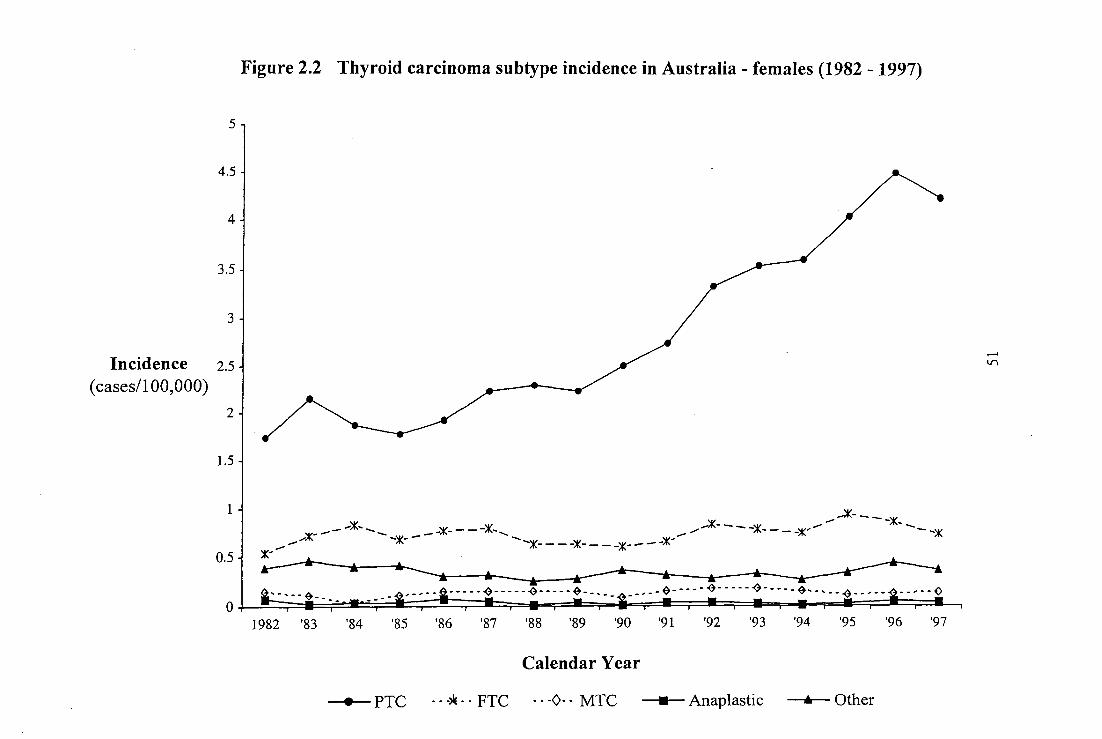
Figure 2.2 Thyroid carcinoma subtype incidence in Australia - females (1982 - 1997)
5-'
4.5 -
4
3.5 -
3
Incidence 2.5 -
(cases/100,000) 2
1.5
1
A' "'" --. - -X- - - -X- ■ -- --"K- -X- - - _x- --- - --X .- --)K- - ----X- - - -X- - - -x- - - -)K- -- -, x-
, 1982 '83
'84 '85 '86 '87 '88 '89 '90 '91 '92 '93 '94 '95 '96 '97
Calendar Year
--*-- PTC - - -* - - FTC - - -0- - MTC —R— Anaplastic --&--- Other
0.5 -
0

Figure 2.3 Thyroid carcinoma subtype incidence in Australia - males (1982 - 1997)
Incidence 2.5 -
(cases/100,000)
1982 '83 '84 '85 '86 '87 '88 '89 '90 '91 '92 '93 '94 '95 '96 '97
Calendar Year
—*-- PTC - - -* - - FTC - - P - - MTC R Anaplastic —A— Other

Figure 2.4 Papillary thyroid carcinoma subtype incidence in Australia - males & females (1982 - 1997)
0
1
1982 '83 '84 '85 '86 '87 '88 '89 '90 '91 '92 '93 '94 '95 '96 '97
Calendar Year
• PTC (Total) —x— PTC (Follicular variant)
4
3
3
2
Incidence (cases/100,000)
2 -

South Pacific Ocean
Western Australia Population 1,798 000
South Australia Population 1,480 000
Figure 2.5 Average annual increase (%pa) in PTC (M+F) - relationship to geographic iodine status
Victoria: Population 4,605 000
Indian Ocean
Tasmania W Population 474 ON 24.7%pa
History of Iodine Deficiency
Moderate Deficiency
Probable Mild Deficiency
References: 214, 217, 218, 219, 224, 225
Mild Deficiency
No history of Deficiency

CHAPTER 3
A review of thyroid carcinoma cases registered by the
Tasmanian Cancer Registry (1978-1998) with particular
reference to the accuracy of case classification
and registration.
55

Prologue
The key questions addressed by this chapter are:
1 Do changes in PTC classification or registration account for observed PTC
incidence trends?
2 What contribution do small (<1cm) tumours make to PTC incidence trends?
3 Could historical variation in risk factors such as iodine nutrition explain observed
PTC incidence trends?
Presentations and publications in relation to this Chapter:
Presented
This work was presented in part at the 42nd meeting of The Endocrine Society of
Australia, Melbourne, Australia, 1999.
Published
Burgess JR, Dwyer T, McArdle K, Tucker P 2000 The changing incidence and spectrum
of thyroid carcinoma in Tasmania (1978-1988) during a transition from iodine
sufficiency to iodine deficiency. J Clin Endocrinol Metab 85: 1513-1517.
In accordance with the criteria for submission of thesis by previously published research,
Chapter 3 is based on the above cited publication.
56
Aim
1 To assess the impact of changing histological diagnostic criteria on thyroid
carcinoma incidence patterns.
2 To determine the relationship between PTC size and overall PTC incidence trends
3 To determine if evidence exists for a relationship between historical changes in
iodine nutrition and PTC incidence trends.
Hypothesis
1 Changes in Cancer Registry case ascertainment and histopathological
classification are unlikely to fully explain observed incidence trends for TC.
2 Increased diagnosis of small PTC accounts for the majority of the observed
increase in PTC incidence.
3 Incidence trends for PTC are influenced by iodine nutrition.
57

Introduction
The previous chapter identified an increasing PTC incidence in Australia over the past
two decades. Furthermore, a state-based geographic gradient for PTC incidence was
identified in which the highest rate of increase was found in Tasmania. Tasmania is an
island state of the Commonwealth of Australia and an area of endemic iodine
deficiency(214, 215).
A number of distinct phases of iodine prophylaxis can be identified in Tasmania.
Initially, potassium iodide tablets were provided to Tasmanian school age children
between 1950 and 1965, whereas in 1966 iodine supplementation via the addition of
potassium iodate to bread commenced(214, 215). Contemporaneous with this, iodine
contamination of milk supplies by iodine residues (from a newly introduced dairy
disinfectant) also provided an additional source of dietary iodine. Consequently, a well-
documented transient increase in the incidence of iodine induced thyrotoxicosis occurred
during the late 1960's and early 1970's(216).
In 1974, iodine supplementation of bread was discontinued and, thereafter, milk provided
the primary method of community iodine repletion(214). For commercial reasons, the use
of iodine containing dairy disinfectants declined in the late 1980s. During the period
1969-1981 only 4% of school children surveyed had urinary iodine to creatinine ratios
less than 75 [ig/g, whereas, during the years 1982-1985 the percentage below 75 1.1g/g
increased to 26%(214). Persistence of mild-moderate iodine deficiency has been
confirmed by subsequent public health studies, including a 1996 analysis in which a
58

median urinary iodine excretion of 42 1.1g/L and in 2000 when median UIC was
84iig/L(217). More recently, studies have indicated the emergence of iodine deficiency
in the other Australian states(19, 218, 219).
In this chapter, the incidence a:nd spectrum of TC in Tasmania during the years 1978—
1998 is evaluated with particular reference to the accuracy of Cancer Registry
documentation and TC initial histopathological diagnosis. This period spans the
transition of the Tasmanian population from iodine sufficiency to iodine deficiency.
59

Subjects and Methods
Data provided by the Australian Bureau of Statistics show that during the period 1978—
1998 Tasmania's population increased by only 15% (62,000 persons) from a 1978
population of 413,538 persons. The male to female ratio remained stable during this
period, ranging between 0.98 and 1.00. Inpatient medical services were provided by one
tertiary referral hospital and eight smaller hospitals distributed throughout the island. By
statutory regulation the Tasmanian Cancer Registry received notification of all cases of
cancer (excluding non-melanoma skin cancer) diagnosed in the Tasmanian population.
Following approval by the Data Release Committee of the Tasmanian Cancer Registry,
all cases of TC diagnosed during the period 1978-1998 were identified. A total of 298
cases of TC were registered in Tasmania between 1978 and 1998. Histopathological
evaluation of tissue specimens has been undertaken by four pathology services. Review
of the original histopathology reports and/or allied clinical details resulted in exclusion of
nine (3%) cases that did not satisfy diagnostic criteria for a primary TC. The remaining
289 cases of primary TC were assigned to one of four diagnostic categories: PTC, FTC,
MTC, and Other TC. To determine the comparability of diagnoses recorded by archival
reports with contemporary diagnostic standards, original histology slides for all cases (n
= 134) of PTC and FTC diagnosed during even numbered years commencing 1978 were
sought for review by two histopathologists blinded to the original diagnosis.
60

Age standardized incidence rates were estimated using the world standard population age
and gender weights(211). All incidence rates are per 100,000 of population. Data were
analyzed using the Student's t test for normally distributed variables and the X2 test for
nonparametric data. Where appropriate, numerical data is presented as mean ± SEM.
61

Results
A total of 289 incident cases of TC were identified in Tasmania during the 21-year period
spanning 1978-1998. PTC, FTC, MTC, and other TC accounted for 180 (62%), 67
(23%), 12 (4%), and 30 (11%) cases, respectively (Table 3.1). Of the 30 cases of other
TC, 21(70%) were classified as either anaplastic or undifferentiated thyroid cancer. The
mean age at diagnosis was 50.8 ± 1.0 year, and the male to female ratio was 1:2.8 (Table
3.1). The median year for diagnosis was 1992. The age standardized incidence of TC
(male and female) increased by 2.3-fold (from 0.75 to 1.76 per 100,000) and 2.2-fold
(from 2.45 to 5.33 per 100,000), respectively, between 1978-1984 and 1992-1998
(Figure 3.1)(P < 0.05).
The overall increase in incidence for TC resulted predominantly from a rise in the
incidence for PTC by 4.5- and 2.1-fold in females (P < 0.05) and males (not significant),
respectively, between the periods 1978-1984 and 1992-1998 (Figure 3.2). The incidence
of other categories of thyroid cancer did not change significantly (Table 3.1). During this
time, the overall FTC/PTC ratio decreased from 0.74 to 0.24 (P < 0.005). The rise in
incidence of PTC was observed in all Tasmanian population regions, spanning all
pathology services. The rise in PTC incidence was, mostly, due to an increase in tumors
of <1 cm in diameter; however, a 3-fold rise in incidence of larger lesions also occurred
during the study period (Table 3.2).
62

Diagnoses of PTC were made at autopsy in eight (4%) cases. For non-PTC carcinoma,
the presence of multinodular histopathology as a co-pathology in thyroid specimens
remained stable during the study period (20% vs. 21%) between 1978-1984 and 1992—
1998, respectively. During this time, the prevalence of multinodular change occurring in
association with PTC increased from 11% to 36% (P < 0.05). Nine (5%) patients with
PTC had an immediate familial history of PTC in which at least one first-degree relative
was affected. This included two pairs of monozygotic twins with concordant
development of PTC. Multifocal and metastatic PTC were identified in 43 (24%) and 33
(18%) patients, respectively (Table 3.2). Seventeen (40%) patients with multifocal PTC
had bilateral tumours.
Histopathological material from 108 patients (diagnosed in the even numbered years)
was available for prospective re-examination by two histopathologists blinded to original
diagnoses. This sample represented 47% of all PTC and 34% of all FTC diagnosed
during the entire time period from 1978-'98. Diagnostic reclassification occurred for five
(6%) PTC and four (17%) FTC (Table 3.3). Of the reclassified PTC's, three were
considered to be FTC and two were classified as papillary oncocytic neoplasms. Of the
reclassified FTC, two were considered to be PTC and one each an adenoma and
anaplastic carcinoma. Reclassifications did not alter the temporal trends observed at the a
priori examination of original pathology reports.
63

Discussion
Consistent with the findings of Chapter 2, there was a significant rise in the incidence of
TC during the study period. This resulted from an increase in incidence of small FTC.
Changes in clinical practice, such as fine sectioning of thyroidectomy specimens resected
for benign indications (such as multinodular goitre, increased use of ultrasonography and
FNAB) may account for this. However, such factors are unlikely to fully explain the
observed trends. By way of example, despite a rise during recent years of multinodular
goiter occurring in association with FTC, the majority of patients with combined PTC
and multinodular goiter had evidence of clinically relevant malignancy. Of the 39 cases
of FTC associated with multinodular goiter diagnosed during the 1992-1998 period, in 8
(21%) cases the FTC was more than 3 cm in diameter, in 11 (33%) cases it was
multifocal, and in 2 (6%) cases it was metastatic. Furthermore, even when lesions of 1
cm or less in diameter are excluded, a 3-fold rise in the incidence of larger FTC was
evident (Table 3.2).
The role of iodine nutrition in the pathogenesis of TC is both complex and
controversial(29, 103, 161, 162, 220). Comparison of incidence rates between iodine-
deficient and iodine-sufficient communities yields conflicting results(95, 162).
Similarly, case control studies have also produced contradictory findings, iodine-rich
diets having been associated with both a heightened and an attenuated risk of TC(221-
223). Despite this, a relatively consistent association has been the link between iodine
nutrition and tumor histology(27, 161, 162). The incidence of FTC and anaplastic TC is
greatest in iodine-deficient populations, whereas the converse is true in regions of iodine
64

sufficiency(29, 161, 163, 220). Consistent with this observation, iodine supplementation
in previously deficient populations is associated with a rise in the proportion of PTC; so
called "papillarization" of TC(28, 29, 161, 163, 220). This is evidenced by a rise in the
proportion PTC relative to FTC(163, 166).
Few studies of iodine nutrition had been undertaken on the Australian mainland prior to
the recent National Iodine Nutrition Study (NINS) (218). Before the 1970's several
localised areas with endemic goitre were recognised on the Australian east coast(17, 19,
214, 224). Agricultural studies of ovine iodine nutrition and thyroid function indicate
iodine deficiency in grazing sheep in Tasmania, Victoria, NSW and southern QLD(17,
225). Similarly, the river systems of Australia's east coast have been shown to be iodine
deplete(17, 224). The recent NINS confirmed a similar distribution of iodine deficiency
in the contemporary human population of Australia(218).
It is likely that iodine nutrition across much of Australia was rendered adequate
unwittingly from the mid 1960's until the late 1980's by use of iodine based disinfectants
(iodophors) in the Australian dairy industry, the residue of which enhanced milk iodine
content(214). However, declining use of iodophor disinfectants over the past two decades
appears to have unmasked underlying Australian regional variations in the ecological
distribution of iodine deficiency(19, 218, 219).
It is therefore notable that in Tasmania, the increase in PTC incidence described in this
chapter has occurred despite the recurrence of iodine deficiency. A complex interaction
between iodine nutrition and thyroid tumorigenesis may account for this. A study by
65

Galanti et al. indicated a possible differential effect for iodine prophylaxis on the
spectrum of TC depending on an individual's age at the time of iodine exposure (27).
The increasing incidence and predominance of PTC despite the fall in contemporary
iodine nutrition could also reflect either a threshold for changes in iodine nutrition or a
latency between changes in iodine intake and the clinical expression of neoplastic
disease.
It is also possible the current rise in PTC incidence relates to a delayed impact of the
relative iodine excess occurring during the late 1960s and early 1970s (214, 215).
Whereas this was associated with an acute increase in the incidence of thyrotoxicosis at
the time, it may also have primed susceptible individuals, then in their childhood, for
subsequent development of PTC as adults (214, 216). If this speculation is correct, a fall
in the incidence of FTC might be expected over the following decade.
Ionising radiation is the external aetiologic factor most clearly associated with an
heightened risk of clinically significant TC(136, 137, 206). Nuclear fallout (radioiodine)
and medical sources of ionising radiation confer an increased risk of both benign and
malignant nodular thyroid disease(134, 136, 137, 139, 144). Exposure to ionising
radiation during childhood increases the likelihood of FTC developing in adult life(133,
137, 138, 209). Tumours occurring in this context are often multicentric and may
develop after a latency of several decades(133, 136, 137, 226). Moreover, PTC rates may
not reach a maximum until 30 years following the exposure(133, 136, 137, 226).
Exposure to radioiodine fallout from nuclear weapon testing is a possible link between
the distribution of ID in Australia and geographic patterns of FTC incidence(134, 226).
66
Atmospheric nuclear weapon testing occurred both in Australia and elsewhere in the
South Pacific between 1950 and 1962 (227). The highest predicted susceptibility to PTC
from fallout related to this maps to birth years 1945-1962. Children born during this
period were exposed potentially to fallout at a time when community iodine nutrition was
suboptimal, enhancing the risk of thyroidal exposure to ionizing radiation(134,214).
Medical use of ionizing radiation in childhood is also associated with an heightened risk
for PTC in adulthood(135). External beam radiotherapy was employed in the mid-
twentieth century for treatment of a diverse range of non-neoplastic conditions (135).
The prevalence of such therapy in Australia is unclear and warrants further consideration
as an etiological risk factor in the context of current PTC incidence trends.
67

Conclusions
The increased incidence of PTC observed by Cancer Registries throughout Australia in
Chapter 2 has been confirmed by detailed review of source documentation for cases
registered by the Tasmanian Cancer Registry. An increase in small (<1cm) PTC is the
major component of the observed change, although a minor increase in the incidence of
large and aggressive PTC has also occurred. Potential explanations for this include both
changes in clinical and diagnostic practice as well a rise in underlying thyroid
tumorigenesis driven by factors associated with changes in iodine nutrition or exposure
to ionising radiation. A more detailed review of clinical practice trends and risk factor
exposure is required to further assess these possibilities.
68

Table 3.1 Characteristics of thyroid carcinoma diagnosed during the calendar
years 1978-1998
Year of Diagnosis Papillary Follicular Medullary Other
1978 — 1984
n (% total TC) 27 (47) 20 (35) 2 (4) 8 (14)
Crude Rate 0.90 0.67 0.06 0.26
Age 46.8±2.9 57.8±3.6 42.5±21.0 71.8±3.9
M:F 1 : 2 1 : 5.7 0 : 2 1: 1.7
1985- 1991
n (% total TC) 43 (54) 21 (26) 3 (4) 13 (16)
Crude Rate 1.36 0.66 0.09 0.41
Age 47.6±2.5 49.4±3.6 50.4±8.8 65.6±4.9
M:F 1: 3.3 1: 3.2 1: 2 1: 1.6
1992 - 1998
n (% total TC) 110 (72) 26 (17) 7 (5) 9 (6)
Crude Rate 3.32 0.78 0.06 0.27
Age 45.9±1.3 54.7±2.3 57.2±5.8 70.9±5.7
M:F 1: 4.2 1: 1.4 1: 0.8 1: 0.8
M:F — Male : Female ratio Crude rate — Crude incidence rate per 100 000 of population
69

Table 3.2 Changes in the spectrum of papillary thyroid carcinoma diagnosed
during the periods 1978-1984, 1985-1991 and 1992-1998
Characteristic
1978 - 1984** 1985 - 1991 1992 - 1998**
n (%) M:F n (%) M:F n (%) M:F
27 1: 2 43 1:3.3 110 1:4.2 Total cases
Size <1.1 7 (26) 1: 2.5 12 (28) 1: 5.0 49 (45) 1: 5.1
Size 1.1-2.0 8 (30) 1: 1.7 15 (35) 1: 4.0 33 (30) 1 : 4.5
Size 2.1-3.0 5 (19) 1: 1.5 9 (21) 1: 3.5 13 (12) 1: 5.5
Size >3.0 5 (19) 1: 1.5 7 (16) 1: 1.3 13 (12) 1: 2.3
Multinodular pathology 3 (11) 1: 0.5 12 (28) 1: 5.0 39* (36) 1: 3.9
Incidental finding 3 (11) 1: 2.0 9 (21) 1: 8.0 33 (30) 1: 6.0
Multifocal primary 1 (4) 0: 1.0 11 (26) 1 : 4.5 31 (28) 1 : 2.9
Regional invasion 3 (11) 1 : 2.0 7 (16) 0 :7.0 12 (11) 0: 12
Metastases 7 (26) 1 : 0.4 10 (23) 1 : 9.0 16 (15) 1: 1.3
Deceased 6 (22) 1: 1 8 (19) 1: 1.7 6 (6) 1: 1
M:F - Male : Female ratio
Size - Tumour size
Multinodular pathology - Histopathological evidence of multinodular change in thyroid tissue.
**Size unknown in 2 cases of papillary carcinoma.
*Of these 39 cases of thyroid carcinoma, 8 were >3cm in diameter, 11 were multifocal, 2 were metastatic, and 16 were incidental findings.
70

Table 3.3 Comparison of original and contemporary histolopathological diagnoses
for 108 cases of differentiated thyroid carcinoma.
1978 - 1984 1985 - 1991 1992 - 1998
n= % n= % n= %
Orignial diagnosis- PTC 18 20 62
Cases available for review* 15 83 16 80 54 87
- PTC confirmed** 13 87 15 94 52 96
Orignial diagnosis- FTC 10 10 14
Cases available for review* 6 60 5 50 12 86
- FTC confirmed** 4 67 5 100 10 83
Original diagnosis - Diagnosis as recorded on the original histopathology report. *Histopathology slides were located and reviewed by pathologist. Percentage represents the proportion of patients for whom slides were located. **Re-examination of histopathology produced a diagnosis concordant with original diagnosis. Percentage represents the proportion of patients for whom the review diagnosis was concordant with the orignial diagnosis.
71

8
6
4 -
2
I I
1977 1980 1983 1986 1989 1992
Calendar Year
0 19195
• 1998
Figure 3.1 Age standardised incidence rate of thyroid cancer in Tasmania 1978 - 1998
Male Ea Female
Inci
denc
e (r
ate
per
10
020
00)

• r- CO ON Os 01
CO 01 0 Csi 1'0 V- 10 h— CO 01 0 •—!.. hi 1'0 LO r- r- co
▪
co CO CO CO CO CO CO CO 01 0% OS 01 0% cr. us a% a% cr. a% ON Ch 01 01 01 01 0% cr. cr. Ch
Figure 3.2 Thyroid cancer histology and year at diagnosis
Calendar Year
Tumour histology • Papillary Follicular in Other
25 -
20 -
15
ncid
ent Ca
ses
Num
ber
01
10
5
0

ncid
ent
Ca
ses
N
um
ber
of
Figure 3.3 Sex distribution for incident cases of PTC
20 —
1 5
1 0
5
0 CO 0% 0 Cs1 1,0 lf) I-- CO Os 0 Cs1 tO !XI r-- CO r- r- co c7-3 co co co co co co co co a, F. Cis Crs Os Os Os Os Os Os as Os CIS as I:Ts 0% Os Os Os 0% 0% Cus Cis OS Crs Os Os OS
Calendar Year
III Female Male
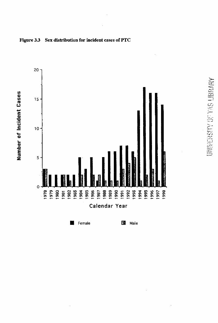
Calendar Year
• Female Male
Num
ber
of In
cide
nt Ca
ses
Figure 3.3 Sex distribution for incident cases of PTC
20 -
15 -
10 -1
5
0

CHAPTER 4
Incidence trends for PTC and their correlation with trends for
thyroid surgery and thyroid FNAB cytology
75

Prologue
The key questions addressed in this Chapter are:
What is the completeness of Tasmanian Cancer Registry case ascertainment for
diagnoses of PTC?
2 Has there been a change in patterns for utilization of FNAB and thyroid surgery?
3 Is there a relationship between thyroid imaging, FNAB and PTC incidence
trends?
Presentations and publications arising from this Chapter:
Presented
Preliminary results resented at the Endocrine Society of Australia, 2002 ASM, Adelaide,
Australia.
Published
Burgess JR, Tucker P. 2006 Incidence trends for papillary thyroid carcinoma and their
correlation with thyroid surgery and thyroid fine needle aspirate cytology. Thyroid 2006;
16: 47-53.
In accordance with the criteria for submission of thesis by previously published research,
Chapter 4 is based on the above cited publication.
76

Aim
1 To confirm the adequacy PTC case ascertainment by the Tasmanian Cancer
Registry.
2 To determine the relationship between thyroid surgical and cytological procedures
and temporal trends for PTC diagnosis.
Hypothesis
Incidence trends for PTC are influenced by changing paradigms for evaluation and
management of nodular thyroid disease.
77

Introduction
Previous chapters demonstrated an increase in incidence of PTC in Australia generally,
and Tasmania in particular(213, 228). Detailed review of source documentation and
tissue samples for cases registered by the Tasmanian Cancer Registry confirmed both the
accuracy of case registration as well as the consistency of histopathological classification
over time. However, the potential for changes in TC case notifications over time could
not be assessed by the methodology utilized in previous chapters. Progressive and
systematic improvements in Cancer Registry case ascertainment is potentially an
important source of bias.
Prior chapters have also linked the ecological distribution of iodine deficiency in
Australia with PTC incidence trends(17, 218, 228). As iodine deficiency is often
associated with goitre monitoring programs, it is possible increased screening accounts
for the majority of observed changes in PTC incidence(71, 200, 229, 230). In support of
this I have previously identified an increase in the incidence of tumours <1.0cm, lesions
which most likely represent asymptomatic and non-palpable PTC (occult PTC) (71, 170,
228, 230). Such PTC generally have a "biologically benign" natural history (41, 71, 202,
205). It is therefore possible that identification of non-palpable nodules by
ultrasonography, and their subsequent cytological evaluation by FNAB, has merely
increased the diagnosis of small and mostly clinically inconsequential tumours (41, 202,
212, 230). At present however, there is a paucity of data directly relating trends for PTC
incidence to trends in the use of neck ultrasonography, thyroid FNAB cytology and
thyroid surgery (230). Such information would assist in determining the role, if any
78

contemporary medical practice has had on PTC incidence trends.
Using the Tasmanian population as a model, this study sought to determine the
relationship between changes in PTC incidence and trends for utilization of neck
ultrasonography, thyroid surgery and thyroid FNAB cytology over an 11-year period
during which PTC incidence increased more than two fold(213).
79

Subjects and Methods
Hospital and pathology services in Tasmania submitted data relating to thyroid surgical,
histopathological and cytological procedures undertaken between January 1988 and
December 1998. This period was selected given the availability of comprehensive data
relating to surgical procedures and pathology outcomes, in conjunction with the
availability of accurate baseline cancer registry data for thyroid carcinoma (213).
Furthermore, during this period Tasmania's population increased by less than 15% (213).
In subsequent years population flux increased and data availability diminished.
Data was cross-referenced against diagnoses of thyroid carcinoma independently
assembled by the Tasmanian Cancer Registry - a statutory authority that receives
notification of all cases of cancer (excluding non-melanoma skin cancer) diagnosed in
the Tasmanian population (213). All tumours designated as primary thyroid carcinoma
were assigned to one of four diagnostic categories; PTC, FTC, MTC, and "Other
diagnoses" (213, 228). The category of "Other diagnoses" comprised anaplastic thyroid
carcinoma, Hurthle cell malignancy and "carcinoma not otherwise specified". Cytology
results from FNAB thyroid were classified into four categories; unsatisfactory sample
(U), benign lesion (B), indeterminate lesion (I) and malignant (M). The indeterminate
category comprised follicular lesions, oncocytic lesions, and other indeterminate or
suspicious cytological appearances.
80
In addition, patients identified by the Tasmanian Cancer Registry with a diagnosis of
PTC during the period 1978-1998 (n=180, Female:Ma1e=3.5, Age at diagnosis 46.4±1.1
yeas) were also sought to determine the preoperative likelihood of TC. Of these, 99
patients (Female:Male=4.8, Age at diagnosis 44.5±1.0 years) were living, contactable
and consented to provide information regarding the original indication for thyroid
surgery.
Normally distributed variables were analysed using the Student's t-test. Time trends in
incidence were analysed by linear regression of the rates on year at diagnosis. Numerical
data is presented as Mean ± Standard Error of Mean (SEM). Age standardised incidence
rates (ASR) were estimated using the World Standard Population age and gender
weights, and are expressed per 100 000 of population (211). The study was approved by
the Royal Hobart Hospital Research Ethics Committee and the Data Release Committee
of the Tasmanian Cancer Registry.
81

Results
In the years 1988-1998 a total of 3452 individuals underwent a thyroid procedure,
comprising 1968 surgical procedures in 1920 patients (F:M 4.5:1) and 1756 FNAB
cytological procedures in 1532 patients (F:M 5.3:1). A new diagnosis of TC was made in
184 patients with a female to male ratio of 3:1. Of these, 175 patients had an
histologically confirmed diagnosis - 121 (65.8%) PTC, 40 (21.7%) FTC, 7 (3.8%) MTC
and 17 (9.2%) "Other diagnoses. When PTC cases registered by the Tasmanian Cancer
Registry were compared with the results of the current study, only 13 previously
unrecognised cases of PTC were identified, indicating Registry ascertainment for the
period 1988-1998 was 93.9% complete (Figure 4.1).
Forty patients with a malignant diagnosis underwent an initial subtotal thyroidectomy
followed by completion thyroidectomy once malignancy was identified. Nine (4.9%)
cases were diagnosed by FNAB cytology alone, without histopathological material being
available for subsequent evaluation. Of 175 patients with an histopathological diagnosis
of TC, 70 (40%) had at least one preoperative assessment using FNAB cytology (14
benign cytology, 42 indeterminate, 12 malignant, 7 unsatisfactory and one result
unavailable for review). Of the 42 patients with indeterminate cytological results -
follicular lesions accounted for 24, oncocytic lesions 4, and other atypical appearances
were reported in 14 cases.
82

During the study period the age standardized rate for thyroidectomy increased by 6.8%
per year for females and 10.9% per year for males (p< 0.05) (Table 4.1, Figure 4.2).
Similarly, FNAB usage increased at a rate of 66.2% per year for females and 17.6% per
year for males (p< 0.005) (Table 4.1, Figure 4.2). During this time the median age at
thyroidectomy and FNAB remained stable at 48 years and 49 years respectively. There
was an 147.6% per year increase in the use of preoperative FNAB prior to thyroidectomy
(p< 0.001) and no significant increase in thyroidectomy without prior FNAB (0.1% per
year increase, p=0.24) (Table 4.1). In the latter part of the study period preoperative
FNAB exhibited less restriction to lesions ultimately found to be malignant than was the
case initially (Table 4.2, 4.3).
The likelihood of diagnosing a PTC in any given thyroidectomy specimen increased from
3.3% in 1988 to 7.7% in 1998 (Table 4.2). Diagnoses of PTC in patients previously
assessed by FNAB increased by 99.7% per year (p<0.005), whilst for those in whom a
prior FNAB was not performed the increase was 10.1% per year(p<0.05). The increase
in PTC for patients assessed by FNAB was evident for tumours <1 cm as well as those
>lcm diameter — although the increase was greater in the former (Table 1). Although not
reaching statistical significance, there was also a trend (10.1% per annum) for increase in
PTC >lcm without history of pre-operative FNAB evaluation (Table 4.1).
To evaluate prevalence trends for incidental diagnoses of PTC, all patients identified by
the Tasmanian Cancer Registry with PTC during the period 1978-1998 were sought for
completion of a questionnaire regarding the preoperative likelihood of malignancy.
There was a 63.6% increase in the diagnoses of PTC _lcm diameter in individuals with a
83

low preoperative likelihood of malignancy, and this accounted for the majority of
increased PTC diagnoses between the time periods 1978-'91 and 1992-'98 (Table 4.4).
84

Discussion
This study suggests an increase in the diagnosis of small PTC resulting from
contemporary clinical practices (increased neck imaging and FNAB) is likely to account
for the majority of the observed increase in PTC incidence (71, 230). Improvements to
Cancer Registry case ascertainment does not account for the rise in PTC incidence.
Whilst it is unlikely my methodology captured every eligible thyroid procedure, this
study does provide a robust estimate of total thyroid surgical and cytological procedures
performed in the Tasmanian population during the study period. Over this time, only
thirteen cases of thyroid carcinoma not originally registered by the Tasmanian Cancer
Registry were identified (Figure 4.1). Similarly, a previous review of tumours recorded
by the Tasmanian Cancer Registry (presented in Chapter 3), determined that incorrect
histological classification either at the Registry, or at the time of original histological
diagnosis, occurred in less than 10% of cases (213). Thus, there is no suggestion that
procedural changes in relation to Cancer Registry case ascertainment have resulted in
observed PTC incidence trends.
Thyroidectomy and FNAB cytology rates have increased by 7% per year and 49.7% per
year respectively in Tasmania (Table 4.1). As evidenced by the increased use of FNAB in
patients ultimately diagnosed with benign lesions and small PTC, there appears to have
been a trend towards decreased restriction of pre-operative FNAB to patients at high a
priori cancer risk (Table 4.2). Particularly in the later years of the study period, not only
was cytology used more frequently, it was more likely to be used less selectively -
preoperative cytological findings and final histology were less concordant.
85

Broader use of FNAB is further suggested by the increasing proportion of patients
undergoing pre-operative FNAB cytology for whom the ultimate diagnosis was a benign
lesion (Table 4.2, 4.3). It appears FNAB was initially reserved for "high" risk patients,
but in more recent years FNAB seems to have been offered more generally to "lower"
risk individuals. This is in keeping with promotion and acceptance of an evaluation
protocol for thyroid nodules that recommends FNAB for the majority of thyroid nodules
found in patients with a non-suppressed TSH (71, 75, 76, 212).
Whilst it is not possible to directly quantify the effect of increased use of neck imaging
(CT and ultrasonography) on the identification of "occult" PTC, there has been a
substantial rise in the use of neck imaging since the early 1980's. Neck ultrasonography
in particular appears to have become widely used in primary medical practice for the
evaluation of non-specific neck symptoms and vascular conditions, as well as for
clinically evident thyroid indications. Australian government data show the number of
neck ultrasound procedures performed in Tasmania increased markedly between 1993 —
1998 (Figure 4.3).
In the context of increased use of medical imaging and contemporary medico-legal
sensitivities it is not surprising that current trends are seen. Impalpable and
asymptomatic thyroid nodules are a common finding when the thyroid is imaged for non-
nodular indications or evaluated at autopsy. The prevalence of PTC diagnosed
antemortem is approximately 0.1%, versus up to 13% for occult PTC in autopsy series
(170, 202, 205). Whilst most nodules identified by neck imaging are benign (colloid
nodules and follicular adenomas), an important minority are PTC, the majority which are
86

5_1cm in diameter. Therefore increased use of neck ultrasound imaging in conjunction
with consequential FNAB cytological evaluation could account for much of the observed
increase in diagnoses of PTC .1cm diameter (201, 205, 210, 228, 230).
Most studies of the natural history of such lesions have concluded these "occult" FTC
mostly follow an indolent course and have an excellent prognosis (83, 182). However a
proportion of incidental thyroid carcinoma are destined to become a clinically relevant.
The study by Nam-Goong et al showed that a proportion of incidental identified PTC
were associated with extrathyroidal extension or metastases (83). At present however,
there is no reliable way of selecting for excision those lesions destined to produce
clinically relevant malignancy, whilst sparing the majority of patients with indolent
disease from an unnecessary thyroidectomy. The standard of care therefore remains
resection of thyroid "incidentalomas" if initial cytology is suspicious for malignancy.
The possibility of a true biological change affecting the incidence of FTC cannot be fully
excluded by this study. The trend for increased incidence of larger (>1cm) FTC, suggests
that improved recognition of "occult" FTC may not account for all of the observed
incidence increase (Table 4.3). In particular, the incidence of PTC >1 cm in patients for
whom a preoperative FNAB had not been undertaken, has also increased by 10.1%
annually. Whilst a proportion of these tumours may have been clinically "occult", it is
likely many were symptomatic or clinically palpable at presentation. This is supported
by the findings presented in Table 4.4.
Therefore, a complex mix of underlying processes is likely to have resulted in the change
to PTC incidence observed during the latter part of the 20 th century (60, 139, 163, 170,
202, 206). The current study suggests the increased PTC incidence is mostly due to
87

identification of "occult" PTC (greater use of ultrasonography and FNAB cytology, lower
thresholds for thyroidectomy and heightened scrutiny during histopathological
assessment). In addition a smaller but real increase in underlying PTC incidence may also
have occurred.
88

Conclusions
Over the past two decades there has been a substantial increase in the use of neck
ultrasonography and consequently thyroid FNAB cytology to evaluate small and
otherwise asymptomatic thyroid nodules. Thyroidectomy rates appear to have risen in
response to increased use of FNAB and the consequential increase in reporting of
suspicious cytology. However, the rise in incidence of PTC >lcm diameter suggests that
a true increase in PTC incidence rates has also occurred. Given parallels between the
overall change in PTC incidence observed in Tasmania as well as other parts of Australia
as well as globally, the findings of the current study are likely to be of value in
understanding the world-wide rise in PTC incidence observed in recent decades.
-, Additional studies are also required to evaluate the role of established risk factors (such
as ionising radiation and genetic susceptibility), as well as further elucidating the role of
iodine nutrition in trends for PTC.
89
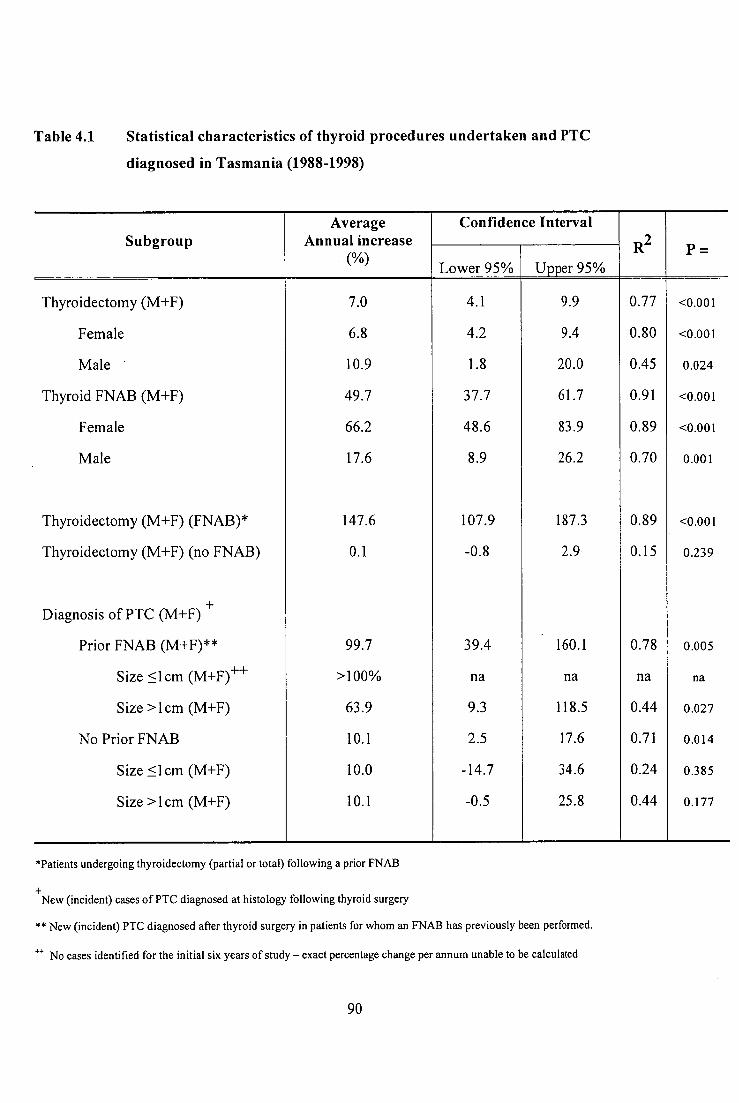
Table 4.1 Statistical characteristics of thyroid procedures undertaken and PTC
diagnosed in Tasmania (1988-1998)
Subgroup Average
Annual increase (%)
Confidence Interval
R2 p = Lower 95% Upper 95%
Thyroidectomy (M+F) 7.0 4.1 9.9 0.77 <0.001
Female 6.8 4.2 9.4 0.80 <0.001
Male 10.9 1.8 20.0 0.45 0.024
Thyroid FNAB (M+F) 49.7 37.7 61.7 0.91 <0.001
Female 66.2 48.6 83.9 0.89 <0.001
Male 17.6 8.9 26.2 0.70 0.001
Thyroidectomy (M+F) (FNAB)* 147.6 107.9 187.3 0.89 <0.001
Thyroidectomy (M+F) (no FNAB) 0.1 -0.8 2.9 0.15 0.239
Diagnosis of PTC (M+F) +
Prior FNAB (M+F)** 99.7 39.4 160.1 0.78 0.005
Size <lcm (M+F) ++ >100% na na na na
Size >lcm (M+F) 63.9 9.3 118.5 0.44 0.027
No Prior FNAB 10.1 2.5 17.6 0.71 0.014
Size lcm (M+F) 10.0 -14.7 34.6 0.24 0.385
Size >lcm (M+F) 10.1 -0.5 25.8 0.44 0.177
*Patients undergoing thyroidectomy (partial or total) following a prior FNAB
New (incident) cases of PTC diagnosed at histology following thyroid surgery
** New (incident) PTC diagnosed after thyroid surgery in patients for whom an FNAB has previously been performed.
++ No cases identified for the initial six years of study - exact percentage change per annum unable to be calculated
90
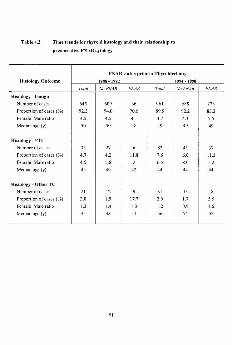
Table 4.2 Time trends for thyroid histology and their relationship to
preoperative FNAB cytology
Histology Outcome
FNAB status prior to Thyroidectomy
1988 - 1992 1994 - 1998 Total No FNAB FNAB Total No FNAB FNAB
Histology - benign Number of cases 645 609 36 961 688 273 Proportion of cases (%) 92.3 94.0 70.6 89.5 92.2 83.2 Female :Male ratio 4.5 4.5 4.1 4.7 4.1 7.5 Median age (y) 50 50 48 49 49 49
Histology - PTC Number of cases 33 27 6 82 45 37 Proportion of cases (%) 4.7 4.2 11.8 7.6 6.0 11.3 Female :Male ratio 4.5 5.8 2 6.5 8.0 5.2 Median age (y) 45 49 42 44 44 44
Histology - Other TC Number of cases 21 12 9 31 13 18 Proportion of cases (%) 3.0 1.9 17.7 2.9 1.7 5.5 Female :Male ratio 1.3 1.4 1.3 1.2 0.9 1.6 Median age (y) 45 48 41 56 74 52
91

Table 4.3 Time trends for thyroid histology and their relationship to pre-
operative cytological findings
Histology Outcome Pre-operative FNAB findings
1988 - 1992 1994 - 1998
Histology - benign Number of cases 32 21 nil 8 148 99 1 46 Proportion of cases (%) 94.2 67.8 100 91.9 75.0 11.1 88.5 Female :Male ratio 3.6 6.0 8:0 6.8 8.9 1:0 5.3 Median age (y) 44 50 47 49 45 17 51
Histology - PTC Number of cases 1 5 1 nil 11 18 7 5 Proportion of cases (%) 2.9 16.1 25.0 6.8 13.6 78.8 9.6 Female :Male ratio 1:0 1.5 1:0 2.7 8.0 6.0 4.0 Median age (y) 36 41 50 45 43 38 48
Histology - Other TC Number of cases 1 5 3 0 2 15 1 1 Proportion of cases (%) 2.9 16.1 75.0 1.3 11.4 11.1 1.9 Female :Male ratio 0:1 1.5 0.5 3:0 1.5 0:1 0:1 Median age (y) 41 41 67 59 52 54 59
92

Table 4.4 Preoperative suspicion of thyroid carcinoma and operative findings for
99 cases of papillary thyroid carcinoma categorized by year of diagnosis.
PTC Characteristics 1978 - 1984 1985 - 1991 1992 - 1998 n (%) M . F n (%) M F n (%) M: F
Total Cases 8 1:7 19 1:3.8 72 1:4.5
Thyroid carcinoma unexpected* 3(37.5) 1:2 8(42.1) 1:3 32 (44.4) 1:5.4
FTC <1.0cm 0 - 3 (15.8) 1:3 17 (23.6) 2:7.5
FTC >3.0cm or Invasive or Metastatic 1(12.5) 1:0 4(21.1) 1:3 6(8.3) 0:6
Thyroid carcinoma expected** 5(62.5) 0:5 10 (52.6) 1:4 38 (52.7) 1:3.8
PTC <1.0cm 2(25.0) 0:2 2(10.5) 0:2 12 (16.7) 1:5
PTC >3.0cm or Invasive or Metastatic 1(12.5) 0:1 4(21.1) 0:4 15 (20.8) 1:2
Based on response to questionnaire completed by 99 patients with PTC diagnosed between 1978-1998. Thyroid carcinoma clinically expected preoperatively
** Thyroid carcinoma not clinically expected preoperatively and identified incidentally at time of thyroidectomy
93

Figure 4.1 Completeness of thyroid carcinoma case ascertainment by Tasmanian Cancer Registry (1988 - 1998)
100
90 -
80 -
70 -
% Registration
Complete
60 -
Ch
50 -
40 -
30 -
20 -
1 0 -
0 1988 '89 '90 '91 '92 '93 '94 '95 '96 '97 '98
Calendar Year
—4°— Total TC (Male+Female) ' -() ' PTC (Male+Female)

Figure II Trends for Thyroid Surgery and FNAB cytology in Tasmania (1988 - 1998)
50 -
45 -
40 -
35 -
30 -
ASR 25 - (Cases / 100000)
20 -
15 -
10 -
5 -
0 1988 '89 '90 '91 '92 '93 '94 '95 '96 '97 '98
Calendar Year
—,i,-- - -•-• - Thyroid surgery (Male + Female) FNAB (Male + Female)

Figure III Trends for Neck ultrasonography in Tasmania (1993 - 1998)
Neck Ultrasound 80 - (Procedures / Month)
Jul- Jan- Jul- Jan- Jul- Jan- Jul- Jan- Jul- Jan- Jul- 93 94 94 95 95 96 96 97 97 98 98
Calendar Months
Data sourced from Australian Health Insurance Commission web site. http://www.hic.gov.au/statistics

CHAPTER 5
The Impact of Birth and Residence in Tasmania on the
Prevalence of Benign and Malignant Thyroid Disease
97

Prologue
The key questions addressed by this chapter are:
1 What is the prevalence of benign and malignant thyroid disease in Tasmania?
2 What is the prevalence of subclinical nodular thyroid disease in Tasmania
3 Is there evidence for an increased risk for thyroid disease based on birth and
subsequent residence in Tasmania?
Presentations and publications arising from this Chapter:
Presented
Preliminary results presented at the 49th meeting of The Endocrine Society of Australia,
Gold Coast, Australia, 2006.
The study described in this chapter was undertaken between 1999-2000 prior to
introduction of community iodine prophylaxis in 2001.
98

Aim
1 To determine the prevalence of clinically diagnosed benign and malignant thyroid
disease in the Tasmanian population.
2 To determine the prevalence subclinical thyroid nodularity in the Tasmanian
population.
3 To determine if longterm residence in Tasmania is associated with an heightened
risk for either benign or malignant thyroid disease.
Hypothesis
1 More severe historical iodine deficiency in Tasmania relative to other
Australian states should manifest as an higher rate of benign and malignant
thyroid disease in long-term Tasmanian residents
2 Increasing rates for PTC diagnosis in Tasmania and other previously iodine
deficient regions of Australia are the result of an high underlying prevalence
of both clinical and subclinical benign thyroid disease, resulting in an
increased likelihood of diagnosing "occult" PTC.
99

Introduction
Factors such as genetic susceptibility iodine deficiency and ionising radiation influence
the aetiogenesis of goitre, thyroid neoplasia and thyroid dysfunction(28, 29, 133, 136,
206, 222). As discussed in prior Chapters, the influence of iodine nutrition on the
pathogenesis and PTC remains poorly understood. Some studies suggest a direct
relationship between increasing iodine nutrition and PTC incidence however, the impact
age at exposure to iodine deficiency/sufficiency has on PTC risk remains unclear(28, 29,
131, 165, 166, 169).
Iodine deficiency may modify the clinical presentation of benign thyroid disease, thereby
increasing the prevalence of goiter and increase the likelihood of diagnosing "occult"
PTC by (45, 50, 57). Greater use of ultrasonography and surgery for multinodular goiter
evaluation and treatment are likely to accentuate this in iodine deficient populations (71,
173).
In this Chapter an health survey and ultrasonographic study was undertaken to determine
the prevalence of both subclinical and previously diagnosed thyroid disease in the
Tasmanian population. Specific attention is given to the influences of birth year, birth
place, and place of long-term residence on the development of thyroid disease to
determine if early life exposure to Tasmanian environmental factors such as iodine
nutrition accounts for the contemporary pattern of TC incidence in Tasmania.
100
Subjects and Methods
Following approval of the Royal Hobart Hospital Research Ethics Committee a random
sample of 10 000 adults registered on the Tasmania electoral role in the year 1999 were
invited to complete a health questionnaire regarding thyroid disease and related risk
factors. The survey was conducted in late-1999 with 5774 respondents available for
assessment.
Of these, 463 age stratified respondents were selected at random for invitation to undergo
thyroid ultrasonography. Of these, 205 agreed to participate and underwent imaging.
Thyroid evaluation was performed by experienced sonographers using a 7.5 mhz
transducer. Scanning was undertaken with the subject supine and the neck
hyperextended. Thyroid volume (m1) was calculated using the formula: width (cm) x
length (cm) x thickness (cm) x 0.479 with the volume of each lobe summed. Goitre was
defined as thyroid volume greater than 18.1m1 in males and 13.0m1 in females(24).
Whilst less sensitive than comparison based on body surface area normative criteria,
insufficient anthropomorphic data was available to undertake this calculation. The level
of early life exposure to the Tasmanian environmental factors such as iodine deficiency
was indirectly assessed by two criteria 1) birthplace Tasmania and <1 year lifetime
residence elsewhere (High exposure), 2) birthplace not Tasmania and >10 years lifetime
residence outside Tasmania (Low exposure).
In addition, patients identified by the Tasmanian Cancer Registry with a diagnosis of
PTC during the period 1978-1998 (n=180, Female:Ma1e=3.5, Age at diagnosis 46.4±1.1)
101

were sought to determine place of birth and long-term residence. Of these, 99 patients
(Female:Male=4.8, Age at diagnosis 44.5±1.0) were living, contactable and consented to
provide this information.
Normally distributed variables were analysed using the Student's t-test. Where
appropriate, numerical data is presented as Mean ± Standard Error of Mean (SEM).
Univariate and multivariate analyses were performed using GraphPad InStat version 3.0a
for Macintosh, GraphPad Software, San Diego California USA.
102

Results
Of 5774 survey respondents (male 2746, female 3028) a past history of TD was reported
by 580 (10.0%), (male 106 (3.9%) and female 474 (15.7%)) (Table 5.1). An history of
thyroid carcinoma was described by 35 (0.6%), (male 10 (0.4%) and female 25 (0.8%)).
Goitre was reported by 241(4.2%) (male 47(1.7%), female 194(6.4%)) and an history of
thyroid imaging by 238 (4.1%). Thyroxine use was reported by 2.3% of survey
respondents (male 12 (0.4%), female 123 (4.1%)); of whom a past history of thyroid
surgery and/or goiter was described by 59.3%. Hyperthyroidism was reported by 167
(2.9%), (male 34 (1.2%), female 133 (4.4%)) (Table 5.1). Thyroid surgery was described
by 162(2.8%) (male 29(1.1%), female 133(4.4%)) of whom, 76 (46.9%) also described
an history of thyroid imaging.
Compared to individuals born and resident life-long in Tasmania (n =3150, F:M ratio =
1.2, age = 51.5±0.3 years), those not born in Tasmania and spending more than ten years
elsewhere (n=1319, F:M ratio 1.0, age 58.210.4 years, mean years lived outside
Tasmania 33.2±0.4 years) had a significantly (p<0.05) lower prevalence of thyroid
problems overall, and goitre in particular (Table 5.2). Thyroid cancer was non-
significantly increased amongst individuals born and life-long resident in Tasmania
(0R=1.3, p=0.751) (Table 5.2).
Ultrasonography revealed thyroid nodules in 43.4% of individuals undergoing
ultrasonography (Table 5.2, 5.3). Nodules were at least as frequent in those born and
long term resident in Tasmania as for individual born elsewhere (Table 5.2). Many
103

patients (88.8%) with thyroid nodules on ultrasound did not report a previously
diagnosed history of thyroid disease (Table 5.4).
Stratification of thyroid disease by birth cohort to reflect likelihood of iodine deficiency
in childhood/ adolescence is presented in Table 5.5. The birth cohort 1925-'40 who were
born and resident in Tasmania can be considered relatively iodine deficient in childhood/
adolescence. Childhood/ adolescent, iodine nutrition had improved and was relatively
similar irrespective of birthplace and subsequent residence for the 1955-'70 birth cohort
due to the nationwide effect of iodophors on iodine nutrition. In a model adjusting for
age and gender, only history of "any thyroid disease" (p=0.002), goitre (p=0.002) and
hyperthyroidism (p=0.024) were significantly associated with birth and residence in
Tasmania for birth cohort 1925-'40. No significant association between thyroid disease
and birth and residence in Tasmania was found for birth cohort 1955-'70 (Table 5.5).
In a sub-study of an additional 99 PTC patients for whom information regarding place of
birth and subsequent residence was available, 57.6% were born in Tasmania and had
never resided elsewhere for more than 12 months. There was no correlation between
PTC size and patient demographic profile, with 18 of 36 (50.0%) PTC _I cm associated
with dominant residence in Tasmania compared to 39 of 62 (62.9%) tumours >1 cm
diameter (ns).
104

Discussion
This study indicates that thyroid disorders (including their investigation and treatment)
are common, occurring with prevalence similar to that of health problems such as
diabetes and hypertension (Table 5.1). These findings are similar to those from a large
Norwegian population survey of thyroid disease in which hyperthyroidism was reported
by 2.5% and 0.6% of females and males respectively and hypothyroidism in 4.8% and
0.9% respectively, and goitre described by 2.9% of females and 0.4% of males(231).
Tasmania's history of relatively more pronounced iodine deficiency appears responsible
for the increased likelihood of thyroid disease and thyroid surgery amongst long-term
residents born prior to the correction of iodine deficiency. Interestingly, subclinical
thyroid nodularity was frequent irrespective of birth place and place of dominant lifetime
residence in the 1960's. Similarly, long-term Tasmanian residence did not confer a
significantly elevated risk for malignant thyroid neoplasms.
The potential for thyroid ultrasonography to reveal "occult" thyroid nodularity is well
recognised and highlighted by the current study(41, 60, 173, 232). In accordance with
prior research, the prevalence of thyroid nodules on ultrasound was approximately ten-
fold higher than that of self reported thyroid disease, with age the key predictor for their
presence (Table 5.2, 5.3) (50, 57, 60). This confirms the anticipated divergence between
the absolute risk for nodular thyroid disease (based on ultrasonography) and the
likelihood of clinically diagnosed thyroid nodularity. Notably, whilst the former appears
uninfluenced by place of birth and residence, the later was increased amongst individuals
105

reporting prolonged residence in Tasmania (particularly in their childhood during the
iodine deficient era prior to the 1950's).
Whilst the current results supports the well established link between iodine deficiency
and goitre (simple, multinodular and functionally autonimous), they do not show a
significant association between the absolute risk for development of nodular thyroid
disease / neoplasia and iodine nutrition. Age as opposed to unique environmental factors
appears to be the key determinant of absolute prevalence of thyroid nodularity.
Thyroid carcinoma was non-significantly higher for in individuals born and resident long
term in Tasmania versus those born elsewhere. However, there was no convincing
evidence for a specific environmental exposure (such as iodine nutrition) to explain PTC
incidence trends. The birth cohort born after introduction of iodine
supplementation/iodophors appear to be at no greater risk of thyroid disease irrespective
of place of birth residence(214, 216). This suggests that the pathogenesis of PTC is not
influenced by the absolute increase (greatest in Tasmania) in iodine nutrition. However,
the relative comparison methodology used by this study does not account for possible
shared exposure to common PTC risk factors affecting both those born and resident long-
term in Tasmania as well as those migrating to Tasmania from elsewhere.
The influence of early life iodine nutrition on thyroid neoplasia could be underestimated
by this study if: a) the historical differential in severity of iodine deficiency between
Tasmania and the other south-eastern Australian states was less pronounced than
generally assumed, b) the assumption that migrants to Tasmania derived predominantly
106

from relatively iodine sufficient regions is incorrect (eg. post second world war migration
from iodine deficient regions of Europe), and c) common levels of iodine nutrition across
Australia in more recent years have modified the impact any difference in childhood
iodine nutrition may have had on nodular thyroid disease and thyroid volume. It is also
notable that whilst the relative increase in TC incidence is highest in previously iodine
deficient regions of Australia, the absolute incidence of PTC in these areas was lower at
baseline. Hence iodine repletion via milk during the 1960's-1980's may have relatively
affected PTC incidence more in previously deficient areas.
Whilst the results from the current study are prone to bias (self-reported medical history
is influenced by the disease in question and the temporal proximity of disease occurrence
to time of survey), thyroid diseases have been shown to be moderately well reported by
self-administered questionnaire compared to in-person interview. In the study by Brix et
al the self-reported diagnosis of hyperthyroidism and hypothyroidism were both 98%
although the specificity was mediocre at 57% and 67% respectively(233). Similarly, an
assessment of self-reported cancer incidence in the California Teachers Study found the
concordance for thyroid cancer between survey results and cancer registry data was at
least 90% sensitive and specific (234).
107

Conclusions
This study confirms that neck imaging has a great capacity to increase identification of
subclinical nodular thyroid disease and that residence in a region with an history of
iodine deficiency increases the likelihood of neck imaging and surgery for benign
conditions. Furthermore, injudicious use of thyroid ultrasonography in patients without
clinical suspicion of thyroid disease could be considered a "risk factor" for thyroid
surgery and consequently the diagnosis of prevalent, yet clinically not apparent, papillary
microcarcinoma.
A direct link between iodine nutrition and the pathogenesis of PTC in Tasmania is not
suggested by this study. However, further evaluation of the role played by other
established risk factors such as ionising radiation and genetic susceptibility will be of
value.
108

Table 5.1 Thyroid and health profile of study participants
Characteristics All questionnaire
respondents Ultraso und sub-
St udy
Male Female Male Female
Number of Subjects n=2746 n=3028 n=76 n=129
Age (yrs) 54.7±0.3 52.5±0.3 55.7±1.7 50.2±1.0
Any past history thyroid disease (%) 3.9 15.7 2.6 13.2
Goitre (%) 1.7 6.4 1.3 5.4
Thyroid surgery (%) 1.1 4.4 0.0 2.3
Thyroid cancer (%) 0.4 0.8 0.0 0.0
Thyroid scan (%) 1.7 6.3 0.0 8.5
Hyperthyroid (%) 1.2 4.4 1.3 3.9
Hypothyroid (%) 1.1 5.8 1.3 3.9
Taking thyroxine (%) 0.4 4.1 0 2.3
Family History thyroid disease (%) 10.4 21.1 15.8 27.1
Hypertension (%) 24.3 27.9 18.4 20.9
Diabetes (%) 5.3 4.5 8.3 3.9
Hypercholesterolaemia (%) 16.7 13.1 13.2 9.3
109

Table 5.2 Prevalence of clinical and subclinical nodular thyroid disease for
questionnaire respondents undergoing thyroid ultrasonography
Ultrasonographic
Characteristics
Age 18-44 years Age 45-64 years Age 65-85 years
Past History
TD*
No Past History
TD**
Past History
TD*
No Past History TD**
Past History
TD*
No Past History TD**
Number of subjects (n----) 6 62 8 82 5 42
Female / Male ratio 6/0 1.8 7.0 2.4 4.0 0.6
Age (yrs) 42.6±0.6 37.9±0.6 53.0±2.4 53.0±0.6 72.2±2.1 71.0±0.6
Thyroid volume (m1) 6.0±2.3 10.2±0.7 7.3±1.6 9.8±0.6 10.5±2.1 11.8±0.9
Nodules (total) (%) 16.7 30.7 50.0 43.9 80.0 57.1
Solitary nodule (%) 16.7 12.9 12.5 12.2 0.0 16.7
Multiple nodules (%) 0.0 17.7 37.5 31.7 80.0 40.5
Nodule size
- <lcm (%) 16.7 27.4 37.5 37.8 60.0 47.6
- 1-2cm (%) 0.0 9.7 12.5 18.3 20.0 26.2
- >2cm (%) 0.0 0.0 0.0 1.2 0.0 4.8
Any past history of thyroid disease (goitre, nodules, surgery, thyroid dysfunction or imaging) reported by patient No past history of thyroid disease reported by patient
110

Table 5.3 Prevalence of thyroid disease based on place of birth and childhood residence for
questionnaire respondents and subjects undergoing thyroid ultrasonography
Characteristic Female Male Born inTas.
<ly Elsewhere**
Not Born in Tas.>10y
Elsewhere" p=
Born in Tas. <ly
Elsewhere**
Not Born in Tas. >10y
Elsewhere" p=
Questionnaire Number of subjects (n=) n=1727 n=650 n=1423 n=669
Age (yrs) 51.1+0.4 56.7+0.6 <0.001 51.9±0.5 59.6±0.6 <0.001 Years lived away Tas. (yrs)* - 32.3±0.6 na - 34.0±0.6 na Any past thyroid disease (%) 16.6 13.5 0.082 4.3 2.5 0.066 Goitre (%) 7.4 4.5 0.013 2.5 0.7 0.013 Thyroid surgery (%) 4.5 3.8 0.547 1.3 0.7 0.404 Thyroid cancer (%) 0.9 0.6 0.719 0.4 0.4 0.930 Thyroid scan (%) *** 5.6 6.6 0.410 1.3 1.5 0.928 Hyperthyroid (%) 4.6 2.9 0.091 1.1 1.2 0.886 Hypothyroid (%) 5.7 6.2 0.770 1.2 0.7 0.481 On thyroxine (%) 3.9 4.0 0.944 0.6 0.3 0.635 Family History TD (%) AA 23.6 14.5 <0.001 11.1 9.0 0.157
Ultrasonography Number of subjects (n=) n=71 n=32 n=38 n=20 Age (yrs) 47.7+1.3 58.0+2.0 <0.001 52.8±2.6 61.8±3.1 <0.001 Years lived away Tas. (yrs)* - 32.9±2.5 na - 35.0±3.7 na Thyroid volume (m1) 8.6±0.6 9.7±0.8 12.7±0.8 11.2±1.2 Goitre (%) 14.1 18.8 0.756 13.2 15.0 0.847 Nodules (total) (%) 46.5 62.5 0.196 28.9 40.0 0.577
Solitary nodule (%) 11.3 12.5 0.857 13.2 15.0 0.847 Multiple nodules (%) 35.2 50.0 0.230 15.7 25.0 0.618
Nodule size - <lcm (%) 39.4 56.3 0.169 26.3 40.0 0.440 - 1-2cm (%) 15.5 25.0 0.381 10.5 10.0 0.950 - >2cm (%) 2.8 3.1 0.931 0 0 na
Number of years of residence away from Tasmania Born in Tasmania and never lived elsewhere for more than 12 months Not born in Tasmania and has lived elsewhere for more than 10 years History of ever undergoing any thyroid imaging reported by patient
AA Family history of thyroid disease (TD) reported by patient ns p values >0.1 reported as ns
111

Table 5.4 Prevalence of thyroid disease based on age for questionnaire respondents
and subjects undergoing thyroid ultrasonography
Characteristics Age 18-44 years Age 45-64 years Age 65-85 years
Male Female Male Female Male Female
Questionnaire Number of subjects (n=) n=808 n=1065 n=1012 n=1089 n=924 n=869 Age (yrs) 33.7±0.3 33.2±0.2 55.0±0.2 54.7±0.2 72.7±0.2 73.4±0.2 Any past thyroid disease (%) 2.5 7.6 3.2 16.8 5.8 24.2 Goitre (%) 1.7 3.5 1.3 6.3 2.2 10.1 Thyroid surgery (%) 0.7 1.7 0.8 5.3 1.6 6.6 Thyroid cancer (%) 0.4 0.8 0.3 1.0 0.4 0.7 Thyroid scan (%)** 1.1 3.9 2.2 6.9 1.7 8.5 Hyperthyroid (%) 1.1 2.8 0.9 3.9 1.7 7.0 Hypothyroid (%) 0.5 3.4 1.4 6.2 1.3 8.2 On thyroxine (%) 0.0 1.9 0.4 5.2 0.9 5.3 Family History TD (%)*** 11.4 20.6 10.1 23.8 10.0 18.4 Born in Tasmania (%)*** 80.1 79.6 65.6 68.2 64.3 64.3
Ultrasonography Number of subjects (n=) n=22 n=46 n=26 n=64 n=28 n=19 Age (yrs) 37.3±1.1 38.8±0.7 54.4±0.9 52.4±0.7 71.4±0.7 70.7±0.9 Thyroid volume (ml) 13.1±1.3 8.2+0.6 11.7±1.2 8.8±0.7 12.4±1.1 10.5±1.1 Goitre (%) 13.6 10.9 15.4 12.5 17.9 26.3 Nodules (total) (%) 22.7 34.8 19.2 56.3 50.0 73.7
Solitary nodule (%) 13.6 15.2 7.7 14.1 17.9 10.5 Multiple nodules (%) 9.0 19.6 11.5 42.2 32.1 63.2
Nodule size - <lcm (%) 22.7 30.4 19.2 46.9 42.9 57.9 - 1-2cm (%) 4.6 10.9 3.9 23.4 25.0 26.3 - >2cm (%) 0.0 0.0 0.0 1.6 0.0 10.5
Family history of thyroid disease (TD) reported by patient History of ever undergoing any thyroid imaging reported by patient Born in Tasmania and never lived elsewhere for more than 12 months
112

Table 5.5 Age and birth cohort stratified prevalence of thyroid disease based on place of
birth and childhood residence for questionnaire respondents and subjects
undergoing thyroid ultrasonography
Characteristic Birth Years 1955 --`70 Birth Years 1925 -`40
Born in Tas. Not Born in p= Born in Tas. Not Born in p= <ly Elsewhere Tas. >10y <ly Elsewhere Tas. >10y
Elsewhere Elsewhere
Questionnaire Number of subjects (n=) 775 229 - 895 512 - Age (yrs) 37.1+0.2 37.80.3 0.06 66.5+0.2 67.1+0.2 0.032 F/M 1.3 1.4 0.649 1.0 0.8 0.052 Years lived away Tas. (yrs) - 25.8+0.5 na - 36.5+0.7 na Any past thyroid disease (%) 5.0 10(4.4 0.813 17.1 8.6 <0.001 Goitre (%) 2.6 2.6 0.974 8.3 2.7 <0.001 Thyroid surgery (%) 1.3 1.3 0.982 4.5 1.6 0.006 Thyroid cancer (%) 0.5 0.4 0.881 0.2 0.2 0.912 Thyroid scan (%)** 2.5 3.5 0.533 5.1 3.9 0.357 Hyperthyroid (%) 1.9 1.3 0.731 4.8 1.8 0.006 Hypothyroid (%) 2.3 2.2 0.902 5.3 4.1 0.402 On thyroxine (%) 1.3 0.9 0.870 3.5 1.8 0.092 Family History TD (%)*** 15.9 12.2 0.211 18.3 10.2 <0.001
Ultrasonography Number of subjects (n=) 43 7 19 22 Age (yrs) 38.5+0.6 41.8+1.1 0.042 65.8+1.2 67.9+0.7 0.124 F/M 1.8 6.0 0.518 0.7 1.8 0.287 Years lived away Tas. (yrs) - 25.1+4.2 na - 37.6+3.7 na Goitre (%) 11.6 0.0 0.786 21.1 18.2 0.817 Nodules (total) (%) 32.6 42.9 0.677 52.6 63.6 0.693
Solitary nodule (%) 11.6 14.3 0.841 15.8 13.6 0.849 Multiple nodules (%) 20.9 28.6 0.651 36.8 50.0 0.595
Nodule size - <lcm (%) 32.6 42.9 0.677 42.1 59.1 0.440 - 1-2cm (%) 7.0 14.3 0.464 21.1 18.2 0.817 - >2cm (%) 0.0 0.0 na 10.5 4.6 0.895
113
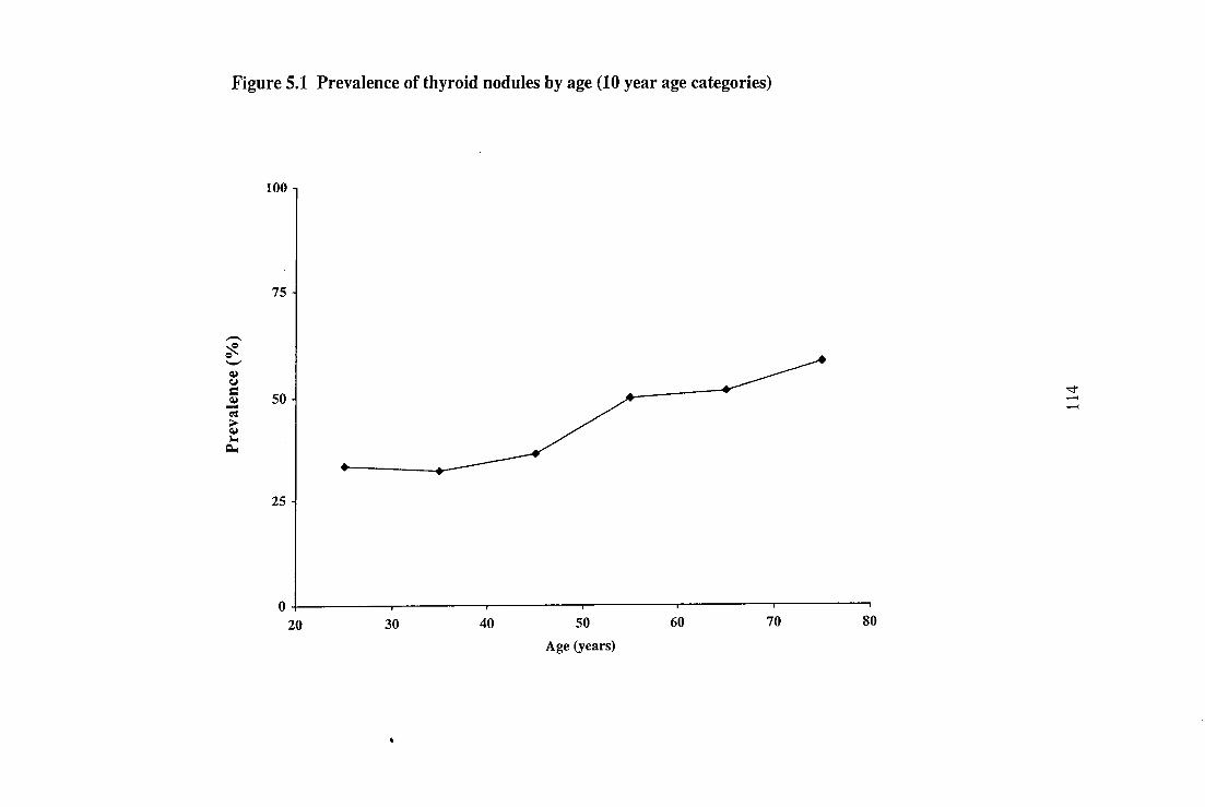
Figure 5.1 Prevalence of thyroid nodules by age (10 year age categories)
loo -
75 -
Prev
alen
ce (
%)
50
25
o 20 30 40 50
Age (years)
60 70 80

CHAPTER 6
An indirect assessment of the role of ionising radiation in the
genesis of contemporary PTC incidence trends
115

Prologue
The key questions addressed by this chapter are:
1 Is there molecular genetic evidence (RET/PTC1 rearrangement) for past ionizing
radiation exposure in priming Tasmanian PTC incidence trends?
2 What are the prevalence trends for RET/PTC1 in Tasmanian PTC?
Presentations and publications arising from this Chapter:
Presented
Preliminary results resented at the International Congress of Endocrinology (ICE 2000),
Sydney, Australia, 2000.
Published
Burgess JR, Skabo S, McArdle K, Tucker P. Temporal trends and clinical correlates for
the ret/PTC mutation in papillary thyroid carcinoma. ANZ J Surg. 2003; 87: 31-5.
In accordance with the criteria for submission of thesis by previously published research,
Chapter 6 is based on the above cited publication.
116

Aim
To determine if evidence exists for past ionizing radiation exposure as a cause for
increasing PTC incidence.
Hypothesis
1 Childhood exposure to radioiodine during the 1950's and early 1960's contributes
to contemporary PTC incidence trends.
2 Time trends for the RET/PTCI rearrangement may provide an indirect indication
of past ionizing radiation exposure as an aetiological factor in contemporary PTC
incidence trends.
117

Introduction
Ionizing radiation is the best characterized aetio logic factor for thyroid neoplasia (134,
136, 206, 235). Benign and malignant tumours are predisposed by prior thyroid
irradiation. PTC is the most frequent malignant sequel of thyroidal irradiation (133, 136,
206). There appears to be no threshold below which thyroid tumorigenesis is not
increased, but the risk is greatest when the exposure occurs during childhood, particularly
during the early years of life (135-137, 206). Although exposure in early childhood
confers the greatest risk, disease may not arise until adulthood because the latency
between exposure and cancer presentation can exceed three decades (133, 136, 137, 209).
Both external beam irradiation and ingestion of radioiodines in childhood are associated
with PTC development(134, 136). In the case of radioiodine, even low levels of exposure
from nuclear fallout can marginally increase long-term thyroid cancer risk (133, 136,
137, 209). It has been estimated that fallout radioiodines from Chernobyl increased
thyroid cancer incidence as far away as Connecticut in the USA (139). When the low
baseline incidence of PTC is considered in conjunction with the size of fallout-exposed
populations, significant relative increases in PTC incidence are a potential long-term
consequence of exposure to low levels of radioiodine fallout.
A number of genes are linked to the development of PTC, including RAS, RET/PTC,
TRK, BRAF, p5.3, MET and PAX8 (49, 109, 110, 115-117). Activating mutations
involving RET, a proto-oncogene encoding a receptor tyrosine kinase, can arise following
thyroidal irradiation (121, 124). Fusion of RET to the promoter region of an unrelated
gene gives rise to mutations collectively termed RET/PTC rearrangements (119, 130).
118

The most prevalent of these are RET/PTC1 and RET/PTC3 (119, 126, 127). Both
mutations result from inversions involving the long arm of chromosome 10 (121, 124,
130). High levels of radioiodine fallout following the Chernobyl accident produced an
early and overt rise in PTC, which was associated with the RET/PTC3 rearrangement
(115, 121, 130). However, subsequent studies have also shown RET/PTC1 to be
associated with PTC developing after a long latency(>10 years) from the time of
radiation exposure (115, 124, 130).
Therefore, the RET/PTC1 rearrangement may occur at higher frequency in thyroid
neoplasms arising after exposure to ionizing radiation, particularly in tumours developing
following a long latency after irradiation (115, 121, 124, 236). The RET/PTC1
rearrangement has also been associated with tumour behaviour and prognosis. Some
studies have suggested an association with a relatively benign clinical pattern of disease
(111, 119, 129, 130).
The influence of past exposure to radioiodine and diagnostic X-rays on contemporary
Australian PTC incidence trends is difficult to establish given the absence of exposure
data. Although RET/PTC1 is not specific to PTC arising after radiation exposure,
prevalence trends for RET/PTC1 in PTC may provide insights into the role of past
ionizing irradiation in the recent rise in national PTC incidence(115, 124).
In this chapter temporal trends and the clinicopathological associations for RET/PTC1
rearrangement in PTC diagnosed in Tasmania during the years 1978-1998 are assessed.
119

Subjects and Methods
Following approval from the Tasmanian Cancer Registry Data Release Committee and
the Royal Hobart Hospital Research Ethics Committee, all cases of PTC recorded by the
Registry during the period 1978-1998 were identified and tissue samples sought for
analysis.
A study sample of 98 cases, representing all cases of PTC diagnosed during the even
numbered years from and including 1978-1998, was defined. Pathology laboratories
around Tasmania were contacted and 62 paraffin-embedded tumour samples were
located. The histology of each sample was reviewed by two histopathologists to confirm
accuracy of the original diagnosis (213).
Ten tissue sections of 5 1.1m thickness were cut from representative areas of each paraffin-
embedded tissue block. Samples were deparaffinized by a 10-min incubation with xylene
followed by a two-stage 100% ethanol wash. Deparaffinized samples then underwent an
overnight (16-h) protease tissue digestion using the DNeasyTM Tissue Kit (Qiagen,
Valencia, CA, USA) standard -protocol. The RNA and DNA were coextracted from
digested samples using the DNeasyTM Tissue Kit (Qiagen) protocol. An additional on-
column DNAase digestion was then used to ensure that the eluted material was free from
DNA contamination.
Reverse transcription-polymerase chain reaction (RT-PCR) for RET/PTC1 was
performed using the One-Step RT-PCR Kit (Qiagen) reagents and standard protocol. The
120

RNA from the TPC-1 cell line was used as the positive control. A negative control (H20)
was included for each primer pair in every RT-PCR run. The primers used to detect the
RET/PTC1 rearrangement were as described by Nikiforov et al. (5'-
GCTGGAGACCTACAAACTGA-3' (nucleotides 425-443) and 5'-
GTTGCCTTGACCACTTTTC-3' (nucleotides 661-685)) (121). Overall RNA integrity
was assessed by RT-PCR using primers for human hypoxanthine
phosphoribosyltransferase (5'-CCTGCTGGAT-TACATCAAAGCACTG-3'(nucleotides
316-340) and 5'-CCTGAAGTATTCATTATAGTCTCAAGG-3' (nucleotides 661-685)]
(236).
One microgram of total RNA was used in each reaction. Reverse transcription was
carried out at 50°C for 30 min followed by a 95°C, 10-min denaturation step.
Amplification consisted of 42 cycles of denaturation at 94°C for 45s, annealing at 56°C
for 1 min, followed by a final 10-min extension step at 72°C.
The RT-PCR products were separated by electrophoresis on agarose gel and visualized
using the ethidium bromide technique prior to photographic documentation (Fig. 6.2).
The identity of products positive for hypoxanthine-guanine phosphoribosyltransferase
(HPRT) and RET/PTC1 and each of the positive controls was confirmed by sequencing
(ABI Prism DNA sequencing kit and ABI PRISM 377 DNA sequencer; Applied
Biosystems, Foster City, CA, USA).
In addition, patients identified by the Tasmanian Cancer Registry with a diagnosis of
PTC during the period 1978-1998 (n=180, Female:Ma1e=3.5, Age at diagnosis
121

46.4±1.1years) were sought to determine history of past head and neck irradiation. Of
these, 99 patients (Female:Male=4.8, Age at diagnosis 44.5±1.0years) were living,
contactable and consented to provide this information.
Data were analysed using the Student's t-test for normally distributed variables and the
chi-squared test for non-parametric data. Where appropriate numerical data are presented
as mean ± SD.
122

Results
The RET/PTC1 mutation was found in 26 (63%) tumours. Forty-three per cent of male
patients and 68% of female patients were positive. The mean age at diagnosis for
RET/PTC/-positive and RET/PTC/-negative tumours was 46.5 ± 15.46 and
41.9 ± 13.45 years, respectively. The RET/PTC1 mutation was significantly associated
with larger tumour size (Table 6.1). The RET/PTC1 mutation was not associated with an
adverse prognosis (Table 6.1).
The prevalence of tumours positive for RET/PTC1 remained stable over the study period
(Table 6.1). Tumours positive for RET/PTC1 did not exhibit birth year or diagnosis year
clustering (Table 6.1;Fig. 6.3). Similarly, RET/PTC1 positivity was not associated with
other clinicopathological characteristics such as presence of multinodular goitre,
lymphocytic infiltration, tumour multicentricity or PTC histological subtype (Table 6.1).
Of the 99 PTC patients who provided information regarding head and neck irradiation, 4
(4.0%) had a past history of this. Two cases related to treatment of haematological
malignancies, one for naevus, and the fourth for a brain tumour. Of these, only one (the
naevus) was associated with childhood radiotherapy.
123

Discussion
In comparison with studies undertaken in other countries, this study identified a relatively
high prevalence for the RET/PTC1 re-arrangement in Tasmanian PTC specimens (130).
However, it is consistent with the findings previously reported by Chua et al. who
described a prevalence of 55% for RET/PTC1 in tumours treated at a. Sydney teaching
hospital (237). However, another Australian study by Learoyd et al. also studied this
issue and reported a prevalence for RET/PTC1 of 8% (120).
Chua et al. also studied PTC from New Caledonia and found a similar frequency of the
RET/PTC1 rearrangement (70%) compared to that observed in NSW (85%) (237). This
is notable given the high rate of PTC incidence New Caledonia (237, 238) . One could
speculate that tumours from New Caledonia might have a higher rate of the RET/PTC1
rearrangement than Australian tumours due to the potential for higher levels of past
exposure to radio-iodine fallout (149). The failure to find a difference between these
populations suggests either: (i) any difference in exposure of the New Caledonian
population relative to that of NSW is not reflected by RET/PTC1 prevalence; (ii) the high
prevalence of RET/PTC1 in NSW samples may indicate a role for past ionizing
irradiation in this Australian population; or (iii) RET/PTC1 is of little or no value as a
radiation signature.
124

Iodine deficiency increases thyrocyte uptake of radioiodines such as 1131, potentially
increasing the likelihood of subsequent neoplasia (144). Mild-to-moderate iodine
deficiency affected Tasmania as well as much of coastal Victoria, New South Wales and
southern Queensland prior to the early 1960s (214, 225). The current rise in PTC
incidence in Australia has been most marked for the population residing on Australia's
eastern seaboard, particularly those regions with a history of iodine deficiency (213, 228).
Prior to the Universal Test Ban Treaty of 1962, atmospheric nuclear weapons testing
during the 1950s and early 1960s resulted in numerous episodes of fallout throughout
Australia (Figure 6.1). Although direct human exposure to radioiodine from
environmental contamination of air, soil and water is likely to have been negligible,
cows' milk is recognised as a vector for radioiodine entering the food chain (144). In
Australia, contamination of this food source by radioiodine was documented by Van
Middlesworth and Melick in the 1950s-1960s. Their research found that episodes of 1131
fallout affected grazing livestock in South Eastern Australia (147, 239).
Evidence for a relationship between RET/PTC1 prevalence and birth cohort was sought
in the present study (Table 6.1). Although exposure of the Australian population to low
levels of radioiodine fallout can be inferred from the available data (Figure. 6.1), the
current findings do not demonstrate an overt relationship between contemporary PTC
incidence trends and the occurrence of RET/PTCI rearrangements (147, 239).
When assessing the possible aetiological role of a risk factor in individual cases of
disease, proof of causation is virtually impossible to establish in the absence of either a
125

direct or surrogate measure of past exposure to that risk factor. While not excluding
ionizing radiation of the Australian population during the 1950s and early 1960s as a
cause for the contemporary rise in PTC incidence, clear support for this hypothesis is not
provided by the current study. Nonetheless, this is the first study to specifically examine
temporal trends for PTC, and it shows a stable prevalence for RET/PTC1 despite a
concurrent rise in PTC incidence.
The present study shows a significant clinicopathological association for the RET/PTC1
rearrangement. Tumours positive for the RET/PTC1 rearrangement were of larger size
and more likely to exhibit lymph node metastases than those without the mutation.
However, this trend was not associated with an increased risk for mortality. These
observations are in keeping with the existing literature that suggests that RET/PTC1 does
not confer heightened tumour aggressiveness (130).
The ret/PTC 1 rearrangement appears to be a relatively early event in PTC
development(130). This could explain the apparent contradiction of the rearrangement
being common in both "occult" papillary microcarcinoma, radiation induced PTC and
follicular adenomas(130). In each case, the early events that initiate tumor formation
may involve ret/PTC 1, but tumour progression requires accrual of additional oncogenic
mutations. Thus, if RET/PTC1 positive tumours acquire concurrent mutations, such as
inactivation of the p53 tumour suppressor gene, a more aggressive phenotype may
develop.
The success of RT-PCR is greatly affected by amplicon size (130, 237). The greater the
size, the greater the chance of false negative results due to sample degradation during
126

RNA extraction. This is particularly so when RT-PCR is performed on RNA derived
from paraffin-embedded tissue samples. In the study by Learoyd et al., which reported an
8% RET/PTC1 prevalence, the amplicon was substantially larger than that sought by the
primer set used both in the present study and that of Chua et al (120, 237). Thus
methodological factors are likely account for the divergent findings of these three
Australian studies.
127

Conclusions
This is the first study to map time trends for the prevalence of RET/PTCI relative to PTC
incidence patterns. There was no clear relationship between PTC incidence trends and
RET/PTC1 prevalence to suggest ionising irradiation was a dominant factor in the
genesis of recent PTC incidence trends. Whilst in some circumstances the RET/PTC1
rearrangement might be an indirect indication of past radiation damage, it may not be
sufficiently specific to be considered a radiation signature. However, the absolute
prevalence of RET/PTC1 (63%) is greater than that reported in many prior studies and is
consistent with levels found in irradiated populations. Further assessment of past
radiation exposure using alternate methodologies is required.
128

Table 6.1 Clinicopathological correlates of RET/PTC1 positive papillary thyroid
carcinoma diagnosed in Tasmania between 1978 and 1998
Characteristic RET/PTC1 (+) (n)
RET/PTC1 (-) (n)
Odds ratio (95% CI)
P value
26 15 Total (n=)
13 (50%) 6 (40%) 1.5 (0.6-2.6) 0.746 Age? 45 years
11(42%) 5(33%) 1.3 (0.6-3.0) 0.742 Multinodular goitre
9 (35%) 8 (53%) 0.5 (0.1-1.7) 0.328 Lymphocitic thyroiditis
3(12%) 2 (13%) 0.9 (0.1-5.8) 1.000 Sclerosing variant
8(31%) 2 (13%) 2.9 (0.5-16.0) 0.277 Follicular variant
5 (19%) 3 (20%) 1.0 (0.2-4.7) 1.000 Multicentric tumour
1(3.9%) nil 1.8 (0.1-48.0) 1.000 Family history of PTC
7 (27%) 1 (7%) 5.2 (0.6-47.0) 0.220 Extra-thyroid invasion
5 (19%) 1(7%) 3.3 (0.4-32.0) 0.388 Metastases present
2(8%) 3 (20%) 0.3 (0.1-2.3) 0.337 Death<1 year post Dx
7(27%) 8 (53%) 0.3 (0.1-1.2) 0.108 Size < 1.0cm
19 (73%) 7(47%) 3.1 (0.8-12 0.108 Size >lcm
13 (50%) nil 31(1.7-570) 0.001 Size >2
11(42%) 7 (47%) 0.8 (0.2-3.0) 1.000 Diagnosed < 1994
15 (58%) 8 (53%) 1.2 (0.3-43.0) 1.000 Diagnosed? 1994
CI - confidence interval Death<1 year post Dx - death within one year of PTCdiagnosis
129

Figure 6.1 Strontium-90 fallout in Hobart, Tasmania 1958-1990
0.4
0.3
Sr" Fallout
(mCi/km 2 ) 0.2 -
•• • 0
0 II:• •
0.1 10% .•
1;1 111:11.51:
8:1616.1 11116110 ID* 41 0 :
0.0 JO, 90 :04 .00004041000
1957 1962 1967 1972 1977 1982 1987
Year
Data from the Fallout Measurements Database, Environmental Measurements Laboratory, Office of Environmental Management, United States Department of Energy. http://www.eml.doe.govidatabases/fallout/fallout_data_search.htm Closed circles represent the level of Se ° Fallout during the individual calendar months of the year.
130
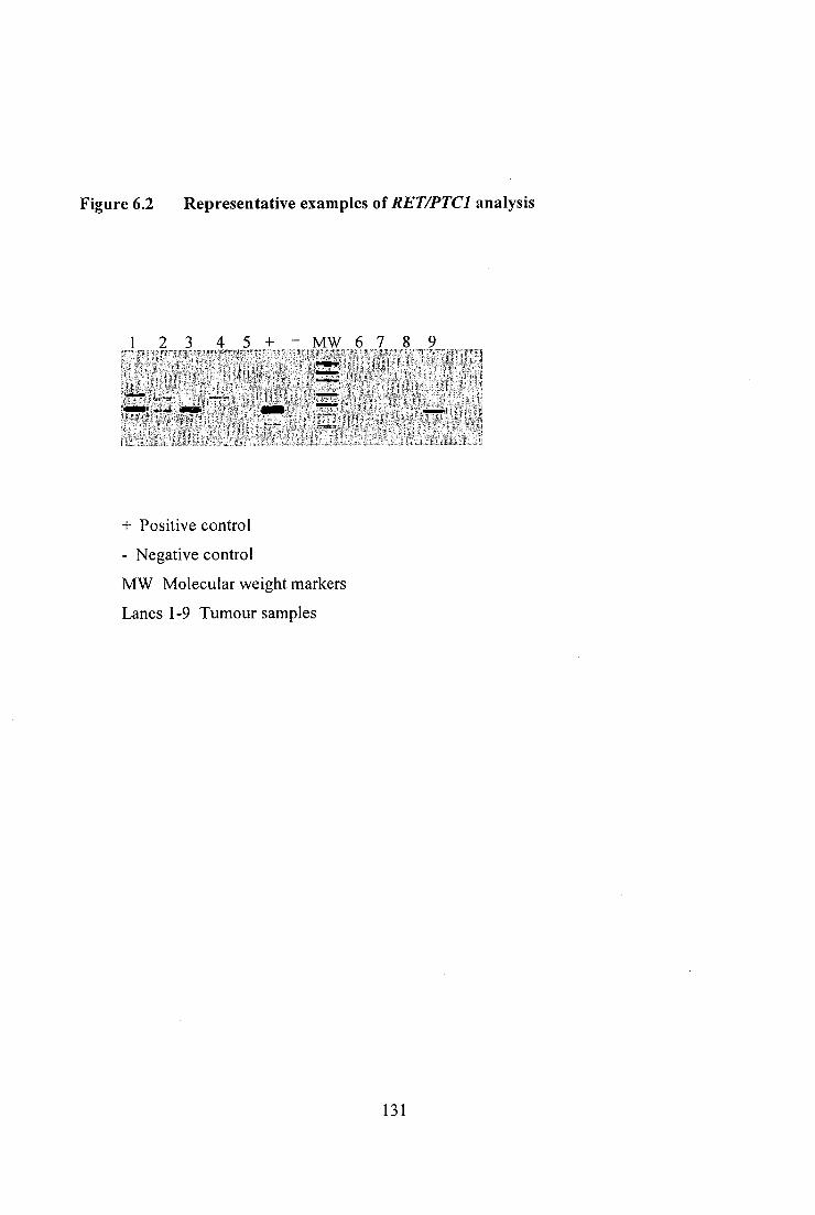
Figure 6.2 Representative examples of RET/PTC1 analysis
+ Positive control
- Negative control
MW Molecular weight markers
Lanes 1-9 Tumour samples
131

Figure 6.3 Birth year distribution of ret/PTC positive PTC
<1940 1940-44 1945-49 1950-'54 1955-59 1960-'64 >1965
Year of birth
132
Chapter 7
Heritable susceptibility to papillary carcinoma of the thyroid
133

PROLOGUE
The key questions addressed by this chapter are:
1 Does heritable susceptibility contribute to the incidence of PTC in Tasmania?
2 What is the inheritance pattern of familial PTC?
Presentations and publications arising from this Chapter:
Published
Burgess JR, Wilkinson R, Ware R, Greenaway TM, Percival J, Hoffman L. Two
families with an autosomal dominant inheritance pattern for papillary carcinoma of the
thyroid. I. Clin. Endocrinol. Metab. 1997; 82: 345-348.
In accordance with the criteria for submission of thesis by previously published research,
Chapter 7 is based on the above cited publication.
134

Aim
To assess the prevalence and inheritance pattern of familial PTC in Tasmania
Hypothesis
1 Germ line mutations modulate the risk for developing PTC.
2 A founder effect does not account for the relatively high incidence of PTC in
Tasmania.
135

Introduction
Understanding the role of germline mutations in modulating PTC risk is of interest for
numerous reasons. It opens an avenue for understanding the pathogenesis of PTC, as
well as influencing clinical management decisions (159). Familial diseases arising in
founder populations also have the potential to influence apparent disease incidence rates
when families are large, multigenerational, and reside in stable and relatively isolated
communities. In Tasmania for example, two key founder mutations predisposing to
Multiple Endocrine Neoplasia Type 1 (MEN 1), have resulted in a prevalence of MEN 1
in Tasmania which is 5-10 fold higher than that reported for other populations (240).
Studies assessing the prevalence of familial forms of PTC indicate that 3.5-6.2% of
patients with PTC have one or more first degree relatives with TC (155, 241, 242). An
association is known to exist between familial adenomatous polyposis (FAP) and
PTC(220, 243, 244). However, a number of reports also describe families in which PTC
occurs as a distinct entity, unrelated to FAP(155, 241, 242, 245, 246). The majority of
such kindreds contain fewer than four family members affected by PTC.
It is possible that either environmental factors or random association explains the
occurrence of PTC in a proportion of these families. However some authors have
postulated an autosomal dominant basis for familial PTC (155, 241, 245). Stoffer et al.
also noted an increased occurrence of nonmalignant thyroid disease in some families
with PTC, but the relationship between benign thyroid disease and the subsequent
development of malignant papillary lesions was unclear(241). In this chapter, the
prevalence and inheritance pattern of PTC in Tasmania is evaluated.
136

Subjects and Methods
Following approval of the Royal Hobart Hospital Human Research Ethics Committee, all
patients identified by the Tasmanian Cancer Registry with a diagnosis of PTC during the
period 1978-1998 (n=180, Female:Ma1e=3.5, Age at diagnosis 46.4±1.1) were sought to
determine the prevalence of familial PTC. Of these, 99 patients (Female:Ma1e=4.8, Age
at diagnosis 44.5±1.0) were living, contactable and consented to provide this
information.
Two large Tasmanian kindreds with a family history of PTC were evaluated in detail.
Where available, the results of isotope imaging and thyroid histopathology of tissue from
fine needle aspiration (FNA) or surgery have been reviewed. A firm diagnosis of PTC
could be made in those patients with characteristic histopathology.
137

Results
In total, 99 PTC patients (male 18, female 81) living, contactable and responded the
survey. A family history of TC was reported by 14 (14.1%). Of these, seven respondents
belonged to two multigenerational PTC pedigrees that are described in detail below.
Pedigree 1
A 62-yr-old female presented with a hoarse voice. A multinodular goiter (MNG) had
been noted 2 yr previously. On examination, left vocal cord paralysis and a hard, left
sided neck mass were detected. Neck exploration demonstrated locally invasive papillary
carcinoma of the thyroid. The patient was treated with total thyroidectomy and 6000
MBq of radio iodine. No evidence of abnormal 1311 uptake was noted on post-ablative
whole body scans. Abnormal areas of thallium uptake in the neck and chest were noted,
becoming less prominent on consecutive scans. She remains well.
Three of the patient's six children presented with MNG in the ensuing six months
(Patients M, N, and P, Figure 7.1) and underwent surgical resection. Surgical
management was undertaken because of mild compressive symptoms, a low thyrotropin
level (indicating unsuitability for thyroxine suppression), or patient anxiety. One patient
had FNA of the thyroid preoperatively that was nondiagnostic. PTC was detected during
pathological examination in all cases, one microscopic, one 10 mm in diameter, and the
other a multifocal PTC. On screening, one of the patient's six children (Patient 0) had a
solitary nodule confirmed as PTC on excision. Screening other family members yielded
two further relatives with MNG who had multifocal PTC (Patients U and W). Patient U
138
had evidence of malignancy on preoperative FNA. Other relatives with abnormal thyroid
glands on screening are shown in Figure 7.1.
The family history revealed two additional relatives who had probable thyroid carcinoma
(patients B and D). Both had thyroidectomies in their 50's (before 1950) and were
reported by relatives and medical practitioners to have thyroid cancer; however,
histological confirmation was not possible. There was no family history of colonic
malignancy or intestinal polyposis and no history of radiation exposure.
Pedigree II
A 49-yr-old male presented with a 3-week history of left-sided neck swelling.
Examination revealed a 1.5 cm mass palpable in the left lobe of the thyroid gland. The
patient's family history was unremarkable for thyroid or bowel malignancy, and there
was no history of radiation exposure. Following ultrasound and isotope imaging (Figure
7.1), the patient underwent hemithyroidectomy, and histopathological examination
revealed papillary carcinoma. Total thyroidectomy was subsequently performed, and the
patient received a 6000 MBq dose of radio iodine. Follow-up studies to 3 yr have not
demonstrated disease recurrence. One year subsequent to presentation of the index case,
the patient's monozygotic twin brother presented with a neck mass, assessment of which
also revealed PTC, metastatic to a cervical lymph node. In 1995, the 23-yr-old daughter
(Patient K) of the index case presented with a right sided thyroid mass, subsequently
proven to be an invasive 12 mm PTC. She was treated by total thyroidectomy, ablative
radio iodine, and thyroxine suppression. The 22-yr-old daughter of the other twin (Patient
L) had a MNG on screening, and a sclerosing papillary microcarcinoma was found on
139

resection. There has been no evidence of recurrence in any of these family members. The
family tree for Pedigree II is also summarized in Figure 7.1. The characteristics of
surgically treated thyroid disease from both pedigrees are summarized in Table 7.1.
140

Discussion
Familial PTC is relatively uncommon in Tasmania, exhibiting a prevalence (14.1%).
Reports by other authors indicate a prevalence in the general population of approximately
5%. The higher prevalence in my study may in part reflect a founder effect associated
with pedigree I and II, as well as bias due to a relatively low survey response rate of 55%.
However, the kindreds presented herein support the existence of an autosomal dominant
inheritance pattern for PTC, separate from the established association with FAP(151).
Pedigree I represents one of the largest kindred with non-FAP familial PTC described
thus far.
In both families, history and physical examination consistent with FAP or Cowden's
syndrome was absent. Although Pedigree II is less extensive, the development of PTC in
monozygotic twins (within one year of each other) and in two of their offspring suggests
an underlying genetic predisposition to thyroid neoplasia. The fact that both parents and a
grandparent of the twins lived to an advanced age without evidence of thyroid cancer
suggests the possibility of a de novo mutation occurring early in the twin's
embryogenesis.
An association may exist between multinodular goitre and PTC. Existing evidence does
not exclude this possibility, as thyroid malignancy is thought to be a multi-step process.
Furthermore, common genetic factors have been implicated in the pathogenesis of both
MNG and PTC (46, 49, 159, 245). It is also possible that the association is indirect, as
the presence of MNG is likely to increase the likelihood of diagnosing "occult"
PTC(210). However, the clinically significant extent of the majority of tumours
141

diagnosed in the families described in this study indicates this is not the dominant
explanation.
Incomplete penetrance of familial PTC may explain both the obligatory carrier status of
patient C in Pedigree I and non-expression of clinically overt thyroid disease. The
influence of secondary modifying factors, such as gender may contribute to this, as was
the case in pedigree I, where six of seven patients with PTC are female. This observation
is similar to that of Lote et al, who described a female predilection for FTC in a
Norwegian family(245). Both families included individuals with multifocal FTC
occurring at a young age, consistent with other reports of familial PTC(159, 247).
The optimal strategy for investigation and management of kindreds with PTC is unclear.
Exclusion of undiagnosed FAP would seem prudent, however in the absence of a family
history of bowel malignancy, or characteristic physical stigmata of Gardner's syndrome,
routine colonic assessment would seem unwarranted. The relatively high risk of PTC
developing in an underlying MNG, and the limited value of ultrasonography, isotope
scanning, and FNAB in excluding malignancy in the setting of multinodularity, argues
strongly for thyroidectomy in family members of high risk pedigrees who have evidence
of MNG. The screening strategy currently recommended for these families is periodic
thyroid ultrasonography and neck palpation by an experienced examiner(160). Patients
with evidence of goiter may then be counseled regarding the potential risk of malignancy
and offered the option of early thyroidectomy(160).
142

Conclusions
Prior to publication of the paper presented in this chapter, there was limited data
available in relation to high penetrance genes associated with autosomal dominant PTC.
Careful characterisation of families expressing such patterns of PTC inheritance should
result in identification of susceptibility loci for PTC. The large pedigree (Pedigree I)
described in this report provided support for the presence of a distinct autosomal
dominant form of PTC as well as being the substrate for subsequent genomic localisation
studies. Whilst a specific gene is yet to be identified, and familial PTC may arise from a
number of distinct genetic loci, the observations reported herein provide impetus for
further research.
143

Table 7.1. Thyroid gland characteristics in patients treated by thyroidectomy
'atient Age Ultrasonography; Characteristics on isotope imaging; Thyroid histopathology
(G) 43 Enlarged gland, heterogenous, consistent with MNG; MNG with lymphocytic thyroiditis. No evidence of malignancy
(K) 62 Enlarged nodular gland, consistent with MNG; no dominant nodule; follicular variant of PTC occurring in a MNG
(L) 34 3 x 2.2 cm solitary solid nodule, cold nodule in otherwise normal gland; MNG with benign dominant nodule
(M) 35
Multiple solid and cystic nodules, consistent with MNG; overall uptake increased; dominant nodule with
cold areas sclerosing PTC occurring in a MNG
(N) 24 Enlarged gland, 4 nodules; Multifocal PTC
(0) 42 Solid nodule; PTC
(P) 39 Consistent with MNG; overall uptake increased; no dominant cold nodule; sclerosing PTC occurring in a MNG;
background of lymphocytic thyroiditis
(T) 58 MNG
(T1) 29 MNG
(T2) 26 2 thyroid nodules, largest 2 cm MNG with lymphocytic thyroiditis
(U) 54 2 thyroid nodules; left lobes; atypical cells on FNA early MNG with multifocal PTC containing follicular variants
(V) 32 2 nodules left lobe early MNG
(W) 23 Multiple nodules, 2-5 mm MNG; multifocal PTC in left lobe
(Z) 27 MNG
I (G) 51 Right lobe enlarged and homogenous, left enlarged; MNG, largest nodule 2 cm large cold area left lobe;
generalized patchy uptake well differentiated PTC metastatic to cervical lymph node; solitary follicular adenoma in
thyroid gland
I (H) 49 Enlarged gland; nodular left lobe containing a 20 mm solid nodule with patchy tracer uptake consistent with MNG
'TC containing follicular elements
I (K) 23 Normal sized gland containing 10 mm and 2 mm cold nodules; PTC containing follicular elements
I (L) 22 Lesions in upper left lobe and heterogeneous texture lower pole right lobe normal sized gland, areas of marked
decrease in uptake sclerosing papillary microcarcinoma
144

PEDIGREE II A 8
Figure 7.1 Pedigree charts for family I and II
KEY CI Male 0 Female ® Normal thyroid • PTC IS Probable PTC Q Goitre 0 Deceased 0 Unknown thyroid status

CHAPTER 8
Conclusion and Future Studies
146

Thesis Conclusions
This thesis provides the first comprehensive evaluation of TC incidence trends in
Australia. Furthermore, the Tasmanian population proved to be a suitable model for
evaluating the factors underlying contemporary TC incidence patterns.
A review of notifications to Australian Cancer Registries during the last two decades of
the twentieth century demonstrated a progressive rise in the reported incidence of TC.
The most pronounced increase occurred during the 1990's and was attributable to an
increasing incidence of PTC. The geographic pattern for this trend was most evident in
Australia's eastern seaboard States, namely Tasmania, Victoria, New South Wales and
Queensland. This distribution co-localised with the ecological pattern of iodine
deficiency in Australia. The population of the Australian south-eastern seaboard was
mild-moderately iodine deficient prior to introduction of iodine-based disinfectants
(iodophors) by the dairy industry in the 1960's. The residual iodine content in milk
(related to iodophor use) serendipitously corrected the deficiency. Progressive phase-out
of these disinfectants in the late 1980's has been associated with a nationwide decline in
iodine nutrition. Over the past decade the population of much of Australia's eastern
seaboard has become iodine deficient. Tasmania, recognised as has having the lowest
pre-odophor level of iodine nutrition and highest prevalence of endemic goitre,
experienced the greatest rise in PTC incidence of all the Australian commonwealth.
However, the information available from the national TC dataset was insufficient to
clarify the role of Cancer Registry ascertainment bias, changing trends in medical
147

practice and altered patterns of PTC risk factor exposure. Addressing these issues
required detailed evaluation of a representative population. The Tasmania community
was selected for this purpose.
Detailed validation of Tasmanian Cancer Registry case ascertainment was undertaken.
This indicated PTC registration was 93.9% complete and without evidence of significant
temporal distortion. As this assessment included the period during which PTC incidence
doubled, I was able to exclude biased Cancer Registry case ascertainment as an
explanation for the observed rise in PTC incidence. This was an important consideration,
as prior to this study, time related variation in case ascertainment by cancer registries had
been postulated as making an important contribution to observed PTC trends.
Furthermore, the morphological classification of tumours over time was found to be
stable when independently reviewed. Changes in tumour classification did not account
for observed trends in TC incidence.
The dominant change identified by this study was a significant increase in diagnosis of
small PTC (<1cm in maximum dimension). Iodine supplementation of iodine deficient
communities has been associated elsewhere with increased diagnosis of PTC of smaller
size and earlier stage. Therefore, given the previously identified geographic linkage
between increased PTC incidence and the distribution of historical iodine deficiency in
Australia, further evaluation of the potential impact changing iodine nutrition on PTC
incidence and pathogenesis was undertaken.
148

Despite two decades of documented iodine sufficiency in Tasmania, PTC incidence
increased more than two-fold at the time when iodine deficiency recurred. Whilst not
excluding a long latency linking improved iodine nutrition in the 1960's to increased
PTC in the 1990's, such a relationship appears unlikely and inconsistent with the
expected time course associated with the concept of "papillarisation" following iodine
supplementation.
A non-biological association between community iodine supplementation occurring in
conjunction with improved community screening for thyroid disease (increasing
availability of ultrasonography and consequential interventions such as FNAB and
thyroid surgery), appears to be a more plausible link between iodine nutrition and
increased incidence of PTC. Similarly, the progressive improvement in medical care and
technology occurring in recent decades could similarly increase the diagnosis of "occult"
PTC.
A survey and ultrasonographic study was undertaken to determine the prevalence of
subclinical and previously diagnosed thyroid disease in Tasmania. The data presented in
Chapter 5 indicate a divergence between the absolute risk for nodular thyroid disease (as
evidenced by ultrasonography) versus the likelihood of clinically diagnosed and treated
thyroid disease. The former was common, irrespective of birth place and residence,
whilst the latter was relatively associated with prolonged residence in Tasmania during
the era of iodine deficiency. The failure to observe a significant association between
place of birth and TC suggests a unique environmental modifier is unlikely to account for
PTC incidence trends in Tasmania.
149
However, the findings were consistent with historically more severe iodine deficiency in
Tasmania increasing the prevalence of benign goiter and thyroid dysfunction. Evaluation
of these may have secondarily increased the likelihood of diagnosing "occult" PTC.
Moreover, thyroid ultrasonography in any person without clinical suspicion of thyroid
disease (irrespective of their iodine nutritional history) may also considered a "risk
factor" for FNAB, thyroid surgery and consequently the diagnosis of "occult" PTC.
My results suggested the increase in PTC incidence observed in Tasmania and more
widely may in large part be attributable to greater diagnosis of small PTC, many of which
are likely to have been asymptomatic, identified by neck ultrasonography and subsequent
FNAB. Iodine deficient (or previously deficient) populations can be expected to have an
higher prevalence of goitre and larger thyroid nodules. The higher prevalence of
clinically apparent structural thyroid disease is likely to increase thyroid imaging, FNAB
and consequently the diagnosis of "occult" PTC. Past and present iodine nutrition need
have no causal link with PTC pathogenesis, merely serve to modify incidental
recognition of existing subclinical nodular thyroid disease.
However, it was also noted that the incidence of PTC larger than lcm in patients without
history of preoperative FNAB had increased in Tasmania, suggesting the occurrence of
clinically relevant tumours increased. It was important therefore to examine trends for
exposure to established risk factors such as ionising radiation. In the absence of case
specific exposure data for fallout to confirm this, Chapter 6 sought to use the RET/PTC1
rearrangement as a surrogate marker for the involvement of ionising radiation in the
150

pathogenesis of individual PTC. Observational studies have suggested the RET/PTC1
rearrangement may occur at higher frequency in PTC arising following thyroidal
irradiation.
I was able to map trends for the prevalence of RET/PTC1 relative to incidence trends for
PTC. Whilst the RET/PTC1 mutation was frequently identified in Tasmanian PTC, there
was no clear relationship between RET/PTC1 and recent PTC incidence trends. This can
be interpreted as either a negative finding (in as much as the fraction of PTC with
RET/PTC1 rearrangement failed to increase significantly in parallel with the rise in
overall PTC incidence) or conversely, suggestive of a proportional and increasing
contribution of ionising radiation to PTC diagnosed in Tasmania. Future research might
address this question by undertaking appropriately powered longitudinal studies
evaluating populations before and after a documented exposure to fallout.
I also examined the prevalence and genetic basis of familial PTC. A founder effect
combined with screening did not account for the rise in PTC incidence observed in
Tasmania. The large Tasmanian pedigree described in Chapter 7 provided support for
the presence of a distinct autosomal dominant form of PTC as well as a basis for
subsequent genomic localisation studies. Whilst a specific gene is yet to be identified,
and familial FTC may arise from a number of distinct genetic loci, this manuscript
provided impetus for further research.
151

In summary, this thesis addressed the questions posed in Chapter 1 and reached the
following conclusions:
1 The incidence of TC increased in Australia during the last two decades of the
twentieth century due to a rise in incidence of PTC.
2 Geographic variation for PTC incidence in Australia was evident, with the
greatest increase occurring in the south-eastern States of Tasmania, Victoria and
NSW.
3 The Tasmanian population proved to be a suitable model for evaluating the basis
for PTC incidence trends given its stable population structure, well documented
history of iodine nutrition and stable health care system.
4 There was no evidence of any substantial systematic modification to
histopathological diagnostic criteria or Cancer Registry case ascertainment and
registration protocols to explain observed PTC incidence trends.
5 Whilst the incidence of both large and small PTC increased over the study period,
greater diagnosis of PTC<1 cm diameter was the dominant factor accounting for
the rise in PTC incidence. Increasing use of neck imaging and FNAB in patients
with asymptomatic nodular thyroid appear to be the main antecedent factors
driving this trend.
6 The geographic patterns for PTC incidence trends were compatible with historical
and contemporary patterns of iodine deficiency in Australia. The severity of
historical deficiency mapped proportionally with the magnitude of recent PTC
incidence increases.
7 \ I cannot exclude a direct biological influence of iodine nutrition on PTC
pathogenesis, however, the influence of improving and subsequently declining
152

levels of iodine nutrition in Tasmania over the past four decades appears to have
been minimal in comparison to the dominant influence of increasing use of neck
ultrasonography and FNAB resulting in diagnosis of "occult" PTC.
8 No direct evidence to support ionising radiation in the genesis of contemporary
PTC incidence trends has been identified. However, the finding of RET/PTC1
mutation as highly prevalent in Tasmanian PTC cases warrants further
consideration of this possibility.
9 Autosomal dominant susceptibility to PTC has been identified in a small
proportion of Tasmanian PTC cases, however the impact of familial disease has
been relatively minimal on overall PTC incidence trends.
10 Based on the data presented in this thesis, it is possible to estimate the relative
contributions of ascertainment bias, and changes in underlying biological
incidence in contemporary PTC incidence trends. Increased ascertainment of
"occult" PTC due to greater use of sensitive neck imaging and contemporary
thyroid nodule evaluation paradigms explains the majority of increased PTC
diagnoses during the 1990's, whilst a biologically significant increase in
underlying PTC incidence appears responsible for the remainder.
153
Future Studies
1
Follow-up of Tasmanian PTC incidence trends in association with
utilization profiles for neck imaging and FNAB to determine if a stable
PTC incidence pattern is established consistent with contemporary
imaging usage.
2 An intervention study based on structured medical practitioner education
regarding appropriate use of thyroid ultrasonography to determine if
decreased non-specific neck imaging is associated with reduced PTC
incidence.
3
A large-scale Australian national study, sufficiently powered to assess the
impact of changes to national iodine nutrition on PTC incidence.
154

BIBLIOGRAPHY
155

1. 2003 Williams Textbook of Endocrinology, 10 ed. Saunders, Philadelphia 2. 2006 Endocrinology, 5 ed. Elsevier Saunders, Philadelphia 3. De Felice M, Di Lauro R 2004 Thyroid development and its disorders: genetics
and molecular mechanisms. Endocr Rev. 25:722-46. 4. Burrow GN, Fisher DA, Larsen PR 1994 Maternal and fetal thyroid function. N
Engl J Med. 331:1072-8. 5. Glinoer D 2006 Iodine nutrition requirements during pregancy. Thyroid. 16:947-
8. 6. Delange F 1994 The disorders induced by iodine deficiency. Thyroid. 4:107-28. 7. Dunn JT, Dunn AD 2001 Update on intrathyroidal iodine metabolism. Thyroid.
11:407-14. 8. Shen DH, Kloos RT, Mazzaferri EL, Jhian SM 2001 Sodium iodide symporter
in health and disease. Thyroid. 11:415-25. 9. Dai G, Levy 0, Carrasco N 1996 Cloning and characterization of the thyroid
iodide transporter. Nature. 379:458 10. Dohan 0, De la Vieja A, Paroder V, et al. 2003 The sodium/iodide Symporter
(NIS): characterization, regulation, and medical significance. Endocr Rev. 24:48- 77.
11. Burger HG, Patel YC 1977 Thyrotrophin releasing hormone--TSH. Clin Endocrinol Metab. 6:83-100.
12. Fisher DA, Nelson JC, Carlton El, Wilcox RB 2000 Maturation of human hypothalamic-pituitary-thyroid function and control. Thyroid. 10:229-34.
13. International Council for Control of Iodine Deficiency Disorders UNCsF, World Health Organisation 2001 Assessment of iodine deficiency disorders and monitoring their elimination.
14. World Health Organisation W, UNICEF, ICCIDD 1994 Indicators for assessing iodine deficiency disorders and their control through salt iodisation. WHO/NUT/94.6. Geneva: WHO
15. Soldin OP 2002 Controversies in urinary iodine determinations. Clin Biochem. 35:575-9.
16. Delange F, Burgi H, Chen ZP, Dunn JT 2002 World status of monitoring iodine deficiency disorders control programs. Thyroid. 12:915-24.
17. Eastman CJ 1993 The status of iodine nutrition in Australia. In: Delange F, Dunn JT, Glinoer D (eds). Iodine Deficiency in Europe: A Continuing Concern. New York: Plenum Press:133-8
18. Delange F, de Benoist B, Pretell E, Dunn JT 2001 Iodine deficiency in the world: where do we stand at the turn of the century? Thyroid. 11:437-47.
19. Eastman CJ 1999 Where has all our iodine gone? Med J Aust 171:455-6 20. Dunn JT 1993 Sources of dietary iodine in industrialised countries. In: Delange
F, Dunn JT, Glinoer D, eds. Iodine Deficiency in Europe. Acontinuing concern. New York: Plenum Press
21. Studer H, Derwahl M 1995 Mechanisms of nonneoplastic endocrine hyperplasia--a changing concept: a review focused on the thyroid gland. Endocr Rev. 16:411-26.
156
22. Sundick RS, Bagchi N, Brown TR 1992 The role of iodine in thyroid autoimmunity: from chickens to humans: a review. Autoimmunity. 13:61-8.
23. Laurberg P, Jorgensen T, Perrild H, et al. 2006 The Danish investigation on iodine intake and thyroid disease, DanThyr: status and perspectives. Eur J Endocrinol. 155:219-28.
24. Aghini-Lombardi F, Antonangeli L, Martino E, et al. 1999 The spectrum of thyroid disorders in an iodine-deficient community: the Pescopagano survey. J Clin Endocrinol Metab. 84:561-6.
25. Laurberg P, Nohr SB, Pedersen KM, et al. 2000 Thyroid disorders in mild iodine deficiency. Thyroid. 10:951-63.
26. Boyages SC 1993 Clinical review 49: Iodine deficiency disorders. J Clin Endocrinol Metab. 77:587-91.
27. Galanti MR, Sparen P, Karlsson A, Grimelius L, Ekbom A 1995 Is residence in areas of endemic goiter a risk factor for thyroid cancer? Int J Cancer 61:615-21
28. Rolon PA 1986 Cancer of the thyroid in an area of endemic goiter. "Papillarization" with prophylactic iodization. Ann Pathol 6:170-5
29. Franceschi S 1998 Iodine intake and thyroid carcinoma--a potential risk factor. Exp Clin Endocrinol Diabetes 106 Suppl 3:S38-44
30. Dunn JT 1996 Seven deadly sins in confronting endemic iodine deficiency, and how to avoid them. J Clin Endocrinol Metab. 81:1332-5.
31. Hofstadter F 1980 Frequency and morphology of malignant tumours of the thyroid before and after the introduction of iodine-prophylaxis. Virchows Arch A Pathol Anat Histol 385:263-70
32. Vermiglio F, Lo Presti VP, Moleti M, et al. 2004 Attention deficit and hyperactivity disorders in the offspring of mothers exposed to mild-moderate iodine deficiency: a possible novel iodine deficiency disorder in developed countries. J Clin Endocrinol Metab. 89:6054-60.
33. Dunn JT, Delange F 2001 Damaged reproduction: the most important consequence of iodine deficiency. J Clin Endocrinol Metab. 86:2360-3.
34. Soriguer F, Milton MC, Munoz R, et al. 2000 The auditory threshold in a school-age population is related to iodine intake and thyroid function. Thyroid. 10:991-9.
35. Vanderpump MP, Tunbridge WM, French JM, et al. 1995 The incidence of thyroid disorders in the community: a twenty-year follow-up of the Whickham Survey. Clin Endocrinol (Oxf). 43:55-68.
36. Roberts CG, Ladenson PW 2004 Hypothyroidism. Lancet. 363:793-803. 37. Cooper DS 2003 Hyperthyroidism. Lancet. 362:459-68. 38. Manji N, Carr-Smith JD, Boelaert K, et al. 2006 Influences of age, gender,
smoking, and family history on autoimmune thyroid disease phenotype. J Clin Endocrinol Metab. 91:4873-80. Epub 2006 Sep 12.
39. Tunbridge WMG, Vanderpump MPJ 2000 Population screening for autoimmune thyroid disease. Endocrinol Metab Clin North Am. 29:239-253.
40. Vanderpump MP, Tunbridge WM 2002 Epidemiology and prevention of clinical and subclinical hypothyroidism. Thyroid. 12:839-47.
41. Wang C, Crapo LM 1997 The epidemiology of thyroid disease and implications for screening. Endocrinol Metab Clin North Am. 26:189-218.
157

42. Topliss DJ, Eastman CJ 2004 5: Diagnosis and management of hyperthyroidism and hypothyroidism. Med J Aust. 180:186-93.
43. 2002 American Association of Clinical Endocrinologists medical guidelines for clinical practice for the evaluation and treatment of hyperthyroidism and hypothyroidism. Endocr Pract. 8:457-69.
44. Walsh JP, Shiels L, Lim EM, et al. 2003 Combined thyroxine/liothyronine treatment does not improve well-being, quality of life, or cognitive function compared to thyroxine alone: a randomized controlled trial in patients with primary hypothyroidism. J Clin Endocrinol Metab. 88:4543-50.
45. Laurberg P, Bulow Pedersen I, Knudsen N, Ovesen L, Andersen S 2001 Environmental iodine intake affects the type of nonmalignant thyroid disease. Thyroid. 11:457-69.
46. Krohn K, Fuhrer D, Bayer Y, et al. 2005 Molecular pathogenesis of euthyroid and toxic multinodular goiter. Endocr Rev. 26:504-24. Epub 2004 Dec 22.
47. Teng W, Shan Z, Teng X, et al. 2006 Effect of iodine intake on thyroid diseases in China. N Engl J Med. 354:2783-93.
48. Laurberg P, Pedersen KM, Hreidarsson A, Sigfusson N, Iversen E, Knudsen PR 1998 Iodine intake and the pattern of thyroid disorders: a comparative epidemiological study of thyroid abnormalities in the elderly in Iceland and in Jutland, Denmark. J Clin Endocrinol Metab. 83:765-9.
49. Learoyd DL, Messina M, Zedenius J, Robinson BG 2000 Molecular genetics of thyroid tumors and surgical decision-making. World J Surg. 24:923-33.
50. Hegedus L, Bonnema SJ, Bennedbaek FN 2003 Management of simple nodular goiter: current status and future perspectives. Endocr Rev. 24:102-32.
51. Zimmermann MB, Molinari L, Spehl M, et al. 2001 Toward a consensus on reference values for thyroid volume in iodine-replete schoolchildren: results of a workshop on inter-observer and inter-equipment variation in sonographic measurement of thyroid volume. Eur J Endocrinol. 144:213-20.
52. Vejbjerg P, Knudsen N, Perrild H, et al. 2007 Effect of a mandatory iodization program on thyroid gland volume based on individuals' age, gender, and preceding severity of dietary iodine deficiency: a prospective, population-based study. J Clin Endocrinol Metab. 92:1397-401. Epub 2007 Jan 30.
53. Hegedus L 2001 Thyroid ultrasound. Endocrinol Metab Clin North Am. 30:339- 60, viii-ix.
54. Reiners C, Wegscheider K, Schicha H, et al. 2004 Prevalence of thyroid disorders in the working population of Germany: ultrasonography screening in 96,278 unselected employees. Thyroid. 14:926-32.
55. Volzke H, Ludemann J, Robinson DM, et al. 2003 The prevalence of undiagnosed thyroid disorders in a previously iodine-deficient area. Thyroid. 13:803-10.
56. Hansen PS, Brix TH, Bennedbaek FN, Bonnema SJ, Kyvik KO, Hegedus L 2004 Genetic and environmental causes of individual differences in thyroid size: a study of healthy Danish twins. J Clin Endocrinol Metab. 89:2071-7.
57. Wiest PW, Hartshorne MF, Inskip PD, et al. 1998 Thyroid palpation versus high-resolution thyroid ultrasonography in the detection of nodules. J Ultrasound Med. 17:487-96.
158
58. Vander JB, Gaston EA, Dawber TR 1954 Significance of solitary nontoxic thyroid nodules; preliminary report. N Engl J Med. 251:970-3.
59. Tunbridge WM, Evered DC, Hall R, et al. 1977 The spectrum of thyroid disease in a community: the Whickham survey. Clin Endocrinol (Oxf). 7:481-93.
60. Tan GH, Gharib H 1997 Thyroid incidentalomas: management approaches to nonpalpable nodules discovered incidentally on thyroid imaging. Ann Intern Med. 126:226-31.
61. Mortensen JD, Bennett WA, Woolner LB 1955 Incidence of carcinoma in thyroid glands removed at 1000 consecutive routine necropsies. Surg Forum. 5:659-63.
62. Levine RA 2004 Something old and something new: a brief history of thyroid ultrasound technology. Endocr Pract. 10:227-33.
63. Marqusee E, Benson CB, Frates MC, et al. 2000 Usefulness of ultrasonography in the management of nodular thyroid disease. Ann Intern Med. 133:696-700.
64. Brander A, Viikinkoski P, Nickels J, Kivisaari L 1991 Thyroid gland: US screening in a random adult population. Radiology. 181:683-7.
65. Brander A, Viikinkoski P, Tuuhea J, Voutilainen L, Kivisaari L 1992 Clinical versus ultrasound examination of the thyroid gland in common clinical practice. J Clin Ultrasound. 20:37-42.
66. Bruneton JN, Balu-Maestro C, Marcy PY, Melia P, Mourou MY 1994 Very high frequency (13 MHz) ultrasonographic examination of the normal neck: detection of normal lymph nodes and thyroid nodules. J Ultrasound Med. 13:87- 90.
67. Knudsen N, Bols B, Bulow I, et al. 1999 Validation of ultrasonography of the thyroid gland for epidemiological purposes. Thyroid. 9:1069-74.
68. Chan BK, Desser TS, McDougall IR, Weigel RJ, Jeffrey RB, Jr. 2003 Common and uncommon sonographic features of papillary thyroid carcinoma. J Ultrasound Med. 22:1083-90.
69. Knudsen N, Bulow I, Jorgensen T, Laurberg P, Ovesen L, Perrild H 2000 Goitre prevalence and thyroid abnormalities at ultrasonography: a comparative epidemiological study in two regions with slightly different iodine status. Clin Endocrinol (Oxf). 53:479-85.
70. Papini E, Guglielmi R, Bianchini A, et al. 2002 Risk of malignancy in nonpalpable thyroid nodules: predictive value of ultrasound and color-Doppler features. J Clin Endocrinol Metab. 87:1941-6.
71. Burguera B, Gharib H 2000 Thyroid incidentalomas. Prevalence, diagnosis, significance, and management. Endocrinol Metab Clin North Am. 29:187-203.
72. Burch HB 1995 Evaluation and management of the solid thyroid nodule. Endocrinol Metab Clin North Am. 24:663-710.
73. Singer PA, Cooper DS, Daniels GH, et al. 1996 Treatment guidelines for patients with thyroid nodules and well-differentiated thyroid cancer. American Thyroid Association. Arch Intern Med. 156:2165-72.
74. Belfiore A, La Rosa GL 2001 Fine-needle aspiration biopsy of the thyroid. Endocrinol Metab Clin North Am 30:361-400
75. Mazzaferri EL 1993 Management of a solitary thyroid nodule. N Engl J Med. 328:553-9.
159
76. Cooper DS, Doherty GM, Haugen BR, et al. 2006 Management guidelines for patients with thyroid nodules and differentiated thyroid cancer. Thyroid. 16:109- 42.
77. Walsh JP, Ryan SA, Lisewski D, et al. 2007 Differences between endocrinologists and endocrine surgeons in management of the solitary thyroid nodule. Clin Endocrinol 15:15
78. Mittendorf EA, McHenry CR 1999 Follow-up evaluation and clinical course of patients with benign nodular thyroid disease. Am Surg 65:653-7; discussion 657- 8
79. Gharib H, Goellner JR 1993 Fine-needle aspiration biopsy of the thyroid: an appraisal. Ann Intern Med. 118:282-9.
80. Nathan AR, Raines KB, Lee YT, Sakas EL, Ribbing JM 1988 Fine-needle aspiration biopsy of cold thyroid nodules. Cancer 62:1337-42
81. Tollin SR, Mery GM, Jelveh N, et al. 2000 The use of fine-needle aspiration biopsy under ultrasound guidance to assess the risk of malignancy in patients with a multinodular goiter. Thyroid 10:235-41
82. Musgrave YM, Davey DD, Weeks JA 1998 Assessment of fine-needle aspiration sampling technique in thyroid nodules. Diagn Cytopathol. 18:76-80
83. Nam-Goong IS, Kim HY, Gong G, et al. 2004 Ultrasonography-guided fine-needle aspiration of thyroid incidentaloma: correlation with pathological findings. Clin Endocrinol (Oxf). 60:21-8.
84. Lin JD, Chao TC, Huang BY, Chen ST, Chang HY, Hsueh C 2005 Thyroid cancer in the thyroid nodules evaluated by ultrasonography and fine-needle aspiration cytology. Thyroid 15:708-17
85. Braga M, Cavalcanti TC, Collaco LM, Graf H 2001 Efficacy of ultrasound-guided fine-needle aspiration biopsy in the diagnosis of complex thyroid nodules. J Clin Endocrinol Metab. 86:4089-91.
86. Alexander EK, Heering JP, Benson CB, et al. 2002 Assessment of nondiagnostic ultrasound-guided fine needle aspirations of thyroid nodules. J Clin Endocrinol Metab. 87:4924-7.
87. Sahin M, Sengul A, Berki Z, Tutuncu NB, Guvener ND 2006 Ultrasound-guided fine-needle aspiration biopsy and ultrasonographic features of infracentimetric nodules in patients with nodular goiter: correlation with pathological findings. Endocr Pathol 17:67-74
88. Ylagan LR, Farkas T, Dehner LP 2004 Fine needle aspiration of the thyroid: a cytohistologic correlation and study of discrepant cases. Thyroid. 14:35-41.
89. Sidoti M, Marino G, Resmini E, et al. 2006 The rational use of fine needle aspiration biopsy (FNAB) in diagnosing thyroid nodules. Minerva Endocrinol 31:159-72
90. Grebe SK, Hay ID 1995 Follicular thyroid cancer. Endocrinol Metab Clin North Am. 24:761-801.
91. Chadwick DR, Harrison BJ 1998 The role of fine-needle aspiration cytology and frozen section histology in management of differentiated thyroid cancer: the UK experience. Langenbecks Arch Surg 383:164-6
92. Mazzaferri EL 1999 An overview of the management of papillary and follicular thyroid carcinoma. Thyroid. 9:421-7.
160

93. Pacini F, Elisei R, Capezzone M, et al. 2001 Contralateral papillary thyroid cancer is frequent at completion thyroidectomy with no difference in low- and high-risk patients. Thyroid. 11:877-81.
94. Whelan SL, Parkin DM, Masuyer Ee 1990 Patterns of Cancer in Five Continents. Lyon: IARC Sci. Publ.,
95. Parkin DM MC, Whelan SL, et al. (eds) 1992 Cancer incidence in Five Continents. Lyon: IARC Sci. Pub!.,
96. Ain KB 1995 Papillary thyroid carcinoma. Etiology, assessment, and therapy. Endocrinol Metab Clin North Am 24:711-60
97. 2004 World Health Organisation Classification of Tumours. Pathology and Genetics of Tumours of Endocrine Organs. IARC Press, Lyon
98. Mulla ZD, Margo CE 2000 Primary malignancies of the thyroid: epidemiologic analysis of the Florida Cancer Data System registry. Ann Epidemiol 10:24-30
99. Levi F, Franceschi S, Te VC, Negri E, La Vecchia C 1990 Descriptive epidemiology of thyroid cancer in the Swiss Canton of Vaud. J Cancer Res Clin Oncol 116:639-47
100. Harach HR, Escalante DA, Day ES 2002 Thyroid cancer and thyroiditis in Salta, Argentina: a 40-yr study in relation to iodine prophylaxis. Endocr Pathol 13:175-81
101. Hundahl SA, Cady B, Cunningham MP, etal. 2000 Initial results from a prospective cohort study of 5583 cases of thyroid carcinoma treated in the united states during 1996. U.S. and German Thyroid Cancer Study Group. An American College of Surgeons Commission on Cancer Patient Care Evaluation study. Cancer. 89:202-17.
102. Agrawal S, Rao RS, Parikh DM, Parikh HK, Borges AM, Sampat MB 1996 Histologic trends in thyroid cancer 1969-1993: a clinico-pathologic analysis of the relative proportion of anaplastic carcinoma of the thyroid. J Surg Oncol 63:251-5
103. Gomez Segovia I, Gallowitsch HJ, Kresnik E, et al. 2004 Descriptive epidemiology of thyroid carcinoma in Carinthia, Austria: 1984-2001. Histopathologic features and tumor classification of 734 cases under elevated general iodination of table salt since 1990: population-based age-stratified analysis on thyroid carcinoma incidence. Thyroid 14:277-86
104. Ezaki H, Ebihara S, Fujimoto Y, et al. 1992 Analysis of thyroid carcinoma based on material registered in Japan during 1977-1986 with special reference to predominance of papillary type. Cancer 70:808-14
105. Albores-Saavedra J, Wu J 2006 The many faces and mimics of papillary thyroid carcinoma. Endocr Pathol. 17:1-18.
106. Takaya K, Masuda T, Mori Y, Matsumoto M, Iguchi A, Mori S 1991 Is the new WHO's histological typing clinically useful for the diagnosis of follicular cancer of the thyroid? Nippon Geka Gakkai Zasshi. 92:1636-41.
107. Vickery AL, Jr. 1983 Thyroid papillary carcinoma. Pathological and philosophical controversies. Am J Surg Pathol. 7:797-807.
108. Liu J, Singh B, Tallini G, et al. 2006 Follicular variant of papillary thyroid carcinoma: a clinicopathologic study of a problematic entity. Cancer. 107:1255- 64.
161

109. DeLellis RA 2006 Pathology and genetics of thyroid carcinoma. J Surg Oncol. 94:662-9.
110. Zhu Z, Gandhi M, Nikiforova MN, Fischer AH, Nikiforov YE 2003 Molecular profile and clinical-pathologic features of the follicular variant of papillary thyroid carcinoma. An unusually high prevalence of ras mutations. Am J Clin Pathol. 120:71-7.
111. Sugg SL, Ezzat S, Rosen IB, Freeman JL, Asa SL 1998 Distinct multiple RET/PTC gene rearrangements in multifocal papillary thyroid neoplasia. J Clin Endocrinol Metab 83:4116-22
112. Jhiang SM, Caruso DR, Gilmore E, et al. 1992 Detection of the PTC/retTPC oncogene in human thyroid cancers. Oncogene 7:1331-7
113. Xing M, Westra WH, Tufano RP, et al. 2005 BRAF mutation predicts a poorer clinical prognosis for papillary thyroid cancer. J Clin Endocrinol Metab. 90:6373- 9. Epub 2005 Sep 20.
114. Kuma K, Matsuzuka F, Yokozawa T, Miyauchi A, Sugawara M 1994 Fate of untreated benign thyroid nodules: results of long-term follow-up. World J Surg. 18:495-8; discussion 499.
115. Rabes HM, Demidchik EP, Sidorow JD, et al. 2000 Pattern of radiation-induced RET and NTRK1 rearrangements in 191 post-chemobyl papillary thyroid carcinomas: biological, phenotypic, and clinical implications. Clin Cancer Res. 6:1093-103.
116. Santoro M, Thomas GA, Vecchio G, et al. 2000 Gene rearrangement and Chernobyl related thyroid cancers. Br J Cancer 82:315-22
117. Tallini G, Santoro M, Helie M, et al. 1998 RET/PTC oncogene activation defines a subset of papillary thyroid carcinomas lacking evidence of progression to poorly differentiated or undifferentiated tumor phenotypes. Clin Cancer Res. 4:287-94.
118. Sugg SL, Zheng L, Rosen IB, Freeman JL, Ezzat S, Asa SL 1996 ret/PTC-1, - 2, and -3 oncogene rearrangements in human thyroid carcinomas: implications for metastatic potential? J Clin Endocrinol Metab 81:3360-5
119. Nikiforov YE 2002 RET/PTC rearrangement in thyroid tumors. Endocr Pathol 13:3-16
120. Learoyd DL, Messina M, Zedenius J, Guinea Al, Delbridge LW, Robinson BG 1998 RET/PTC and RET tyrosine kinase expression in adult papillary thyroid carcinomas. J Clin Endocrinol Metab 83:3631-5
121. Nikiforov YE, Rowland JM, Bove KE, Monforte-Munoz H, Fagin JA 1997 Distinct pattern of ret oncogene rearrangements in morphological variants of radiation-induced and sporadic thyroid papillary carcinomas in children. Cancer Res 57:1690-4
122. Nakazawa T, Kondo T, Kobayashi Y, et al. 2005 RET gene rearrangements (RET/PTC1 and RET/PTC3) in papillary thyroid carcinomas from an iodine-rich country (Japan). Cancer 104:943-51
123. Pisarchik AV, Ermak G, Fomicheva V, Kartel NA, Figge J 1998 The ret/PTC1 rearrangement is a common feature of Chernobyl-associated papillary thyroid carcinomas from Belarus. Thyroid 8:133-9
162

124. Smida J, Salassidis K, Hieber L, et al. 1999 Distinct frequency of ret rearrangements in papillary thyroid carcinomas of children and adults from Belarus. Int J Cancer 80:32-8
125. Mizuno T, Iwamoto KS, Kyoizumi S, et al. 2000 Preferential induction of RET/PTC1 rearrangement by X-ray irradiation. Oncogene 19:438-43
126. Caudill CM, Zhu Z, Ciampi R, Stringer JR, Nikiforov YE 2005 Dose-dependent generation of RET/PTC in human thyroid cells after in vitro exposure to gamma-radiation: a model of carcinogenic chromosomal rearrangement induced by ionizing radiation. J Clin Endocrinol Metab 90:2364-9
127. Collins BJ, Chiappetta G, Schneider AB, et al. 2002 RET expression in papillary thyroid cancer from patients irradiated in childhood for benign conditions. J Clin Endocrinol Metab 87:3941-6
128. Bounacer A, Wicker R, Caillou B, et al. 1997 High prevalence of activating ret proto-oncogene rearrangements, in thyroid tumors from patients who had received external radiation. Oncogene 15:1263-73
129. Puxeddu E, Moretti S, Giannico A, et al. 2003 Ret/PTC activation does not influence clinical and pathological features of adult papillary thyroid carcinomas. Eur J Endocrinol 148:505-13
130. Tallini G, Asa SL 2001 RET oncogene activation in papillary thyroid carcinoma. Adv Anat Pathol 8:345-54
131. Farahati J, Geling M, Mader U, et al. 2004 Changing trends of incidence and prognosis of thyroid carcinoma in lower Franconia, Germany, from 1981-1995. Thyroid 14:141-7
132. Farahati J, Demidchik EP, Biko J, Reiners C 2000 Inverse association between age at the time of radiation exposure and extent of disease in cases of radiation-induced childhood thyroid carcinoma in Belarus. Cancer. 88:1470-6.
133. Schneider AB, Ron E, Lubin J, Stovall M, Gierlowski TC 1993 Dose-response relationships for radiation-induced thyroid cancer and thyroid nodules: evidence for the prolonged effects of radiation on the thyroid. J Clin Endocrinol Metab 77:362-9
134. Robbins J, Schneider AB 2000 Thyroid cancer following exposure to radioactive iodine. Rev Endocr Metab Disord. 1:197-203.
135. Ron E, Lubin JH, Shore RE, et al. 1995 Thyroid cancer after exposure to external radiation: a pooled analysis of seven studies. Radiat Res. 141:259-77.
136. Ron E 1998 Ionizing radiation and cancer risk: Evidence from epidemiology. Radiat Res 150(Suppl):S30-41
137. Becker DV, Robbins J, Beebe GW, Bouville AC, Wachholz BW 1996 Childhood thyroid cancer following the Chernobyl accident: a status report. Endocrinol Metab Clin North Am 25:197-211
138. Gilbert ES, Tarone R, Bouville A, Ron E 1998 Thyroid cancer rates and 1311 doses from Nevada atmospheric nuclear bomb tests. J Natl Cancer Inst. 90:1654- 60.
139. Mangano JJ 1996 A post-Chernobyl rise in thyroid cancer in Connecticut, USA. Eur J Cancer Prey 5:75-81
140. Montanaro F, Pury P, Bordoni A, Lutz JM 2006 Unexpected additional increase in the incidence of thyroid cancer among a recent birth cohort in Switzerland. Eur J Cancer Prey 15:178-86
163

141. Cotterill SJ, Pearce MS, Parker L 2001 Thyroid cancer in children and young adults in the North of England. Is increasing incidence related to the Chernobyl accident? Eur J Cancer 37:1020-6
142. Pacini F, Vorontsova T, Demidchik EP, et al. 1997 Post-Chernobyl thyroid carcinoma in Belarus children and adolescents: comparison with naturally occurring thyroid carcinoma in Italy and France. J Clin Endocrinol Metab. 82:3563-9.
143. Mahoney MC, Lawvere S, Falkner ICL, et al. 2004 Thyroid cancer incidence trends in Belarus: examining the impact of Chernobyl. Int J Epidemiol 33:1025- 33
144. Robbins J, Dunn JT, Bouville A, et al. 2001 Iodine nutrition and the risk from radioactive iodine: a workshop report in the chernobyl long-term follow-up study. Thyroid 11:487-91
145. Szybinski Z, Huszno B, Zemla B, et al. 2003 Incidence of thyroid cancer in the selected areas of iodine deficiency in Poland. J Endocrinol Invest 26:63-70
146. Cardis E, Kesminiene A, Ivanov V, et al. 2005 Risk of thyroid cancer after exposure to 1311 in childhood. J Natl Cancer Inst. 97:724-32.
147. Van Middlesworth L 1975 Radioiodine (I131) in animal thyroids during nuclear test in both hemispheres. Health Phys 29:861-3
148. Bouville A, Dreicer M, Beck HL, Hoecker WH, Wachholz BW 1990 Models of radioiodine transport to populations within the continental U.S. Health Phys. 59:659-68.
149. de Vathaire F, Le Vu B, Vathaire CC 2000 Thyroid cancer in French Polynesia between 1985 and 1995: influence of atmospheric nuclear bomb tests performed at Mururoa and Fangataufa between 1966 and 1974. Cancer Causes Control 11:59-63
150. Alsanea 0, Clark OH Familial thyroid cancer. 151. Malchoff CD, Sarfarazi M, Tendler B, Forouhar F, Whalen G, Malchoff DM
1999 Familial papillary thyroid carcinoma is genetically distinct from familial adenomatous polyposis coli. Thyroid 9:247-52
152. Tsilchorozidou T, Vafiadou E, Yovos JG, et al. 2005 A Greek family with a follicular variant of familial papillary thyroid carcinoma: TCO, MNG1, fPTC/PRN, and NMTC1 excluded as susceptibility loci. Thyroid 15:1349-54
153. Sturgeon C, Clark OH 2005 Familial nonmedullary thyroid cancer. Thyroid. 15:588-93.
154. Galanti MR, Ekbom A, Grimelius L, Yuen J 1997 Parental cancer and risk of papillary and follicular thyroid carcinoma. Br J Cancer. 75:451-6.
155. Hrafnkelsson J, Tulinius H, Jonasson JG, Olafsdottir G, Sigvaldason H 1989 Papillary thyroid carcinoma in Iceland. A study of the occurrence in families and the coexistence of other primary tumours. Acta Oncol. 28:785-8.
156. Frich L, Glattre E, Akslen LA 2001 Familial occurrence of nonmedullary thyroid cancer: a population-based study of 5673 first-degree relatives of thyroid cancer patients from Norway. Cancer Epidemiol Biomarkers Prey. 10:113-7.
157. McKay JD, Lesueur F, Jonard L, et al. 2001 Localization of a susceptibility gene for familial nonmedullary thyroid carcinoma to chromosome 421. Am J Hum Genet 69:440-6
164

158. McKay JD, Williamson J, Lesueur F, et al. 1999 At least three genes account for familial papillary thyroid carcinoma: TCO and MNG1 excluded as susceptibility loci from a large Tasmanian family. Eur J Endocrinol 141:122-5
159. Musholt TJ, Musholt PB, Petrich T, Oetting G, Knapp WH, Klempnauer J 2000 Familial papillary thyroid carcinoma: genetics, criteria for diagnosis, clinical features, and surgical treatment. World J Surg 24:1409-17
160. Uchino S, Noguchi S, Yamashita H, et al. 2004 Detection of asymptomatic differentiated thyroid carcinoma by neck ultrasonographic screening for familial nonmedullary thyroid carcinoma. World J Surg. 28:1099-102.
161. Lind P, Langsteger W, Molnar M, Gallowitsch HJ, Mikosch P, Gomez 1 1998 Epidemiology of thyroid diseases in iodine sufficiency. Thyroid 8:1179-83
162. Salabe GB 1994 Aetiology of thyroid cancer: an epidemiological overview. Thyroidology 6:11-9
163. Pettersson B, Coleman MP, Ron E, Adami HO 1996 Iodine supplementation in Sweden and regional trends in thyroid cancer incidence by histopathologic type. Int J Cancer 65:13-9
164. Parkin DM WS, Ferlay J et al. (eds) 2002 Cancer Incidence in five continents. Vol VIII. IARC Sci Publ 5, Lyon, 2002.
165. Feldt-Rasmussen U 2001 Iodine and cancer. Thyroid 11:483-6 166. Bacher-Stier C, Riccabona G, Totsch M, Kemmler G, Oberaigner W,
Moncayo R 1997 Incidence and clinical characteristics of thyroid carcinoma after iodine prophylaxis in an endemic goiter country. Thyroid 7:733-41
167. Gyory F, Balazs G, Nagy EV, et al. 2004 Differentiated thyroid cancer and outcome in iodine deficiency. Eur J Surg Oncol 30:325-31
168. Verkooijen HM, Fioretta G, Pache JC, et al. 2003 Diagnostic changes as a reason for the increase in papillary thyroid cancer incidence in Geneva, Switzerland. Cancer Causes Control 14:13-7
169. Sehestedt T, Knudsen N, Perrild H, Johansen C 2006 Iodine intake and incidence of thyroid cancer in Denmark. Clin Endocrinol (Oxf) 65:229-33
170. Colonna M, Grosclaude P, Remontet L, et al. 2002 Incidence of thyroid cancer in adults recorded by French cancer registries (1978-1997). Eur J Cancer 38:1762-8
171. Miccoli P, Minuto MN, Galleri D, et al. 2006 Incidental thyroid carcinoma in a large series of consecutive patients operated on for benign thyroid disease. ANZ J Surg 76:123-6
172. Carpi A, Nicolini A, Casara D, Rubello D, Rosa Pelizzo M 2003 Nonpalpable thyroid carcinoma: clinical controversies on preoperative selection. Am J Clin Oncol 26:232-5
173. Seehofer D, Rayes N, Klupp J, et al. 2005 Prevalence of thyroid nodules and carcinomas in patients operated on for renal hyperparathyroidism: experience with 339 consecutive patients and review of the literature. World J Surg 29:1180- 4
174. Ottino A, Pianzola HM, Castelletto RH 1989 Occult papillary thyroid carcinoma at autopsy in La Plata, Argentina. Cancer 64:547-51
175. Ito Y, Uruno T, Nakano K, et al. 2003 An observation trial without surgical treatment in patients with papillary microcarcinoma of the thyroid. Thyroid. 13:381-7.
165

176. Lo CY, Chan WF, Lang BH, Lam KY, Wan KY 2006 Papillary microcarcinoma: is there any difference between clinically overt and occult tumors? World J Surg. 30:759-66.
177. Roti E, Rossi R, Trasforini G, et al. 2006 Clinical and histological characteristics of papillary thyroid microcarcinoma: results of a retrospective study in 243 patients. J Clin Endocrinol Metab 91:2171-8
178. Martinez-Tello FJ, Martinez-Cabruja R, Fernandez-Martin J, Lasso C, Ballestin-Careavilla C 1993 Occult carcinoma of the thyroid. A systematic autopsy study from Spain of two series performed with two different methods. Cancer 71:4022-9
179. Renshaw AA 2005 Papillary carcinoma of the thyroid <or=1.0 cm: rarely incidental or occult any more. Cancer 105:217-9
180. Barbaro D, Simi U, Meucci G, Lapi P, Orsini P, Pasquini C 2005 Thyroid papillary cancers: microcarcinoma and carcinoma, incidental cancers and non-incidental cancers - are they different diseases? Clin Endocrinol (Oxf) 63:577-81
181. Price R, Horvath K, Moore FD, Jr. 1993 Surgery for solitary thyroid nodules: assessment of methods to select patients at low risk for unsuspected malignancy in the unaffected lobe and the possible utility of preoperative thyroid ultrasound. Thyroid 3:87-92
182. Chow SM, Law SC, Chan JK, Au SK, Yau S, Lau WH 2003 Papillary microcarcinoma of the thyroid-Prognostic significance of lymph node metastasis and multifocality. Cancer. 98:31-40.
183. Hay ID, Grant CS, van Heerden JA, Goellner JR, Ebersold JR, Bergstralh EJ 1992 Papillary thyroid microcarcinoma: a study of 535 cases observed in a 50- year period. Surgery. 112:1139-46; discussion 1146-7.
184. Hundahl SA, Fleming ID, Fremgen AM, Menck HR 1998 A National Cancer Data Base report on 53,856 cases of thyroid carcinoma treated in the U.S., 1985- 1995 [see commetns]. Cancer. 83:2638-48.
185. Hay ID, Thompson GB, Grant CS, et al. 2002 Papillary thyroid carcinoma managed at the Mayo Clinic during six decades (1940-1999): temporal trends in initial therapy and long-term outcome in 2444 consecutively treated patients. World J Surg 26:879-85
186. Sherman SI, Brierley JD, Sperling M, et al. 1998 Prospective multicenter study of thyrois]carcinoma treatment: initial analysis of staging and outcome. National Thyroid Cancer Treatment Cooperative Study Registry Group. Cancer. 83:1012- 21.
187. Mazzaferri EL, Robbins RJ, Spencer CA, et al. 2003 A consensus report of the role of serum thyroglobulin as a monitoring method for low-risk patients with papillary thyroid carcinoma. J Clin Endocrinol Metab. 88:1433-41.
188. Serpell JVV, Phan D 2007 Safety of total thyroidectomy. ANZ J Surg. 77:15-9. 189. Rosato L, Avenia N, Bernante P, et al. 2004 Complications of thyroid surgery:
analysis of a multicentric study on 14,934 patients operated on in Italy over 5 years. World J Surg. 28:271-6. Epub 2004 Feb 17.
190. Lundgren CI, Hall P, Ekbom A, Frisell J, Zedenius J, Dickman PW 2003 Incidence and survival of Swedish patients with differentiated thyroid cancer. Int J Cancer 106:569-73
166

191. Mazzaferri EL 2000 Long-term outcome of patients with differentiated thyroid carcinoma: effect of therapy. Endocr Pract. 6:469-76.
192. Goodman MT, Yoshizawa CN, Kolonel LN 1988 Descriptive epidemiology of thyroid cancer in Hawaii. Cancer 61:1272-81
193. Smailyte G, Miseikyte-Kaubriene E, Kurtinaitis J 2006 Increasing thyroid cancer incidence in Lithuania in 1978-2003. BMC Cancer 6:284
194. Christensen SB, Ljungberg 0, Tibblin S 1984 Thyroid carcinoma in Malmo, 1960-1977. Epidemiologic, clinical, and prognostic findings in a defined urban population. Cancer 53:1625-33
195. Pettersson B, Adami HO, Wilander E, Coleman MP 1991 Trends in thyroid cancer incidence in Sweden, 1958-1981, by histopathologic type. Int J Cancer 48:28-33
196. Lubina A, Cohen 0, Barchana M, et al. 2006 Time trends of incidence rates of thyroid cancer in Israel: what might explain the sharp increase. Thyroid 16:1033- 40
197. Zheng T, Holford TR, Chen Y, et al. 1996 Time trend and age-period-cohort effect on incidence of thyroid cancer in Connecticut, 1935-1992. Int J Cancer 67:504-9
198. Levi F, Randimbison L, Te VC, La Vecchia C 2002 Thyroid cancer in Vaud, Switzerland: an update. Thyroid 12:163-8
199. Haselkorn T, Bernstein L, Preston-Martin S, Cozen W, Mack WJ 2000 Descriptive epidemiology of thyroid cancer in Los Angeles County, 1972-1995. Cancer Causes Control 11:163-70
200. Brawley OW 1997 Prostate carcinoma incidence and patient mortality: the effects of screening and early detection. Cancer. 80:1857-63.
201. Black WC, Welch HG 1993 Advances in diagnostic imaging and overestimations of disease prevalence and the benefits of therapy. N Engl J Med. 328:1237-43.
202. Chidiac RM, Aron DC 1997 Incidentalomas. A disease of modern technology. Endocrinol Metab Clin North Am. 26:233-53.
203. Reynolds RM, Weir J, Stockton DL, Brewster DH, Sandeep TC, Strachan MW 2005 Changing trends in incidence and mortality of thyroid cancer in Scotland. Clin Endocrinol (Oxf) 62:156-62
204. Ron E, Lubin J, Schneider AB 1992 Thyroid cancer incidence. Nature. 360:113.
205. Thorvaldsson SE, Tulinius H, Bjornsson J, Bjarnason 0 1992 Latent thyroid carcinoma in Iceland at autopsy. Pathol Res Pract 188:747-50
206. Negri E, Ron E, Franceschi S, et al. 1999 A pooled analysis of case-control studies of thyroid cancer. I. Methods. Cancer Causes Control 10:131-42
207. Hodgson NC, Button J, Solorzano CC 2004 Thyroid cancer: is the incidence still increasing? Ann Surg Oncol 11:1093-7
208. dos Santos Silva I, Swerdlow AJ 1993 Thyroid cancer epidemiology in England and Wales: time trends and geographical distribution. Br J Cancer 67:330-40
209. Lund E, Galanti MR 1999 Incidence of thyroid cancer in Scandinavia following fallout from atomic bomb testing: an analysis of birth cohorts. Cancer Causes Control 10:181-7
167

210. Fahey TJ, 3rd, Reeve TS, Delbridge L 1995 Increasing incidence and changing presentation of thyroid cancer over a 30-year period. Br J Surg 82:518-20
211. Doll R 1976 Comparison between registers, age-standardised rates. In: Waterhouse J, Muir C, Correa P, et al., eds. Cancer incidence in five continents. IARC Sci Publ. 3:453:459.
212. Oertel YC 1996 Fine-needle aspiration and the diagnosis of thyroid cancer. Endocrinol Metab Clin North Am. 25:69-91.
213. Burgess JR, Dwyer T, McArdle K, Tucker P, Shugg D 2000 The changing incidence and spectrum of thyroid carcinoma in Tasmania (1978-1998) during a transition from iodine sufficiency to iodine deficiency. J Clin Endocrinol Metab 85:1513-7
214. Gibson HB Surveillance of iodine deficiency disorders in Tasmania 1949-1984. Report held by the University of Tasmania Library, Hobart
215. Richards PA 1995 Iodine nutrition in two Tasmanian cultures. Med J Aust 163:628-30
216. Connolly RJ, Vidor GI, Stewart JC 1970 Increase in thyrotoxicosis in endemic goitre area after iodation of bread. Lancet 1:500-2
217. Guttikonda K, Burgess JR, Hynes K, Boyages S, Byth K, Parameswaran V 2002 Recurrent iodine deficiency in Tasmania, Australia: a salutary lesson in sustainable iodine prophylaxis and its monitoring. J Clin Endocrinol Metab 87:2809-15
218. Li M, Eastman CJ, Waite KY, et al. 2006 Are Australian children iodine deficient? Results of the Australian National Iodine Nutrition Study. Med J Aust. 184:165-9.
219. Gunton JE, Hams G, Fiegert M, McElduff A 1999 Iodine deficiency in ambulatory participants at a Sydney teaching hospital: is Australia truly iodine replete? Med J Aust 171:467-70
220. Farid NR, Shi Y, Zou M 1994 Molecular basis of thyroid cancer. Endocr Rev 15:202-32
221. Galanti MR, Hansson L, Bergstrom R, et al. 1997 Diet and the risk of papillary and follicular thyroid carcinoma: a population-based case-control study in Sweden and Norway. Cancer Causes Control 8:205-14
222. Franceschi S, Levi F, Negri E, Fassina A, La Vecchia C 1991 Diet and thyroid cancer: a pooled analysis of four European case-control studies. Int J Cancer 48:395-8
223. Bosetti C, Kolonel L, Negri E, et al. 2001 A pooled analysis of case-control studies of thyroid cancer. VI. Fish and shellfish consumption. Cancer Causes Control. 12:375-82.
224. Hetzel B, Hales IB 1980 New Zealand, Australia, Papua New Guinea. In: Stanbury JB, Hetzel BS (ed) Endemic Goitre and Endemic Cretinism. Wiley Medical Publication, New York: 123-129
225. Wallace AL, Gleeson AR, Hopkins PS, Mason RW, White RR 1978 Plasma thyroxine concentrations in grazing sheep in several areas of Australia. Aust J Biol Sci 31:39-47
226. Conard RA, Dobyns BM, Sutow WW 1970 Thyroid neoplasia as late effect of exposure to radioactive iodine in fallout. Jama 214:316-24
168

227. Darby SC, Kendall GM, Fell TP, et al. 1988 A summary of mortality and incidence of cancer in men from the United Kingdom who participated in the United Kingdom's atmospheric nuclear weapon tests and experimental programmes. Br Med J (Clin Res Ed) 296:332-8
228. Burgess JR 2002 Temporal trends for thyroid carcinoma in Australia: an increasing incidence of papillary thyroid carcinoma (1982-1997). Thyroid 12:141- 9
229. Liu S, Semenciw R, Ugnat AM, Mao Y 2001 Increasing thyroid cancer incidence in Canada, 1970-1996: time trends and age-period-cohort effects. Br J Cancer 85:1335-9
230. Leenhardt L, Bernier MO, Boin-Pineau MH, et al. 2004 Advances in diagnostic practices affect thyroid cancer incidence in France. Eur J Endocrinol 150:133-139
231. Bjoro T, Holmen J, Kruger 0, et al. 2000 Prevalence of thyroid disease, thyroid dysfunction and thyroid peroxidase antibodies in a large, unselected population. The Health Study of Nord-Trondelag (HUNT). Eur J Endocrinol. 143:639-47.
232. Lin JD, Huang BY, Chao TC, Hsueh C 1997 Diagnosis of occult thyroid carcinoma by thyroid ultrasonography with fine needle aspiration cytology. Acta Cytol 41:1751-6
233. Brix TH, Kyvik KO, Hegedus L 2001 Validity of self-reported hyperthyroidism and hypothyroidism: comparison of self-reported questionnaire data with medical record review. Thyroid. 11:769-73.
234. Parikh-Patel A, Allen M, Wright WE 2003 Validation of self-reported cancers in the California Teachers Study. Am J Epidemiol. 157:539-45.
235. Kung TM, Ng WL, Gibson JB 1981 Volcanoes and carcinoma of the thyroid: a possible association. Arch Environ Health 36:265-7
236. Thomas GA, Bunnell H, Cook HA, et al. 1999 High prevalence of RET/PTC rearrangements in Ukrainian and Belarussian post-Chernobyl thyroid papillary carcinomas: a strong correlation between RET/PTC3 and the solid-follicular variant. J Clin Endocrinol Metab 84:4232-8
237. Chua EL, Wu WM, Tran KT, et al. 2000 Prevalence and distribution of ret/ptc 1, 2, and 3 in papillary thyroid carcinoma in New Caledonia and Australia. J Clin Endocrinol Metab 85:2733-9
238. Ballivet S, Salmi LR, Dubourdieu D, Bach F 1995 Incidence of thyroid cancer in New Caledonia, South Pacific, during 1985-1992. Am J Epidemiol 141:741-6
239. Van Middlesworth L, Melick R 1959 Thyroid 1131 content of Victorian lambs in relation to nuclear weapon tests. Med J Aust 46:664-6
240. Wilkinson S, Young M, Shepherd JJ 1996 The prevalence of MEN-1 in Tasmania. Aust N Z J Surg. 66:141-3.
241. Stoffer SS, Van Dyke DL, Bach JV, Szpunar W, Weiss L 1986 Familial papillary carcinoma of the thyroid. Am J Med Genet 25:775-82
242. Kwok CG, McDougall IR 1995 Familial differentiated carcinoma of the thyroid: report of five pairs of siblings. Thyroid 5:395-397
243. Hospital CrotMG 1982 (Case. 50 - 1982). N Engl J Med. 307:1566-1573 244. Camiel MR, Mule JE, Alexander LI, Beninghoff DL 1968 Association of
thyroid carcinoma with Gardener's syndrome in siblings. N Engl J Med. 278:1056-1058
169

245. Lote K, Andersen K, Nordal E, Brennhovd 10 1980 Familial occurrence of papillary thyroid carcinoma. Cancer 46:1291-1297
246. Gorson D 1992 Familial papillary carcinoma of the thyroid. Thyroid 2:131-132 247. Grossman RF, Shih-Hsin T, Quan-Yang D, al. e 1995 Familial nonmedullary
thyroid cancer. Arch Surg. 130:892-899
170