Embed Size (px)
Citation preview
The Endocrine System
Chapter 18
Function of the Endocrine System
• Maintain homeostatic balance of the body
• Endocrine system – the body’s second great controlling system which influences metabolic activities of cells by means of hormones
• The endocrine system is a collection of “ductless” glands and tissues
• The products of these glands are hormones
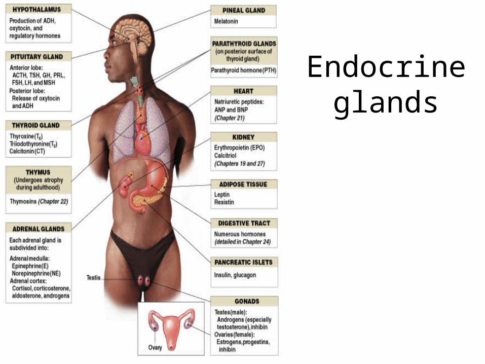
Endocrine glands
Hormones
• Functions– Regulation:
• Water balance and body fluid chemistry
• Metabolic rate and energy balance
• Cardiac and smooth muscle activity
• Immune system activity
– Control growth and development– Reproductive organ function and cycles– Circadian rythms
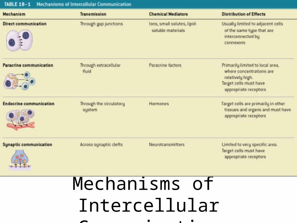
Mechanisms of Intercellular CommunicationTable 18–1
Hormones
• Can be divided into 3 groups:
– amino acid derivatives
– peptide hormones
– lipid derivatives
Amino Acid Derivatives
• Small molecules structurally related to amino acids
• Synthesized from the amino acids tyrosine and tryptophan
Tyrosine Derivatives
• Thyroid hormones
• Compounds:– epinephrine (E)– norepinephrine (NE)– dopamine, also called catecholamines
Tryptophan Derivative
• Melatonin: – produced by pineal gland
Peptide Hormones
• Chains of amino acids
• Synthesized as prohormones:– inactive molecules converted to active
hormones before or after secretion
2 Groups of Peptide Hormones
• Group 1:
– glycoproteins:
• more than 200 amino acids long, with carbohydrate side chains:
–thyroid-stimulating hormone (TSH)
–luteinizing hormone (LH)
–follicle-stimulating hormone (FSH)
2 Groups of Peptide Hormones• Group 2:
– all hormones secreted by:• hypothalamus
• heart
• thymus
• digestive tract
• pancreas
• posterior lobe of pituitary gland
• anterior lobe of pituitary gland
2 Classes of Lipid Derivatives
• Eicosanoids: – derived from arachidonic acid
• Steroid hormones: – derived from cholesterol
Eicosanoids
• Are small molecules with five-carbon ring at one end
• Are important paracrine factors
• Coordinate cellular activities
• Affect enzymatic processes in extracellular fluids
Leukotrienes
• Are eicosanoids released by activated white blood cells, or leukocytes
• Important in coordinating tissue responses to injury or disease
Prostaglandins
• A second group of eicosanoids produced in most tissues of body
• Are involved in coordinating local cellular activities
• Sometimes converted to thromboxanes and prostacyclins
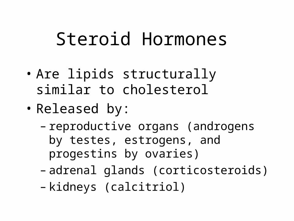
Steroid Hormones
• Are lipids structurally similar to cholesterol
• Released by:– reproductive organs (androgens by testes,
estrogens, and progestins by ovaries)– adrenal glands (corticosteroids)– kidneys (calcitriol)
Steroid Hormones
• Remain in circulation longer than peptide hormones
• Are absorbed gradually by liver
• Are converted to soluble form
• Are excreted in bile or urine
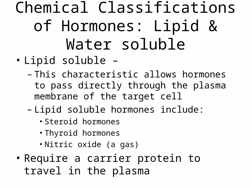
Chemical Classifications of Hormones: Lipid & Water soluble
• Lipid soluble –– This characteristic allows hormones to pass directly
through the plasma membrane of the target cell– Lipid soluble hormones include:
• Steroid hormones
• Thyroid hormones
• Nitric oxide (a gas)
• Require a carrier protein to travel in the plasma
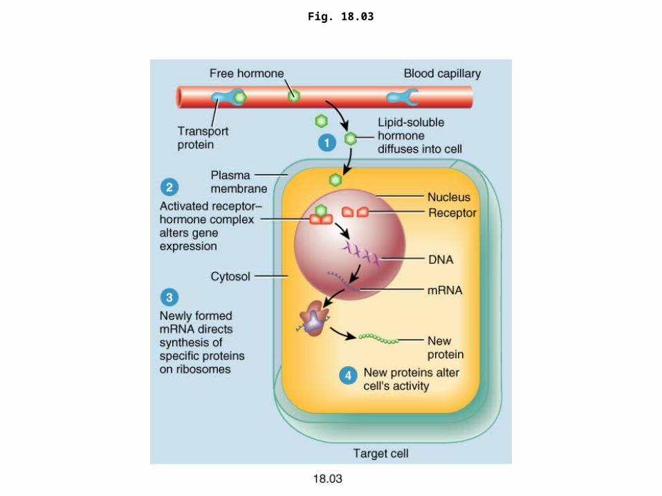
Fig. 18.03
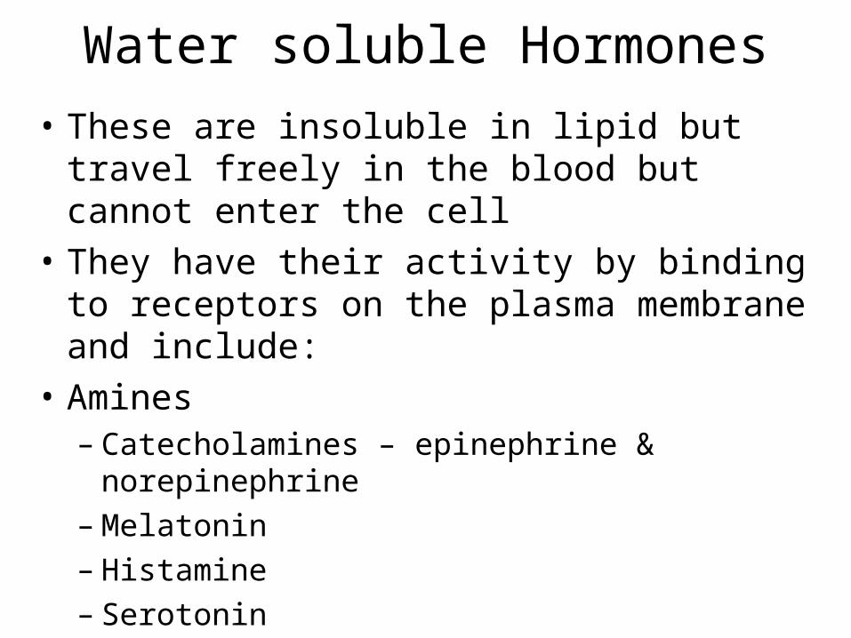
Water soluble Hormones
• These are insoluble in lipid but travel freely in the blood but cannot enter the cell
• They have their activity by binding to receptors on the plasma membrane and include:
• Amines– Catecholamines – epinephrine & norepinephrine– Melatonin– Histamine– Serotonin
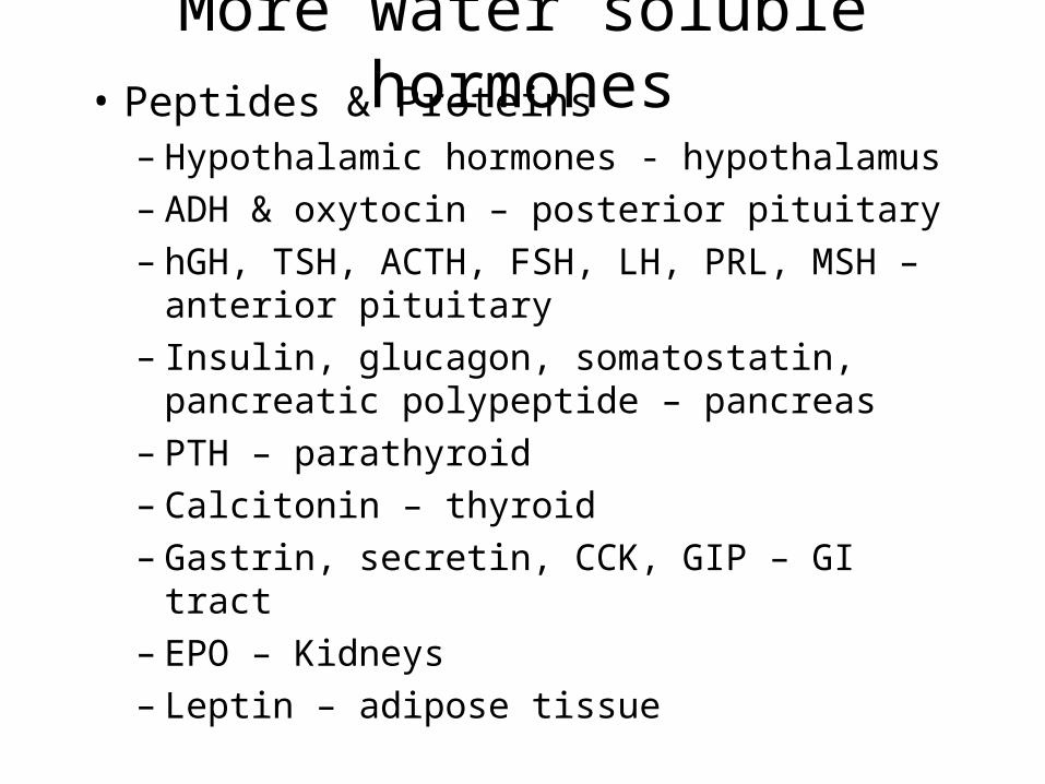
More water soluble hormones• Peptides & Proteins
– Hypothalamic hormones - hypothalamus– ADH & oxytocin – posterior pituitary– hGH, TSH, ACTH, FSH, LH, PRL, MSH –
anterior pituitary– Insulin, glucagon, somatostatin, pancreatic
polypeptide – pancreas– PTH – parathyroid– Calcitonin – thyroid– Gastrin, secretin, CCK, GIP – GI tract– EPO – Kidneys– Leptin – adipose tissue
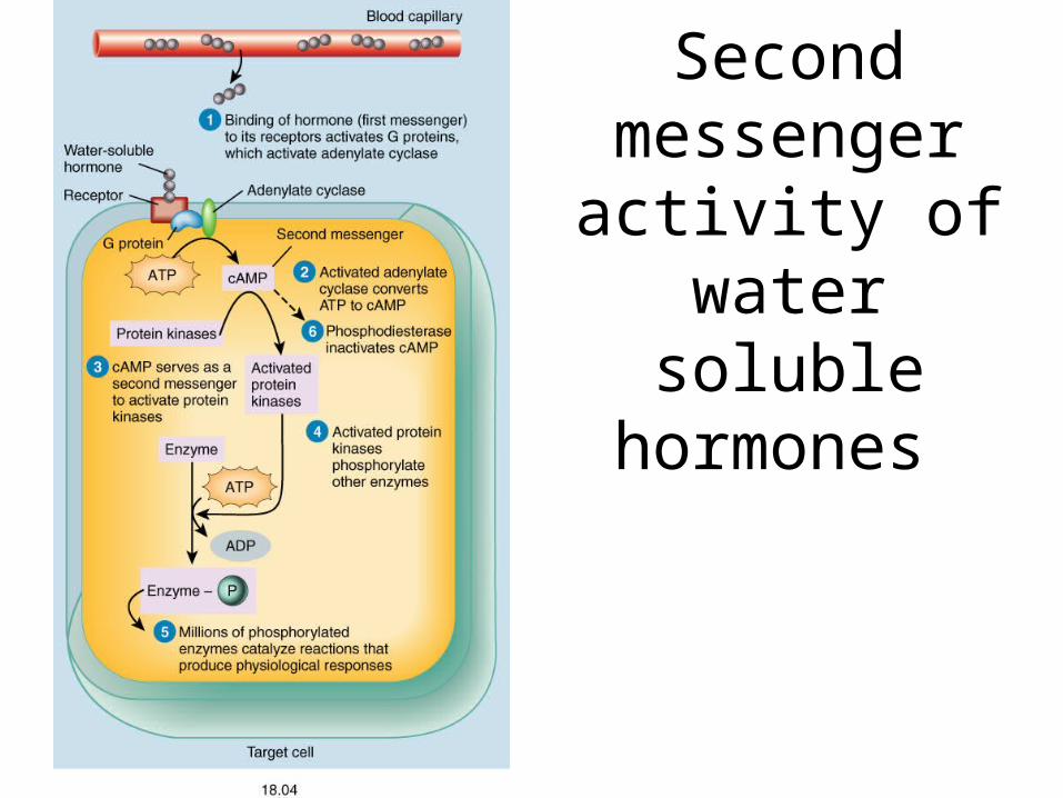
Second messenger activity of water
soluble hormones
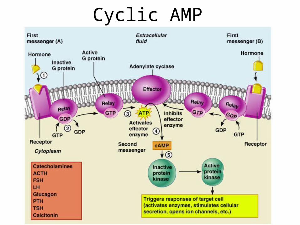
Cyclic AMP
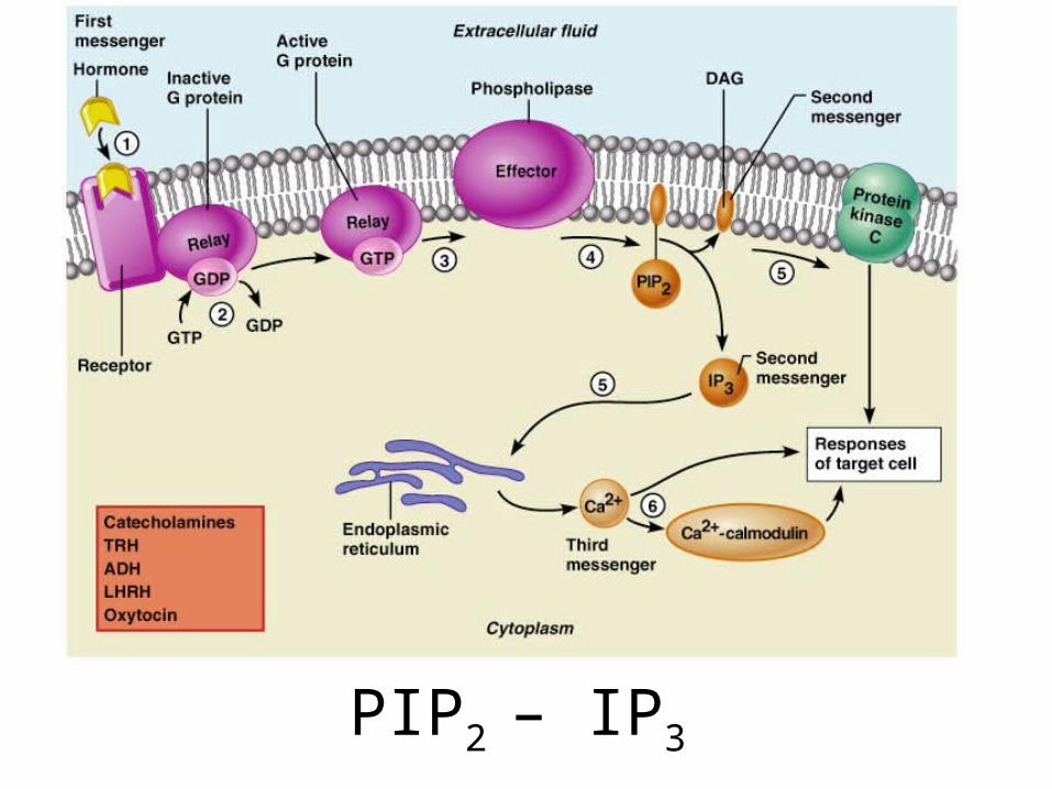
PIP2 – IP3
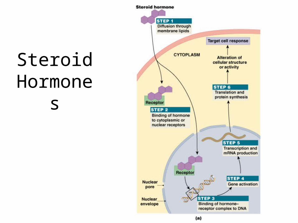
Figure 18–4a
Steroid Hormones
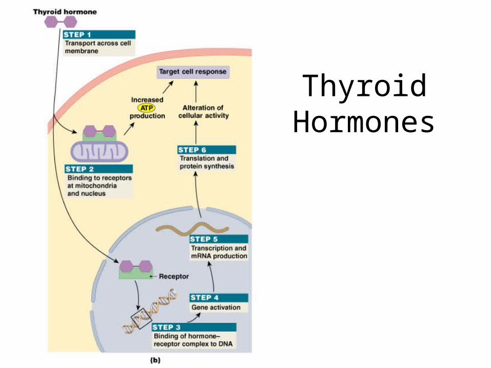
Thyroid Hormones
Figure 18–4b
Endocrine Reflexes
• Functional counterparts of neural reflexes
• In most cases, controlled by negative feedback mechanisms
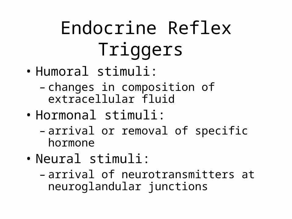
Endocrine Reflex Triggers
• Humoral stimuli: – changes in composition of extracellular fluid
• Hormonal stimuli: – arrival or removal of specific hormone
• Neural stimuli: – arrival of neurotransmitters at neuroglandular
junctions
Simple Endocrine Reflex
• Involves only 1 hormone
• Controls hormone secretion by:– heart– pancreas– parathyroid gland– digestive tract
Complex Endocrine Reflex
• Involves:– 1 or more intermediary steps– 2 or more hormones
Hormone activity
• Down-regulation –– Excessively high concentrations of hormones
reduces the number of receptors on the target tissues. This makes the tissue less sensitive to that hormone
• Up-regulation –– Low concentrations cause cells to produce more
receptors resulting in increased sensitivity of the tissue
The Hormones and their glands
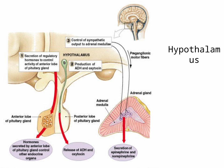
Figure 18–5
Hypothalamus

the Pituitary
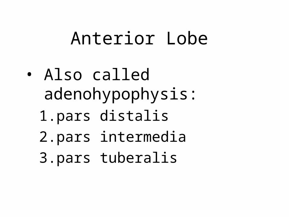
Anterior Lobe
• Also called adenohypophysis:1. pars distalis
2. pars intermedia
3. pars tuberalis
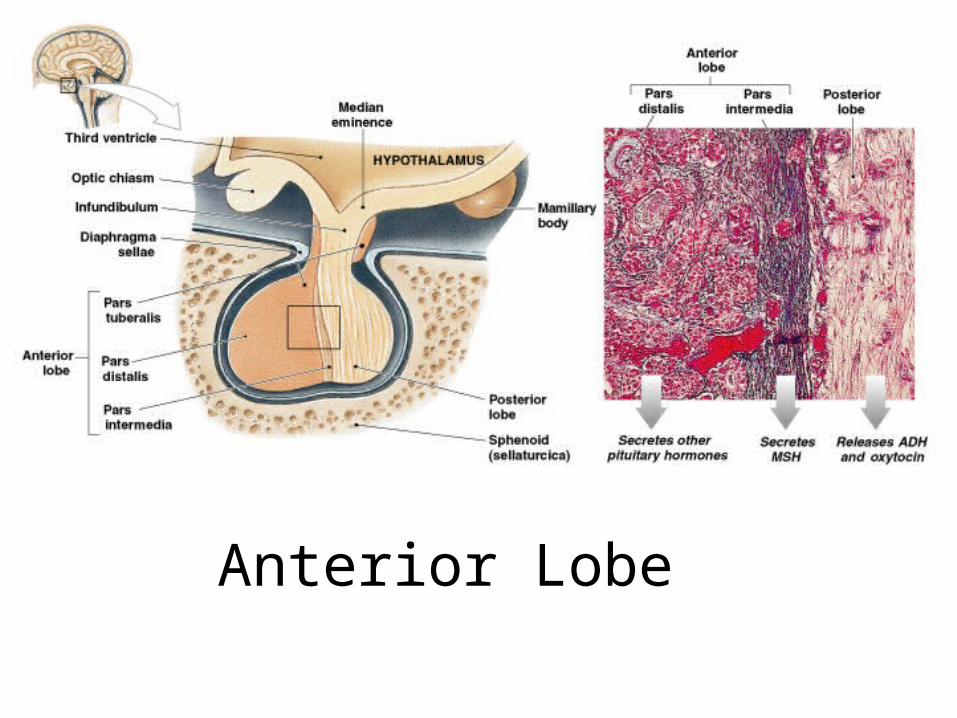
Figure 18–6
Anterior Lobe
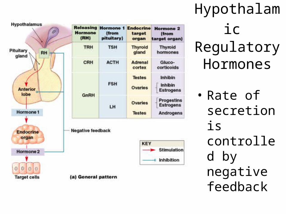
Figure 18–8a
Hypothalamic Regulatory Hormones
• Rate of secretion is controlled by negative feedback

Figure 18–8b
Hypothalamic
Regulatory
Hormones
Thyroid-Stimulating Hormone (TSH)
• Also called thyrotropin
• Triggers release of thyroid hormones
Adrenocorticotropic Hormone (ACTH)
• Also called corticotropin
• Stimulates release of steroid hormones by adrenal cortex
• Targets cells that produce glucocorticoids
Gonadotropins
• Regulate activities of gonads (testes, ovaries)
• Follicle-stimulating hormone
• Luteinizing hormone
Follicle-Stimulating Hormone (FSH)
• Also called follitropin
• Stimulates follicle development and estrogen secretion in females
• Stimulates sustentacular cells in males: – promotes physical maturation of sperm
• Production inhibited by inhibin: – peptide hormone released by testes and ovaries
Luteinizing Hormone (LH)
• Also called lutropin
• Causes ovulation and progestin production in females
• Causes androgen production in males
FSH and LH Production
• Stimulated by gonadotropin-releasing hormone (GnRH) from hypothalamus:– GnRH production inhibited by estrogens,
progestins, and androgens
Prolactin (PRL)
• Also called mammotropin
• Stimulates development of mammary glands and milk production
• Production inhibited by prolactin-inhibiting hormone (PIH)
Prolactin (PRL)
• Stimulates PIH release
• Inhibits secretion of prolactin-releasing factors (PRF)
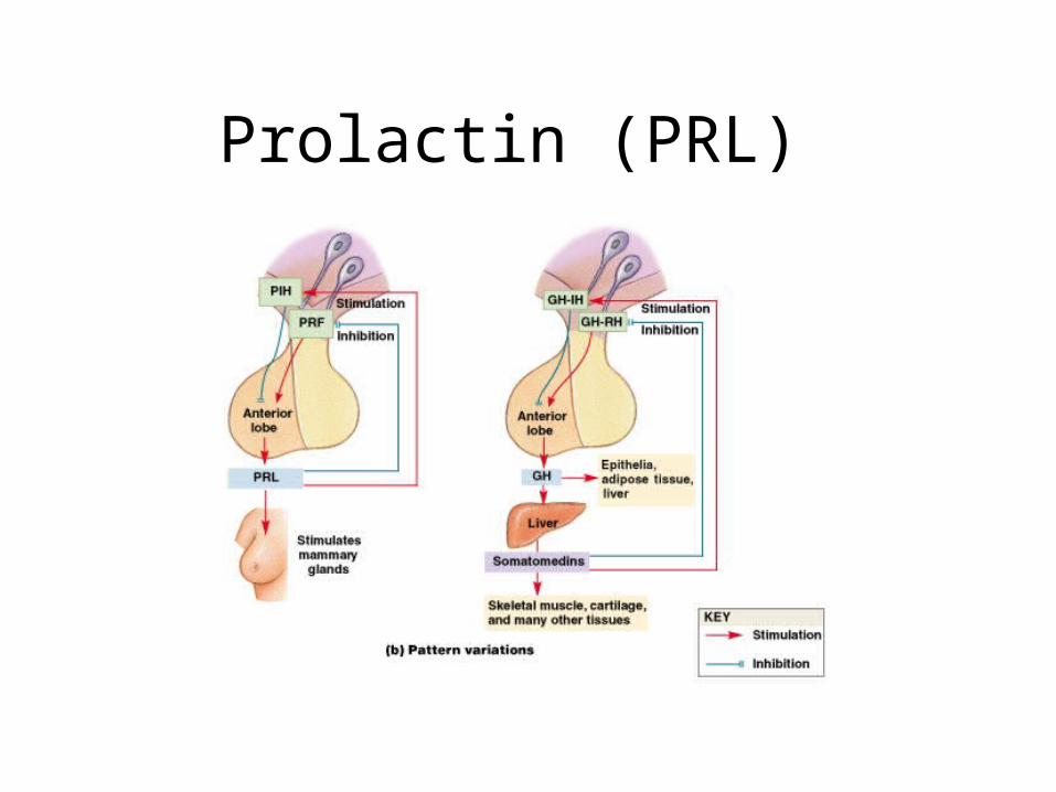
Figure 18–8b
Prolactin (PRL)
Growth Hormone (GH)
• Also called somatotropin
• Stimulates cell growth and replication
• Production regulated by:
– growth hormone–releasing hormone (GH–RH)
– growth hormone–inhibiting hormone (GH–IH)
Melanocyte-Stimulating Hormone (MSH)
• Also called melanotropin
• Stimulates melanocytes to produce melanin
• Inhibited by dopamine
Melanocyte-Stimulating Hormone (MSH)
• Secreted by pars intermedia during:
– fetal development
– early childhood
– pregnancy
– certain diseases
Posterior Lobe
• Also called neurohypophysis
• Contains unmyelinated axons of hypothalamic neurons
• Supraoptic and paraventricular nuclei manufacture:– antidiuretic hormone (ADH)– oxytocin (OT)
Antidiuretic Hormone
• Decreases amount of water lost at kidneys
• Elevates blood pressure
• Release inhibited by alcohol
Oxytocin
• Stimulates contractile cells in mammary glands
• Stimulates smooth muscles in uterus
• Secretion and milk ejection are part of neuroendocrine reflex
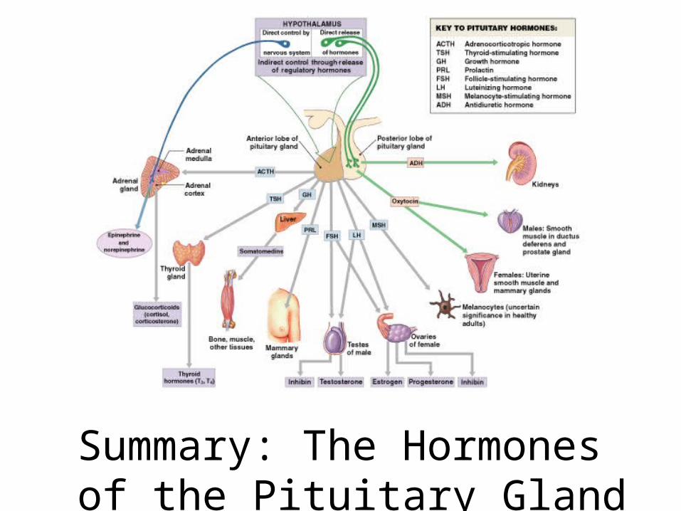
Figure 18–9
Summary: The Hormones of the Pituitary Gland

Table 18–2
Summary: The Hormones of the Pituitary Gland
Fig. 18.10
The Thyroid

Thyroid Gland Figure 18–10a, b
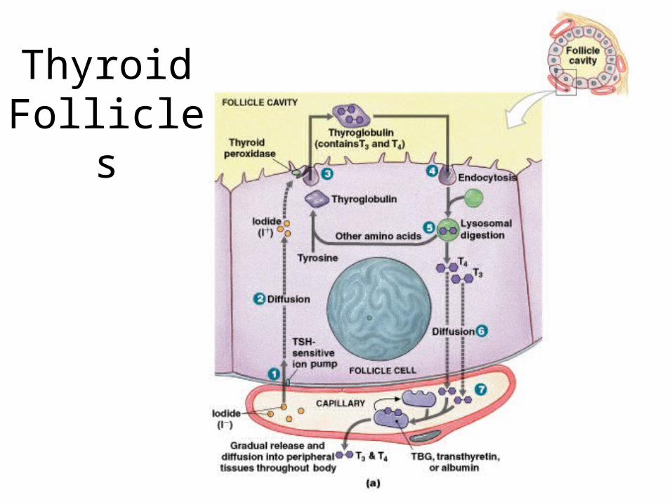
Thyroid Follicles
Figure 18–11a, b
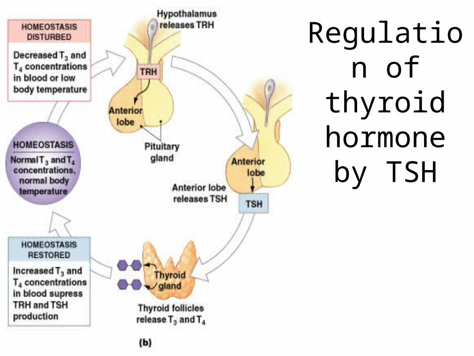
Regulation of thyroid
hormone by TSH
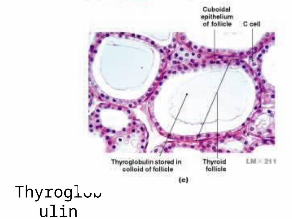
Figure 18–10c
Thyroglobulin
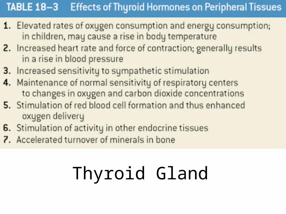
Thyroid Gland
Table 18–3
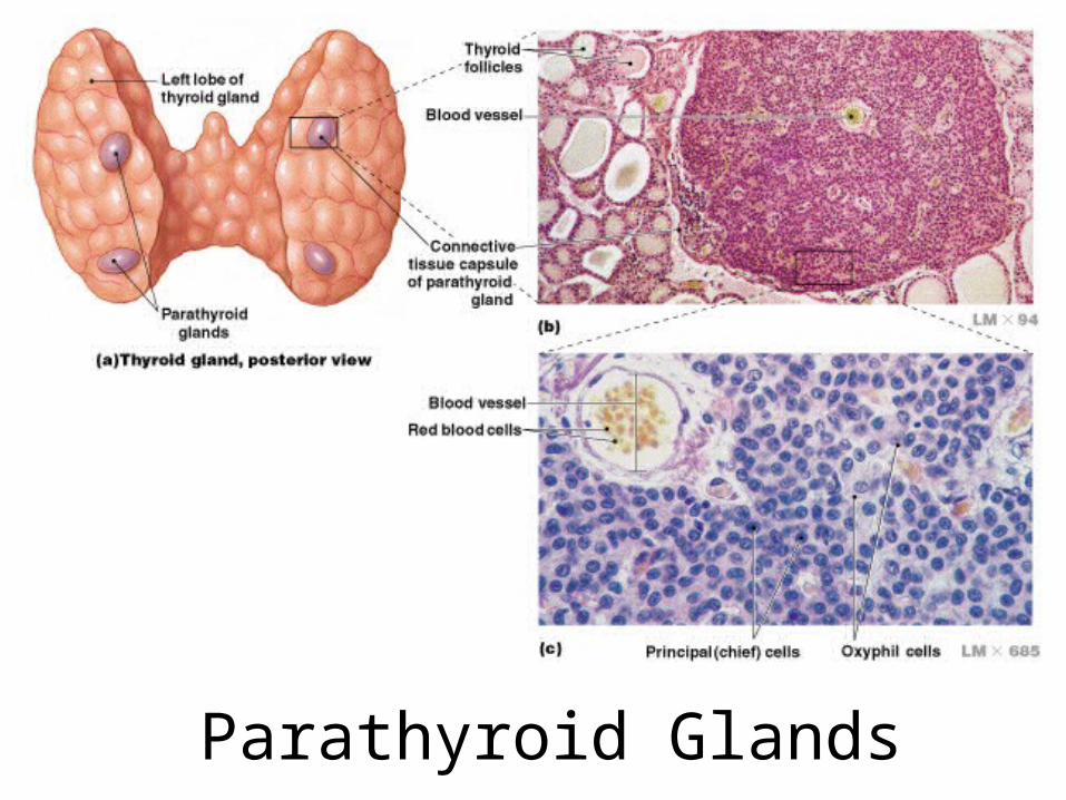
Parathyroid GlandsFigure 18–12
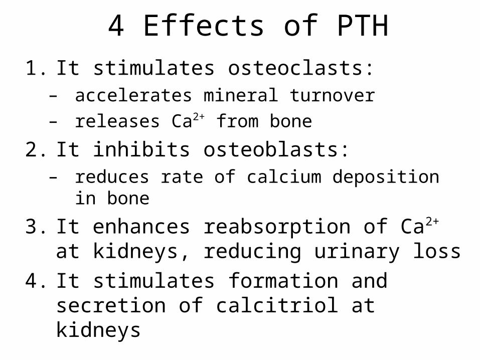
4 Effects of PTH1. It stimulates osteoclasts:
– accelerates mineral turnover– releases Ca2+ from bone
2. It inhibits osteoblasts:– reduces rate of calcium deposition in bone
3. It enhances reabsorption of Ca2+ at kidneys, reducing urinary loss
4. It stimulates formation and secretion of calcitriol at kidneys
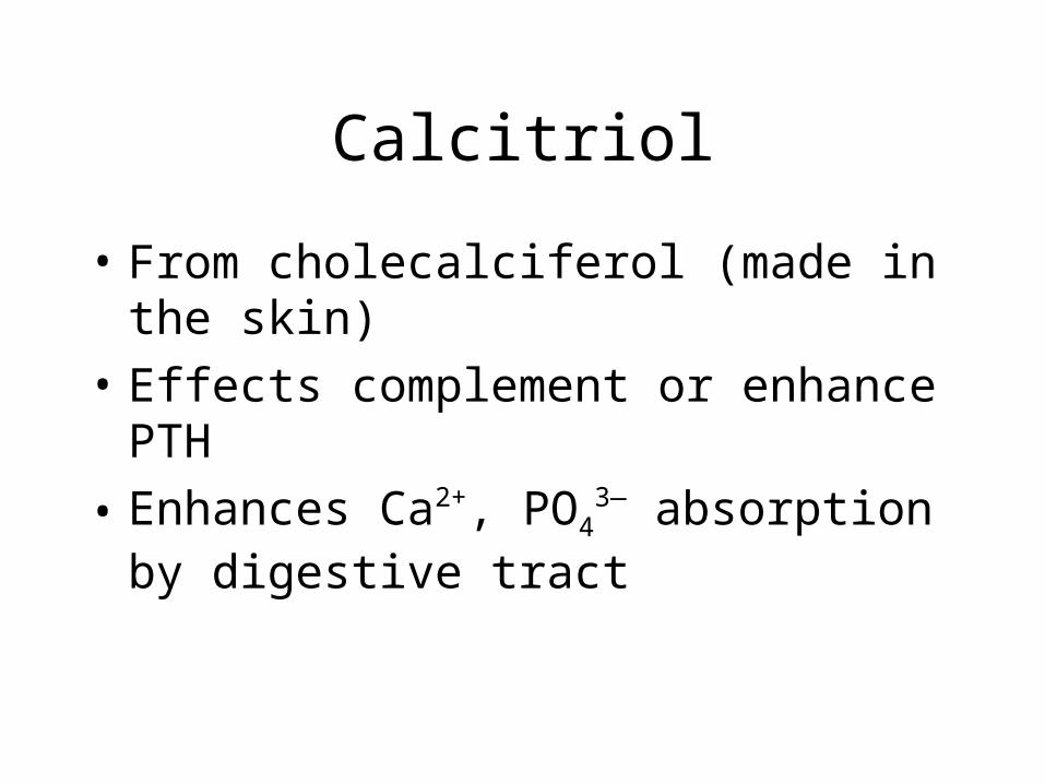
Calcitriol
• From cholecalciferol (made in the skin)
• Effects complement or enhance PTH
• Enhances Ca2+, PO43— absorption by
digestive tract
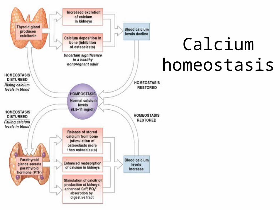
Calcium homeostasis
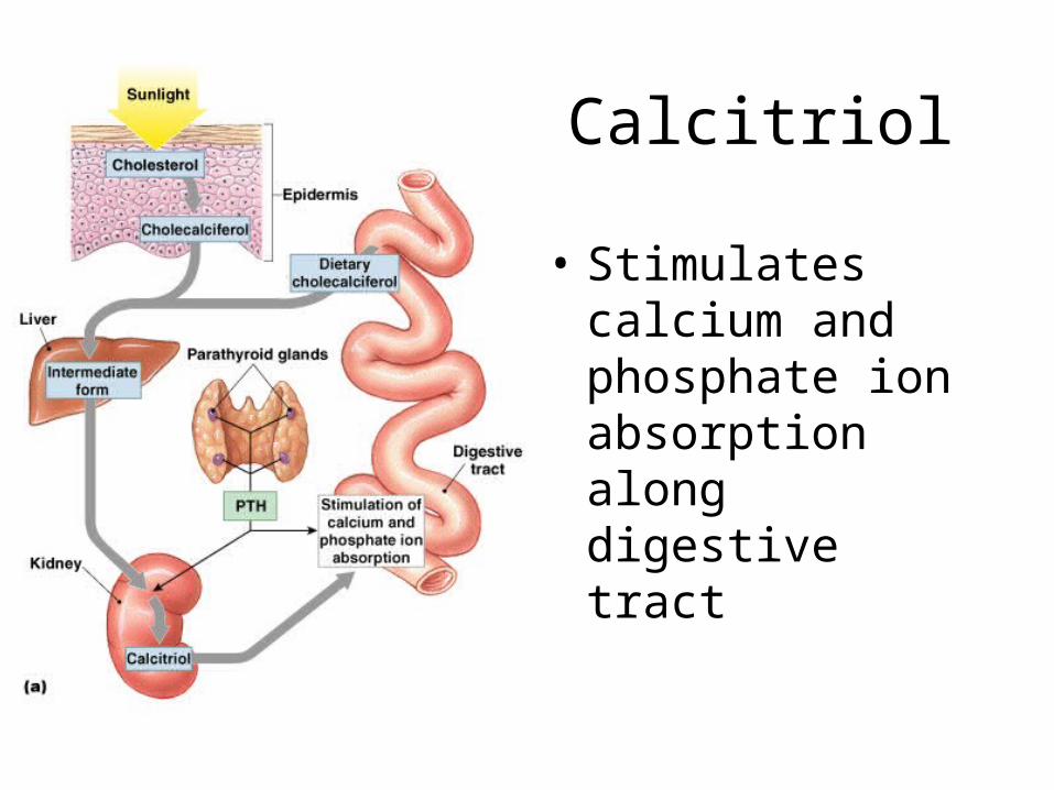
Calcitriol
• Stimulates calcium and phosphate ion absorption along digestive tract
Figure 18–17a
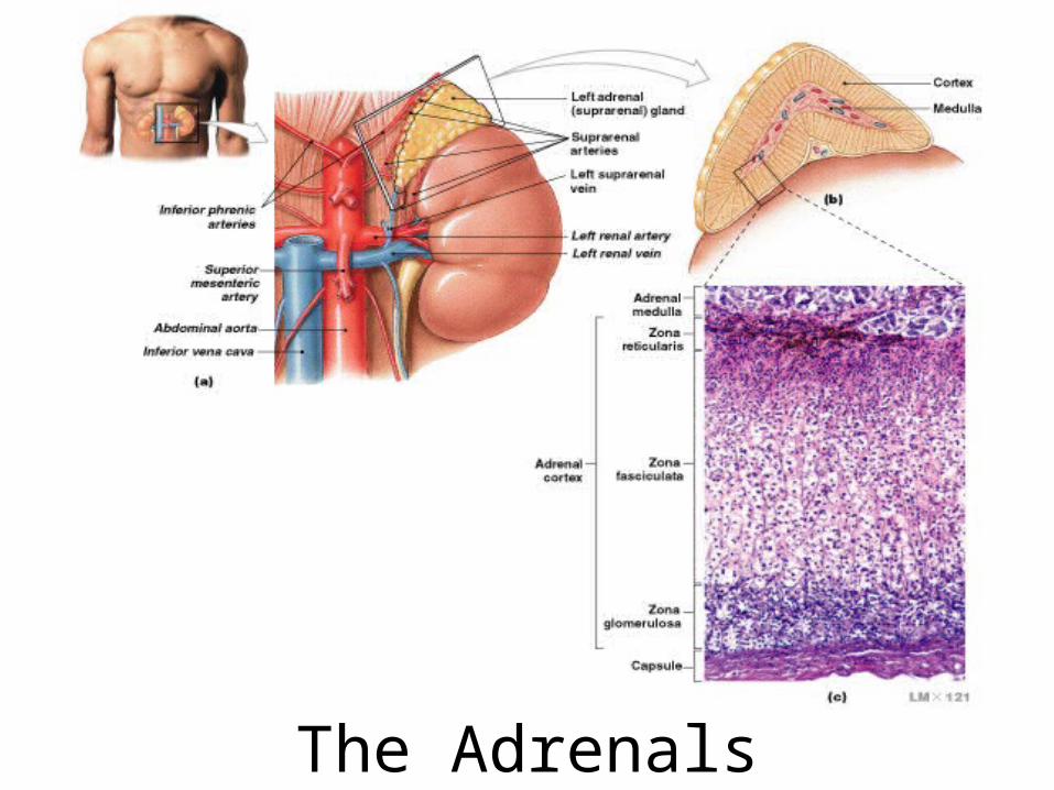
The Adrenals
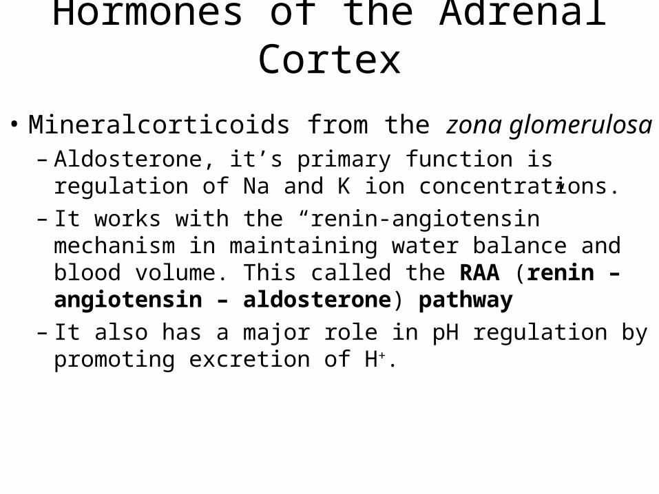
Hormones of the Adrenal Cortex
• Mineralcorticoids from the zona glomerulosa– Aldosterone, it’s primary function is regulation of Na and
K ion concentrations.– It works with the “renin-angiotensin” mechanism in
maintaining water balance and blood volume. This called the RAA (renin – angiotensin – aldosterone) pathway
– It also has a major role in pH regulation by promoting excretion of H+.
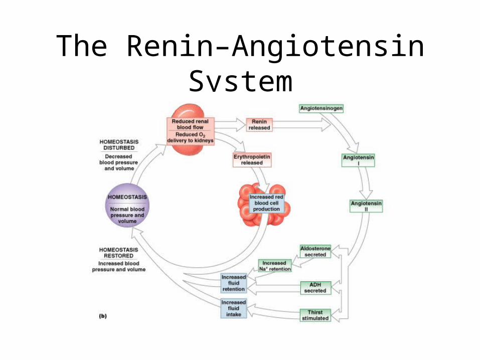
The Renin–Angiotensin System
Figure 18–17b
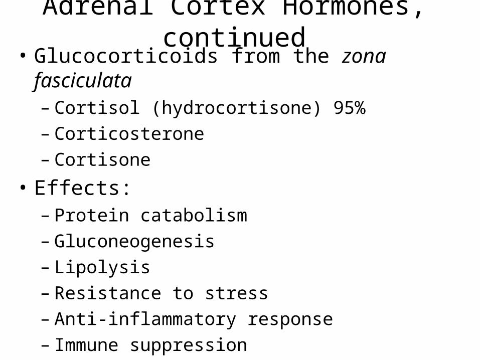
Adrenal Cortex Hormones, continued• Glucocorticoids from the zona fasciculata
– Cortisol (hydrocortisone) 95%– Corticosterone– Cortisone
• Effects:– Protein catabolism– Gluconeogenesis– Lipolysis– Resistance to stress– Anti-inflammatory response– Immune suppression
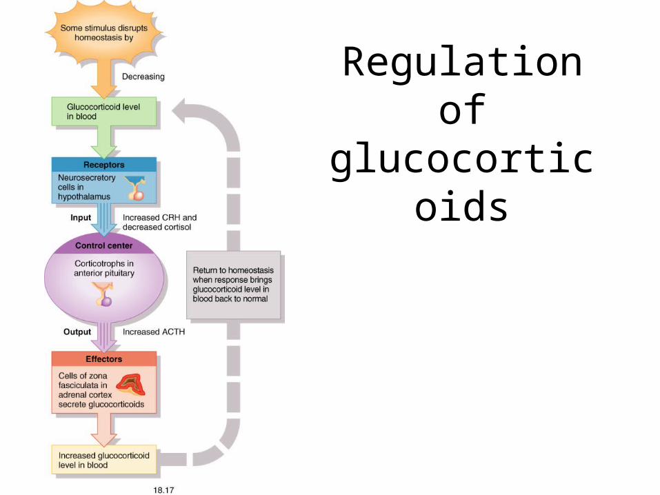
Regulation of glucocorticoids

Adrenal cortex hormones iii• Sterocorticoids (Androgens) from the zona
reticularis
• Mostly DHEA (dehydroepiandrosterone)– Unimportant in males after puberty– Promote libido in females and converted to estrogen– Following menopause this is the source of estrogens for
women

Adrenal Cortex
Table 18–5
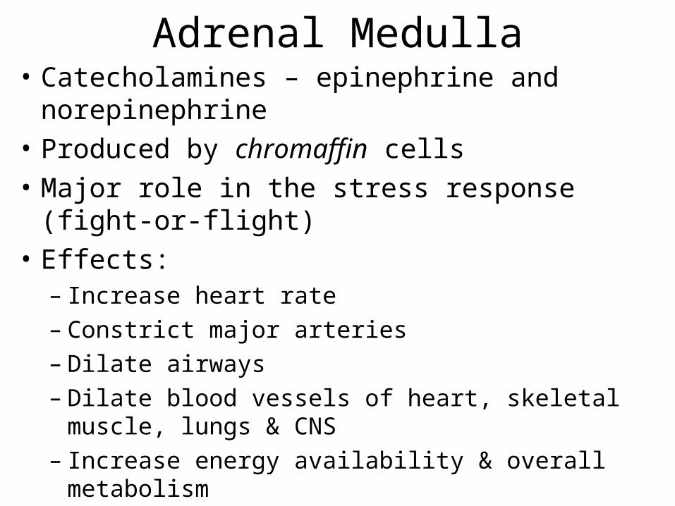
Adrenal Medulla• Catecholamines – epinephrine and norepinephrine
• Produced by chromaffin cells
• Major role in the stress response (fight-or-flight)
• Effects:– Increase heart rate– Constrict major arteries– Dilate airways– Dilate blood vessels of heart, skeletal muscle, lungs &
CNS– Increase energy availability & overall metabolism
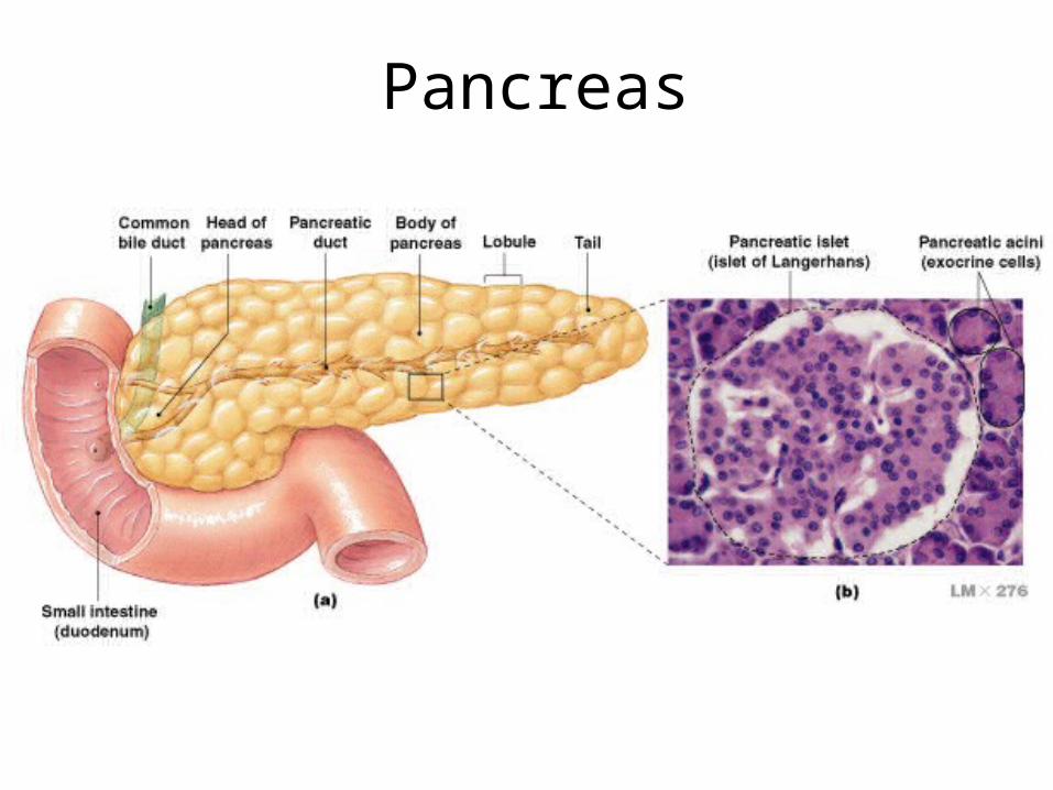
Figure 18–15
Pancreas
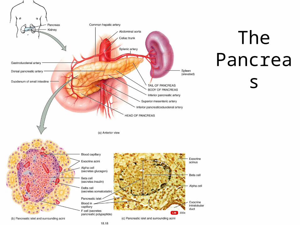
Fig. 18.18
The Pancreas
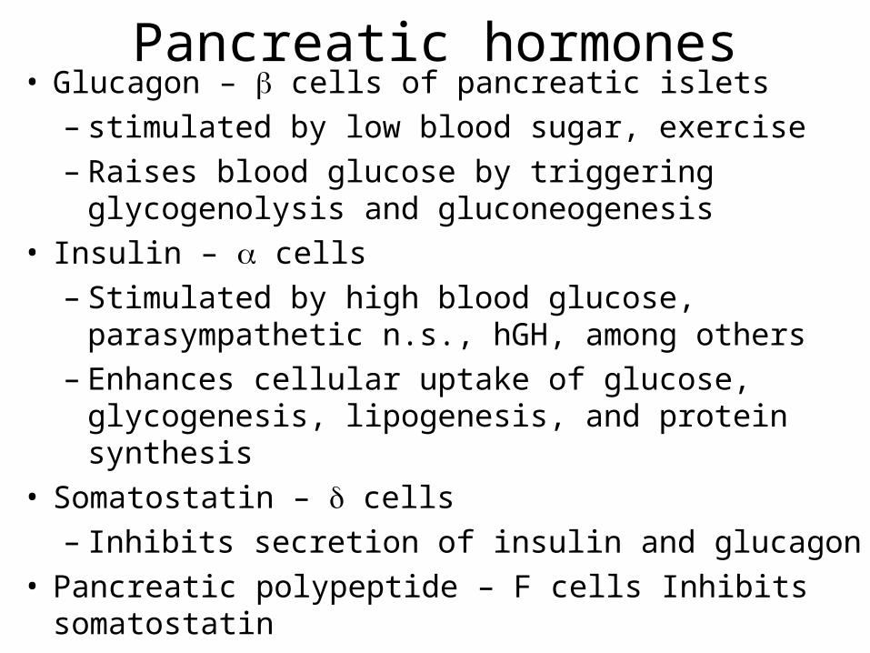
Pancreatic hormones• Glucagon – cells of pancreatic islets
– stimulated by low blood sugar, exercise– Raises blood glucose by triggering glycogenolysis and
gluconeogenesis• Insulin – cells
– Stimulated by high blood glucose, parasympathetic n.s., hGH, among others
– Enhances cellular uptake of glucose, glycogenesis, lipogenesis, and protein synthesis
• Somatostatin – cells– Inhibits secretion of insulin and glucagon
• Pancreatic polypeptide – F cells Inhibits somatostatin
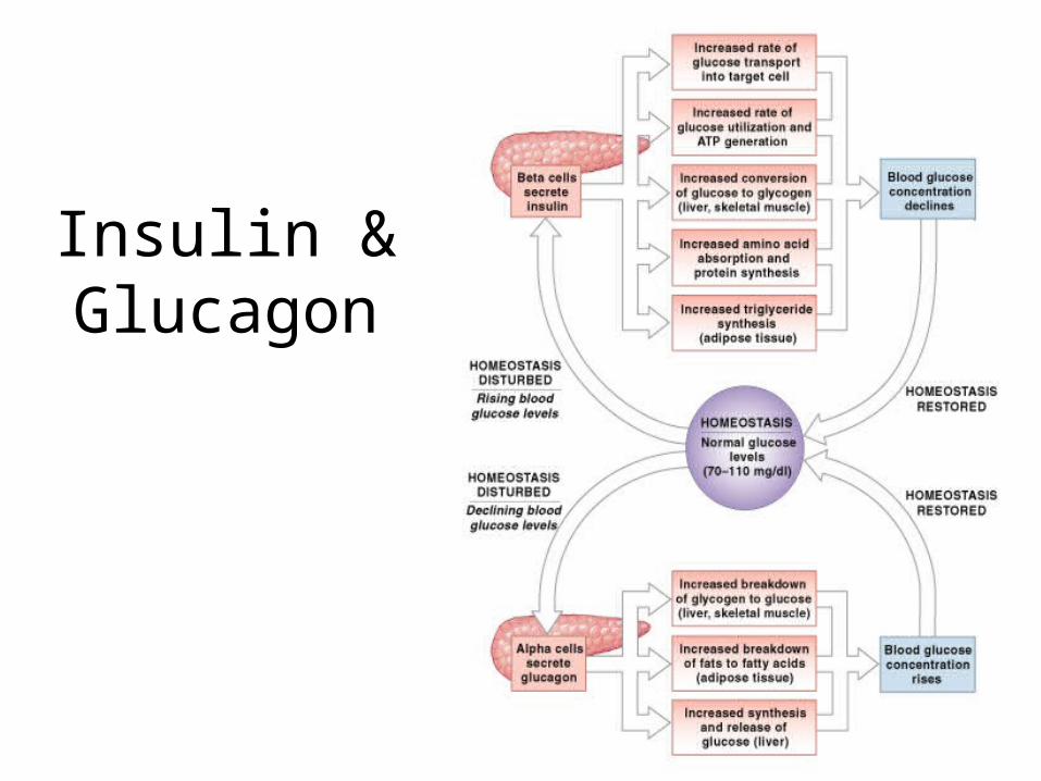
Insulin & Glucagon
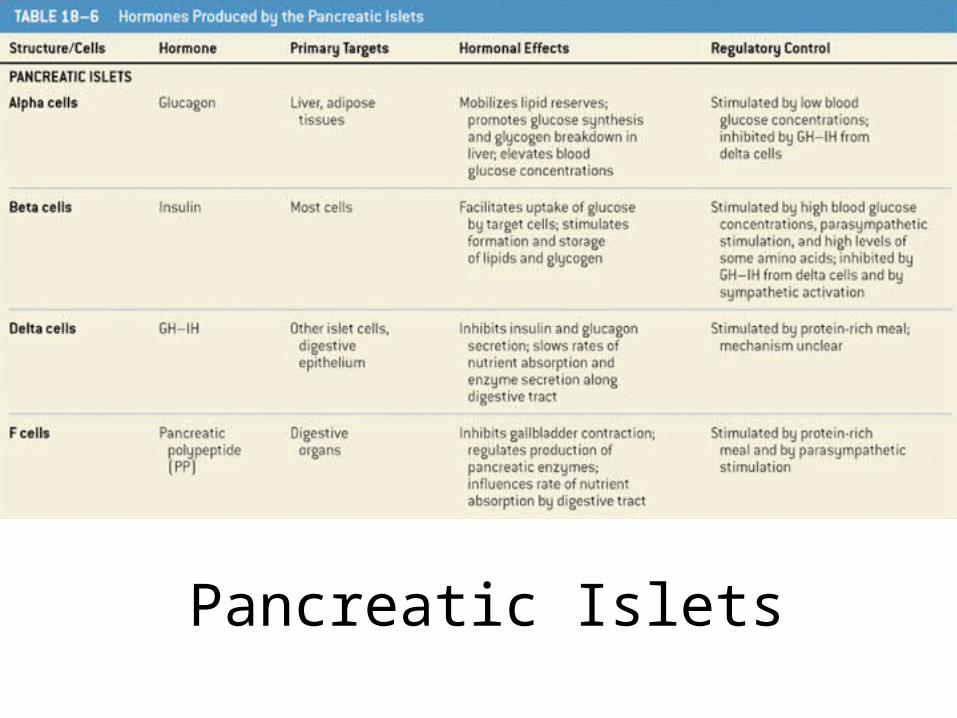
Table 18–6
Pancreatic Islets
Effects of Diabetes Mellitus
• Results from hyposecretion or hypoactivity of insulin
• The three cardinal signs of DM are:– Polyuria – huge urine output– Polydipsia – excessive thirst– Polyphagia – excessive hunger and food
consumption
• Hyperinsulinism – excessive insulin secretion, resulting in hypoglycemia
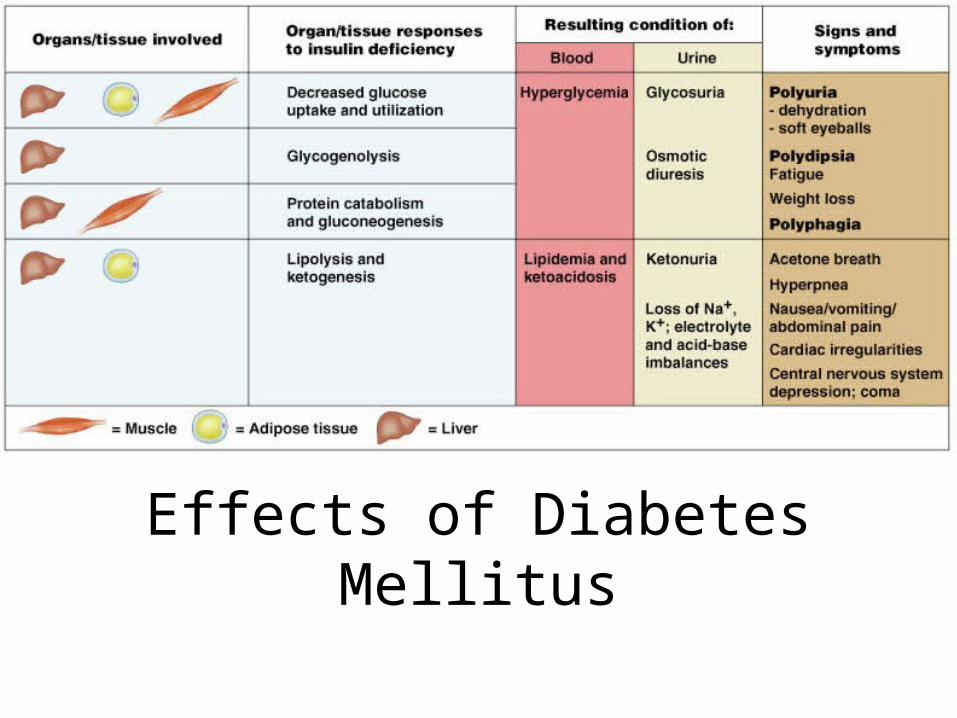
Effects of Diabetes Mellitus
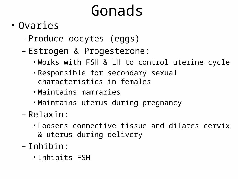
Gonads• Ovaries
– Produce oocytes (eggs)– Estrogen & Progesterone:
• Works with FSH & LH to control uterine cycle
• Responsible for secondary sexual characteristics in females
• Maintains mammaries
• Maintains uterus during pregnancy
– Relaxin:• Loosens connective tissue and dilates cervix & uterus
during delivery
– Inhibin:• Inhibits FSH
The Testis
• Produce sperm
• Testosterone– Responsible for secondary sexual characteristics
• Inhibin – Controls testosterone release
Pineal Gland
• Produces melatonin– Stimulated by darkness– Contributes to maintaining day/cycles– Linked to seasonal affective disorder
Thymus
• Secretes thymosin, thymic humoral factor & thymopoietin– These are involved with the development and
immunocompetence of T-lymphocytes

Other endocrine cells• Erythropoietin (EPO) from kidney
– Stimulates red blood cell production
• Atrial Natruetic Peptide (ANP) from heart– Antagonistic to aldosterone
• The placenta – produces sex hormones, human chorionic gonadotropin (hCG), hCS and relaxin during pregnancy
• Eicsanoids – produced by many cells as autocrine and paracrine hormones
• GI tract – produces GIP,CCK, Gastrin
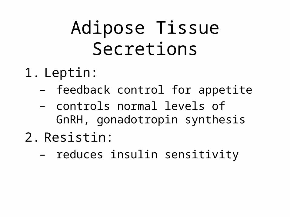
Adipose Tissue Secretions
1. Leptin: – feedback control for appetite– controls normal levels of GnRH,
gonadotropin synthesis
2. Resistin: – reduces insulin sensitivity
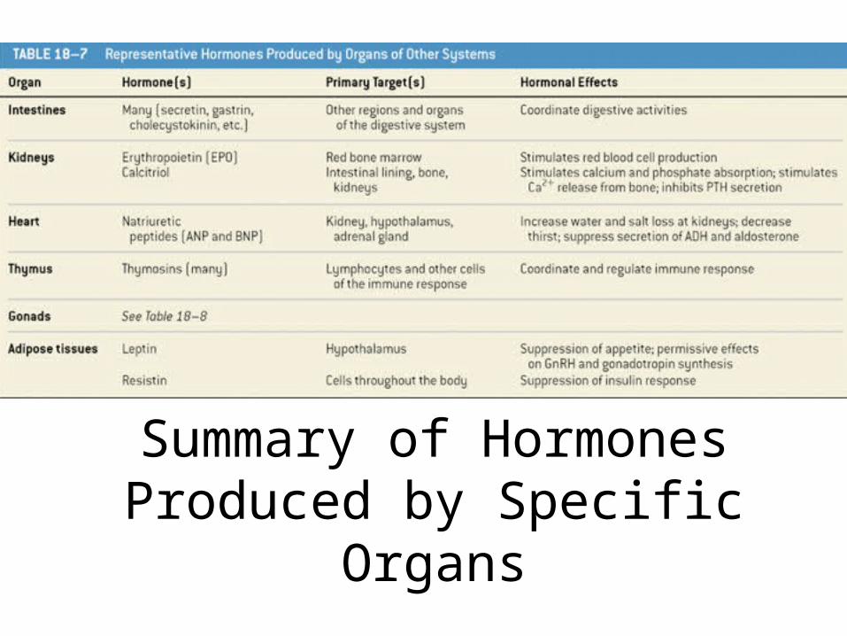
Summary of Hormones Produced by Specific Organs
Table 18–7
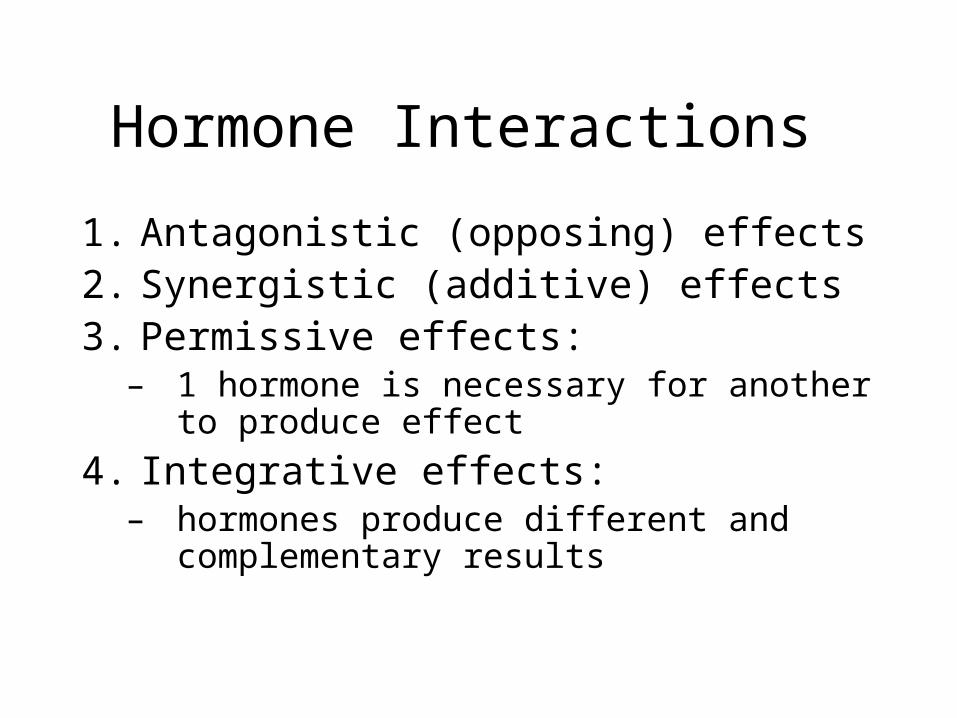
Hormone Interactions
1. Antagonistic (opposing) effects2. Synergistic (additive) effects3. Permissive effects:
– 1 hormone is necessary for another to produce effect
4. Integrative effects: – hormones produce different and
complementary results
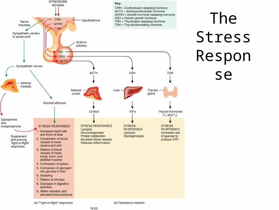
The Stress
Response
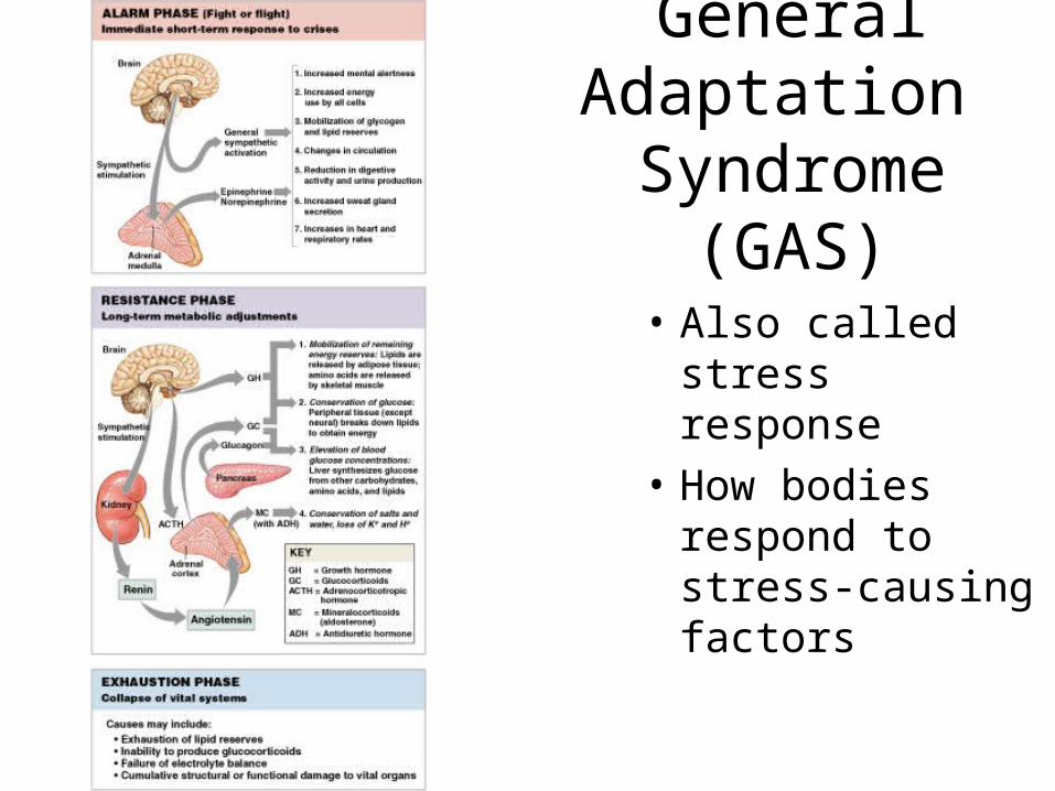
General Adaptation
Syndrome (GAS)
• Also called stress response
• How bodies respond to stress-causing factors
Figure 18–18
General Adaptation Syndrome (GAS)
• Is divided into 3 phases: 1. alarm phase
2. resistance phase
3. exhaustion phase
Alarm Phase
• Is an immediate response to stress
• Is directed by ANS
• Energy reserves mobilized (glucose)
• “Fight or flight” responses
• Dominant hormone is epinephrine
7 Characteristics of Alarm Phase
1. Increased mental alertness
2. Increased energy consumption
3. Mobilization of energy reserves (glycogen and lipids)
7 Characteristics of Alarm Phase
4. Circulation changes: – increased blood flow to skeletal muscles– decreased blood flow to skin, kidneys, and
digestive organs
7 Characteristics of Alarm Phase
5. Drastic reduction in digestion and urine production
6. Increased sweat gland secretion
7. Increases in blood pressure, heart rate, and respiratory rate
Resistance Phase
• Entered if stress lasts longer than few hours
• Dominant hormones are glucocorticoids
• Energy demands remain high
• Glycogen reserves nearly exhausted after several hours of stress
Effects of Resistance Phase
1. Mobilize remaining lipid and protein reserves
2. Conserve glucose for neural tissues
3. Elevate and stabilize blood glucose concentrations
4. Conserve salts, water, and loss of K+, H+
Exhaustion Phase
• Begins when homeostatic regulation breaks down
• Failure of 1 or more organ systems will prove fatal
• Mineral imbalance
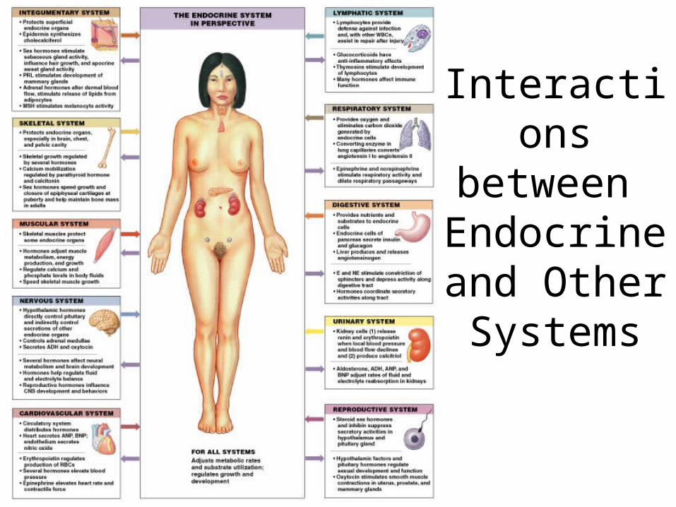
Interactions between
Endocrine and Other Systems
Figure 18–19
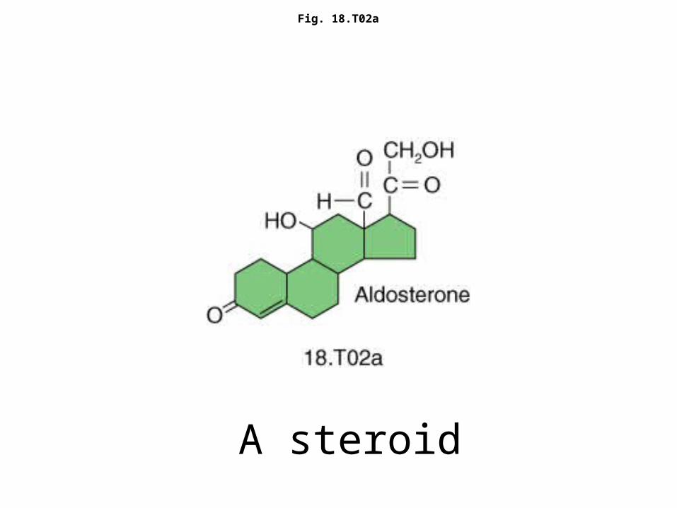
Fig. 18.T02a
A steroid
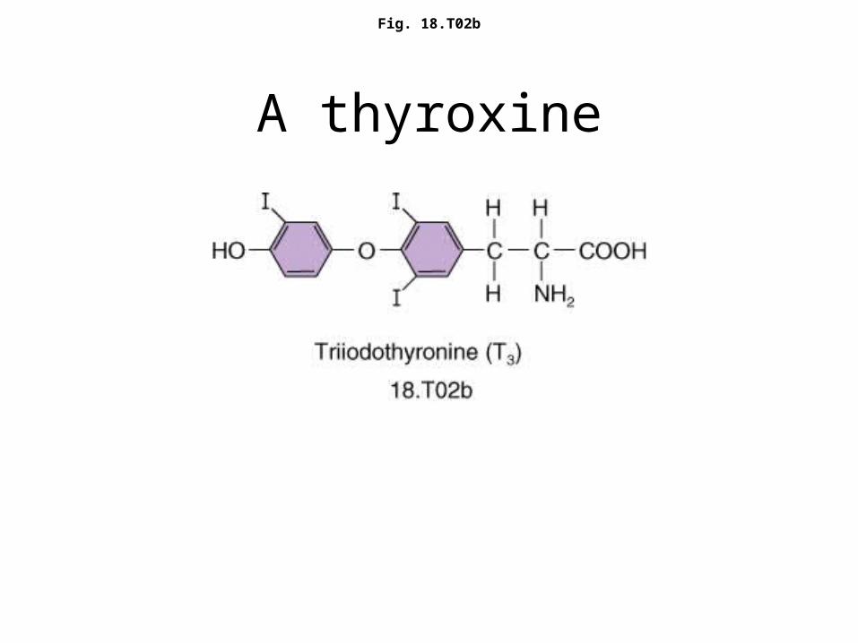
Fig. 18.T02b
A thyroxine
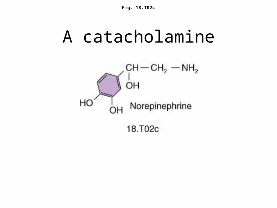
Fig. 18.T02c
A catacholamine
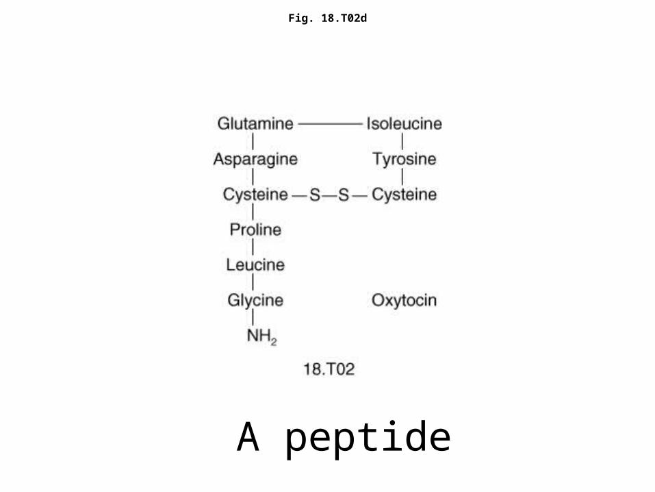
Fig. 18.T02d
A peptide
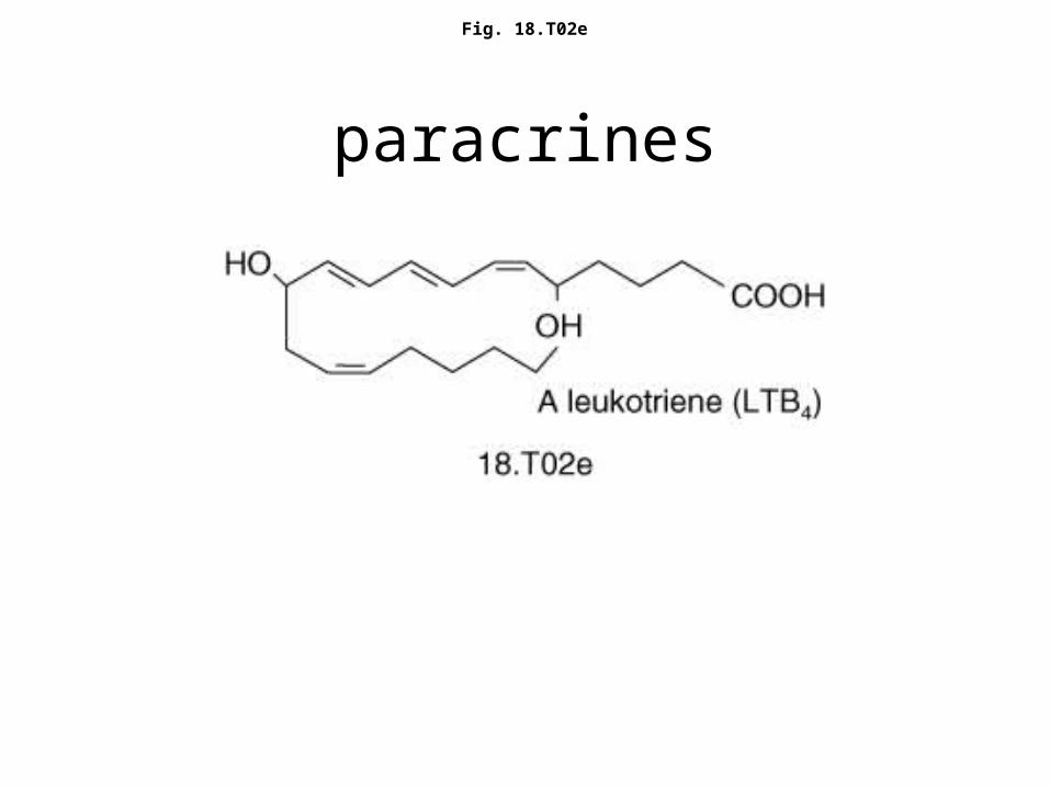
Fig. 18.T02e
paracrines