Embed Size (px)
Citation preview

Journal of Equine Veterinary Science xxx (2012) 1-11
Journal of Equine Veterinary Science
journal homepage: www.j -evs.com
Original Research
The Effect of Exercise and Nutritional Supplementation onProinflammatory Cytokine Expression in Young RacehorsesDuring Training
David W. Horohov PhD a, Stephen T. Sinatra MDb, Raj K. Chopra MS c, Stanley Jankowitz MA c,Alejandra Betancourt BS a, Richard J. Bloomer PhD d
aDepartment of Veterinary Science, Maxwell Gluck Equine Research Center, University of Kentucky, Lexington, KYbDepartment of Medicine, University of Connecticut School of Medicine, Farmington, CTcGelTec/Tishcon Corporation, Westbury, NYdCardiorespiratory Metabolic Laboratory, University of Memphis, Memphis, TN
a r t i c l e i n f o
Article history:Received 29 February 2012Received in revised form23 March 2012Accepted 28 March 2012Available online xxx
Keywords:ExerciseInflammationAdaptationMalondialdehydeLactateSupplementation
Corresponding author at: David W. Horohov, Depnary Science, 128H Maxwell Gluck Equine Research CKentucky, 40546-0099 Lexington, KY.
E-mail address: [email protected] (D.W. Horoho
0737-0806/$ - see front matter � 2012 Elsevier Inc. Adoi:10.1016/j.jevs.2012.03.017
a b s t r a c t
The inflammatory response to vigorous exercise ranges from the mild symptoms ofdelayed-onset muscle soreness to debilitating injuries affecting soft tissue, joint, andbone. Although there is a great deal of information available on the inflammatoryresponse to exercise in human athletes, less information is available regarding theinflammatory response to exercise in young horses undergoing training for racingcareers. Here, we assessed the cytokine response to exercise in a group of young Thor-oughbred racehorses during their initial training. Because there is interest in non-pharmacologic approaches to control or ameliorate exercise-induced inflammation, wealso examined the anti-inflammatory effect of a nutritional supplement fed to half of thehorses undergoing training. Twenty-five Thoroughbred horses aged 2 years were fol-lowed through their initial race training. Peripheral blood samples were collected atvarious times during the exercise for the quantitation of lactic acid, oxidative stress, andinflammatory cytokine gene expression. There was an intensity-dependent effect ofexercise on lactate, malondialdehyde, and proinflammatory cytokine gene expression.Although training itself was associated with an overall reduction in inflammatorymarkers, horses receiving the supplement exhibited further reductions in their indicatorsof inflammation. As such, this study provides novel evidence of nutritional supplemen-tation reducing postexercise inflammation.
� 2012 Elsevier Inc. All rights reserved.
1. Introduction
Lameness in horses is the most common cause ofdiminished athletic performance, as well as of economicand animal loss [1]. In the early 1980s, it was reported that67.6% of the lost training days for racehorses was becauseof lameness [2]. Recent data indicated that more than 80%
artment of Veteri-enter, University of
v).
ll rights reserved.
of total days lost from training Thoroughbred racehorsesaged 2 and 3 years were because of lameness issues [3]. Inanother study, race records showed that less than 80% ofthe 2-year-old racehorses in training completed theirtraining and went on to race [1]. Although the causes oflameness are varied, increased attention has been focusedon the role of inflammatory mediators in this process [4-6].Inflammation is associated with the production of proin-flammatory cytokines, which are small hormone-likeproteins produced in response to external stimuli. Proin-flammatory cytokines such as interleukin (IL)-1b, IL-6, andtumor necrosis factor-a (TNF-a) are induced by vigorous

Table 1Supplement componentsa
1.6 g Curcumin (BCM-95)1.6 g Boswellia (Bospure)400 mg coenzyme Q10 (HydroQSorb)4 g Glycine propionyl-L-carnitine HCL (GlycoCarn)10 g D-Ribose
aDouble diamond (peak performance), Equine Nutriceuticals, L.L.C.,Franklin Lakes, NJ.
D.W. Horohov et al. / Journal of Equine Veterinary Science xxx (2012) 1-112
exercise [7-10]. The consequences associated with thisincreased expression of inflammatory mediators post-exercise can range from the mild symptoms of delayed-onset muscle soreness [11] to debilitating problemsrelated to soft tissue, joint, and bone damage [12-14].Although an acute inflammatory response is a part of thehealing process, chronic inflammation can lead to tissuedestruction, subsequent poor performance, and perhaps anend to the athletic career [15]. Although numerous studieshave evaluated proinflammatory cytokine expression inresponse to different modes and intensities of exercise inhumans [16-18], limited work has been done in horses[19-21]. In general, equine athletes exhibit evidence ofincreased inflammation subsequent to exercise as charac-terized by increased expression of mRNA for TNF-a, IL-1b,and IL-6 in peripheral blood mononuclear cells [21,22].However, most of these studies have focused on treadmill-based exercise protocols typically involving a single bout ofintensive exercise. The use of a treadmill allows for stan-dardized testing, precise control of exercise parameters,and frequent blood collection and other sampling [23];however, there are both physiological and locomotiondifferences when comparing exercise on the trackwith thaton a treadmill [24]. Differences between treadmills can alsoaffect somemeasurements of performance [25]. Because allyoung Thoroughbred horses are trained on tracks, we wereinterested in using this approach to assess proin-flammatory cytokine gene expression during their exerciseat a training facility.
Given the negative impact of exercise-induced inflam-mation, there is a widespread use of nonsteroidal anti-inflammatory drugs (NSAIDS) in racehorses [26-33]. Arecent study reported that the plasma concentrations ofNSAIDS were highest in injured horses [33]. Although anti-inflammatory medication may relieve muscle soreness inhuman athletes, they do not affect recovery of musclefunction or markers of inflammation [34]. Besides concernsregarding the possibility that excessive use of NSAIDS maycontribute to race-related injuries [33], long-term use ofNSAIDs is associated with other adverse effects [35]. Thus,there is growing interest in nonpharmacologic approachesto ameliorate exercise-induced inflammation [36-39].Various dietary supplements have been proposed to alle-viate exercise-related injuries and illnesses [40-50].Although many of these same compounds have been usedin horses [37,48-58], their effect on exercise-inducedproinflammatory responses has not been investigated indetail [55]. Here, we have examined the effect of a nutri-tional supplement on the proinflammatory response toexercise in 2-year-old Thoroughbred racehorses in training.A second population of 2-year-old Thoroughbred race-horses training at the same facility was not given thesupplement, and these horses were included as controlhorses.
2. Materials and Methods
2.1. Horses
Twenty-five Thoroughbred horses aged 2 years werehoused and trained at a farm in Darlington, MD. A dirt ovaltrack of 1.0 km (total length) was used for training and
tests. A licensed trainer oversaw the training of the horses.All horses remained under routine management, withveterinary care provided by the farm’s regular veterinarian.Breaking to saddle and basic preparatory fitness workbegan when the horses were aged 18-21 months whichlasted for approximately 8 weeks. In general, the training ofthe 2-year-old horses consisted of 6 days/week slow can-tering (7-9 m/s) for 4 weeks, fast cantering (9-11 m/s) foranother month, and then continuation of fast canteringwith galloping (14-15 m/s) three times a week. A dailyexercise log was maintained, with specific mention of anydays of missed training and the reason. Each horse wastrained according to its individual abilities, as assessed bythe trainer, to achieve each horse’s optimal race potential.All training and testing of the horses occurred at the sametraining facility. All horses received the same daily rations.Health records were maintained for all horses, includingspecific information regarding injuries and treatments. Theuse of any medication was duly noted, and any horsereceiving NSAIDS or other treatments within a week of theexercise test was excluded from testing.
2.2. Nutritional Supplement
The nutritional supplement (Table 1) tested is designedto reduce joint injuries by preventing inflammation andenhancing energy production. The supplement was fed tothe horses (n ¼ 13) twice daily, with meals providedeveryday throughout the study period. Control horses (n ¼12) received daily meals without the supplement. Horseswere randomly assigned to treatment or control groups atthe beginning of their training period and remained ontreatment throughout the testing period.
2.3. Exercise Tests and Blood Sampling
The study consisted of four tests of increasing exerciseintensity (Table 2) during the training period. These testscoincided with each of the phases of the training period. Atthe beginning of the training period, all horses wereinitially sampled at rest to assess baseline proinflammatorycytokine responses before training. Each test consisted ofan initial warm-up period, followed by galloping at thespecified speed and distance for that particular test, andthen by a cool-down period (Table 2). The first test wasperformed before the beginning of the initial trainingperiod. All horses completed the tests on the scheduledday. The first peripheral blood samples (Pre) were collectedwithin 1 hour of the horses beginning the exercise test. Asecond set of samples were collected within 4-5 minutes ofthe completion of the exercise test (Post). Owing to thenature of training on a track, it was not possible to collect

Table 2Exercise tests
Date Distance Speed
Test 1 June 12 3.2 km 8.0 m/sTest 2 July 31 400 m 13.3 m/sTest 3 August 14 600 m 14.3 m/sTest 4 August 21 1,000 m 14.2 m/s
Table 3RT-PCR analysis of gene expression
Gene Target Function
IL-1 Proinflammatory cytokineIL-6 Proinflammatory cytokineTNF-a Proinflammatory cytokineIFNg LAK cell markerGranzyme B LAK cell marker
IL-1, interleukin 1; IL-6, interleukin 6; TNF-a, tumor necrosis factor-a;IFNg, interferon gamma; RT-PCR, real-time polymerase chain reaction.
D.W. Horohov et al. / Journal of Equine Veterinary Science xxx (2012) 1-11 3
all samples at the same time, but every effort was made tocollect themwithin a few minutes of each other. A third setof samples were collected at 2 hours postexercise when thehorses were in their stall (after 2 hours). Specific notationswere made on the length and time of the workout sessionduring which the sample was collected. This samplingschedule was repeated for each of the subsequent tests atthe intervals indicated in Table 2. Each test consisted of aninitial warm-up period, followed by the specified speed anddistance. The final test was performed at the end of thetraining period.
2.4. Blood Measures
Peripheral blood (w3 mL) was aseptically collectedfrom the jugular vein into a sodium-heparinized evacu-ated tube using an 18-g needle and hub. Lactate wasmeasured in plasma using an automated analyzer (LactatePlus; Nova Biomedical, Waltham, MA). Results areexpressed in millimeter concentrations. The Lactate Plusunit provides repeatable measures with precision, as hasbeen reported for similar units when compared withtraditional laboratory-based tools used for the assessmentof blood lactate [59]. However, it has been suggested thatsuch tools may not have precise linearity when used toassess lactate in blood samples obtained under extremeintensities of exercise. Because horses were pushed hardin the current design, yielding lactate values close to 20mM, this potential limitation should be noted [60].Malondialdehyde (MDA) was measured in plasmafollowing the modified procedures of Jentzsch et al. [61],using reagents purchased from Northwest Life ScienceSpecialties (Vancouver, WA). Specifically, 75 mL of plasmawas added to microcentrifuge reaction tubes, with theaddition of 3 mL of butylated hydroxytoluene in methanolto minimize ex vivo lipid peroxidation. Seventy-fivemicroliters of 1-M phosphoric acid and 75 mL of2-thiobarbituric acid reagent was added to each reactiontube and mixed thoroughly. Samples and reagents werethen incubated for 60 minutes at 60�C. Following incu-bation, tubes were removed, and the reaction mixture wastransferred to a microplate. The absorbance was then readusing a spectrophotometer at both 535 and 572 nm tocorrect for baseline absorption. MDA equivalents werecalculated using the difference in absorption at the twowavelengths. Quantification was performed with a cali-bration curve using tetramethoxypropane in a stabilizingbuffer. Samples were analyzed in duplicate on first thaw.The coefficient of variationwas 5.6%. Results are expressedin micrometer concentrations.
Peripheral blood (w3mL) was aseptically collected fromthe jugular vein into the PAXgene tube (Qiagen, Valencia,
CA) using an 18-g needle and hub. Once collected, thePAXgene tubes were inverted 20 times to ensure propermixing; they were then kept at room temperature for24 hours and then refrigerated until shipped. If held formore than 7 days, the tubes were stored frozen (�4�C) untilshipped. Each tube was labeled with the following infor-mation: horse ID, date of collection (MM/DD/YY), andsample (pre, post, 2 hours).
2.5. Gene Expression Analyses
Total RNA was isolated from the PAXgene tubes andanalyzed for gene expression of proinflammatory cytokinesand Lymphokine activated Killer (LAK) cell markers usingreal-time polymerase chain reaction and equine-specificprimers and probes (Table 3) [62]. The relative quantita-tion method was used to analyze gene expression wherethe average of all the horses’ initial pretraining sampleswasused as a calibrator against which all subsequent sampleswere compared [63]. The results were expressed as foldincreases above that of the initial sample.
2.6. Statistical Analysis
The effect of treatment and exercise on the lactate, MDA,and the expression of proinflammatory cytokines post-exercise was determined using a 3-way analysis of variancewith exercise test, time (before, immediately after, andafter 2 hours), and nutritional supplement included as themain variables. Results were considered significant at a Pvalue �.05 and power �.80. When necessary, data weretransformed to obtain normality of distribution and equalvariances.
3. Results
3.1. Lactate and Lipid Peroxidation Postexercise
Figure 1 shows blood lactic acid (mM) and MDA (mM)concentrations throughout the exercise test for both thecontrol (left panels) and supplemented (right panels)horses. As expected, lactic acid accumulation was observedimmediately after exercise, which returned to pre-exerciselevels after 2 hours. In general, there was an intensity-related increase in lactic acid production, with the thirdand fourth tests being different from the first and secondtest, and the second test being significantly different fromthe first. Although a similar pattern is observed for MDA,this intensity-related increase was only significant at thefourth test. The difference in the mean values between thecontrol and supplement groups were not great enough to
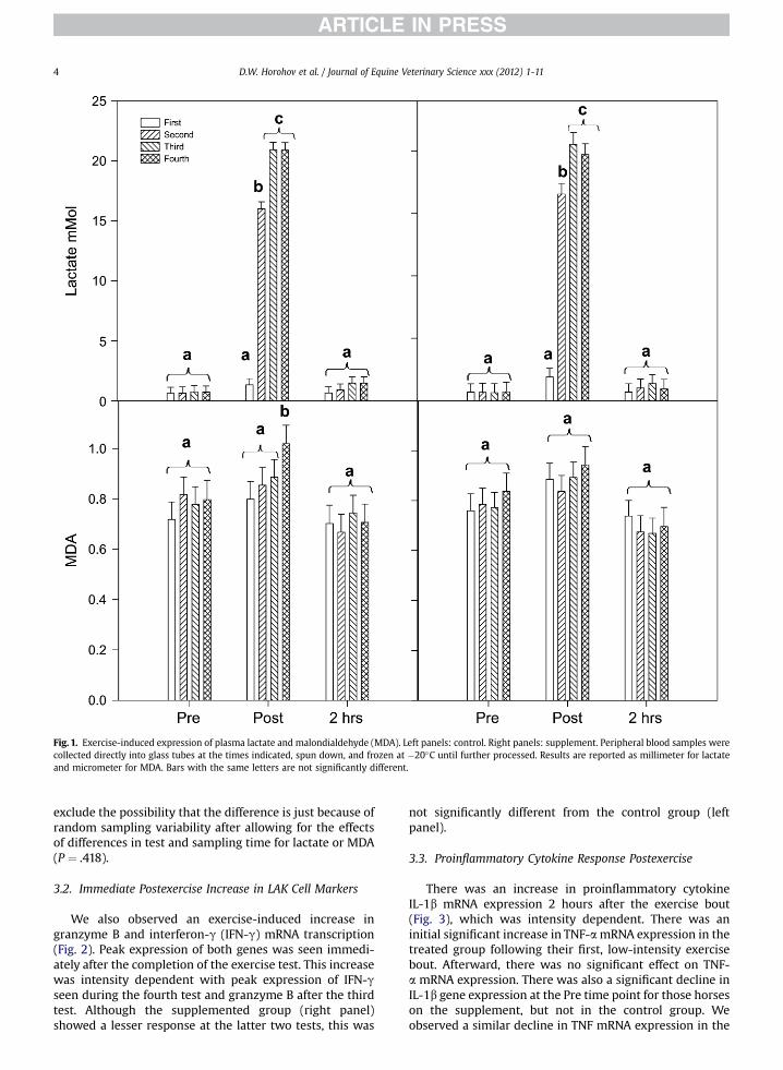
Fig. 1. Exercise-induced expression of plasma lactate and malondialdehyde (MDA). Left panels: control. Right panels: supplement. Peripheral blood samples werecollected directly into glass tubes at the times indicated, spun down, and frozen at �20�C until further processed. Results are reported as millimeter for lactateand micrometer for MDA. Bars with the same letters are not significantly different.
D.W. Horohov et al. / Journal of Equine Veterinary Science xxx (2012) 1-114
exclude the possibility that the difference is just because ofrandom sampling variability after allowing for the effectsof differences in test and sampling time for lactate or MDA(P ¼ .418).
3.2. Immediate Postexercise Increase in LAK Cell Markers
We also observed an exercise-induced increase ingranzyme B and interferon-g (IFN-g) mRNA transcription(Fig. 2). Peak expression of both genes was seen immedi-ately after the completion of the exercise test. This increasewas intensity dependent with peak expression of IFN-gseen during the fourth test and granzyme B after the thirdtest. Although the supplemented group (right panel)showed a lesser response at the latter two tests, this was
not significantly different from the control group (leftpanel).
3.3. Proinflammatory Cytokine Response Postexercise
There was an increase in proinflammatory cytokineIL-1b mRNA expression 2 hours after the exercise bout(Fig. 3), which was intensity dependent. There was aninitial significant increase in TNF-amRNA expression in thetreated group following their first, low-intensity exercisebout. Afterward, there was no significant effect on TNF-a mRNA expression. There was also a significant decline inIL-1b gene expression at the Pre time point for those horseson the supplement, but not in the control group. Weobserved a similar decline in TNF mRNA expression in the

Fig. 2. Exercise-induced increase in LAK cell markers. Peripheral blood samples were collected directly into PAXgene tubes at the times indicated and laterprocessed for real-time polymerase chain reaction (RT-PCR) analysis of granzyme B (Granz B) and interferon gamma (IFNg) mRNA expression. Left panels: control.Right panels: supplement.
D.W. Horohov et al. / Journal of Equine Veterinary Science xxx (2012) 1-11 5
Pre time point of the supplemented group, but not thecontrol group. There was a significant increase in IL-6mRNA expression in both groups of horses over the sixtests, although no specific effect of sample time (Fig. 4) wasobserved. However, TNF mRNA expression was inverselycorrelated with IL-6 mRNA expression (r ¼ 0.253, P < .001).
4. Discussion
Numerous studies have documented alterations inproinflammatory cytokine expression in response to exer-cise in humans [16-18,64]. Equine athletes likewise exhibitaltered inflammatory responses subsequent to exercise[22,62,63,65-68]. The proposed mechanism for thisexercise-induced inflammation is high-intensity training-induced muscle, skeletal, or joint trauma [14,69]. A role for
reactive oxygen species in exercise-induced damage tomuscle has also been proposed [41,70,71]. Free radicalgeneration can be estimated by the measurement ofproducts of lipid peroxidation such as MDA whose level isincreased in equine blood and tissues during exercise [72].The accumulation of lactic acid is a functional measure ofanaerobic metabolism associated with high-intensityexercise [73]. Together, these multiple insults activatecirculating monocytes and other cells to produce proin-flammatory cytokines [74]. These changes in proin-flammatory gene expression occur in skeletal muscle andperipheral blood cells [62]. Peripherally produced cyto-kines can in turn augment or exacerbate localized inflam-matory responses in joints and muscles, thus leading to anamplification of the inflammatory cascade [10]. Althoughmost previous work has used treadmill-based exercise

Fig. 3. Exercise-induced increase in proinflammatory markers. Peripheral blood samples were collected directly into PAXgene tubes at the times indicated andlater processed for RT-PCR analysis of interleukin 1b (IL-1b) and tumor necrosis factor-a (TNF-a) mRNA expression. Left panels: control. Right panels: supplement.
D.W. Horohov et al. / Journal of Equine Veterinary Science xxx (2012) 1-116
models, in this study, we have looked for similar effect inyoung Thoroughbreds exercising at a training facility.
For each of the tests performed, the initial effect of theexercise bout was the intensity-dependent accumulation oflactic acid. Because it was only possible to perform limitedsampling on these horses, we could only detect lactateaccumulation within 5 minutes of the completion of thespeed portion of the exercise test. Thus, peak lactateproduction can only be estimated. Nevertheless, the levelsof lactate in the blood sample were proportional to theeffort expended in each of the tests, with maximal accu-mulation occurring in response to the higher speed tests.This relationship between peak lactate accumulation andexercise intensity is well established [73]. Therewas also anincrease in MDA in the same postexercise sample, althoughthis was only significant for the last test. A similar transient
increase in MDA was also observed in horses 5 minutesafter high-speed treadmill exercise [75]. The role of reactiveoxygen species in the mediation of exercise-inducedoxidative damage to muscle has been well studied and isthought to contribute to the inflammatory response[72,76]. As each of the first four tests was of increasingintensity, it was not possible to use either of thesemeasuresas a means of assessing a conditioning effect. Instead, theyprovided evidence of the intensity of the exercise at thetime of sample collection. Therewas no difference in lactateproduction between the third and fourth tests, likelyreflecting the fact that they were performed at similarspeeds.
Concurrent with the increase in lactate and MDA, wealso observed a significant increase in two indirectmarkers for LAK activity in the horse, granzyme B and

Fig. 4. IL-6 mRNA expression increases over time. Peripheral blood samples were collected directly into PAXgene tubes at the times indicated and later processedfor RT-PCR analysis of interleukin 6 (IL-6) mRNA expression. Tests with the same letter are not significantly different using a 2-way analysis of variance. There wasno effect of treatment (P ¼ .781).
D.W. Horohov et al. / Journal of Equine Veterinary Science xxx (2012) 1-11 7
IFN-g [77], immediately after exercise. Increased LAKactivity immediately following peak exercise has beenreported for humans [64,78,79] and horses [67]. Thisincrease in LAK function is likely the result of a reparti-tioning of the cells in response to exercise and theincreased cytotoxic activity of the cells in response toincreased cytokine or other mediator production [80-82].The overall effectwe observedwas intensity-dependent, astherewas no effect of the initial light exercise test on eithermarker, similar to what occurs to LAK function followinglow-intensity exercise in thehorse [67]. Therewasno effectof exercise training on pre-exercise or baseline IFN-g andgranzyme B expression throughout the training period.These results are consistent with data indicating thatexercise training had no effect on natural killer (NK) cellactivity in mice [83], although it is in contrast to otherreports stating that exercise training could augmentbaseline NK cell function in humans and mice [84,85].
Two hours after completing the exercise tests, thehorses exhibited increased expression of IL-1b mRNA intheir peripheral blood. This parallels our previous resultsfollowing treadmill- [62] and race-based exercise studies[86]. The expression of IL-1b was exercise intensity-dependent. This postexercise increase in IL-1b productionis well documented [7-10]. The intensity-related associa-tion between cytokine production andmarkers of oxidativedamage supports the notion that damage to muscle fibersstimulates the production of inflammatory cytokines [87].
Given the role of IL-1b in tissue repair [88], its increasedexpression postexercise is expected, although chronic orelevated expression of this cytokine can lead to degenera-tive changes and loss of function [69,88]. We did not seea significant increase in TNF-amRNA expression at 2 hoursafter exercise, possibly because its expression had not yetpeaked [62], although there is an overall trend for itsexpression to increase during the 2-hour sampling period.Limitations in the study design prevented us from collect-ing samples at later time points.
During the training period, there was a significantdecrease in baseline expression of TNF-a, and this was alsoassociated with an overall trend for decrease in the IL-1bbaseline expression. This overall decline in proin-flammatory cytokine expression was significant for horsesreceiving the supplement. It is known that adaptation toexercise leads to reduced inflammatory responses intrained human athletes [89,90]. Thus, exercise training isassociated with an overall “anti-inflammatory state”[17,91]. A recent study reported that IL-6 and IL-6R weresignificantly upregulated in both humans and horsesfollowing training [92]. Here, too, we saw increasedexpression of IL-6 in both groups of horses. This upregu-lation of IL-6 expression in peripheral blood mononuclearcells in horses could reflect a mechanism that generated ananti-inflammatory environment, thereby reducing theexpression of the proinflammatory cytokines. Our obser-vation that IL-6 mRNA expression was inversely correlated
D.W. Horohov et al. / Journal of Equine Veterinary Science xxx (2012) 1-118
with proinflammatory cytokine mRNA expression isconsistent with this proposed mechanism.
Because oxidative stress and inflammation have tradi-tionally been associated with fatigue and impairedrecovery from exercise [45], we evaluated the effect of thesupplement on markers of inflammation (e.g., cytokines)and oxidative damage (MDA). The main difference betweenthe supplemented horses and the control horses was thesignificant reduction in proinflammatory gene expressionin the supplemented group. This was evidenced by both anoverall decline in baseline IL-1b and TNF-a mRNA in thepre-exercise samples and a reduced postexercise increasein both IL-1b and TNF-a during subsequent exercise tests.The supplement contained Boswellia whose anti-inflammatory and proapoptotic activities have been welldocumented [93], with the primary mode of actionappearing to be the inhibition of nuclear factor-kB [93,94].Similarly, the anti-inflammatory activities of curcuminhave been widely studied [95-97] and shown to affectintracellular signaling pathways involved in proin-flammatory cytokine and other mediator production [98].In vitro, curcuminoids neutralized free radical speciesgenerated by activated equine neutrophils portendinga therapeutic possibility for equine pathologies associatedwith excessive inflammatory reactions [99]. Although thereare no in vivo data for either curcumin or Boswellia in thehorse, there is information regarding the other componentsof the nutritional supplement; coenzyme Q10 (CoQ10),glycine propionyl-L-carnitine (GPLC), and ribose. Thesenutrients have a fundamental role in cellular bioenergetics.GPLC transports fatty acids to the mitochondria for oxida-tion, CoQ10 is essential for adenosine-50-triphosphate(ATP) production as a cofactor in the mitochondrial elec-tron transport chain, and D-ribose is an intergral structuralcomponent of ATP. These three nutrients have beendemonstrated to increase exercise capacity in humans[100-103] and equids [58,104], presumably by stimulatingATP production and recovery while supporting ATP energysubstrates at the same time [105]. Daily ribose supple-mentation (0.07 g/kg BW, twice daily) for 14 days in the dietof exercising geldings resulted in lower blood ammonia-Nand plasma lactic acid during recovery after a standard-ized exercise test. CoQ10 can also serve as a protectiveagent against lipid peroxidation [106] and has an anti-inflammatory activity [107]. There are no data in the liter-ature on CoQ10 dosage in horses; our results indicateda marked increase from a baseline value of 0.187 mg/mL to0.407 mg/mL at 30 days and 0.511 mg/mL after 60 days ata daily dose of 800 mg [108]. Besides its role as a buffer indifferent tissues during ketosis and hypoxic muscularactivity, carnitine protects membrane structures andreduces lactate production [109]. GPLC has been previouslyreported to lower resting concentrations of MDA, suggest-ing a potential antioxidant effect of this nutrient [110]. Ananti-inflammatory function for carnitine has also beendescribed in several model systems [111-113]. The concen-tration of carnitine in blood plasma of horses variesmarkedly between animals and with increasing exerciseintensity [109]. Although acute exercise does not havea marked effect on the content of total carnitine in skeletalmuscle, training elevates its total concentration in themiddle gluteal muscle [114]. Oral supplementation with
10 g/d L-carnitine can elevate carnitine concentration inequine blood plasma [104]. The daily dose of carnitine usedin this study was 8 g/d, which could achieve similar effects.Thus, the reduced proinflammatory cytokine response inthe supplemented horses in this study likely reflected anadaptation to exercise, which was enhanced by the nutri-tional components present in the supplement. However, itis not possible to identify which specific component orcomponents mediated this effect.
5. Conclusions
Although exercise-induced changes in cytokine geneexpression have beenwidely studied using treadmill-basedexercise tests, the cytokine response to race training hadnot been investigated in Thoroughbred racehorses. Here,we have shown that the exercise under race trainingconditions is associated with the temporal induction ofcytokines characteristic of the initial elevation in LAK cellactivity immediately after exercise and the subsequentexpression of proinflammatory cytokines 2 hours later.These time- and intensity-dependent changes in cytokinegene expression parallel data from previous studies usingtreadmill-based exercise testing. We also observed signs ofadaptation to exercise over the training period as indicatedby an overall reduction in the expression of proin-flammatory cytokines and increased expression of IL-6.Although dietary supplements have been used in horsesin the past for various reasons, including performanceenhancement, their effect on race training has not beeninvestigated. The nutritional supplement used in thepresent study containing antioxidant nutrients and anti-inflammatory agents did not affect the immediateresponse to exercise; however, it was associated with anenhanced adaptation to exercise in terms of a significantreduction in proinflammatory cytokine expression beforeand after exercise. This underscores the potential fornutritional supplementation to reduce exercise-inducedinflammatory pathologies in racehorses.
Acknowledgments
The expertise of Coby Caiazzo at Murmur Farm inDarlington Maryland is gratefully acknowledged by theauthors. The horses involved in this study were madeavailable by Sinatra Racing. Partial funding for this studywas provided by the W.R. Mills Endowment and Dr.Stephen Sinatra. The authors also appreciate the reviewsand suggestions by Dr. Hemmi N. Bhagavan and Ken Hassen.
References
[1] Wilsher S, Allen WR, Wood JL. Factors associated with failureof thoroughbred horses to train and race. Equine Vet J 2006;38:113-8.
[2] Rossdale PD, Hopes R, Digby NJ, Offord K. Epidemiological study ofwastage among racehorses 1982 and 1983. Vet Rec 1985;116:66-9.
[3] Dyson PK, Jackson BF, Pfeiffer DU, Price JS. Days lost from trainingby two- and three-year-old Thoroughbred horses: a survey ofseven UK training yards. Equine Vet J 2008;40:650-7.
[4] Ley C, Ekman S, Roneus B, Eloranta ML. Interleukin-6 and highmobility group box protein-1 in synovial membranes and osteo-chondral fragments in equine osteoarthritis. Res Vet Sci 2009;86:490-7.

D.W. Horohov et al. / Journal of Equine Veterinary Science xxx (2012) 1-11 9
[5] Clutterbuck AL, Mobasheri A, Shakibaei M, Allaway D, Harris P.Interleukin-1 beta-induced extracellular matrix degradation andglycosaminoglycan release is inhibited by curcumin in an explantmodel of cartilage inflammation. Ann N Y Acad Sci 2009;1171:428-35.
[6] Ley C, Ekman S, Elmen A, Nilsson G, Eloranta ML. Interleukin-6 andtumour necrosis factor in synovial fluid from horses with carpaljoint pathology. J Vet Med A Physiol Pathol Clin Med 2007;54:346-51.
[7] Cuzzocrea S, Reiter RJ. Pharmacological action of melatonin inshock, inflammation and ischemia/reperfusion injury. Eur J Phar-macol 2001;426:1-10.
[8] Haahr PM, Pedersen BK, Fomsgaard A, Tvede N, Diamant M,Klarlund K, et al. Effect of physical exercise on in vitro productionof interleukin 1, interleukin 6, tumour necrosis factor-alpha,interleukin 2 and interferon-gamma. Int J Sports Med 1991;12:223-7.
[9] Nieman DC, Henson DA, Davis JM, Dumke CL, Utter AC, Murphy EA,et al. Blood leukocyte mRNA expression for IL-10, IL-1Ra, and IL-8,but not IL-6, increases after exercise. J Interferon Cytokine Res2006;26:668-74.
[10] Suzuki K, Nakaji S, Yamada M, Totsuka M, Sato K, Sugawara K.Systemic inflammatory response to exhaustive exercise. Cytokinekinetics. Exerc Immunol Rev 2002;8:6-48.
[11] MacIntyre DL, Reid WD, McKenzie DC. Delayed muscle soreness.The inflammatory response to muscle injury and its clinicalimplications. Sports Med 1995;20:24-40.
[12] Auer DE. An hypothesis on the etio-pathogenesis of equineinflammatory joint disease. Vet Clin Pathol 1989;18:21-6.
[13] Smith LL. Tissue trauma: the underlying cause of overtrainingsyndrome? J Strength Cond Res 2004;18:185-93.
[14] Nieman DC, Dumke CL, Henson DA, McAnulty SR, Gross SJ, Lind RH.Muscle damage is linked to cytokine changes following a 160-kmrace. Brain Behav Immun 2005;19:398-403.
[15] Petersen EW, Ostrowski K, Ibfelt T, Richelle M, Offord E, Halkjaer-Kristensen J, et al. Effect of vitamin supplementation on cytokineresponse and on muscle damage after strenuous exercise. Am JPhysiol Cell Physiol 2001;280:C1570-5.
[16] Nielsen AR, Pedersen BK. The biological roles of exercise-inducedcytokines: IL-6, IL-8, and IL-15. Appl Physiol Nutr Metab 2007;32:833-9.
[17] Woods JA, Vieira VJ, Keylock KT. Exercise, inflammation, and innateimmunity. Immunol Allergy Clin North Am 2009;29:381-93.
[18] Handschin C, Spiegelman BM. The role of exercise andPGC1alpha in inflammation and chronic disease. Nature 2008;454:463-9.
[19] Colahan PT, Kollias-Bakert C, Leutenegger CM, Jones JH. Doestraining affect mRNA transciption for cytokine production incirculating leucocytes? Equine Vet J Suppl 2002;34:154-8.
[20] Ainsworth DM, Appleton JA, Eicker SW, Luce R, Julia Flaminio M,Antczak DF. The effect of strenuous exercise on mRNA concentra-tions of interleukin-12, interferon-gamma and interleukin-4 inequine pulmonary and peripheral blood mononuclear cells. VetImmunol Immunopathol 2003;91:61-71.
[21] Lamprecht EL, Bagnell C, Williams C. Inflammatory responses tothree modes of intense exercise in Standardbred maresda pilotstudy. Comp Ex Physiol 2009;5:115-25.
[22] Donovan DC, Jackson CA, Colahan PT, Norton N, Hurley DJ. Exer-cise-induced alterations in pro-inflammatory cytokines and pros-taglandin F2alpha in horses. Vet Immunol Immunopathol 2007;118:263-9.
[23] Seeherman HJ, Morris E, O’Callaghan MW. The use of sportsmedicine techniques in evaluating the problem equine athlete. VetClin North Am Equine Pract 1990;6:239-74.
[24] Courouce A, Geffroy O, Barrey E, Auvinet B, Rose RJ. Comparison ofexercise tests in French trotters under training track, racetrack andtreadmill conditions. Equine Vet J Suppl 1999;30:528-32.
[25] Jones JH, Ohmura H, Stanley SD, Hiraga A. Energetic cost of loco-motion on different equine treadmills. Equine Vet J Suppl 2006;38:365-9.
[26] Heinecke LF, Grzanna MW, Au AY, Mochal CA, Rashmir-Raven A,Frondoza CG. Inhibition of cyclooxygenase-2 expression andprostaglandin E(2) production in chondrocytes by avocadosoybean unsaponifiables and epigallocatechin gallate. Osteoar-thritis Cartilage 2010;18:220-7.
[27] Goodrich LR, Nixon AJ. Medical treatment of osteoarthritis in thehorseda review. Vet J 2006;171:51-69.
[28] Morton AJ, Campbell NB, Gayle FM, Redding WR, Blikslager AT.Preferential and non-selective cyclooxygenase inhibitors reduceinflammation during lipopolysaccharide-induced synovitis. ResVet Sci 2005;78:189-92.
[29] Thomasy SM, Slovis N, Maxwell LK, Kollias-Baker C. Transdermalfentanyl combined with nonsteroidal anti-inflammatory drugs foranalgesia in horses. J Vet Intern Med 2004;18:550-4.
[30] Kallings P, Johnston C, Drevemo S. Effects of flunixin on movementand performance of standardbred trotters on the track. Equine VetJ Suppl 1999;30:270-3.
[31] Taylor MR, Westwood SA. Quantitation of phenylbutazone andoxyphenbutazone in equine plasma by high-performance liquid-chromatography with solid-phase extraction. J Chrom 1995;697:389-96.
[32] Kallings P. Nonsteroidal anti-inflammatory drugs. Vet Clin NorthAm Equine Pract 1993;9:523-41.
[33] Dirikolu L, Woods WE, Boyles J, Lehner AF, Harkins JD, Fisher M,et al. Nonsteroidal anti-inflammatory agents and musculoskeletalinjuries in Thoroughbred racehorses in Kentucky. J Vet PharmacolTher 2009;32:271-9.
[34] Paulsen G, Egner IM, Drange M, Langberg H, Benestad HB, Fjeld JG,et al. A COX-2 inhibitor reduces muscle soreness, but does notinfluence recovery and adaptation after eccentric exercise. Scand JMed Sci Sports 2010;20:e195-207.
[35] Reed SK, Messer NT, Tessman RK, Keegan KG. Effects of phenylbu-tazone alone or in combination with flunixin meglumine on bloodprotein concentrations in horses. Am J Vet Res 2006;67:398-402.
[36] McMeniman NP, Hintz HF. Effect of vitamin E status on lipid per-oxidation in exercised horses. Equine Vet J 1992;24:482-4.
[37] Williams CA, Carlucci SA. Oral vitamin E supplementation onoxidative stress, vitamin and antioxidant status in intensely exer-cised horses. Equine Vet J Suppl 2006;38:617-21.
[38] Ducharme NG, Fortier LA, Kraus MS, Hobo S, Mohammed HO,McHugh MP, et al. Effect of a tart cherry juice blend on exercise-induced muscle damage in horses. Am J Vet Res 2009;70:758-63.
[39] Clarke JO, Mullin GE. A review of complementary and alternativeapproaches to immunomodulation. Nutr Clin Pract 2008;23:49-62.
[40] Nieman DC. Exercise immunology: nutritional countermeasures.Can J Appl Physiol 2001;26(Suppl):S45-55.
[41] Urso ML, Clarkson PM. Oxidative stress, exercise, and antioxidantsupplementation. Toxicology 2003;189:41-54.
[42] Walson T, Callister R, Taylor R, Sibbritt D, MacDonald L, Garg M.Antioxidant restriction and oxidative stress in short-durationexhaustive exercise. Med Sci Sports Exerc 2005;37:63-71.
[43] Aguilo A, Tauler P, Fuentespina E, Villa G, Cordova A, Tur JA, et al.Antioxidant diet supplementation influences blood iron status inendurance athletes. Int J Sport Nutr Exerc Metab 2004;14:147-60.
[44] Margaritis I, Rousseau AS. Does physical exercise modify antioxi-dant requirements? Nutr Res Rev 2008;21:3-12.
[45] Peake JM, Suzuki K, Coombes JS. The influence of antioxidantsupplementation on markers of inflammation and the relationshipto oxidative stress after exercise. J Nutr Biochem 2007;18:357-71.
[46] Moreira A, Kekkonen RA, Delgado L, Fonseca J, Korpela R,Haahtela T. Nutritional modulation of exercise-induced immuno-depression in athletes: a systematic review and meta-analysis. EurJ Clin Nutr 2007;61:443-60.
[47] Margaritis I, Palazzetti S, Rousseau AS, Richard MJ, Favier A.Antioxidant supplementation and tapering exercise improveexercise-induced antioxidant response. J Am Coll Nutr 2003;22:147-56.
[48] Pearson W, Orth MW, Lindinger MI. Evaluation of inflammatoryresponses induced via intra-articular injection of interleukin-1 inhorses receiving a dietary nutraceutical and assessment of theclinical effects of long-term nutraceutical administration. Am J VetRes 2009;70:848-61.
[49] Forsyth RK, Brigden CV, Northrop AJ. Double blind investigation ofthe effects of oral supplementation of combined glucosaminehydrochloride (GHCL) and chondroitin sulphate (CS) on stridecharacteristics of veteran horses. Equine Vet J Suppl 2006;38:622-5.
[50] Oke S, Aghazadeh-Habashi A, Weese JS, Jamali F. Evaluation ofglucosamine levels in commercial equine oral supplements forjoints. Equine Vet J 2006;38:93-5.
[51] De Moffarts B, Portier K, Kirschvink N, Coudert J, Fellmann N, vanErck E, et al. Effects of exercise and oral antioxidant supplemen-tation enriched in (n-3) fatty acids on blood oxidant markers anderythrocyte membrane fluidity in horses. Vet J 2007;174:113-21.
[52] Deaton CM, Marlin DJ. Exercise-associated oxidative stress. ClinTech Equine Pract 2004;2:278-91.
[53] de Moffarts B, Kirschvink N, Art T, Pincemail J, Lekeux P. Effect ofexercise on blood oxidant/antioxidant markers in standardbredhorses: comparison between treadmill and race track tests. EquineVet J Suppl 2006;38:254-7.
[54] Hargreaves BJ, Kronfeld DS, Waldron JN, Lopes MA, Gay LS,Saker KE, et al. Antioxidant status and muscle cell leakage duringendurance exercise. Equine Vet J Suppl 2002;34:116-21.

D.W. Horohov et al. / Journal of Equine Veterinary Science xxx (2012) 1-1110
[55] Kirschvink N, de Moffarts B, Lekeux P. The oxidant/antioxidantequilibrium in horses. Vet J 2008;177:178-91.
[56] Portier K, de Moffarts B, Fellman N, Kirschvink N, Motta C,Letellierw C, et al. The effects of dietary N-3 and antioxidantsupplementation on erythrocyte membrane fatty acid compo-sition and fluidity in exercising horses. Equine Vet J Suppl2006;38:279-84.
[57] Williams CA, Lamprecht ED. Some commonly fed herbs and otherfunctional foods in equine nutrition: a review. Vet J 2008;178:21-31.
[58] Kavazis AN, Sobota JS, Kivipelto J, Porter MB, Colahan PT, Ott EA.Ribose supplementation in maximally exercising Thoroughbreds.Equine Vet J Suppl 2002;34:191-6.
[59] Bonavolontà V, Emerenziani G, Gallotta M, Silva A, Guidetti L.Accuracy, reliability, linearity of Accutrend and Lactate Pro versusEBIO plus analyzer. Eur J Appl Physiol 2009;107:105-11.
[60] Tanner RK, Fuller KL, Ross ML. Evaluation of three portable bloodlactate analysers: lactate pro, lactate scout and lactate plus. Eur JAppl Physiol 2010;109:551-9.
[61] Jentzsch AM, Bachmann H, Furst P, Biesalski HK. Improved analysisof malondialdehyde in human body fluids. Free Radic Biol Med1996;20:251-6.
[62] Liburt NR, Adams AA, Betancourt A, Horohov DW, McKeever KH.Exercise-induced increases in inflammatory cytokines in muscleand blood of horses. Equine Vet J 2010;42(Suppl 38):280-8.
[63] Lamprecht ED, Bagnell CA, Williams CA. Superoxide dismutasesupplementation does not affect erythrocyte superoxide dismutaseactivity or systemic inflammatory cytokine responses in an equineexercise model. J Equine Vet Sci 2009;29:298-9.
[64] Walsh NP, Gleeson M, Shephard RJ, Woods JA, Bishop NC,Fleshner M, et al. Position statement. Part one: immune functionand exercise. Exerc Immunol Rev 2011;17:6-63.
[65] Folsom RW, Littlefield-Chabaud MA, French DD, Pourciau SS,Mistric L, Horohov DW. Exercise alters the immune response toequine influenza virus and increases susceptibility to infection.Equine Vet J 2001;33:664-9.
[66] Horohov DW. Is exercise bad for the immune system? Equine Vet J2003;35:113-6.
[67] Horohov DW, Dimock A, Guirnalda P, Folsom RW, McKeever KH,Malinowski K. Effect of exercise on the immune response of youngand old horses. Am J Vet Res 1999;60:643-7.
[68] Lunn DP, Hussey S, Sebing R, Rushlow KE, Radecki SV, Whitaker-Dowling P, et al. Safety, efficacy, and immunogenicity of a modi-fied-live equine influenza virus vaccine in ponies after inductionof exercise-induced immunosuppression. J Am Vet Med Assoc2001;218:900-6.
[69] Smith LL. Cytokine hypothesis of overtraining: a physiologicaladaptation toexcessive stress?MedSci Sports Exerc 2000;32:317-31.
[70] Mills PC, Ng JC, Thornton J, Seawright AA, Auer DE. Exercise-induced connective tissue turnover and lipid peroxidation inhorses. Br Vet J 1994;150:53-63.
[71] Brancaccio P, Lippi G, Maffulli N. Biochemical markers of musculardamage. Clin Chem Lab Med 2010;48:757-67.
[72] Chiaradia E, Avellini L, Rueca F, Spaterna A, Porciello F,Antonioni MT, et al. Physical exercise, oxidative stress and muscledamage in racehorses. Comp Biochem Physiol B Biochem Mol Biol1998;119:833-6.
[73] Auvinet B. Performance testing and improvement in humanathletes. Pferdeheilkunde 1996;12:455-6.
[74] Izquierdo M, Ibanez J, Calbet JA, Navarro-Amezqueta I, Gonzalez-Izal M, Idoate F, et al. Cytokine and hormone responses to resis-tance training. Eur J Appl Physiol 2009;107:397-409.
[75] Matsuki N, Tamura S, Ono K, Watari T, Goitsuka R, Yamanobe A,et al. Exercise-induced phospholipid degradation in the equineskeletal muscle and erythrocytes. J Vet Med Sci 1991;53:1001-7.
[76] Pyne DB. Exercise-induced muscle damage and inflammation:a review. Aust J Sci Med Sport 1994;26:49-58.
[77] Liu C, Betancourt A, Cohen DA, Adams AA, Sun L, Horohov DW.Granzyme B-mRNA expression by equine lymphokine activatedkiller (LAK) cells is associated with the induction of apoptosis intarget cells. Vet Immunol Immunopathol 2011;143:108-15.
[78] Hoffman-Goetz L, Arumugam Y, Sweeny L. Lymphokine activatedkiller cell activity following voluntary physical activity in mice.J Sports Med Phys Fitness 1994;34:83-90.
[79] Shephard RJ, Rhind S, Shek PN. Exercise and the immune system.Natural killer cells, interleukins and related responses. Sports Med1994;18:340-69.
[80] Pedersen BK, Bruunsgaard H, Klokker M, Kappel M, MacLean DA,Nielsen HB, et al. Exercise-induced immunomodulationdpossibleroles of neuroendocrine and metabolic factors. Int J Sports Med1997;18(Suppl 1):S2-7.
[81] Pedersen BK, Ullum H. NK cell response to physical activity:possible mechanisms of action. Med Sci Sports Exerc 1994;26:140-6.
[82] Horohov DW, Keadle TL, Pourciau SS, Littlefield-Chabaud MA,Kamerling SG, Keowen ML, et al. Mechanism of exercise-inducedaugmentation of lymphokine activated killer (LAK) cell activity inthe horse. Vet Immunol Immunopathol 1996;53:221-33.
[83] Lee EG, Fox ME, Meadows GG, Johnson EC, Blank SE. Relationshipbetween moderate intensity endurance training volume andnatural killer cell cytolytic activity. J Sports Med Phys Fitness1999;39:275-81.
[84] Shinkai S, Konishi M, Shephard RJ. Aging, exercise, training, and theimmune system. Exerc Immunol Rev 1997;3:68-95.
[85] Blank SE, Johansson JO, Pfister LJ, Gallucci RM, Lee EG,Meadows GG. Mechanistic differences in NK cell cytolytic activityin treadmill-trained and chronic ethanol-consuming mice. J ApplPhysiol 1994;76:2031-6.
[86] Saulez MN, Godfroid J, Bosman A, Stiltner JL, Breathnach CC,Horohov DW. Cytokine mRNA expressions after racing at a highaltitude and at sea level in horses with exercise-induced pulmo-nary hemorrhage. Am J Vet Res 2010;71:447-53.
[87] Pedersen BK, Ostrowski K, Rohde T, Bruunsgaard H. The cytokineresponse to strenuous exercise. Can J Physiol Pharmacol 1998;76:505-11.
[88] Dennis RA, Trappe TA, Simpson P, Carroll C, Huang BE, Nagarajan R,et al. Interleukin-1 polymorphisms are associated with theinflammatory response in human muscle to acute resistanceexercise. J Physiol 2004;560:617-26.
[89] Pedersen BK, Hoffman-Goetz L. Exercise and the immune system:regulation, integration, and adaptation. Physiol Rev 2000;80:1055-81.
[90] Goldhammer E, Tanchilevitch A,Maor I, Beniamini Y, Rosenschein U,Sagiv M. Exercise training modulates cytokines activity in coronaryheart disease patients. Int J Cardiol 2005;100:93-9.
[91] Pedersen BK. The anti-inflammatory effect of exercise: its role indiabetes and cardiovascular disease control. Essays Biochem2006;42:105-17.
[92] Capomaccio S, Cappelli K, Spinsanti G, Mencarelli M, Muscettola M,Felicetti M, et al. Athletic humans and horses: comparative analysisof interleukin-6 (IL-6) and IL-6 receptor (IL-6R) expression inperipheral blood mononuclear cells in trained and untrainedsubjects at rest. BMC Physiol 2011;11:3.
[93] Moussaieff A, Mechoulam R. Boswellia resin: from religious cere-monies to medical uses; a review of in-vitro, in-vivo and clinicaltrials. J Pharm Pharmacol 2009;61:1281-93.
[94] Moussaieff A, Shohami E, Kashman Y, Fride E, Schmitz ML,Renner F, et al. Incensole acetate, a novel anti-inflammatorycompound isolated from Boswellia resin, inhibits nuclear factor-kappa B activation. Mol Pharmacol 2007;72:1657-64.
[95] Aggarwal BB, Harikumar KB. Potential therapeutic effects of cur-cumin, the anti-inflammatory agent, against neurodegenerative,cardiovascular, pulmonary, metabolic, autoimmune and neoplasticdiseases. Int J Biochem Cell Biol 2009;41:40-59.
[96] Anand P, Thomas SG, Kunnumakkara AB, Sundaram C,Harikumar KB, Sung B, et al. Biological activities of curcumin andits analogues (Congeners) made by man and Mother Nature.Biochem Pharmacol 2008;76:1590-611.
[97] Bengmark S, Mesa MD, Gil A. Plant-derived health: the effects ofturmeric and curcuminoids. Nutr Hosp 2009;24:273-81.
[98] Camacho-Barquero L, Villegas I, Sanchez-Calvo JM, Talero E,Sanchez-Fidalgo S, Motilva V, et al. Curcumin, a Curcuma longaconstituent, acts on MAPK p38 pathway modulating COX-2 andiNOS expression in chronic experimental colitis. Int Immuno-pharmacol 2007;7:333-42.
[99] Franck T, Kohnen S, Grulke S, Neven P, Goutman Y, Peters F, et al.Inhibitory effect of curcuminoids and tetrahydrocurcuminoids onequine activated neutrophils and myeloperoxidase activity. PhysiolRes 2008;57:577-87.
[100] Cooke M, Iosia M, Buford T, Shelmadine B, Hudson G, Kerksick C,et al. Effects of acute and 14-day coenzyme Q10 supplementationon exercise performance in both trained and untrained individuals.J Int Soc Sports Nutr 2008;5:8.
[101] Wagner DR, Gresser U, Zollner N. Effects of oral ribose on musclemetabolism during bicycle ergometer in AMPD-deficient patients.Ann Nutr Metab 1991;35:297-302.
[102] Kolodziejczyk J, Saluk-Juszczak J, Wachowicz B. l-carnitine protectsplasma components against oxidative alterations. Nutrition 2011;27:693-9.
[103] Jacobs PL, Goldstein ER, Blackburn W, Orem I, Hughes JJ. Glycinepropionyl-L-carnitine produces enhanced anaerobic work capacity

D.W. Horohov et al. / Journal of Equine Veterinary Science xxx (2012) 1-11 11
with reduced lactate accumulation in resistance trained males. J IntSoc Sports Nutr 2009;6:9.
[104] Rivero JL, Sporleder HP, Quiroz-Rothe E, Vervuert I, Coenen M,Harmeyer J. Oral L-carnitine combined with training promoteschanges in skeletal muscle. Equine Vet J Suppl 2002;34:269-74.
[105] Sinatra ST. Metabolic cardiology: the missing link in cardiovasculardisease. Altern Ther Health Med 2009;15:48-50.
[106] Abd El-Gawad HM, Khalifa AE. Quercetin, coenzyme Q10, andL-canavanine as protective agents against lipid peroxidation andnitric oxide generation in endotoxin-induced shock in rat brain.Pharmacol Res 2001;43:257-63.
[107] Schmelzer C, Lindner I, Rimbach G, Niklowitz P, Menke T, Doring F.Functions of coenzyme Q10 in inflammation and gene expression.Biofactors 2008;32:179-83.
[108] Sinatra ST, Chopra RK, Jankowitz S, Horohov DW, Bhagavan H.Coenzyme Q10 in equine serum: response to supplementation.J Equine Vet Sci (in press).
[109] Zeyner A, Harmeyer J. Metabolic functions of L-carnitine and itseffects as feed additive in horses. A review. Arch Tierernahr1999;52:115-38.
[110] Bloomer RJTL, Smith WA. Glycine propionyl-L-carnitine modulateslipid peroxidation and nitric oxide in human subjects. Int J VitamNutr Res 2009;79:131-41.
[111] Rosa FT, Zulet MA, Marchini JS, Martinez JA. Bioactive compoundswith effects on inflammation markers in humans. Int J Food SciNutr; 2012 Jan 17 [Epub ahead of print].
[112] Fortin G. L-Carnitine and intestinal inflammation. Vitam Horm2011;86:353-66.
[113] Cave MC, Hurt RT, Frazier TH, Matheson PJ, Garrison RN, McClain CJ,et al. Obesity, inflammation, and the potential application ofpharmaconutrition. Nutr Clin Pract 2008;23:16-34.
[114] Foster CV, Harris RC. Total carnitine content of the middle glutealmuscle of thoroughbred horses: normal values, variability andeffect of acute exercise. Equine Vet J 1992;24:52-7.

DOUBLE DIAMOND® I FORMULA FOR PEAK-EQUINE-PERFORMANCE
*PATENTED INGREDIENTSIN A PROPRIETARY PATENT PENDING
DOUBLE-DIAMOND® I FORMULA
EQUINE ENERGY & RECOVERY-PowerhouseSM
Studies Have Shown This Formula Helps Support & Maintain Healthy Inflammatory-Responses In Equine*
®
• Mitochondrial Powerhouse
• Helps Energize & Maintain Optimal Mitochondrial Functioning & Peak-Equine-Performance
• Helps Support Recovery After Performance Events
• Studies Show That Supplementation Helps Support & Maintain A Healthy Response In Young Race Horses During TrainingSM ††
• Patented Technologies For Enhanced Bioavailability
PRODUCT USED IN STUDY
The WINNING EDGESM
©2012 Equine Nutriceuticals, LLC. 257 East Center Street, Manchester, CT 06040 - (855) 647-9729
For more information please refer to: www.EquineNutriceuticals.com All trademarks, service marks and trade names referenced in this material are the property of their respective owners.
VET
ERIN
ARIAN FORMULATED
D R U G F R E E
TM
Journal of Equine Veterinary ScienceStudies published in the:
Original ResearchCoenzyme Q10 in Equine Serum: Response to Supplementation
Stephen T. Sinatra MD a, Raj K. Chopra MS b, Stanley Jankowitz MBA b, David W. Horohov PhD c,
Hemmi N. Bhagavan PhD ba University of Connecticut School of Medicine, Farmington, CT 06030, USA
b GelTec/Tishcon Corporation, Westbury, NY 11590, USA
c University of Kentucky, Lexington, KY 40546
a r t i c l e i n f oArticle history:Received 18 January 2012Received in revised form10 April 2012
Accepted 2 May 2012Available online xxx
Keywords:Coenzyme Q10Ubiquinone
Thoroughbred horsesSerum coenzyme Q10Coenzyme Q10 supplementation
a b s t r a c tAlthough the importance of coenzyme Q10 (CoQ10) in health and disease and its critical
role in cellular bioenergetics have been well established in various species, there is a total
lack o�nformation on the role of CoQ10 in horses. Therefore, the present study was
undertaken first to ascertain the occurrence of CoQ10 and/or possibly other homologs of
coenzyme Q (CoQ) in horse serum, and then to examine the response to CoQ10
supplementation on serum CoQ10 concentration. The study was carried out with 2-year-
old Thoroughbred horses. Total CoQ (reduced and oxidized forms) in serum was assayed
by high-performance liquid chromatography. The data show that horse serum contains
CoQ as CoQ10, at a much lower concentration as compared with that of humans and
several other species. There is no evidence for the presence of coenzyme Q9 or other
homologs of CoQ. On supplementation with CoQ10 at 800 mg a day (1.47 mg/kg body
weight), there was a signi ficant increase in serum CoQ10 concentration, approximately
2.7-fold at 60 days. CoQ10 supplementation at this dosage was found to be safe and well
tolerated. Additional studies are needed to examine whether maintaining higher serum
CoQ10 concentrations is of potential health bene fit to the horses, as assessed by selected
biochemical markers and also in terms of performance.2012 Elsevier Inc. All rights reserved.
1. Introduction
Coenzyme Q (CoQ) refers to a homologous series of
naturally occurring compounds that contain a benzoquinone
ring structure with an isoprenoid side chain of varying
lengths. CoQ is also known as ubiquinone because o�ts
ubiquitous distribution in nature. In humans and several
other mammalian species, the side chain is composed of 10
isoprene units, hence it is called coenzyme Q10 (CoQ10).
The chemical nomenclature of CoQ10 is 2,3-dimethoxy-5-
methyl-6-decaprenyl-1,4-benzoquinone. Although CoQ10
functions like a vitamin, it is not considered one because
CoQ10 (or its homolog) is synthesized in the body in humans
and other species of animals.
CoQ10 has a fundamental role in cellular bioenergetics
as a cofactor in the mitochondrial electron transport chain
(respiratory chain) and is therefore essential for the
production of biological/chemical energy in the form of
adenosine triphosphate [1] . It functions as a mobile redox
agent shuttling electrons and protons in the electron
transport chain. The redox functions of CoQ10 extend
beyond its role in the mitochondria. Furthermore, CoQ10 in
its reduced form as the hydroquinone (called ubiquinol) is
a potent lipophilic antioxidant and is capable of recycling
and regenerating other antioxidants such as tocopherol and
ascorbate. Other important functions of CoQ10 such as cell
signaling and gene expression have also been recognized
[2] . The role of CoQ10 in health and disease and the bene fits
of CoQ10 supplementation in various conditions have been
documented [3,4] .Although the importance of CoQ10 in humans and
several other species of animals has been well recognized,
Corresponding author at: Hemmi N. Bhagavan, PhD, Tishcon Corpo-
ration, 50 Sylvester Street, Westbury, NY 11590.E-mail address: [email protected] (H.N. Bhagavan).
Journal of Equine Veterinary Sciencej o u r n a l h o me p a g e : w w w . j - e v s . c o m
0737-0806/$ - see front matter 2012 Elsevier Inc. All rights reserved.
doi: 10.1016/j.jevs.2012.05.001
Journal of Equine Veterinary Science xxx (2012) 1-3
Original Research
The E�ect of Exercise and Nutritional Supplementation on
Proin flammatory Cytokine Expression in Young Racehorses
During Training
David W. Horohov PhDa , Stephen T. Sinatra MD
b, Raj K. Chopra MSc , Stanley Jankowitz MA
c ,
Alejandra Betancourt BSa , Richard J. Bloomer PhD
d
a Department of Veterinary Science, Maxwell Gluck Equine Research Center, University of Kentucky, Lexington, KY
b Department of Medicine, University of Connecticut School of Medicine, Farmington, CT
c GelTec/Tishcon Corporation, Westbury, NY
d Cardiorespiratory Metabolic Laboratory, University of Memphis, Memphis, TN
a r t i c l e i n f o
Article history:
Received 29 February 2012
Received in revised form
23 March 2012
Accepted 28 March 2012
Available online xxx
Keywords:
Exercise
In flammation
Adaptation
Malondialdehyde
Lactate
Supplementation
a b s t r a c t
The in flammatory response to vigorous exercise ranges from the mild symptoms of
delayed-onset muscle soreness to debilitating injuries a�ecting soft tissue, joint, and
bone. Although there is a great deal o�nformation available on the inflammatory
response to exercise in human athletes, less information is available regarding the
in flammatory response to exercise in young horses undergoing training for racing
careers. Here, we assessed the cytokine response to exercise in a group of young Thor-
oughbred racehorses during their initial training. Because there is interest in non-
pharmacologic approaches to control or ameliorate exercise-induced inflammation, we
also examined the anti-in flammatory e�ect of a nutritional supplement fed to half of the
horses undergoing training. Twenty- five Thoroughbred horses aged 2 years were fol-
lowed through their initial race training. Peripheral blood samples were collected atdna,ssertsevitadixo,dicacitcalfonoitatitnauqehtrofesicrexeehtgnirudsemitsuoirav
in flammatory cytokine gene expression. There was an intensity-dependent e�ect of
exercise on lactate, malondialdehyde, and proin flammatory cytokine gene expression.
Although training itself was associated with an overall reduction in inflammatory
markers, horses receiving the supplement exhibited further reductions in their indicators
o�n flammation. As such, this study provides novel evidence of nutritional supplemen-
tation reducing postexercise in flammation. 2012 Elsevier Inc. All rights reserved.
1. Introduction
Lameness in horses is the most common cause of
diminished athletic performance, as well as of economic
and animal loss [1] . In the early 1980s, it was reported that
67.6% of the lost training days for racehorses was because
o�ameness [2] . Recent data indicated that more than 80%
of total days lost from training Thoroughbred racehorses
aged 2 and 3 years were because o�ameness issues [3] . In
another study, race records showed that less than 80% of
the 2-year-old racehorses in training completed their
training and went on to race [1] . Although the causes of
lameness are varied, increased attention has been focused
on the role o�n flammatory mediators in this process [4-6] .
In flammation is associated with the production of proin-
flammatory cytokines, which are small hormone-like
proteins produced in response to external stimuli. Proin-
flammatory cytokines such as interleukin (IL)-1b, IL-6, and
tumor necrosis factor- a (TNF- a) are induced by vigorous
Corresponding author at: David W. Horohov, Department of Veteri-
nary Science, 128H Maxwell Gluck Equine Research Center, University of
Kentucky, 40546-0099 Lexington, KY.
E-mail address: [email protected] (D.W. Horohov).
Journal of Equine Veterinary Science
j o u r n a l h o me p a g e : w w w . j - e v s . c o m
0737-0806/$ - see front matter2012 Elsevier Inc. All rights reserved.
doi: 10.1016/j.jevs.2012.03.017
Journal of Equine Veterinary Science xxx (2012) 1-11