Embed Size (px)
Citation preview

The Plant Cell, Vol. 15, 694–705, March 2003, www.plantcell.org © 2003 American Society of Plant Biologists
The
DORNRÖSCHEN/ENHANCER OF SHOOT REGENERATION1
Gene of Arabidopsis Acts in the Control of Meristem Cell Fateand Lateral Organ Development
Thomas Kirch,
a
Rüdiger Simon,
a,b
Margit Grünewald,
a
and Wolfgang Werr
a,1
a
Institut für Entwicklungsbiologie, Universität zu Köln, Gyrhofstrasse 17, D-50923 Köln, Germany
b
Institut für Genetik, Heinrich-Heine-Universität Düsseldorf, Universitätstraße 1, D-40225 Düsseldorf, Germany
The two main tasks of a meristem, self-perpetuation and organ initiation, are separated spatially. Slowly dividing cells in themeristem center act as pluripotent stem cells, and only their derivatives in the meristem periphery specify new organs. Mer-istem integrity and cellular proliferation are controlled in part by regulatory interactions between genes that are expressedin specific subdomains of the meristem. Using transposon-mediated activation tagging, we have identified
Dornröschen
(
drn-D
) mutants of Arabidopsis that prematurely arrest shoot meristem activity with the formation of radialized lateral or-gans. The mutated gene (
DRN/ESR1
), which encodes an AP2/ERF protein, is expressed in a subdomain of meristem stemcells, in lateral organ anlagen, and transiently in the distal domain of organ primordia. During the development of
drn-D
mu-tants, expression of the homeobox gene
SHOOTMERISTEMLESS
is downregulated and later reactivated in an altered do-main. In addition, we found increased expression of
CLAVATA3
and
WUSCHEL
, two genes that antagonistically regulatestem cell fate in meristems. These findings suggest that the
DRN/ESR1
gene product is involved in the regulation of geneexpression patterns in meristems. Furthermore, specific misexpression of
DRN
in meristem stem cells affects organ polar-ity and outgrowth in the meristem periphery, indicating that
DRN/ESR1
itself, or a process regulated by
DRN/ESR1
, can actnon-cell-autonomously. We elaborate on the role of
DRN/ESR1
in meristem and organ development and discuss its possi-ble role in the process of shoot regeneration.
INTRODUCTION
The shoot apical meristem (SAM) is initiated during embryo-genesis and subsequently produces the basic elements of theplant shoot structure: leaves and stems. Along the radial axis,the SAM is subdivided into a central zone that harbors a reser-voir of pluripotent stem cells and a peripheral zone in which ap-pendages such as leaves and flowers are formed (Bowman andEshed, 2000; Brand et al., 2001). Leaves originate as small pri-mordia at the meristem flanks; along the radial axis of the pri-mordium, the cells that are closer to the meristem center willform the adaxial (or upper) side of the leaf, and cells that aredistal to the meristem center will form the abaxial (or lower) sideof the leaf (Bowman et al., 2002). Leaves of
Antirrhinum
seed-lings mutant for the
PHANTASTICA
gene are abaxialized andradially symmetric (Waites et al., 1998), indicating that the jux-taposition of adaxial and abaxial domains is required for the lat-eral and distal outgrowth of the leaf blade.
Several observations indicate an intimate relationship be-tween the establishment of radial axes in the shoot meristemand lateral organs. Mutations in
PINHEAD
(
PNH
) cause an ar-rest of SAM development after initiation, terminating in a flatmeristem accompanied by the formation of abaxialized, radially
symmetric organs (Lynn et al., 1999). Expression of
PNH
isfound in the vasculature, the SAM, and the adaxial side of leafprimordia, suggesting that these regions may share positionalidentity. Similarly, abaxial leaf fates are replaced by adaxialfates in the dominant
phabulosa
(
phb
) and
phavoluta
(
phv
) mu-tants of Arabidopsis (McConnell et al., 2001). The
PHB
and
PHV
genes are members of a small gene family that encodeshomeodomain-Leu zipper proteins.
PHB
is expressed in thecentral region of the SAM and the adaxial domain of leaf pri-mordia. In the gain-of-function
phb-1D
mutant, adaxialized leavesare formed that initiate new meristems all around their bases(McConnell and Barton, 1998). Interestingly, loss-of-functionmutations in
PHB
and
PHV
are aphenotypic, indicating thatgenes of this family may have overlapping or redundant func-tions. Several genes have been identified in Arabidopsis thatpromote abaxial identity.
KANADI
(
KAN
) is expressed in theperipheral region of embryos and the abaxial side of leaves(Eshed et al., 2001). When
KAN
was expressed ectopically froma constitutive promoter, the embryos showed peripheral iden-tity even in the central zone. These results suggest that the for-mation of both radial axes, the adaxial-abaxial axis in leaf pri-mordia and the central-peripheral axis in the SAM, may becontrolled by the same mechanism and involve genes that canact in both processes.
During development, cell loss from the meristem attributableto organogenesis in the peripheral zone needs to be balancedby stem cell divisions in the central zone. In Arabidopsis, thisbalance is maintained by the antagonistic activities of the
1
To whom correspondence should be addressed. E-mail [email protected]; fax 49-221-470-5164. Article, publication date, and citation information can be found atwww.plantcell.org/cgi/doi/10.1105/tpc.009480.

DRN/ESR1
in Cell Fate and Development 695
WUSCHEL
(
WUS
) gene (Mayer et al., 1998; Schoof et al., 2000)and the
CLAVATA
(
CLV
) gene (Brand et al., 2000).
WUS
, a ho-meobox gene, is expressed in a small group of cells under-neath the stem cells and inhibits them from differentiating.Stem cells at the meristem tip express the
CLV3
gene, whichencodes a secreted protein that activates the CLV1/CLV2 re-ceptor complex, resulting in a restriction of
WUS
expression(Fletcher et al., 1999; Brand et al., 2000). In the absence of
WUS
function (e.g., in
wus
mutant seedlings or when
CLV3
ac-tivity is increased), stem cells are lost prematurely from thecentral zone and SAM development arrests with the formationof radialized organs.
We have used a transposon-based activation tagging system(Tissier et al., 1999) to identify new genes that control shoot mer-istem development in Arabidopsis. Activation-tagged lines weregenerated by transposition of a modified
Spm
element,
dSpm-Act
, that carries four copies of the 35S transcriptional enhancerelement of
Cauliflower mosaic virus
(CaMV35S). One of the dom-inant mutants isolated in this screen,
Dornröschen-D
(
drn-D
)(Eckardt et al., 2001), exhibits specific defects in meristem main-
tenance and lateral organ formation. Cloning of the
DRN
generevealed that it encodes an AP2/EREB-type transcription factor.Banno et al. (2001) recently identified the same gene as
EN-HANCER OF SHOOT REGENERATION1
(
ESR1
), because itsmisexpression accelerates the regeneration of shoots from cul-tured root tissue. We report here on the dynamics of
DRN/ESR1
expression during embryogenesis and meristem and organ de-velopment. We also analyzed the consequences of
DRN/ESR1
misexpression in plants for gene expression in meristems andpropose a role for
DRN/ESR1
in the control of cell fate.
RESULTS
Isolation of the
drn-D
Mutant by Activation Tagging
The early vegetative phase of
drn-D
mutants until the four- tofive-leaf stage is unaffected. From leaves 6 to 8 and onward, allleaves appear increasingly radialized, and
�
3 weeks after ger-mination, leaf initiation terminates with the formation of filamen-tous organs that lack the central vasculature (Figures 1A to 1C).
Figure 1. Development of drn-D Mutants and Comparison with Transgenic 35S::DRN/ESR1 and 35S::DRN/ESR1-GR Plants.
(A) Wild-type Arabidopsis seedling.(B) drn-D mutant seedling at 20 DAG. The shoot meristem has lost activity after the development of seven leaves.(C) Close-up of the drn-D apex in (B). Filamentous organs surround the enlarged meristem.(D) drn-D inflorescence originating from an axillary meristem with a characteristic stop-and-go phenotype. The primary inflorescence meristem ar-rests, and axillary shoots grow out.(E) Most drn-D plants develop stunted flowers after initiating numerous cauline leaves and several stop-and-go cycles.(F) Siliques of drn-D mutants (top) are shorter and broader than wild-type siliques (bottom).(G) Phenotype of a class-1 35S::DRN/ESR1 seedling. The SAM is arrested after the development of a single filament.(H) 35S::DRN/ESR1-GR T2 plant at 7 days after dexamethasone treatment. SAM activity stops after the development of a single radialized organ.(I) to (K) Dellafield staining of longitudinal sections through wild-type (I) and drn-D shoot meristems at two developmental stages ([J] and [K]). In theearly seedling (6 DAG), the meristem is still active and initiates three to four additional leaves (J), whereas the late (20 DAG) drn-D SAM has stoppedactivity (K). A single filament is sectioned to the right of the apex. Note the size increase in the early and late drn-D SAM relative to the wild-type SAM.The characteristic L1, L2, and L3 layering is lost already in the early (6 DAG) seedling, although the SAM remains active.(L) Dellafield-stained cross-section through a filamentous organ that appears fully radialized.Bars � 5 mm in (A) and (B), 1 mm in (C) and (E) to (H), 10 mm in (D), and 100 �m in (I) to (L).

696 The Plant Cell
Approximately 3 months after germination, secondary inflores-cence shoots emerge from axillary meristems, which againcease organ formation after the initiation of a few cauline leaveswith the formation of radialized lateral organs (Figure 1D). Inmost
drn-D
plants, inflorescences bearing flowers are formedfrom axillary meristems after several “stop-and-go” cycles. Flo-ral organ number is normal, but
drn-D
flowers appear stuntedand develop short, broad siliques (Figures 1E and 1F).
Compared with the wild type, histological sections show thatthe
drn-D
SAM is flat and wider in diameter already in the earlymutant seedling (Figures 1I and 1J). Meristem size increasesfurther before the SAM finally fails to initiate lateral organs (Fig-ure 1K). Instead of small, meristematic cells, which are typicalfor the outer cell layers of the wild-type SAM, large and vacu-olated cells, indicative of premature cellular differentiation,cover the
drn-D
mutant apex. The filamentous organs that areproduced before meristem arrest are radially symmetric (Figure 1L).
Molecular Analysis
As expected for a dominant gain-of-function allele, the
drn-D
phenotype is transmitted to subsequent generations with a 1:1or 3:1 segregation ratio in backcrosses to the wild type or self-ings, respectively. Genomic DNA flanking the
dSpm-Act
trans-posable element insertion was isolated by inverse PCR and se-quenced. The
dSpm-Act
element is located 305 bp 5
�
to theATG translation start codon of
DRN/ESR1
(At1g12980; Figure2A), which encodes a putative AP2/ERF-type transcription fac-tor (Riechmann and Meyerowitz, 1998). On RNA gel blots withtotal RNA, high
DRN/ESR1
transcript levels were detected in
drn-D
mutant seedlings but not in wild-type controls (Figure2B), indicating that the
drn-D
phenotype was caused by in-creased transcription of
DRN/ESR1
.Database searches identified a closely related gene, DRN-
like (At1g24590), that is located 24 centimorgan from DRN/ESR1 on chromosome 1 (Figure 2C). Two adjacent genes,At1g13000 and At1g24570, are conserved flanking both ERFs,suggesting that one of these loci arose via extended intrachro-mosomal duplication. In a phylogenetic tree based on the con-served AP2 domain, DRN/ESR1 and DRN-like are located onone distinct branch, together with five additional AP2/ERF-typegenes (Figure 2D).
Ectopic Expression of DRN/ESR1 in Transgenic Arabidopsis Plants
To confirm that increased expression of DRN/ESR1 causes thedrn-D phenotype, we expressed the coding region under thecontrol of the CaMV35S promoter in transgenic plants. Of 179primary transformants carrying the transgene, 9 seedlingsstopped development after the emergence of two cotyledons(class 1). A second class comprising 22 plantlets was darkgreen, small, and stunted. After the emergence of three to fourtiny leaflets, growth arrested with a single radialized organ (Fig-ure 1G). Sections through seedlings showed an enlarged anddisorganized SAM, resembling the meristems of drn-D mutantplants (similar to those in Figure 1K). A third class of transgenic
plants (18 of 179) was characterized by strong dwarfism, epi-nastic leaves, delayed bolting, and short but broad siliques.The majority of transgenic plants (130 of 179; class 4), weremildly dwarfed but formed short and broad siliques. Plants ofall four phenotypic classes showed at least some of the pheno-typic alterations that we observed in drn-D mutants: alterationsin silique shape (classes 3 and 4), growth retardation (classes 2,3, and 4), and meristem arrest (classes 1 and 2). RNA gel blotanalysis confirmed that differences between phenotypic classeswere attributable to differences in the level of transgene ex-pression (data not shown). Loss of SAM activity and radializa-tion of leaves were detected only in plants that expressed thetransgene at high levels.
To control ectopic DRN/ESR1 gene activity, we expressed afusion protein of DRN/ESR1 and the hormone binding domainof the rat glucocorticoid receptor (GR) in Arabidopsis (Lloyd etal., 1994). The development of transgenic plants (T1) express-ing the DRN/ESR1-GR fusion from the CaMV35S promoter ap-peared normal. Application of dexamethasone to 30 individuallines of the T2 progeny at the two-leaf stage resulted in SAMarrest within 1 week, after the development of three to four ad-ditional leaflets (Figure 1H). In most cases, SAM activity termi-nated with the initiation of radialized organs, as in drn-D mutantplants.
In summary, we have shown that misexpression of DRN/ESR1 is responsible for the severe meristem defects in drn-Dmutants. In addition, DRN/ESR1 activity requires access of theprotein to the nuclear compartment, supporting a role in tran-scriptional control.
Expression Pattern of DRN/ESR1 during Development
We used RNA in situ hybridizations and a �-glucuronidase(GUS) reporter gene for expression analysis of DRN/ESR1. Toconstruct the GUS reporter, the DRN/ESR1 coding region wasreplaced by the GUS open reading frame in a genomic clonecarrying 4.8-kb DNA sequences upstream of the ATG and 1.5-kb DNA sequences downstream of the termination codon. GUSstaining patterns were comparable to those revealed by RNA insitu hybridization.
During embryogenesis, expression was detected first in theproembryo at the four-cell stage (Figure 3A) and throughout theembryo at the globular stage. At the early heart stage, tran-scripts accumulated at high levels in the emerging cotyledonsand at lower levels at the position of the prospective SAM (Fig-ure 3B). Expression in cotyledons was only transient, and fromthe torpedo stage onward, transcripts were found exclusively inthe L1 layer of the SAM (Figure 3C). When the seedling hadgerminated, expression was found in young leaf primordia (Fig-ure 3D) and in the two outer cell layers, L1 and L2, of the SAM.After floral induction, RNA was detected in the central zone ofthe inflorescence meristem and in the flower primordia P�1, P0,and P2 (Figures 3E to 3L). Expression extended from the centerof the SAM into the P�1 primordium (Figures 3F to 3H) and wasfound in the P0 and P2 positions. In the P2 primordium, high lev-els of RNA were confined to the apical region of the primordium(cf. Figures 3I, 3J, and 3K with 3L). Interestingly, we failed todetect RNA at the P1 position. During floral development,

DRN/ESR1 in Cell Fate and Development 697
expression always was found in three to four cell layers in thecenter of floral meristems (Figures 3E to 3H) and in the anlagenof sepals and stamens (Figure 3L). DRN/ESR1 transcripts werefound in single L1 layer cells marking ovule anlagen at stag-gered positions of the two placentae (Schneitz et al., 1995), and
expression persisted in a single apical cell during further devel-opment of the ovule primordia (Figures 3M and 3N).
To confirm RNA expression in the central zone of meristems,we introduced the DRN/ESR1-GUS reporter line into clv3-2mutants, which accumulate stem cells in the meristem center.
Figure 2. Structure of the drn-D Allele and Phylogeny.
(A) Position and orientation of the activating en/En transposable element insertion in drn-D relative to the DRN/ESR1 transcription unit and the neigh-boring At1g12970 and At1g12990 genes. The CaMV35S enhancer elements direct towards the DRN/ESR1 coding region. In the loss-of-function drn-1allele, a modified dSpm element is inserted into the coding region. The 5� and 3� untranslated regions are shown as gray boxes. In the DRN/ESR1-GUS reporter construct, the DRN/ESR1 coding region was replaced by the GUS coding region.(B) RNA gel blot with 10 �g of total RNA from wild-type (wt) and drn-D seedlings probed for DRN/ESR1 expression. DRN/ESR1 is expressed at highlevels in drn-D mutant seedlings. Failure to detect DRN/ESR1 RNA in the wild type is attributable to the very restricted expression domain in the SAMand early primordia anlagen.(C) DRN/ESR1 protein sequence compared with the sequence of its closest relative, DRN-like (At1g24590). The highly conserved AP2 domain startsat position 56 and ends at residue 116. Within the 68–amino acid AP2 domain, 58 residues are conserved, and a scaffold of Pro (7) and Ser/Thr (11)residues is shared in the C-terminal region. Identical residues are highlighted in black, and isomorphic replacements are highlighted in gray. The opentriangle at position 107 indicates the insertion of the dSpm element into the AP2 domain in the drn-1 allele.(D) DRN/ESR1 is located on a distinct phylogenetic branch of the AP2/ERF transcription factor family. Only one other protein of this family, LEAFYPETIOLE (LEP; van der Graaff et al., 2000), has been analyzed to date.

698 The Plant Cell
GUS activity was found in an enlarged domain, consistent withthe expression of DRN/ESR1 in the stem cell domain (Figure 3P).
In summary, DRN/ESR1 was expressed from embryogenesisonward in the central zone of the shoot apical and floral mer-istems, in organ anlagen, and (transiently) in the distal domainsof organ primordia.
SAM Organization in the drn-D Mutant
Arrest of meristem activity in drn-D mutants could be causedby changes in the activity or expression levels of genes that arerequired for meristem function. For example, an active shootmeristem was not maintained in shootmeristemless (stm) andwus mutants or when CLV3 was expressed at increased levels.To characterize the changes in the size and organization of drn-Dmeristems, we analyzed the expression patterns of DRN/ESR1,STM, CLV3, and WUS in drn-D mutant seedlings by RNA in situhybridization.
DRN/ESR1
In the wild type, DRN/ESR1 was expressed in the L1 and L2layers of the central zone and in young organ primordia. Mer-istems of drn-D mutant seedlings at 7 days after germination(DAG) expressed DRN/ESR1 in an enlarged domain that ex-tended into the differentiated stem tissue underneath (Figure4A). In the inactive SAM at 20 DAG, expression was restrictedto the deeper regions of the enlarged apex (Figure 4B). The in-sertion of CaMV enhancer elements upstream of the drn-Dtranscription unit thus resulted in an enlarged expression do-main but not in general ectopic expression throughout theplant.
WUS
Previous studies have shown that WUS promotes both stemcell fate and CLV3 expression. In the wild type, WUS was ex-pressed in the organizing center, a small group of cells under-neath the stem cell zone of shoot and floral meristems (Mayeret al., 1998). In the inactive meristem of drn-D mutant seed-lings, we found WUS RNA in an enlarged basal region, whichlargely overlaps with the late DRN/ESR1 pattern (Figure 4C).
CLV3
In the wild type, CLV3 is expressed only in the putative stemcells in the central zone of shoot and floral meristems (Fletcheret al., 1999). Although a small increase in the size of the CLV3expression domain was detectable in early drn-D seedlings(data not shown), it still was confined to the apical tip of theSAM. Later in development, CLV3 expression was shifted into adeeper region of the mutant seedling meristem, where it wascoexpressed with DRN/ESR1 and WUS (Figure 4D).
STM
The STM gene was expressed in all cell layers of the wild-typeSAM but was lacking in cells that were recruited into lateral or-
gan primordia at the flanks of the meristem (Long et al., 1996)(Figure 4E). In drn-D seedlings at 6 DAG, STM expression wascomparable to that in the wild type (Figure 4F). However, wefailed to detect any expression of STM in drn-D seedlings at 8to 10 DAG (Figure 4G). In the arrested apex (20 DAG), STM ex-pression was restored in an inverted cup–shaped domain con-sisting of small and potentially meristematic cells (Figure 4H).Although DRN/ESR1 and STM were expressed in overlappingdomains in early drn-D seedlings, both patterns appeared mu-tually exclusive in the inactive apex.
In summary, we observed a drastic reorganization of geneexpression domains and of the structure of drn-D shoot mer-istems. During seedling development, DRN/ESR1 expressionexpanded from an initially small group of cells at the meristemtip into deeper regions; a similar change of gene expressionwas found for CLV3 and WUS. Furthermore, STM expressionwas first lost in the meristem but then reactivated in a novelpattern in the deeper meristem region. At the same time, newlayers of apparently differentiated cells were formed at the apexthat failed to express STM.
DRN/ESR1 Acts Independently of STM, WUS, and CLV
To determine if the inactivity of drn-D meristems is caused bymisexpression of STM, WUS, or CLV3, we created double mu-tants of drn-D with stm, wus, and clv3.
wus drn-D
The SAM of wus-1 mutants appeared flat and arrested after theformation of the first leaves (Figure 4I); new leaf primordia andsecondary meristems were initiated from the leaf axils. wusdrn-D seedlings displayed a more extreme phenotype: the firsttwo leaves were converted to radialized filaments, and no sec-ondary meristems were formed (Figure 4J). Thus, ectopic ex-pression of DRN/ESR1 abolished residual SAM activity thatwas maintained in the absence of WUS function.
clv drn-D
Increased activity of the CLV pathway (e.g., increased expres-sion of CLV3) results in the downregulation of WUS and stemcell loss (Brand et al., 2000). To determine whether the arrest ofmeristem activity in the drn-D mutant is mediated by increasedCLV signaling, we created double mutants of drn-D with theloss-of-function mutant clv3-2. We observed an additional in-crease in meristem size and a subsequent arrest of meristemfunction, comparable to the effects of drn-D in a wild-typebackground. Inflorescences that formed during later develop-ment initiated radially symmetric filaments and flowers with anincreased number of floral organs. In most clv mutants, floralmeristems were not consumed entirely by the formation of car-pels from the meristem center. Ectopic DRN/ESR1 expressionfurther enhanced this phenotype: in clv3-2 drn-D mutants, dis-organized tissue grew out from the center of floral meristems,indicating that DRN/ESR1 can promote cell proliferation in themeristem center (Figure 4K).

DRN/ESR1 in Cell Fate and Development 699
stm drn-D
Loss-of-function mutations in STM result in a failure to initiate ashoot meristem during embryogenesis. In strong stm-5 mu-tants, secondary meristems that arise at the base of the cotyle-dons may form leaves with axillary meristems, resulting in aseedling that consists of a disorganized leaf rosette (Endrizzi et
al., 1996). In stm-5 drn-D double mutants, these secondarymeristems initiated four to six leaves; some of these were atleast partly radialized, and leaf initiation stopped with the for-mation of small, filamentous organs (Figure 4L). Thus, the de-fect in meristem maintenance that is characteristic of stm mu-tants was enhanced by DRN/ESR1 upregulation.
Misexpression of DRN/ESR1 in drn-D affected meristem
Figure 3. DRN/ESR1 Expression Pattern.
(A) Early embryo. DRN/ESR1 transcripts are confined to the four-cell embryo proper.(B) At the heart stage, DRN/ESR1 is expressed in the emerging cotyledons and at low levels where the SAM will be formed.(C) Longitudinal section through a walking-stick stage embryo. DRN/ESR1 transcripts are restricted to the SAM.(D) Whole mount of a DRN/ESR1-GUS seedling at 4 DAG showing high GUS activity in both leaf primordia and weak expression in the SAM.(E) to (H) Serial longitudinal sections through inflorescence and floral meristems. The DRN/ESR1 expression domain extends from the P0 primordium([E] and [F]) through the central zone (G) into the P�1 primordium (H). In a stage-4 flower ([E] to [H], at left), DRN/ESR1 is expressed in the centralzone.(I) to (L) Transverse sections through the inflorescence. DRN/ESR1 activity extends from the center ([I] and [J]) into the P�1 primordium (I). In the sec-tion below, DRN/ESR1 activity is high in P�1 at the periphery of the inflorescence meristem but weaker toward the center (J), which is consistent withthe superficial expression pattern seen in the longitudinal sections. In the same section (J), strong expression is seen in the P2 primordium, but notranscripts are found in P1. The signal in P0 already is disappearing. DRN/ESR1 transcripts are restricted to the apical half of the P2 primordium ([J] to[L]). (K) and (L) show expression in the sepals of a P4 floral meristem.(M) and (N) Sections through placental tissue in the developing gynoecium. DRN/ESR1 is expressed in the interdigitating ovule anlagen of a stage-7flower (M) and in the most apical cell of emerging ovule primordia at later stages (N).(O) Cross-section through flowers showing the expression of DRN-like in petals and stamens.(P) clv3-2 mutant inflorescence. The DRN/ESR1-GUS marker is expressed in the expanded stem cell domain at the inflorescence tip.CZ, central zone; P�1 to P4, primordia; Pe, petal; St, stamen. Bars � 10 �m in (A), (B), and (D) and 50 �m in all other panels.

700 The Plant Cell
function and lateral organ development in all double mutantcombinations, indicating that DRN/ESR1 can act independentlyof STM, WUS, and CLV3.
A Loss-of-Function Allele Is Aphenotypic
The Dornröschen phenotype reflects the consequences of ec-topic or increased gene expression. We isolated a loss-of-func-tion allele, drn-1, from the SLAT collection (Tissier et al., 1999)carrying a single dSpm transposable element inserted in thecoding region (Figure 2C). The dSpm insertion disrupts theconserved RAYD element in the AP2 domain (Riechmann andMeyerowitz, 1998); therefore, the insertion allele is likely to be anull allele. To date, we were unable to detect any phenotypic al-teration in plants homozygous for the potential null allele com-pared with the wild type.
A candidate gene that may act redundantly with DRN/ESR1is DRN-like (At1g24590), which shows the highest sequencesimilarity. DRN-like was expressed in organ anlagen in a similarpattern to DRN/ESR1 (Figure 3O) but not in the stem cell do-main of meristems. Like CaMV35S-DRN/ESR1 transgenic plants,plants that expressed DRN-like from the CaMV35S promoterexhibited dwarfism or alterations in silique shape but main-tained a functional shoot meristem during development (datanot shown). Thus, DRN/ESR1 and DRN-like may have only par-tially overlapping functions, because both expression in thecentral zone and shoot meristem arrest in overexpression lineswere specific for DRN/ESR1.
Expression of DRN/ESR1 in the Central Zone Affects Organ Development in the Periphery
Our previous experiments had shown that misexpression ofDRN/ESR1 affects both meristem maintenance and the devel-opment of organs in the meristem periphery. Here, we used theCLV3 promoter to misexpress DRN/ESR1 only in the centralzone of meristems. At 10 DAG, all transgenic T1 plants (n �
154) had formed two to four normal leaves (Figure 5A). In 60%of the plants (class 1, n � 92), the apical dome enlarged pro-gressively, which was accompanied by the initiation of a fewradialized organs. Later organ primordia (18 DAG) remained re-duced to small protrusions (Figure 5B). Scanning electron mi-croscopy revealed that the shoot meristem, resting on a ball-shaped shoot axis, still was able to initiate organ primordia in aspiral phyllotaxis. However, during displacement to the periph-ery, these organs failed to grow out and were overgrown by themassively enlarging shoot axis (Figures 5C and 5D). We wereunable to detect a functional meristem at 25 DAG. Class-2transgenic plants (25%, n � 38) developed initially like class-1plants, but the highly enlarged shoot axis bolted after evoca-tion, and floral primordia were initiated (Figure 5E) that pro-duced viable seeds. The remaining transgenic plants (class 3, n �24) developed normally during the vegetative phase. After bolt-ing, cauline leaves were replaced by filamentous organs (Figure5F). Transmission of the phenotype to the subsequent genera-tion was confirmed in 32 lines that produced seeds. The recov-ery of strong class-1 phenotypes among T2 progeny of class-2
and -3 primary transformants indicates that heterozygosity andhomozygosity (i.e., differences in transgene expression levels)could account for the phenotypic differences.
Thus, the expression of DRN/ESR1 in the central zone of mer-istems is sufficient to affect organ development in the peripheralzone, indicating that DRN/ESR1 can act non-cell-autonomously.
DISCUSSION
Role of DRN/ESR1 in Gene Regulation in the Meristem
We have shown that ectopic expression of DRN/ESR1 through-out the plant, and also misexpression from the CLV3 promoterin the central zone where DRN/ESR1 is expressed normally,have dramatic consequences for meristem and organ develop-ment. The SAM arrests the formation of lateral organs with theinitiation of radialized leaves. One simple explanation for thefailure to maintain a functional meristem could be that cells inthe meristem stop dividing. However, compared with the wild-type SAM, the shoot apex of drn-D mutants was enlarged mas-sively, with several layers of large and differentiated cells at thetip. This finding suggests that cells in drn-D meristems still di-vide but are unable to maintain their appropriate identity. At�21 DAG, several new layers of apparently differentiating cellswere found at the apex of drn-D seedlings that did not expressany of the meristem markers we tested (CLV3, STM, and WUS).This proliferation and differentiation of cells at the meristem tipcould indicate that DRN/ESR1 acts by promoting cell divisionand ultimately cellular differentiation. Alternatively, the struc-tural changes in drn-D meristems and the defects in organ for-mation could be caused by the misregulation of genes that arecrucial for meristem function.
One of the potential target genes for regulation by DRN/ESR1 is STM. In young drn-D meristems, STM was downregu-lated when DRN/ESR1 expression increased. At later stages,STM expression was reactivated in a narrow region of cells,forming an inverted cup–shaped domain. DRN/ESR1 and STMRNAs then were found in adjacent, nonoverlapping domains (asimilar relationship between STM and DRN/ESR1 expressionwas found in wild-type organ primordia that express only DRN/ESR1 but not STM). Thus, cellular differentiation at the apex ofdrn-D meristems may result from the loss of STM expression atthe meristem apex, indicating that DRN/ESR1 can repress STMexpression. Alternatively, increased expression of DRN/ESR1may promote cell differentiation, resulting in an indirect down-regulation of STM expression at the meristem tip.
As described in Introduction, the mutual regulation of CLV3and WUS is required to maintain an appropriate pool of stemcells in meristems. We found that in addition to STM, the ex-pression patterns of CLV3 and WUS also were altered in drn-Dmutants. In wild-type meristems, CLV3 was expressed in stemcells and WUS was expressed in a separate, subjacent domain.In drn-D mutants, the expression domains of both genes in-creased in size and shifted to a basal region, where DRN/ESR1,CLV3, and WUS were coexpressed. Given that CLV3 and DRN/ESR1 were coexpressed in the stem cell zone of wild-typemeristems, one role of DRN/ESR1 may be to promote CLV3expression. Consistent with this idea, we found that CLV3

DRN/ESR1 in Cell Fate and Development 701
expression levels increased in drn-D mutants even beforegross changes in meristem structure became apparent (datanot shown). However, it is puzzling that increased expression ofCLV3 in drn-D mutants did not result in the expected downreg-ulation of WUS (Brand et al., 2000). This finding may be ex-plained if increased DRN/ESR1 expression interferes with orabolishes signaling via the CLV pathway. Alternatively, DRN/ESR1 could promote the expression of both CLV3 and WUS inthe meristem. However, DRN/ESR1 misexpression affected shootdevelopment in all mutant backgrounds analyzed to date, indi-cating that DRN/ESR1 may not act exclusively by regulatingSTM, WUS, or CLV3 expression.
To date, the data have shown that DRN/ESR1, which en-codes an AP2/ERF-type transcription factor, can regulate theexpression patterns of STM, CLV3, and WUS in the SAM. Inaddition, we found that increased DRN/ESR1 expression notonly interfered with the maintenance of an active meristem but
also affected leaf polarity. These organ polarity defects couldbe caused by increased DRN/ESR1 expression within leaf pri-mordia, resulting in the misregulation of genes that control theestablishment of adaxial or abaxial leaf domains. However, ra-dialized organs that failed to grow out also were formed inCLV3-DRN/ESR1 transgenic plants, in which the misexpres-sion of DRN/ESR1 was confined to the central zone of mer-istems. The establishment of dorsoventral polarity in lateral or-gans requires the generation and perception of positionalinformation along the radius of the meristem, and several Arabi-dopsis mutants that fail to establish or maintain a SAM termi-nate development with the formation of radially symmetricleaves (Lynn et al., 1999; McConnell et al., 2001). Thus, the for-mation of filamentous organs in drn-D may result from the dra-matic changes in meristem structure and gene expression pat-terns, interfering with the acquisition of dorsoventral polarity inleaf founder cells.
Figure 4. Gene Expression Patterns in drn-D Mutants and Double Mutant Analysis.
Expression of DRN/ESR1, WUS, and CLV3 in the shoot meristem of drn-D mutants was analyzed by RNA in situ hybridization. STM patterns were de-tected using a STM::GUS reporter line.(A) DRN/ESR1 expression in drn-D seedlings at 7 DAG.(B) At 20 DAG, the DRN/ESR1 expression domain has shifted to a central position in the meristem.(C) and (D) Both WUS (C) and CLV3 (D) are expressed in an expanded domain in drn-D mutants at 20 DAG.(E) to (H) STM expression patterns were analyzed using a STM::GUS reporter. Compared with that in the wild type (E), the STM pattern remains nor-mal in drn-D seedlings at 6 DAG (F), although the SAM has already lost its dome shape. At 10 DAG (G), STM is no longer expressed. In arrested mer-istems at 20 DAG (H), STM is reexpressed in an inverted cup–shaped domain. Note that DRN/ESR1, WUS, and CLV3 all are expressed in a commoncentral domain of the SAM at 20 DAG, whereas STM is expressed in overlying cells (cf. [B] with [D] and [H]).(I) to (L) Analysis of double mutant combinations with drn-D.(I) wus-1 seedling with three fully developed leaves. The shoot meristem has terminated organ initiation.(J) wus-1 drn-D double mutant. The first two leaf primordia are radialized (arrow) and the SAM has lost activity.(K) Callus-like tissue proliferates from the center of a clv3-2 drn-D flower.(L) stm-5 drn-D double mutant with a lateral shoot (arrow) emerging from the hypocotyl after the formation of a few leaves. The arrow in the close-upat right shows a filamentous organ that developed when the axillary meristem arrested.Bars � 50 �m.

702 The Plant Cell
Does DRN/ESR1 Act in a Signaling Pathway That Controls Cell Proliferation?
Although the SAM of drn-D mutants arrests organ formationcomparable to stm or wus mutants, it is surprising that the mer-istem continues to grow. Notably, we found that meristem sizealso increased when DRN/ESR1 was expressed only in thecentral zone of the meristem, indicating that DRN/ESR1 mayact non-cell-autonomously to promote tissue growth in themeristem periphery. For example, DRN/ESR1 could be in-volved in the generation or perception of signals that promotecell division, such as cytokinins.
In tissue culture experiments, the regeneration of shoots fromroot cultures requires a balanced supply of auxins and cytokinins.Transient overexpression of DRN/ESR1 in Arabidopsis root ex-plants was shown recently to permit shoot regeneration even inthe absence of exogenous cytokinin (Banno et al., 2001). In thepresence of cytokinin, DRN/ESR1 overexpression increased theoverall efficiency of shoot regeneration; these experiments sug-gested that DRN/ESR1 may act synergistically with cytokinins.Furthermore, the authors reported on the cytokinin inducibility ofESR1 expression in roots after pretreatment with auxin analogs,concluding that DRN/ESR1 regulates the induction of shoot re-generation after the acquisition of competence for organogenesis.
Is there a similar interdependence between DRN/ESR1 activ-ity and phytohormones during normal development? AlthoughBanno et al. (2001) reported that DRN/ESR1 expression can beinduced in root cultures, we did not detect DRN/ESR1 expres-sion in roots of wild-type Arabidopsis seedlings. Because cy-tokinins are present throughout plant meristems and organs(Jacqmard et al., 2002), we regard it as unlikely that the com-plex and cell type–specific expression pattern of DRN is con-trolled mainly by cytokinin levels. Furthermore, we also showedthat increased DRN/ESR1 expression represses STM in themeristem, whereas increased cytokinin signaling promotes theexpression of STM and other homeobox genes (Rupp et al.,1999). It may be premature to speculate on a direct connectionbetween DRN/ESR1 and cytokinin signaling. From our analysis,we propose that enhanced shoot regeneration from DRN/ESR1-expressing roots is caused by the activation of genesthat promote meristem formation and activity.
Control and Consequences of DRN/ESR1 Expression
The expression pattern of DRN/ESR1 mRNA is very dynamic.DRN/ESR1 is expressed first at the four-cell stage of embryo-genesis. From a ubiquitous distribution in the globular embryo,the expression domain focuses on the emerging cotyledonsduring the heart stage. Toward the end of embryogenesis andat later stages in the vegetative, inflorescence, and floral mer-istems, DRN/ESR1 transcripts are found consistently at theapical tip of shoot and floral meristems, where expression isconfined to the central zone. Furthermore, DRN/ESR1 is ex-pressed in the anlagen of lateral organs, where expression ismaintained for a short period at the tip of the primordium. Forexample, DRN/ESR1 is expressed in single epidermal cells ofthe ovule anlagen and remains expressed in the most apicalcell of the growing ovule.
What are the candidate genes that may specify the DRN/ESR1 expression pattern? Two observations indicate thatDRN/ESR1 expression in the meristem center may be regulatedby WUS activity, comparable to the control of CLV3 expres-sion. First, both CLV3 and DRN/ESR1 mRNAs are found only inthe putative stem cells at the meristem tip. Second, in clv mu-tant meristems that accumulate stems cells as a result of unre-stricted WUS expression, both CLV3 (Fletcher et al., 1999) andDRN/ESR1 (Figure 3P) are expressed in an expanded domain.The expression pattern of DRN/ESR1 in the meristem centerand in organ anlagen resembles the distribution of PHB mRNA(McConnell et al., 2001), indicating that PHB may regulatesome aspects of DRN/ESR1 expression or vice versa or thatboth genes interpret the same positional information.
Figure 5. DRN/ESR1 Misexpression in the Central Zone Arrests Mer-istem Activity and Affects Organ Development in the Periphery.
Transgenic plants expressing DRN/ESR1 from the CLV3 promoter.(A) Class-1 CLV3-DRN/ESR1 seedling at 10 DAG. Two normal leaveshave developed.(B) At 18 DAG, the expanded shoot apex is surrounded by radialized or-gans.(C) and (D) Scanning electron micrographs reveal that organs are initi-ated but fail to grow out during later stages (arrows).(E) Inflorescence of a class-2 seedling.(F) Radialized cauline leaf formed on the inflorescence of a class-3seedling.Bars � 100 �m.

DRN/ESR1 in Cell Fate and Development 703
There is a common theme in the spatial distribution of DRN/ESR1 mRNA: in all meristems and organs, DRN/ESR1 expres-sion becomes confined to apical regions. However, the tips oflateral organs are the first to cease cell division and start differ-entiation, whereas meristem tips are the source of nondifferen-tiating stem cells. Increased DRN/ESR1 expression in the drn-Dmutant results in cellular differentiation at the meristem tip,suggesting that DRN/ESR1 plays a role in repressing stem cellfate. In wild-type meristems, DRN/ESR1 could counteract theWUS-dependent stem cell–promoting signal and foster the exitfrom stem cell fate in the immediate descendants of stem cells.DRN/ESR1 activity during the early stages of organ develop-ment then has to be antagonized by other proteins, allowingDRN/ESR1 to promote cell differentiation only at later stages.
To date, we have not been able to associate a loss-of-func-tion phenotype with DRN/ESR1. Several genes that are relatedclosely to DRN/ESR1 may be redundant to one another, andmultiple mutations could be required to obtain a phenotype.Consequently, the drn-D gain-of-function allele provided aunique resource to determine the role of DRN/ESR1 in shootmeristem development. Notably, our double mutant analysis in-dicates that ectopic DRN/ESR1 activity can act independentlyof STM, WUS, and CLV3 functions; thus, DRN/ESR1 may be aredundant component of a new signaling pathway in the Arabi-dopsis SAM.
METHODS
Growth Conditions
Arabidopsis thaliana plants were grown on soil or 0.5 � Murashige andSkoog (1962) medium supplemented with 1% Suc under either a 10-h-light/14-h-dark regime (short-day conditions) at 18�C or a 16-h-light/8-h-darkregime (long-day conditions) at 22�C.
Genetics
Details of the TAMARA transposable element activation tagging systemare available upon request. The drn-D mutant line identified in theTAMARA population carried a single dSpm/En transposon insertion inthe Columbia background. For the generation of double mutant lines, thegenetic background of drn-D was homogenized with the Landsbergerecta background of stm-5, wus-1, clv1-4, clv2-1, and clv3-2 for a leastthree generations by the selection of seedlings with Columbia character-istics among BASTA-resistant progeny. The transmission of the drn-Dallele in various mutant backgrounds was followed by use of the BASTAresistance marker in the enhancing dSpm-Act element. All phenotypeswere analyzed among BASTA-resistant F2 progeny.
Chimeric Constructs and Plant Transformation
The DRN/ESR1 (At1g12980) and DRN-like (At1g24590) coding se-quences were amplified from genomic DNA by PCR with the primers 5�-ACCAACCATGGAAAAAGCCTTGAGAAAC-3� and 5�-ACCAAAACT-CAAAACATAATC-3� (for DRN/ESR1) or 5�-GGTCAACCATGGAAGAAG-CAATCA-3� and 5�-GATAAGCACGTAAAAAGTAGAACA-3� (for DRN-like). After subcloning into the vector pCRII-TOPO (Invitrogen, Carlsbad,CA), the open reading frame was cloned directionally behind theCaMV35S promoter into the vector pRTNOT/AscI; in a second step, it
was transferred to a pGPTV binary vector using the AscI sites flankingthe expression cassette (Überlacker and Werr, 1996).
For CaMV35S-DRN/ESR1-GR, the DRN/ESR1 open reading framewas inserted as a BamHI-XbaI fragment into the binary vector pBI-GR(Lloyd et al., 1994) in frame with the hormone binding domain of the glu-cocorticoid receptor at the C terminus after amplifying the At1g24590open reading frame with the primers 5�-ACCAACCATGGAAAAAGCCTT-GAGAAAC-3� (forward) and 5�-GGATCCCACGATCTTCGGCAAG-3� (re-verse) and subcloning as described above.
For the DRN/ESR1 promoter–�-glucuronidase (GUS) fusion, a 4.8-kbDNA sequence upstream from the DRN/ESR1 translation start and 1.5kb downstream of the translational stop codon were amplified by PCR(Expand-Polymerase; Roche, Mannheim, Germany) using BAC F13K23(Resource Centre, Berlin, Germany) as a template with the followingprimers: 5�-TTTGGTTCCTAGGGTTTTGGTTTG-3�, 5�-CGTTTGTTCATC-TTTCGTTTCAGC-3�, 5�-GGAGAGCTCGATATTCATCATGATTATG-3�, and5�-AGCTTGGAGCTCGAATAGAGTTCAAC-3�. The 5� and 3� fragmentswere inserted upstream and downstream, respectively, of the GUS cod-ing region in pGPTV-BAR (Becker et al., 1992).
For CLV3-DRN/ESR1, the DRN/ESR1 coding region was inserted be-tween the 1.5-kb CLV3 upstream promoter and the 1.2-kb downstreamenhancer sequences. For details of the construct, see Brand et al.(2002). For STM-GUS, 5.5 kb of the STM gene 5� to the putative tran-scription start site were fused to the GUS gene in binary vector pGPTV.Transgenic plants were generated by vacuum infiltration of Arabidopsisecotype Columbia recipient plants using Agrobacterium tumefaciensstrain GV3101 (Bechtold and Pelletier, 1998). Transgenic plants were se-lected using resistance against the herbicides kanamycin and BASTA.
RNA Gel Blot Analysis and in Situ Hybridization
Total RNA was extracted from 3-week-old seedlings (Chomczynski andSacchi, 1987), separated on a 1.2% formaldehyde gel, and transferredonto Hybond N�. Hybridization was performed according to standardprotocols with a 670-bp DNA probe extending from an internal PvuII site3� of the AP2 domain to the translational stop codon, which was labeledby nick translation with �-32P-dCTP (Amersham). Nonradioactive in situhybridization experiments were performed essentially as described pre-viously (Bradley et al., 1993).
PCR Conditions and Primers
The position of the dSpm-Act element in the Arabidopsis genome wasdetermined by inverse PCR. Genomic DNA (0.2 to 0.5 �g) was digestedwith Sau3AI or RsaI and precipitated after phenol/chloroform extraction.Autoligation was performed overnight at 16�C in a total volume of 300 �Lwith 5 units of T4 DNA ligase and stopped by phenol/chloroform extrac-tion before PCR amplification. PCR conditions were as follows: 5 min at94�C; 39 cycles of 30 s at 94�C, 45 s at 60�C, and 2 min at 72�C; and 5min at 72�C. Flanking sequences 5� to the dSpm element were amplifiedwith the following primer pairs: 5�-CCTGATTACGAGATGACAACACTG-3�
and 5�-GCACGACGGCTGTAGAATAGG-3� (for Sau3AI) or 5�-CGCGCA-CCTCCAAGTAGC-3� and 5�-GCACGACGGCTGTAGAATAGG-3� (forRsaI). Sequences flanking the 3� end of the Spm insertion were obtainedwith primer combinations 5�-ATTCATTCTGTTGGTGGGTCATTG-3� and5�-CTTAGAGTGTCGGCTTATTTCAGT-3� (for Sau3AI) or 5�-GGACCG-ACGCTCTTATGTTAAAAG-3� and 5�-CAGTAAGAGTGTGGGGTTTTGG-3�
(for RsaI). Gel-purified PCR products were subcloned into pCRII-TOPO(Invitrogen) for DNA sequencing. To identify the transcription start andpolyadenylation site of DRN/ESR1, 5� and 3� rapid amplification of cDNAends (RACE) experiments were performed as described (Comelli et al.,1999). Sequences of the DRN/ESR1-specific nested primers for 5� RACEwere 5�-AATTAGTACGAGCCTTTGC-3� and 5�-GGTTTCTAGGGTTTT-
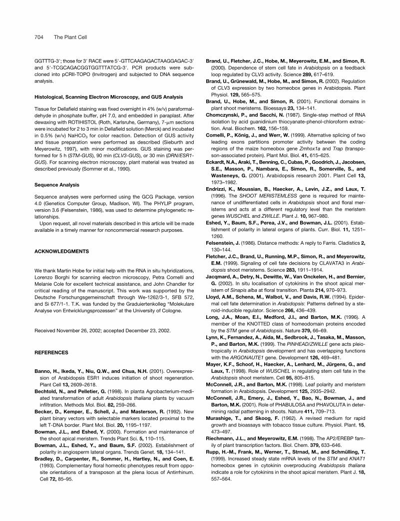
704 The Plant Cell
GGTTTG-3�; those for 3� RACE were 5�-GTTCAAGAGACTAAGGAGAC-3�
and 5�-TCGCAGACGGTGGTTTATCG-3�. PCR products were sub-cloned into pCRII-TOPO (Invitrogen) and subjected to DNA sequenceanalysis.
Histological, Scanning Electron Microscopy, and GUS Analysis
Tissue for Dellafield staining was fixed overnight in 4% (w/v) paraformal-dehyde in phosphate buffer, pH 7.0, and embedded in paraplast. Afterdewaxing with ROTIHISTOL (Roth, Karlsruhe, Germany), 7-�m sectionswere incubated for 2 to 3 min in Dellafield solution (Merck) and incubatedin 0.5% (w/v) NaHCO3 for color reaction. Detection of GUS activityand tissue preparation were performed as described (Sieburth andMeyerowitz, 1997), with minor modifications. GUS staining was per-formed for 5 h (STM-GUS), 90 min (CLV3-GUS), or 30 min (DRN/ESR1-GUS). For scanning electron microscopy, plant material was treated asdescribed previously (Sommer et al., 1990).
Sequence Analysis
Sequence analyses were performed using the GCG Package, version4.0 (Genetics Computer Group, Madison, WI). The PHYLIP program,version 3.6 (Felsenstein, 1986), was used to determine phylogenetic re-lationships.
Upon request, all novel materials described in this article will be madeavailable in a timely manner for noncommercial research purposes.
ACKNOWLEDGMENTS
We thank Martin Hobe for initial help with the RNA in situ hybridizations,Lorenzo Borghi for scanning electron microscopy, Petra Comelli andMelanie Cole for excellent technical assistance, and John Chandler forcritical reading of the manuscript. This work was supported by theDeutsche Forschungsgemeinschaft through We-1262/3-1, SFB 572,and Si 677/1-1. T.K. was funded by the Graduiertenkolleg “MolekulareAnalyse von Entwicklungsprozessen” at the University of Cologne.
Received November 26, 2002; accepted December 23, 2002.
REFERENCES
Banno, H., Ikeda, Y., Niu, Q.W., and Chua, N.H. (2001). Overexpres-sion of Arabidopsis ESR1 induces initiation of shoot regeneration.Plant Cell 13, 2609–2618.
Bechtold, N., and Pelletier, G. (1998). In planta Agrobacterium-medi-ated transformation of adult Arabidopsis thaliana plants by vacuuminfiltration. Methods Mol. Biol. 82, 259–266.
Becker, D., Kemper, E., Schell, J., and Masterson, R. (1992). Newplant binary vectors with selectable markers located proximal to theleft T-DNA border. Plant Mol. Biol. 20, 1195–1197.
Bowman, J.L., and Eshed, Y. (2000). Formation and maintenance ofthe shoot apical meristem. Trends Plant Sci. 5, 110–115.
Bowman, J.L., Eshed, Y., and Baum, S.F. (2002). Establishment ofpolarity in angiosperm lateral organs. Trends Genet. 18, 134–141.
Bradley, D., Carpenter, R., Sommer, H., Hartley, N., and Coen, E.(1993). Complementary floral homeotic phenotypes result from oppo-site orientations of a transposon at the plena locus of Antirrhinum.Cell 72, 85–95.
Brand, U., Fletcher, J.C., Hobe, M., Meyerowitz, E.M., and Simon, R.(2000). Dependence of stem cell fate in Arabidopsis on a feedbackloop regulated by CLV3 activity. Science 289, 617–619.
Brand, U., Grünewald, M., Hobe, M., and Simon, R. (2002). Regulationof CLV3 expression by two homeobox genes in Arabidopsis. PlantPhysiol. 129, 565–575.
Brand, U., Hobe, M., and Simon, R. (2001). Functional domains inplant shoot meristems. Bioessays 23, 134–141.
Chomczynski, P., and Sacchi, N. (1987). Single-step method of RNAisolation by acid guanidinium thiocyanate-phenol-chloroform extrac-tion. Anal. Biochem. 162, 156–159.
Comelli, P., König, J., and Werr, W. (1999). Alternative splicing of twoleading exons partitions promoter activity between the codingregions of the maize homeobox gene Zmhox1a and Trap (transpo-son-associated protein). Plant Mol. Biol. 41, 615–625.
Eckardt, N.A., Araki, T., Benning, C., Cubas, P., Goodrich, J., Jacobsen,S.E., Masson, P., Nambara, E., Simon, R., Somerville, S., andWasteneys, G. (2001). Arabidopsis research 2001. Plant Cell 13,1973–1982.
Endrizzi, K., Moussian, B., Haecker, A., Levin, J.Z., and Laux, T.(1996). The SHOOT MERISTEMLESS gene is required for mainte-nance of undifferentiated cells in Arabidopsis shoot and floral mer-istems and acts at a different regulatory level than the meristemgenes WUSCHEL and ZWILLE. Plant J. 10, 967–980.
Eshed, Y., Baum, S.F., Perea, J.V., and Bowman, J.L. (2001). Estab-lishment of polarity in lateral organs of plants. Curr. Biol. 11, 1251–1260.
Felsenstein, J. (1986). Distance methods: A reply to Farris. Cladistics 2,130–144.
Fletcher, J.C., Brand, U., Running, M.P., Simon, R., and Meyerowitz,E.M. (1999). Signaling of cell fate decisions by CLAVATA3 in Arabi-dopsis shoot meristems. Science 283, 1911–1914.
Jacqmard, A., Detry, N., Dewitte, W., Van Onckelen, H., and Bernier,G. (2002). In situ localisation of cytokinins in the shoot apical mer-istem of Sinapis alba at floral transition. Planta 214, 970–973.
Lloyd, A.M., Schena, M., Walbot, V., and Davis, R.W. (1994). Epider-mal cell fate determination in Arabidopsis: Patterns defined by a ste-roid-inducible regulator. Science 266, 436–439.
Long, J.A., Moan, E.I., Medford, J.I., and Barton, M.K. (1996). Amember of the KNOTTED class of homeodomain proteins encodedby the STM gene of Arabidopsis. Nature 379, 66–69.
Lynn, K., Fernandez, A., Aida, M., Sedbrook, J., Tasaka, M., Masson,P., and Barton, M.K. (1999). The PINHEAD/ZWILLE gene acts pleio-tropically in Arabidopsis development and has overlapping functionswith the ARGONAUTE1 gene. Development 126, 469–481.
Mayer, K.F., Schoof, H., Haecker, A., Lenhard, M., Jürgens, G., andLaux, T. (1998). Role of WUSCHEL in regulating stem cell fate in theArabidopsis shoot meristem. Cell 95, 805–815.
McConnell, J.R., and Barton, M.K. (1998). Leaf polarity and meristemformation in Arabidopsis. Development 125, 2935–2942.
McConnell, J.R., Emery, J., Eshed, Y., Bao, N., Bowman, J., andBarton, M.K. (2001). Role of PHABULOSA and PHAVOLUTA in deter-mining radial patterning in shoots. Nature 411, 709–713.
Murashige, T., and Skoog, F. (1962). A revised medium for rapidgrowth and bioassays with tobacco tissue culture. Physiol. Plant. 15,473–497.
Riechmann, J.L., and Meyerowitz, E.M. (1998). The AP2/EREBP fam-ily of plant transcription factors. Biol. Chem. 379, 633–646.
Rupp, H.-M., Frank, M., Werner, T., Strnad, M., and Schmülling, T.(1999). Increased steady state mRNA levels of the STM and KNAT1homeobox genes in cytokinin overproducing Arabidopsis thalianaindicate a role for cytokinins in the shoot apical meristem. Plant J. 18,557–564.

DRN/ESR1 in Cell Fate and Development 705
Schneitz, K., Hülskamp, M., and Pruitt, R.E. (1995). Wild-type ovuledevelopment in Arabidopsis thaliana: A light microscope study ofcleared whole-mount tissue. Plant J. 7, 731–748.
Schoof, H., Lenhard, M., Haecker, A., Mayer, K.F., Jurgens, G., andLaux, T. (2000). The stem cell population of Arabidopsis shoot mer-istems is maintained by a regulatory loop between the CLAVATA andWUSCHEL genes. Cell 100, 635–644.
Sieburth, L.E., and Meyerowitz, E.M. (1997). Molecular dissection ofthe AGAMOUS control region shows that cis elements for spatial reg-ulation are located intragenically. Plant Cell 9, 355–365.
Sommer, H., Beltran, J.P., Huijser, P., Pape, H., Lonnig, W.E.,Saedler, H., and Schwarz-Sommer, Z. (1990). Deficiens, a homeoticgene involved in the control of flower morphogenesis in Antirrhinummajus: The protein shows homology to transcription factors. EMBO J.9, 605–613.
Tissier, A.F., Marillonnet, S., Klimyuk, V., Patel, K., Torres, M.A.,Murphy, G., and Jones, J.D. (1999). Multiple independent defectivesuppressor-mutator transposon insertions in Arabidopsis: A tool forfunctional genomics. Plant Cell 11, 1841–1852.
Überlacker, B., and Werr, W. (1996). Vectors with rare-cutter restric-tion enzyme sites for expression of open reading frames in transgenicplants. Mol. Breeding 2, 293–295.
van der Graaff, E., Dulk-Ras, A.D., Hooykaas, P.J., and Keller, B.(2000). Activation tagging of the LEAFY PETIOLE gene affects leafpetiole development in Arabidopsis thaliana. Development 127, 4971–4980.
Waites, R., Selvadurai, H.R., Oliver, I.R., and Hudson, A. (1998). ThePHANTASTICA gene encodes a MYB transcription factor involved ingrowth and dorsoventrality of lateral organs in Antirrhinum. Cell 93,779–789.

DOI 10.1105/tpc.009480; originally published online February 21, 2003; 2003;15;694-705Plant Cell
Thomas Kirch, Rüdiger Simon, Margit Grünewald and Wolfgang Werrthe Control of Meristem Cell Fate and Lateral Organ Development
Gene of Arabidopsis Acts inDORNRÖSCHEN/ENHANCER OF SHOOT REGENERATION1The
This information is current as of September 17, 2018
References /content/15/3/694.full.html#ref-list-1
This article cites 34 articles, 11 of which can be accessed free at:
Permissions https://www.copyright.com/ccc/openurl.do?sid=pd_hw1532298X&issn=1532298X&WT.mc_id=pd_hw1532298X
eTOCs http://www.plantcell.org/cgi/alerts/ctmain
Sign up for eTOCs at:
CiteTrack Alerts http://www.plantcell.org/cgi/alerts/ctmain
Sign up for CiteTrack Alerts at:
Subscription Information http://www.aspb.org/publications/subscriptions.cfm
is available at:Plant Physiology and The Plant CellSubscription Information for
ADVANCING THE SCIENCE OF PLANT BIOLOGY © American Society of Plant Biologists