Embed Size (px)
Citation preview

Current Concepts
The Disabled Throwing Shoulder: Spectrum of Pathology
Part III: The SICK Scapula, Scapular Dyskinesis, the Kinetic
Chain, and Rehabilitation
Stephen S. Burkhart, M.D., Craig D. Morgan, M.D., and W. Ben Kibler, M.D.
We use the acronym SICK to refer to the findings
one sees in this syndrome (Scapular malposi-
tion, Inferior medial border prominence, Coracoid
pain and malposition, and dysKinesis of scapular
movement). This recently recognized overuse muscu-
lar fatigue syndrome is yet another cause of shoulder
pain in the throwing athlete who presents with dead
arm complaints.1 The hallmark feature of this syn-
drome is asymmetric malposition of the scapula in the
dominant throwing shoulder, which usually appears
on examination as if one shoulder is lower than the
other. This statically observable position is suggestive
of underlying muscle activation alterations that pro-
duce altered kinematics of the scapula upon dynamic
use. The altered kinematics fall into 3 clinically rec-
ognizable patterns of scapular dyskinesis, 2 of which
are most commonly associated with labral pathology:
type I, inferior medial scapular border prominence,
and type II, medial scapular border prominence.
The type III pattern, which is associated with im-
pingement and rotator cuff lesions rather than labral
lesions, displays prominence of the superomedial bor-
der of the scapula. In the SICK scapula syndrome,
scapular asymmetry is measured statically, but ac-
tively produces scapular dyskinesis as the shoulder
goes through the throwing cycle. The malpositioned
dyskinetic scapula, in turn, dynamically produces al-
tered kinematics of the glenohumeral and acromiocla-
vicular joints and the muscles that insert on the scap-
ula. Because of these complex interrelationships,
scapular dyskinesis, including the SICK scapula syn-
drome, can cause a spectrum of clinical complaints
originating from any or all of these anatomic loca-
tions.
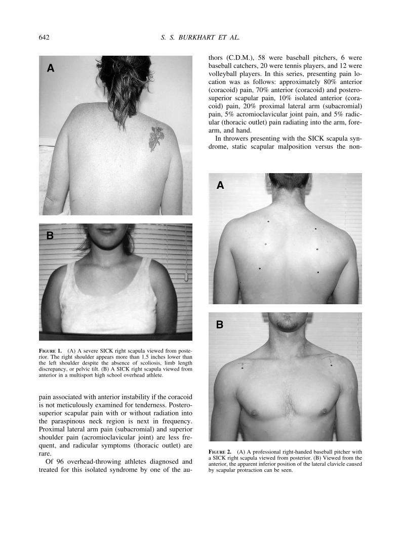
A thrower with this syndrome presents with an
apparent “dropped” scapula in his dominant symptom-
atic shoulder compared with the contralateral shoul-
der’s scapular position. In actuality, the scapula ini-
tially protracts, rotating about a horizontal axis, with
the upper scapula rotating anteroinferiorly. However,
the clinical appearance, with the arms relaxed in ad-
duction at the side, is that the involved scapula is
lower than the scapula on the uninvolved side (Figs 1
and 2). Viewing from behind, the inferior medial
scapular border appears very prominent, with the su-
perior medial border and acromion less prominent.
When viewed from the front, this tilting (protraction)
of the scapula makes the shoulder appear to be lower
than the opposite side. The pectoralis minor tightens
as the coracoid tilts inferiorly and shifts laterally away
from the midline, and its insertion at the coracoid
becomes very tender.
Symptomatic patients with an isolated SICK scap-
ula may complain of anterior shoulder pain, postero-
superior scapular pain, superior shoulder pain, proxi-
mal lateral arm pain, or any combination of the above.
In addition, posterosuperior scapular pain may radiate
into the ipsilateral paraspinous cervical region or the
patient may complain of radicular/thoracic outlet type
symptoms into the affected arm, forearm, and hand or
any combination of the above. The onset of symptoms
with the SICK scapula syndrome is almost always
insidious. By far, the most common presenting com-
plaint is anterior shoulder pain in the region of the
coracoid, which can easily be confused with anterior
Address correspondence and reprint requests to Stephen S.Burkhart, M.D., 540 Madison Oak Dr, Suite 620, San Antonio, TX78258, U.S.A.
© 2003 by the Arthroscopy Association of North America0749-8063/03/1906-3489-3$30.00/0doi:10.1016/S0749-8063(03)00389-X
641Arthroscopy: The Journal of Arthroscopic and Related Surgery, Vol 19, No 6 (July-August), 2003: pp 641-661
pain associated with anterior instability if the coracoid
is not meticulously examined for tenderness. Postero-
superior scapular pain with or without radiation into
the paraspinous neck region is next in frequency.
Proximal lateral arm pain (subacromial) and superior
shoulder pain (acromioclavicular joint) are less fre-
quent, and radicular symptoms (thoracic outlet) are
rare.
Of 96 overhead-throwing athletes diagnosed and
treated for this isolated syndrome by one of the au-
thors (C.D.M.), 58 were baseball pitchers, 6 were
baseball catchers, 20 were tennis players, and 12 were
volleyball players. In this series, presenting pain lo-
cation was as follows: approximately 80% anterior
(coracoid) pain, 70% anterior (coracoid) and postero-
superior scapular pain, 10% isolated anterior (cora-
coid) pain, 20% proximal lateral arm (subacromial)
pain, 5% acromioclavicular joint pain, and 5% radic-
ular (thoracic outlet) pain radiating into the arm, fore-
arm, and hand.
In throwers presenting with the SICK scapula syn-
drome, static scapular malposition versus the non-
FIGURE 2. (A) A professional right-handed baseball pitcher witha SICK right scapula viewed from posterior. (B) Viewed from theanterior, the apparent inferior position of the lateral clavicle causedby scapular protraction can be seen.
FIGURE 1. (A) A severe SICK right scapula viewed from poste-rior. The right shoulder appears more than 1.5 inches lower thanthe left shoulder despite the absence of scoliosis, limb lengthdiscrepancy, or pelvic tilt. (B) A SICK right scapula viewed fromanterior in a multisport high school overhead athlete.
642 S. S. BURKHART ET AL.

throwing shoulder is objectively measured in 3 cate-
gories: (1) infera, which is the visual appearance of a
dropped scapula due to scapular tilting or protraction;
(2) lateral displacement; and (3) abduction. All mea-
surements are made statically with the patients stand-
ing erect with arms relaxed in adduction at their side.
The measurement of infera is the difference in vertical
height of the superomedial scapular angle of the
dropped scapula in centimeters compared with the
contralateral superomedial angle (Figs 3 and 4). This
measurement is most accurately performed with a
bubble goniometer that uses the same bubble chamber
as a carpenter’s level. Although the measuring process
would appear to quantify linear displacement of the
scapula inferiorly, the malposition is actually a rota-
tional displacement (forward tilting, protraction).
Even so, the linear measurements are useful in esti-
mating how severe the dysfunction is and allowing
objective measurement of recovery during rehabilita-
tion. Scapular lateral displacement is the difference in
centimeters of the superomedial scapular angle from
the midline between the SICK and contralateral scap-
ula (Figs 3 and 4). Scapular abduction is the difference
in angular degrees of the medial scapular margin from
plumb midline between the SICK and contralateral
scapula measured with a goniometer (Fig 5).
FIGURE 3. Three categories of static scapular malposition seenwith the SICK scapula syndrome. (A) All 3 malpositions—infera,lateral protraction, and abduction—are illustrated in a high schoolquarterback’s right shoulder. (B) One or any combination of the 3malpositions may occur, as seen in the right shoulder of a collegebaseball pitcher with an isolated abduction component of the SICKscapula syndrome.
FIGURE 4. (A) A high-level woman tennis player with a SICKright scapula. (B) Note that the severity of the problem is moreevident when the scapular margins are outlined.
643THE DISABLED THROWING SHOULDER

Most throwers with the SICK scapula syndrome
present with static scapular malposition in all 3 cate-
gories, but single or dual combinations of any of the 3
have been seen. Although one of the authors (C.D.M.)
has devised a grading system for the severity of scap-
ular malposition based on these measurements, we
recognize that the use of superficial landmarks can
make these measurements less reliable and less repro-
ducible than we would like. Nonetheless, they give us
a qualitative sense of the severity of this dyskinetic
syndrome and a method of measuring progress with a
rehabilitation program (Table 1).
On physical examination, patients with anterior
shoulder complaints and a SICK scapula are found to
have marked coracoid tenderness, more medial than
lateral on the coracoid tip, at the point of insertion of
the pectoralis minor tendon. Throwers with anterior
coracoid pain can easily be confused with throwers
with other causes of anterior shoulder pain, such as
anterior instability or SLAP lesions, unless the cora-
coid is meticulously examined. SICK scapula patients
with coracoid pain usually lack full forward flexion on
the affected side and have accentuated coracoid pain
with attempts at gaining maximum passive forward
flexion by the examiner (Fig 6). With manual reposi-
tioning of the scapula in retraction and posterior tilt by
the examiner (the scapular retraction test), full for-
ward flexion without coracoid pain is usually attained,
which is diagnostic of this syndrome (Fig 6B).
The pathophysiology behind why the dropped SICK
scapula presents with coracoid pain is explained by
the coracoid static malposition and the dyskinesis that
it produces. Because of the ellipsoid shape of the
thorax, as the scapula tilts anteriorly, protracts, and
abducts, it tends to ride “up and over” the top of the
thorax.2,3 As it does, the coracoid tilts anteroinferiorly
and moves laterally from the midline. The pectoralis
minor and short head of the biceps become adaptively
tight. This tightness increases the scapular malposi-
tion, lowers the leading edge of the acromion, and
decreases the ability to achieve full forward flexion of
the arm. Impingement-like symptoms result from the
anteroinferior angulation of the acromion because of
scapular protraction.
Overhead athletes with SICK scapulas who present
with posterosuperior periscapular and lower paracer-
vical pain are usually found to have marked tender-
ness at the superomedial angle of the affected scapula
in the area of insertion of the levator scapulae muscle.
As the scapula tilts and rotates laterally, traction on
the levator scapulae creates pain and muscle spasm
(Fig 7). This can often be relieved on physical exam-
ination by correcting the tilted scapula by means of the
scapular retraction test.
TABLE 1. SICK Scapula Rating Scale: StaticMeasurements: 0–20 Points
FIGURE 5. Scapular abduction is measured with a goniometer onthe affected medial scapular margin in reference to vertical midlineversus the uninvolved medial scapular margin.
644 S. S. BURKHART ET AL.

Subacromial origin pain in the SICK throwing
shoulder will present with positive subacromial im-
pingement tests that bring out proximal lateral arm
pain. However, the true cause of these findings is a
malpositioned dyskinetic acromion resulting from
scapular protraction during all phases of the throwing
cycle, rather than true mechanical subacromial im-
pingement produced by a type III acromion with an
anterior osteophyte. Likewise, acromioclavicular joint
pain is caused by a relatively discongruous position of
the distal clavicle in reference to the acromion as a
result of scapular malposition. As the scapula tilts and
protracts, its acromion process moves anterior, de-
creasing the acromioclavicular angle and increasing
compressive stress to the acromioclavicular joint.4
The altered kinematics at the acromioclavicular joint
(and occasionally the sternoclavicular joint) cause
pain with repetitive overhead use.
Finally, the rare thrower with radicular or thoracic
outlet symptoms associated with a malpositioned
SICK scapula develops these symptoms due to a shift
in position of the clavicle in reference to the upper
chest wall, particularly the first rib. As the scapula
shifts, the lateral clavicle also drops anteroinferiorly,
resulting in a decreased subclavian chest wall space.
This space restriction may impinge the brachial plexus
as it crosses this zone resulting clinically in the picture
of thoracic outlet syndrome.
A 20-point clinical rating scale for the SICK scap-
ula syndrome has been developed by one of the au-
thors (C.D.M.) to statically assess the severity of the
syndrome at the time of presentation and to objec-
tively monitor clinical improvement during the treat-
ment phase (Table 1). The rating scale awards points
for subjective complaints and objective findings in the
categories previously discussed, as well as points for
the presence and severity of static scapular malposi-
tion in the 3 modes (“apparent” infera, lateral trans-
lation, and abduction). A healthy symmetrical asymp-
FIGURE 6. The scapular retraction test. (A) SICK scapula patientswith infera and abduction components and resultant coracoid painpresent with lack of full forward flexion because of pseudotight-ness of the pectoralis minor caused by coracoid malposition. (B)With manual repositioning of the scapula (retraction) the coracoidpain resolves and full forward flexion is attained.
FIGURE 7. Levator scapulae insertional pain caused by a SICKscapula. As the scapula abducts and protracts, the insertion of thelevator scapulae at the superomedial angle of the scapula becomesovertensioned and produces a painful tendinopathy in this area.
645THE DISABLED THROWING SHOULDER

tomatic scapula receives a score of 0, and the worst
SICK malpositioned scapula with all the pathologic
clinical components is scored as 20.
In an attempt to determine if components of scap-
ular malposition could be a normal adaptive phenom-
enon in the throwing athlete, a group of asymptomatic
professional baseball pitchers who denied previous
shoulder problems were prospectively evaluated for
presence or absence of SICK scapulas and scored on
the SICK scapula rating scale (Table 1) during spring
training and at the end of 2 consecutive pain-free
baseball seasons (P. Donley, J. Cooper, personal com-
munication, 2000). Nineteen pitchers meeting these
criteria were studied and found to have scores of 0
during the entire testing period (P. Donley, J. Cooper,
personal communication, 2000). These findings sup-
port the concept that the healthy throwing shoulder
exhibits no component of the SICK scapula syndrome
(no scapular asymmetry), and that this syndrome is
abnormal and predisposes the throwing shoulder to
pathologic symptomatology as previously discussed.
Treatment for the SICK scapula syndrome regard-
less of presenting symptoms or severity of scapular
malposition is nonoperative and focused on scapular
muscle rehabilitation. Initially, the thrower is re-
stricted from all throwing and begun on a regimented
daily strengthening and re-education program for all
the scapular stabilizer muscles (P. Donley, J. Cooper,
personal communication, 2000). Details of a scapular
rehabilitation program are discussed in the section
dealing with rehabilitation. During the treatment pe-
riod, scapular position is monitored on a weekly basis.
When the affected scapula is 50% or more improved
in position from its initial pathologic position, the
thrower is begun on an interval throwing program, if
asymptomatic, and continues the scapular program
until the scapula is symmetric with the other side (Fig
8). At that time, return to sport and unrestricted throw-
ing is allowed and the thrower is strongly encouraged
to maintain an every-other-day scapular muscle-
strengthening program to prevent recurrence of the
syndrome.
In general, most throwers with symptomatic SICK
scapulas present with scores between 10 and 14. In-
terval throwing usually begins with scores in the 4 to
6 range, and return to sport at the thrower’s previous
level of performance is attained when the score drops
between 0 and 2.
In an adherent patient who commits to doing the
rehabilitation exercises 3 times per day, the 50% re-
positioned scapula can be routinely attained within 2
to 3 weeks. Completion of the interval throwing pro-
gram usually takes 3 to 4 weeks, and complete sym-
metrical scapular repositioning usually takes 3
months. In general, the anterior tilt (apparent infera)
component is the first to resolve, the lateral translation
goes away second, and the abduction component (loss
of protraction control) is the last and most difficult to
resolve. Of the 96 overhead athletes treated for this
syndrome and followed up for more than 1 year, all
successfully returned to asymptomatic throwing at
their preinjury level of performance by 4 months.
Resolution of symptoms during the treatment period
was directly proportional to the rehabilitation pro-
gram’s ability to reposition the scapula symmetrical to
the other side. In addition, compliance with an every-
other-day scapular stabilizer muscle-strengthening
program prevented recurrence of the syndrome. Nine
of the 96 patients developed recurrence of the syn-
drome after 3 to 4 months of symptom-free throwing.
However, all 9 of these throwers admitted to total
nonadherence with the maintenance scapular program
for more than 3 months before developing symptoms
related to recurrent SICK scapula syndrome.
PRE-SLAP PRODROME AND THE
“SHOULDER AT RISK”
On persistent questioning, most throwers with ar-
throscopically proven posterior type II SLAP lesions
and the picture of internal impingement admit to a
pre-SLAP prodrome of ill-defined symptoms that they
ignored. During the early prodromal phase, the
thrower senses tightness in the back of his or her
dominant shoulder, oftentimes described as an inabil-
ity to “get loose.” As the player tries to “play through”
these prodromal symptoms and continues to throw,
the posteroinferior capsular contracture that caused
the tight symptomatology gets worse, to a point where
posterosuperior discomfort is now present with the
sense of tightness or stiffness. As the magnitude of the
capsular contracture continues to increase with con-
tinued throwing in the face of these prodromal symp-
toms, the SLAP event will then occur at which time
the shoulder develops sudden onset of mechanical
symptoms that were absent during the prodrome.
Once mechanical symptoms are present, treatment
becomes a surgical issue directed toward SLAP repair
and correction of the internal rotation deficit as dis-
cussed in the section dealing with posterior inferior
capsular contracture.
The “shoulder at risk” of developing dead arm
complaints is the asymptomatic shoulder that exhibits
small to moderate amounts of either a throwing-ac-
646 S. S. BURKHART ET AL.

quired glenohumeral internal rotation deficit (GIRD),
a malpositioned SICK scapula, or both.
In our experience, if the athlete with a shoulder at
risk keeps throwing, the magnitude of the shoulder
dysfunction will increase to a point at which intra-
articular structural damage occurs and the patient be-
comes symptomatic. Unfortunately, at this point the
problem has usually become a surgical issue. The
shoulder most at risk is one with a SICK scapula and
GIRD. This combination is particularly dangerous to
the posterosuperior labrum, the undersurface of the
posterior supraspinatus tendon, and the anterior infe-
rior capsular structures. The reason for this is that both
problems cause the thrower to abduct in extension
(toward second base), rather than in the plane of the
scapula, and hyperangulate in external rotation with a
low arm body angle (below the horizontal) during the
late cocking phase of throwing (Fig 9). This hyperan-
gulation is made more acute if the dyskinetic scapula
is in a position of protraction and glenoid antetilting,
thereby bringing the posterior edge of the glenoid
toward the humerus. More proper mechanics seen in
FIGURE 8. The SICK scapula before and after treatment. A professional right-handed baseball pitcher with a symptomatic SICK rightscapula seen (A) at the time of initial presentation and (B) 6 weeks after scapular rehabilitation. The 1999 National League All Star startingpitcher and 2001 World Series Most Valuable Player seen in the late 1999 season with a (C) symptomatic SICK right scapula and (D) 6 weeksafter scapular rehabilitation.
647THE DISABLED THROWING SHOULDER

healthy shoulders consist of abduction in the plane of
a well-positioned scapula and a greater arm–body
abduction angle during the late cocking and early
acceleration phases of the throwing cycle (Fig 10).
Early recognition of the shoulder at risk and insti-
tution of internal rotation stretching and scapular sta-
bilizer strengthening protocols have been shown to be
highly effective in converting the shoulder at risk to
the shoulder not at risk and, in so doing, avoiding the
shoulder at surgery. For these reasons, we encourage
and recommend screening examinations to find these
problems at the beginning and during each season for
all overhead athletes. In addition, these athletes need
to be educated by their respective athletic trainers and
doctors regarding these issues so that they are willing
to commit to the stretching and strengthening that will
keep their shoulders healthy.
THE ROTATIONAL UNITY RULE
The healthy throwing shoulder has normal rota-
tional kinematics without any form of glenohumeral
instability throughout the throwing cycle as long as its
GIRD is less than or equal to its external rotation gain.
However, if the GIRD exceeds the external rotation
gain (ERG) with a GIRD/ERG ratio greater than 1, the
shoulder then becomes headed for trouble because of
a posterosuperior shift of the glenohumeral rotation
point with abduction and external rotation during the
late cocking phase of throwing as previously dis-
cussed. The risk of structural injury is directly propor-
tional to the increase in the GIRD/ERG ratio past
unity. Although the absolute rotational numbers in
degrees will vary from patient to patient because of
variability in congenital laxity, GIRD that exceeds
10% of the contralateral shoulder’s total rotation arc
will usually produce a GIRD/ERG ratio significantly
greater than 1. It is important to stabilize the scapula
with the arm at 90° abduction when measuring inter-
nal and external rotation.
KINETIC CHAIN CONTRIBUTIONS TO
DEAD ARM
Shoulder function in throwing requires contribu-
tions from all body segments to generate the forces
necessary to propel the ball and position the bones of
the joints to minimize the loads each joint structure
must bear, and pass the forces and loads to the distal
segments.5 This coordinated sequencing of the seg-
ments is termed the kinetic chain. In the normal ki-
netic chain of throwing, the ground, legs, and trunk act
as the force generators; the shoulder acts as a funnel
and force regulator; and the arm acts as the force
delivery mechanism. The labrum is a key structure in
providing glenohumeral stability in this sequence and
may be injured by excessive or imbalanced forces that
may occur at the shoulder if regional or distant areas
of the kinetic chain are abnormal.
Clinical studies have shown that alterations in flex-
ibility or muscle imbalance are common in patients
with shoulder injury. Kinetic chain alterations have
been documented in shoulder impingement,6,7,8 rota-
tor cuff injury,6,9 and instability.9,10 One study specif-
ically evaluated distant findings that were present in
throwers with arthroscopically proven posterosuperior
labral tears.11 Of 64 patients, 38 had isolated postero-
superior labral tears, and 26 had combined anterior
and posterior lesions. On physical examination, 64 of
64 (100%) had restricted goniometrically measured
internal rotation (with the shoulder at 90° abduc-
tion and the scapula stabilized) of more than 25°
compared with the opposite side (mean, 32.6°;
range, 26° to 58°). A total of 60 of 64 (94%) had
patterns of dynamic scapular dyskinesis, or asymmet-
ric motion or position of the scapula on rest or abduc-
tion motion.6,12
The SICK scapula is an extreme form of scapular
dyskinesis. A total of 41 subjects had the type I
dyskinetic pattern of winging of the inferomedial
scapular border and depression of the acromion with
rest or motion, whereas 19 showed the type II pattern
of true lateral slide, in which the entire medial scap-
ular border is winged with the scapula protracted and
laterally translated with rest or motion.13 The mean
lateral slide asymmetry measurement12 in 60 patients
was 2.2 cm (range, 0.7 to 3.1 cm). A total of 46 of 64
patients (72%) showed weakness of the infraspinatus
or teres minor on resisted external rotation. A total of
31 of 64 (48%) exhibited lower-back inflexibility,
with reach deficits of greater than 5 cm on sit-and-
reach examination. A total of 28 of 64 (44%) were
unable to complete a nondominant leg stability se-
quence of one-legged stance with no Trendelenburg
sign, 1-legged squat with pelvic stability, and 1-legged
step-up and step-down with pelvic stability. Finally,
25 of 64 subjects (39%) had asymmetric decreased
internal rotation on the nondominant hip.
These alterations in normal kinetic chain motions
and positions affect the shoulder by altering proximal
force generation and altering distal joint positioning.
These alterations are especially important at the shoul-
der, which does not have a large force-generating
648 S. S. BURKHART ET AL.

capability and whose bony anatomy is dependent on
body positioning and muscular activation to control
excessive joint translations.
Leg and Trunk
The leg and trunk are important to provide a stable
base for arm motion,13,14 provide rotational momen-
tum for force generation,14,15 and generate 50% to
55% of the total force and kinetic energy in the tennis
serve.3 Inflexibility of the nondominant hip or of trunk
rotation, or weakness of hip abductors or trunk flexors
“breaks the kinetic chain.” This breakage increases
lumbar lordosis in acceleration, which places the arm
behind the body. This “slow arm” creates a hyperab-
duction/external rotation position at the shoulder and
increases posterior compression loads on the struc-
tures including the labrum. It moves the arm out of the
safe zone of glenohumeral angulation that has been
advocated by Jobe et al.16 and shown by Happee17 to
be the most stable angle for the joint.
Scapula
The scapula plays many roles in throwing or serving
that may affect labral injury.9 First, the glenoid must
be positioned and stabilized in 3-dimensional space to
act as a congruous socket for the humeral head as it
rotates at the high velocities necessary for throwing or
serving. Second, it must smoothly retract and protract
around the thoracic wall as the arm moves from cock-
ing through full cocking and then into acceleration and
follow-through. The scapula must move in relation to
FIGURE 9. This pitcher shows abduction in extension, with angu-lation of the arm posterior to the plane of the scapula rather than inthe plane of the scapula. Note the “dropped elbow” in this pitcher,causing the arm-body angle to drop below the horizontal.
FIGURE 10. Ideal mechanics involve abduction in the plane of thescapula (A, dotted line) with the elbow high enough to keep theupper arm at or above the horizontal plane. (B) With a “droppedelbow” (solid line), the upper arm hyperangulates posterior to theplane of the scapula. (C) This pitcher has excellent mechanics, withthe arm abducted in the plane of the scapula and positioned abovethe horizontal plane.
649THE DISABLED THROWING SHOULDER

the moving humerus to maintain a safe zone of gle-
nohumeral angulation and avoid hyperangulation of
the humerus on the glenoid.16 Finally, it acts as a
stable base for origin of the extrinsic and intrinsic
muscles that control arm motion and provide gleno-
humeral compression. These roles require active po-
sitioning and active motion by the periscapular mus-
cles. Alterations of normal position or motion, which
have been termed scapular dyskinesis,13 can be caused
by inflexibility, weakness, or activation imbalance of
the muscles.
Two patterns of dynamic scapular dyskinesis are
associated with posterosuperior labral lesions. The
type I pattern consists of inferomedial scapular border
prominence at rest, with increasing prominence, lack
of acromial elevation, and lack of full retraction on
cocking (Fig 11). It is associated with inflexibility of
the pectoralis major and minor, and weakness of the
lower trapezius and serratus anterior. The type II pat-
tern consists of entire medial border winging at rest,
which becomes more prominent with cocking or ele-
vation (Fig 12). It is associated with upper and lower
trapezius and rhomboid weakness, with little anterior
inflexibility. Both patterns create an abnormal position
of excessive scapular protraction at rest and an abnor-
mal motion of decreased scapular retraction and de-
creased acromial elevation during cocking and early
acceleration. The type III pattern, which is not asso-
ciated with superior labral lesions, displays promi-
nence of the superomedial border of the scapula. It is
associated with impingement and rotator cuff symp-
toms.
Scapular dyskinesis that creates excessive protrac-
tion and decreased cocking and elevation alters nor-
mal scapular biomechanics and plays a role in pos-
FIGURE 11. Type I scapular dyskinesis, with prominence of theinferomedial scapular border.
FIGURE 12. Type II scapular dyskinesis, with prominence of theentire medial border.
FIGURE 13. Scapular assistance test. The examiner stabilizes theupper scapular border and assists upward rotation of the inferome-dial border.
650 S. S. BURKHART ET AL.

terosuperior labral injuries. Excessive protraction
increases glenohumeral angulation outside of the safe
zone, creating excessive anterior tension and posterior
compression. Increased glenohumeral angulation also
may increase humeral external rotation in cocking and
acceleration as the arm lags behind the body, increas-
ing the peel-back effect. Decreased retraction in-
creases the posterior glenohumeral compression on
the labrum and rotator cuff and decreases the scapular
role as a stable base for muscle origin, thereby effec-
tively decreasing muscle strength. Decreased acromial
elevation due to scapular protraction creates rotator
cuff impingement in cocking and acceleration as the
arm is abducted.
Clinical Implications
Evaluation of athletes with suspected superior gle-
noid labral lesions should include tests to check ki-
netic chain functioning. History taking should include
questions about previous leg or back problems, espe-
cially on the nondominant side. A screening exami-
FIGURE 14. Scapular retraction test. The examiner stabilizes theretracted scapula against the thorax.
FIGURE 15. One-leg stance, trunk rotation, and scapular retraction combined movements.
651THE DISABLED THROWING SHOULDER

nation for the legs and trunk should include a posture
examination for legs and trunk, seated range-of-mo-
tion examination of both hips and knees, the one-leg
stability series (i.e., 1-legged stance with no Tren-
delenburg sign and 1-legged squat with no pelvic tilt
or rotation), low back flexion and extension and side-
bending, and situps and back extensions. Further test-
ing is indicated for patients with positive tests.
The screening scapular examination should include a
posture check for cervical and thoracic areas, scapular
symmetry at rest and on ascending and descending arm
motion in flexion and abduction, active scapular retrac-
tion and elevation, lateral slide measurements, and gle-
nohumeral internal rotation measurements.
Special scapular tests include the scapular assis-
tance test (SAT) and the scapular retraction test
(SRT). The SAT (Fig 13) involves assisting scapular
upward rotation by manually stabilizing the upper
medial border and rotating the inferomedial border as
the arm is abducted. The test is positive when it gives
relief of symptoms of impingement, clicking, or rota-
tor cuff weakness.13 The scapular retraction test (Fig
14) involves manually positioning and stabilizing the
entire medial border of the scapula. This test is helpful
in 2 groups of patients. The first group has decreased
retraction and apparent rotator cuff weakness. The test
is positive when retesting reveals increased muscle
strength with the scapula in the stabilized position.
The second group has a positive Jobe relocation test
for posterosuperior labral injury. The test is positive
when scapular retraction decreases the pain or im-
pingement associated with the Jobe relocation test.
When positive, these tests show that specific scapular
alterations are present and should be addressed as part
of the rehabilitation.
REHABILITATION OF THE OVERHEAD
ATHLETE
Rehabilitation of patients with superior glenoid la-
bral lesions and scapular dyskinesis should also in-
clude the kinetic chain. This aspect of the rehabilita-
tion may be started early, even while shoulder
evaluation and treatment is being done. Leg, back, and
trunk flexibility and strength should be normalized,
and exercises that emphasize kinetic chain activation
of the leg, trunk, and scapula should be instituted.13,18
Useful combinations of movements to allow activa-
tion include trunk extension and scapular retraction,
trunk rotation and scapular retraction, and 1-legged
stance and diagonal trunk rotation and scapular retrac-
tion (Fig 15). All of these exercises facilitate lower
trapezius muscle activation.
Scapular exercises start with scapular punches and
isometric scapular retractions. A very safe exercise is
the “low row” (Fig 16), an exercise that involves trunk
extension, scapular retraction, and arm extension as
the patient pushes against resistance in a posterior
direction. More advanced closed-chain exercises in-
clude the scapular clock (Fig 17), in which the hand is
placed on the wall, eliminating the weight of the arm,
and moving the scapula in elevation and depression
(the 12 and 6 o’clock positions) and retraction and
protraction (the 9 and 3 o’clock positions). These
exercises are generally safe and do not seem to in-
crease labral injury. All preoperative patients may be
placed on this program, similar to knee exercises
before anterior cruciate ligament reconstruction. By
stabilizing the kinetic chain and scapular base, the
FIGURE 16. The “low-row” trunk extension, scapular retraction,and arm extension. This can be initially done as an isometricexercise, progressing to an isotonic movement.
652 S. S. BURKHART ET AL.

patient can move more rapidly through the early
stages of rehabilitation, regain the normal patterns of
trunk-scapula-arm integration, eliminate the “shrug”
that is common when arm abduction is attempted, and
move more rapidly into rotator cuff strengthening.
Rotator cuff exercises can be integrated into the
rehabilitation program after the proximal base has
been established.18,19 Maximal rotator cuff activation
requires a stable scapular base, since all the rotator
cuff muscles have their origin on the scapula; ade-
quate scapular elevation, to eliminate impingement;
closed-chain activation to eliminate excessive shear in
the early stages of rehabilitation; and coordinated ac-
tivation of all of the components in force couples,
rather than isolated activation of individual muscles.
Closed-chain rotator cuff exercises include humeral
head depressions and rotations on a ball (Fig 18),
FIGURE 17. Scapular clock: The hand is placed on the wall or aball, with varying degrees of abduction and flexion.
FIGURE 18. Humeral head depressions and rotations with thehand on a ball.
FIGURE 19. “Wall washes”: (A) Starting position and (B) move-ment pattern. The arm is then brought back to the starting position.
653THE DISABLED THROWING SHOULDER

“wall washes,” which combine trunk and scapula ac-
tivation with rotator cuff activations (Fig 19), and
punches, which combine closed-chain shoulder acti-
vation with open-chain arm motion (Fig 20). This
progression of exercises creates a progression of chal-
lenge to the rotator cuff, and a resulting progression of
muscle activation (Table 2).20,21 Closed-chain exer-
cises require less activation than open-chain exercises,
and vertical patterns with the arm closer to the body,
creating a shorter lever arm, requires less activation
than diagonal patterns, with a long lever arm. This
progression allows a more rapid but safe progression
of rehabilitation that can be characterized as “accel-
erated,” similar to the accelerated anterior cruciate
ligament programs that take advantage of the same
physiologic and biomechanical principles of stable
fixation, early protected range of motion, closed-chain
activation of cocontraction force couples, and early
activation of muscles in their physiologic positions.22
Rehabilitation for the SICK Scapula
The SICK scapula, as well as other types of scap-
ular dyskinesis, must be aggressively treated with a
focused scapular rehabilitation program. Rehabilita-
tion consists of both stretching and strengthening.
The 2 areas of tightness that accompany the SICK
scapula are (1) pectoralis minor tightness anteriorly
and (2) posteroinferior capsular tightness with GIRD.
FIGURE 20. Punches: (A) Punch out. The motion may be varied-diagonal, upward, or downward. (B) Return position should always be“elbows in the back pocket” to facilitate scapular retraction.
TABLE 2. Progression of Muscle Activation
Exercise
Percent of MaximalMuscle Activation
Supraspinatus Infraspinatus
Humeral head rotations
(Codman exercise on a ball) �10 �10
Vertical wall wash 12.8 8.6
Diagonal wall wash 18.0 10.4
Vertical punch 16.9 9.8
Diagonal punch 21.6 14.6
654 S. S. BURKHART ET AL.
The anterior tightness is treated by placing a rolled
towel between the shoulder blades of the supine pa-
tient and steadily pushing posteriorly on the shoulders
to stretch the pectoralis minor (Fig 21). The postero-
inferior capsular tightness is treated by “sleeper
stretches” in which the athlete lies on his side with the
shoulder in 90° flexion and the elbow in 90° flexion.
The shoulder is passively internally rotated by pushing
the forearm toward the table around a fixed elbow,
which acts as the pivot point (Fig 22).
Strengthening for the SICK scapula patient consists
of exercises to regain control of scapular protraction,
retraction, depression, elevation, and rotation. Closed-
chain exercises without weight are initiated to regain
scapular control (Fig 23). Open-chain forward and
lateral lunges and diagonal pulls are added, first with-
out weights and then with 2- to 3-lb wrist weights or
dumbbells (Fig 24). Blackburn retraction exercises are
used to strengthen the scapular retractors and posterior
rotator cuff (Fig 25). Seated push-ups strengthen scap-
ular depressors or retractors, triceps, latissimus dorsi,
and teres major (Fig 26). Rowing exercises, both
standard row and low row, strengthen scapular retrac-
tors and the posterior rotator cuff. The low row is
more specific for strengthening the serratus anterior
(Fig 27).
SUMMARY OF THE ENTIRE CURRENT
CONCEPTS: PARTS I, II, AND III
The disabled throwing shoulder comprises a spec-
trum of pathology. At the most dramatic and severe
end of this spectrum is the dead arm, a pathologic
shoulder condition in which the thrower is unable to
throw with preinjury level velocity and control. The
most common cause of the dead arm syndrome is a
type II SLAP lesion, although the SICK scapula may
cause a reversible type of dead arm with different
clinical findings from those of the SLAP lesion.
FIGURE 21. Pectoralis minor tightness is treated by placing arolled towel between the shoulder blades of the supine patient andpushing posteriorly on the shoulders.
FIGURE 22. Sleeper stretch: (A) The patient lies on the side with the involved arm against the table and perpendicular to the body. The elbowis flexed 90°. (B) The patient pushes the forearm toward the table, stretching the posteroinferior capsule. (See Fig 4 in Part I of this CurrentConcepts in the April issue for other variations of the “sleeper stretch.”)
655THE DISABLED THROWING SHOULDER
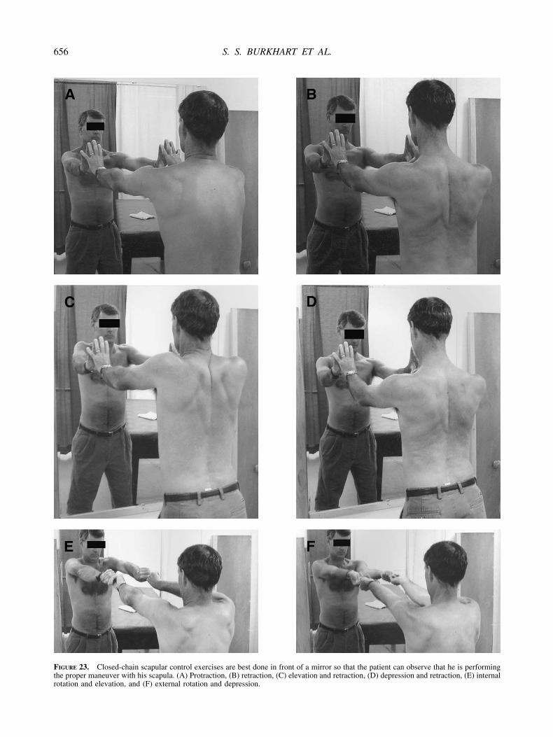
FIGURE 23. Closed-chain scapular control exercises are best done in front of a mirror so that the patient can observe that he is performingthe proper maneuver with his scapula. (A) Protraction, (B) retraction, (C) elevation and retraction, (D) depression and retraction, (E) internalrotation and elevation, and (F) external rotation and depression.
656 S. S. BURKHART ET AL.

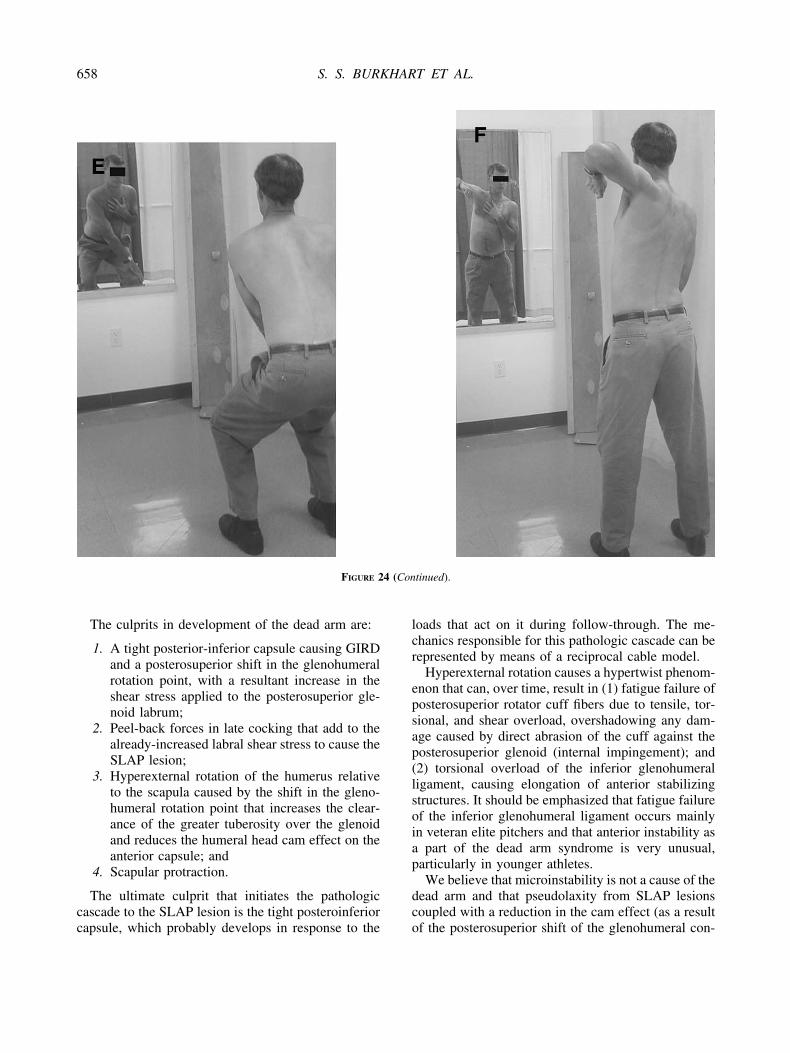
FIGURE 24. Open-chain scapular strengthening exercises. (A, B) Forward lunges to strengthen scapular protractors and retractors. (C, D)Lateral lunges to strengthen scapular retractors and upward rotators (upstroke) as well as scapular depressors and downward rotators(downstroke). (E, F) Diagonal pulls (lawnmower pulls) to strengthen scapular protractors and depressors as well as retractors and elevators.
657THE DISABLED THROWING SHOULDER
The culprits in development of the dead arm are:
1. A tight posterior-inferior capsule causing GIRD
and a posterosuperior shift in the glenohumeral
rotation point, with a resultant increase in the
shear stress applied to the posterosuperior gle-
noid labrum;
2. Peel-back forces in late cocking that add to the
already-increased labral shear stress to cause the
SLAP lesion;
3. Hyperexternal rotation of the humerus relative
to the scapula caused by the shift in the gleno-
humeral rotation point that increases the clear-
ance of the greater tuberosity over the glenoid
and reduces the humeral head cam effect on the
anterior capsule; and
4. Scapular protraction.
The ultimate culprit that initiates the pathologic
cascade to the SLAP lesion is the tight posteroinferior
capsule, which probably develops in response to the
loads that act on it during follow-through. The me-
chanics responsible for this pathologic cascade can be
represented by means of a reciprocal cable model.
Hyperexternal rotation causes a hypertwist phenom-
enon that can, over time, result in (1) fatigue failure of
posterosuperior rotator cuff fibers due to tensile, tor-
sional, and shear overload, overshadowing any dam-
age caused by direct abrasion of the cuff against the
posterosuperior glenoid (internal impingement); and
(2) torsional overload of the inferior glenohumeral
ligament, causing elongation of anterior stabilizing
structures. It should be emphasized that fatigue failure
of the inferior glenohumeral ligament occurs mainly
in veteran elite pitchers and that anterior instability as
a part of the dead arm syndrome is very unusual,
particularly in younger athletes.
We believe that microinstability is not a cause of the
dead arm and that pseudolaxity from SLAP lesions
coupled with a reduction in the cam effect (as a result
of the posterosuperior shift of the glenohumeral con-
FIGURE 24 (Continued).
658 S. S. BURKHART ET AL.

FIGURE 25. Blackburn exercises to strengthen scapular retractors and posterior rotator cuff. (A) Position 1, (B) position 2, (C) position 3,(D) position 4, (E) position 5, and (F) position 6.
659THE DISABLED THROWING SHOULDER

tact point) has been misinterpreted as microinstability.
Furthermore, we believe that internal impingement is
a normal phenomenon that is not usually pathologic in
the throwing shoulder.
For a thrower with a dead arm and a SLAP lesion,
repair of the SLAP lesion combined with an ongoing
stretching program of the posteroinferior capsule is
usually curative, returning the thrower to his preinjury
level of competition in 87% of cases.23 For successful
SLAP repair, the surgeon must arthroscopically con-
firm elimination of the peel-back sign and elimination
of the drive-through sign. For throwers who have been
“stretch nonresponders,” the surgeon may consider
performing an arthroscopic release of the posteroinfe-
rior capsule. If there is greater than 130° of external
rotation with the scapula stabilized, one should con-
sider electrothermal shrinkage versus arthroscopic
capsular plication of the anterior band of the inferior
glenohumeral ligament.
The SICK scapula syndrome, an extreme form of
scapular dyskinesis, can be a cause of dead arm.
Extreme protraction and anterior tilting of the scapula
gives the impression that it is inferiorly displaced.
This syndrome has unique clinical characteristics and
generally responds to a focused rehabilitation of the
shoulder.
Scapular biomechanics are vitally important to the
throwing athlete and can be adversely affected by
derangements at any point in the kinetic chain, includ-
ing lower extremity function. The surgeon who treats
throwing athletes must have a thorough understanding
of the kinetic chain as well as an appreciation for the
need for a well-focused rehabilitation program in re-
storing these athletes to the high level of function that
their sport demands. In addition, closed-chain exer-
cises can restore function while decreasing stresses to
damaged tissues, resulting in more rapid rehabilita-
tion.
The biomechanical and anatomic factors responsi-
FIGURE 26. Seated push-ups: (A) Strengthening scapular retractors and elevators. (B) Strengthening scapular retractors and depressors.
FIGURE 27. The low row is performed with the patient pullingposteriorly (shoulder extension and retraction) with the elbowlocked in full extension. This exercise strengthens scapular retrac-tors, particularly the serratus anterior.
660 S. S. BURKHART ET AL.

ble for the dead arm have only recently been identi-
fied. As a result, appropriate surgical and nonsurgical
treatments, including rehabilitation, can now be more
precisely directed at specific pathophysiologic ele-
ments. The dead arm syndrome, so mysterious and so
elusive for so long, is finally giving up its secrets.
Editor’s Note: This concludes this three-part Cur-
rent Concepts series published in Vol. 19, Nos. 4, 5,
and 6 of Arthroscopy.
Acknowledgment: The authors wish to thank Jeff Coo-per, P.T., A.T.C. (Head Athletic Trainer, Philadelphia Phil-lies baseball team), and Phil Donley, P.T., A.T.C., M.S.(Consultant, Philadelphia Phillies) for their assistance in thepreparation of this manuscript.
REFERENCES
1. Morgan CD. The thrower’s shoulder. Two perspectives. In:McGinty JB, et al. eds. Operative arthroscopy. Ed 3. Phila-delphia: Lippincott Williams & Wilkins, 2003; 570-584.
2. Lukasiewicz AC, McClure PW, Michener LA. Comparison ofthree dimensional scapular position and orientation betweensubjects with and without shoulder impingement. J OrthopSports Phy Ther 1999;29:574-586.
3. McClure PW, Michener LA, Sennett BJ, Karduna AR. Directthree-dimensional measurement of scapular kinematics duringdynamic movements in vivo. J Shoulder Elbow Surg 2001;10:269-278.
4. Bagg SD, Forrest WJ. A biomechanical analysis of scapularrotation during arm abduction in the scapular plane. Am J PhysMed 1988;67:238-245.
5. Kibler WB. Biomechanical analysis of the shoulder duringtennis activities. Clin Sports Med 1995;14:79-86.
6. Warner JJP, Micheli L, Arslenian L. Scapulothoracic motionin normal shoulders and shoulders with glenohumeral insta-bility and impingement syndrome. Clin Orthop 1992;285:191-199.
7. McQuade KJ, Dawson J, Smidt GL. Scapulothoracic musclefatigue associated with alterations in scapulohumeral rhythmkinematics during maximum resistive shoulder elevation.J Orthop Sports Phys Ther 1998;28:74-80.
8. Tyler TF, Nicholas SJ, Roy T, et al. Quantification of posteriorcapsule tightness and motion loss in patients with shoulderimpingement. Am J Sports Med 2000;28:668-674.
9. Paletta GA, Warner JJP, Warren RF, et al. Shoulder kineticswith two-plane x-ray evaluation in patients with anterior in-stability or rotator cuff tears. Shoulder Elbow Surg 1997;6:516-527.
10. Weiser WM, Lee TQ, McMaster WC. Effects of simulatedscapular protraction on anterior glenohumeral stability. Am JSports Med 1999;27:801-805.
11. Burkhart SS, Morgan CD, Kibler WB. Shoulder injuries inoverhead athletes. Clin Sports Med 2000;19:125-158.
12. Kibler WB. The role of the scapula in athletic shoulder func-tion. Am J Sports Med 1998;26:325-337.
13. Kibler WB, Livingston BP. Closed chain rehabilitation for theupper and lower extremity. J Am Acad Orthop Surg 2001;9:412-421.
14. Young JL, Herring SA, Press JM, et al. The influence of thespine on the shoulder in the throwing athlete. J Back Muscu-loskeletal Rehab 1996;7:5-17.
15. Watkins RG, Dennis S, Dillin WH, et al. Dynamic EMGanalysis of torque transfer in professional baseball pitchers.Spine 1989;14:404-408.
16. Pink MM, Perry J. Biomechanics of the shoulder. In: Jobe FW,ed. Operative techniques in upper extremity sports injuries. St.Louis: Mosby, 1996; 10–124.
17. Happee R, Van Der Helm FC. Control of shoulder musclesduring goal-directed movements, an inverse dynamic analysis.J Biomech 1995;28:1179-1191.
18. McMullen J, Uhl TL. A kinetic chain approach for shoulderrehabilitation. J Athlet Train 2000;35:329-337.
19. Kibler WB. Shoulder rehabilitation: Principles and practice.Med Sci Sports Exerc 1998;30:540-550 (suppl).
20. Lephart SM, Henry TJ. The physiological basis for open andclosed kinetic chain rehabilitation for the upper extremity.J Sport Rehabil 1996;5:71-87.
21. Uhl TL, Kibler WB. Rotator cuff activation in closed and openchain exercises. Presented at the 18th Open Meeting of theAmerican Shoulder and Elbow Surgeons, Dallas, TX, Febru-ary 16, 2002.
22. Kibler WB, McMullen JB. Accelerated postoperative shoulderrehabilitation. In: Norris TR, ed. Orthopaedic knowledge up-date: Shoulder and elbow 2. Rosemont, IL: American Acad-emy of Orthopaedic Surgeons, 2002; 403-409.
23. Morgan CD, Burkhart SS, Palmeri M, et al. Type II SLAPlesions: Three subtypes and their relationship to superior in-stability and rotator cuff tears. Arthroscopy 1998;14:553-565.
661THE DISABLED THROWING SHOULDER