Embed Size (px)
Citation preview
Current Concepts
The Disabled Throwing Shoulder: Spectrum of PathologyPart I: Pathoanatomy and Biomechanics
Stephen S. Burkhart, M.D., Craig D. Morgan, M.D., and W. Ben Kibler, M.D.
Prologue: Several years ago, when we began to question microinstability as the universal cause ofthe disabled throwing shoulder, we knew that we were questioning a sacrosanct tenet of Americansports medicine. However, we were comfortable in our skepticism because we were relying onarthroscopic insights, clinical observations, and biomechanical data, thereby challenging unverifiedopinion with science. In so doing, we assembled a unified concept of the disabled throwing shoulderthat encompassed biomechanics, pathoanatomy, kinetic chain considerations, surgical treatment, andrehabilitation. In developing this unified concept, we rejected much of the conventional wisdom ofmicroinstability-based treatment in favor of more successful techniques (as judged by comparativeoutcomes) that were based on sound biomechanical concepts that had been scientifically verified.Although we have reported various components of this unified concept previously, we have beenurged by many of our colleagues to publish this information together in a single reference for easyaccess by orthopaedic surgeons who treat overhead athletes. We are grateful to the editors ofArthroscopy for allowing us to present our view of the disabled throwing shoulder. Part I: Patho-anatomy and Biomechanics is presented in this issue. Part II: Evaluation and Treatment of SLAPLesions in Throwers will be presented in the May-June issue. Part III: The “SICK” Scapula, ScapularDyskinesis, the Kinetic Chain, and Rehabilitation will be presented in the July-August issue. Wehope you find it thought-provoking and compelling.
The medical community’s fascination with the dis-abled throwing shoulder derives from the pub-
lic’s fascination with the intact throwing shoulder.The ability to throw a baseball with pinpoint accuracyat speeds above 90 miles an hour defines the upperechelon of athletic achievement. The sudden loss ofthat ability, as occurs in the so-called “dead arm,” isnothing short of an athletic tragedy.
The dead arm has long been recognized as a career-ending affliction in the overhead athlete, but onlyrecently have we understood this condition enough toprovide effective treatment. We define the “dead arm”
as any pathologic shoulder condition in which thethrower is unable to throw with preinjury velocity andcontrol because of a combination of pain and subjec-tive unease in the shoulder. The athlete usually relatesthe discomfort to the late cocking or early accelerationphase of the throwing sequence, when the arm beginsto move forward. At this point, the thrower feels asudden sharp pain, the arm “goes dead,” and theathlete is unable to throw the ball with his usualvelocity. The story of the dead arm is the story of thedisabled throwing shoulder; we examine that story, inall its forms, in this 3-part Current Concepts series.
SLAP LESIONS AS A CAUSE OF THEDEAD ARM
Two of the authors (C.D.M., S.S.B.) reported on 53baseball players, 44 of whom were pitchers, who hadtype 2 SLAP lesions that were surgically repaired after
Address correspondence and reprint requests to Stephen S.Burkhart, M.D., 540 Madison Oak Dr, Suite 620, San Antonio, TX78258, U.S.A.
© 2003 by the Arthroscopy Association of North America0749-8063/03/1904-3489-1$30.00/0doi:10.1053/jars.2003.50128
404 Arthroscopy: The Journal of Arthroscopic and Related Surgery, Vol 19, No 4 (April), 2003: pp 404-420
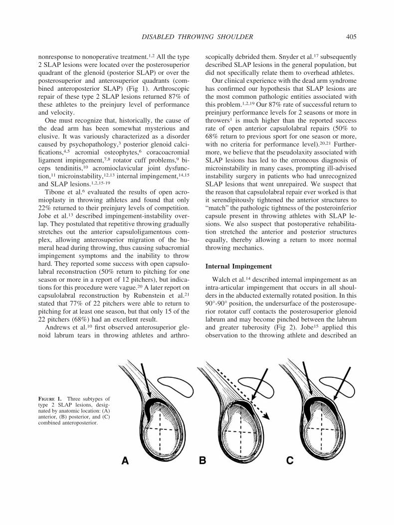
nonresponse to nonoperative treatment.1,2 All the type2 SLAP lesions were located over the posterosuperiorquadrant of the glenoid (posterior SLAP) or over theposterosuperior and anterosuperior quadrants (com-bined anteroposterior SLAP) (Fig 1). Arthroscopicrepair of these type 2 SLAP lesions returned 87% ofthese athletes to the preinjury level of performanceand velocity.
One must recognize that, historically, the cause ofthe dead arm has been somewhat mysterious andelusive. It was variously characterized as a disordercaused by psychopathology,3 posterior glenoid calci-fications,4,5 acromial osteophytes,6 coracoacromialligament impingement,7,8 rotator cuff problems,9 bi-ceps tendinitis,10 acromioclavicular joint dysfunc-tion,11 microinstability,12,13 internal impingement,14,15
and SLAP lesions.1,2,15-19
Tibone et al.6 evaluated the results of open acro-mioplasty in throwing athletes and found that only22% returned to their preinjury levels of competition.Jobe et al.13 described impingement-instability over-lap. They postulated that repetitive throwing graduallystretches out the anterior capsuloligamentous com-plex, allowing anterosuperior migration of the hu-meral head during throwing, thus causing subacromialimpingement symptoms and the inability to throwhard. They reported some success with open capsulo-labral reconstruction (50% return to pitching for oneseason or more in a report of 12 pitchers), but indica-tions for this procedure were vague.20 A later report oncapsulolabral reconstruction by Rubenstein et al.21
stated that 77% of 22 pitchers were able to return topitching for at least one season, but that only 15 of the22 pitchers (68%) had an excellent result.
Andrews et al.10 first observed anterosuperior gle-noid labrum tears in throwing athletes and arthro-
scopically debrided them. Snyder et al.17 subsequentlydescribed SLAP lesions in the general population, butdid not specifically relate them to overhead athletes.
Our clinical experience with the dead arm syndromehas confirmed our hypothesis that SLAP lesions arethe most common pathologic entities associated withthis problem.1,2,19 Our 87% rate of successful return topreinjury performance levels for 2 seasons or more inthrowers1 is much higher than the reported successrate of open anterior capsulolabral repairs (50% to68% return to previous sport for one season or more,with no criteria for performance level).20,21 Further-more, we believe that the pseudolaxity associated withSLAP lesions has led to the erroneous diagnosis ofmicroinstability in many cases, prompting ill-advisedinstability surgery in patients who had unrecognizedSLAP lesions that went unrepaired. We suspect thatthe reason that capsulolabral repair ever worked is thatit serendipitously tightened the anterior structures to“match” the pathologic tightness of the posteroinferiorcapsule present in throwing athletes with SLAP le-sions. We also suspect that postoperative rehabilita-tion stretched the anterior and posterior structuresequally, thereby allowing a return to more normalthrowing mechanics.
Internal Impingement
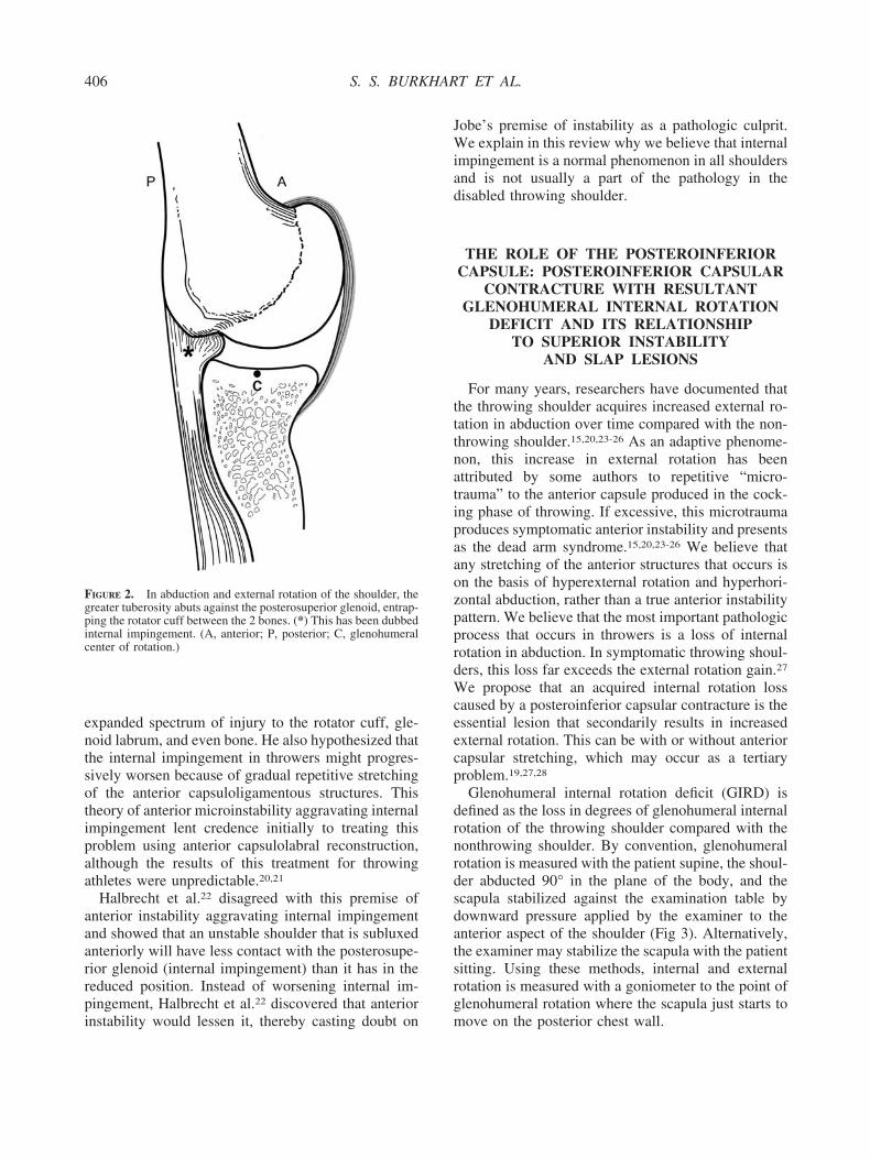
Walch et al.14 described internal impingement as anintra-articular impingement that occurs in all shoul-ders in the abducted externally rotated position. In this90°-90° position, the undersurface of the posterosupe-rior rotator cuff contacts the posterosuperior glenoidlabrum and may become pinched between the labrumand greater tuberosity (Fig 2). Jobe15 applied thisobservation to the throwing athlete and described an
FIGURE 1. Three subtypes oftype 2 SLAP lesions, desig-nated by anatomic location: (A)anterior, (B) posterior, and (C)combined anteroposterior.
405DISABLED THROWING SHOULDER
expanded spectrum of injury to the rotator cuff, gle-noid labrum, and even bone. He also hypothesized thatthe internal impingement in throwers might progres-sively worsen because of gradual repetitive stretchingof the anterior capsuloligamentous structures. Thistheory of anterior microinstability aggravating internalimpingement lent credence initially to treating thisproblem using anterior capsulolabral reconstruction,although the results of this treatment for throwingathletes were unpredictable.20,21
Halbrecht et al.22 disagreed with this premise ofanterior instability aggravating internal impingementand showed that an unstable shoulder that is subluxedanteriorly will have less contact with the posterosupe-rior glenoid (internal impingement) than it has in thereduced position. Instead of worsening internal im-pingement, Halbrecht et al.22 discovered that anteriorinstability would lessen it, thereby casting doubt on
Jobe’s premise of instability as a pathologic culprit.We explain in this review why we believe that internalimpingement is a normal phenomenon in all shouldersand is not usually a part of the pathology in thedisabled throwing shoulder.
THE ROLE OF THE POSTEROINFERIORCAPSULE: POSTEROINFERIOR CAPSULAR
CONTRACTURE WITH RESULTANTGLENOHUMERAL INTERNAL ROTATION
DEFICIT AND ITS RELATIONSHIPTO SUPERIOR INSTABILITY
AND SLAP LESIONS
For many years, researchers have documented thatthe throwing shoulder acquires increased external ro-tation in abduction over time compared with the non-throwing shoulder.15,20,23-26 As an adaptive phenome-non, this increase in external rotation has beenattributed by some authors to repetitive “micro-trauma” to the anterior capsule produced in the cock-ing phase of throwing. If excessive, this microtraumaproduces symptomatic anterior instability and presentsas the dead arm syndrome.15,20,23-26 We believe thatany stretching of the anterior structures that occurs ison the basis of hyperexternal rotation and hyperhori-zontal abduction, rather than a true anterior instabilitypattern. We believe that the most important pathologicprocess that occurs in throwers is a loss of internalrotation in abduction. In symptomatic throwing shoul-ders, this loss far exceeds the external rotation gain.27
We propose that an acquired internal rotation losscaused by a posteroinferior capsular contracture is theessential lesion that secondarily results in increasedexternal rotation. This can be with or without anteriorcapsular stretching, which may occur as a tertiaryproblem.19,27,28
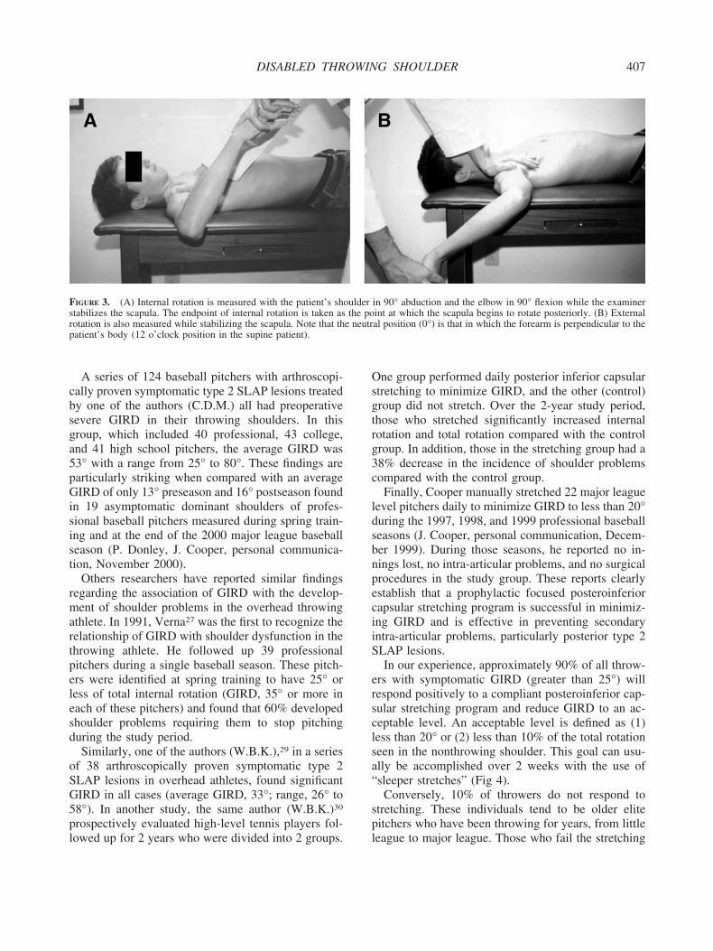
Glenohumeral internal rotation deficit (GIRD) isdefined as the loss in degrees of glenohumeral internalrotation of the throwing shoulder compared with thenonthrowing shoulder. By convention, glenohumeralrotation is measured with the patient supine, the shoul-der abducted 90° in the plane of the body, and thescapula stabilized against the examination table bydownward pressure applied by the examiner to theanterior aspect of the shoulder (Fig 3). Alternatively,the examiner may stabilize the scapula with the patientsitting. Using these methods, internal and externalrotation is measured with a goniometer to the point ofglenohumeral rotation where the scapula just starts tomove on the posterior chest wall.
FIGURE 2. In abduction and external rotation of the shoulder, thegreater tuberosity abuts against the posterosuperior glenoid, entrap-ping the rotator cuff between the 2 bones. (*) This has been dubbedinternal impingement. (A, anterior; P, posterior; C, glenohumeralcenter of rotation.)
406 S. S. BURKHART ET AL.
A series of 124 baseball pitchers with arthroscopi-cally proven symptomatic type 2 SLAP lesions treatedby one of the authors (C.D.M.) all had preoperativesevere GIRD in their throwing shoulders. In thisgroup, which included 40 professional, 43 college,and 41 high school pitchers, the average GIRD was53° with a range from 25° to 80°. These findings areparticularly striking when compared with an averageGIRD of only 13° preseason and 16° postseason foundin 19 asymptomatic dominant shoulders of profes-sional baseball pitchers measured during spring train-ing and at the end of the 2000 major league baseballseason (P. Donley, J. Cooper, personal communica-tion, November 2000).
Others researchers have reported similar findingsregarding the association of GIRD with the develop-ment of shoulder problems in the overhead throwingathlete. In 1991, Verna27 was the first to recognize therelationship of GIRD with shoulder dysfunction in thethrowing athlete. He followed up 39 professionalpitchers during a single baseball season. These pitch-ers were identified at spring training to have 25° orless of total internal rotation (GIRD, 35° or more ineach of these pitchers) and found that 60% developedshoulder problems requiring them to stop pitchingduring the study period.
Similarly, one of the authors (W.B.K.),29 in a seriesof 38 arthroscopically proven symptomatic type 2SLAP lesions in overhead athletes, found significantGIRD in all cases (average GIRD, 33°; range, 26° to58°). In another study, the same author (W.B.K.)30
prospectively evaluated high-level tennis players fol-lowed up for 2 years who were divided into 2 groups.
One group performed daily posterior inferior capsularstretching to minimize GIRD, and the other (control)group did not stretch. Over the 2-year study period,those who stretched significantly increased internalrotation and total rotation compared with the controlgroup. In addition, those in the stretching group had a38% decrease in the incidence of shoulder problemscompared with the control group.
Finally, Cooper manually stretched 22 major leaguelevel pitchers daily to minimize GIRD to less than 20°during the 1997, 1998, and 1999 professional baseballseasons (J. Cooper, personal communication, Decem-ber 1999). During those seasons, he reported no in-nings lost, no intra-articular problems, and no surgicalprocedures in the study group. These reports clearlyestablish that a prophylactic focused posteroinferiorcapsular stretching program is successful in minimiz-ing GIRD and is effective in preventing secondaryintra-articular problems, particularly posterior type 2SLAP lesions.
In our experience, approximately 90% of all throw-ers with symptomatic GIRD (greater than 25°) willrespond positively to a compliant posteroinferior cap-sular stretching program and reduce GIRD to an ac-ceptable level. An acceptable level is defined as (1)less than 20° or (2) less than 10% of the total rotationseen in the nonthrowing shoulder. This goal can usu-ally be accomplished over 2 weeks with the use of“sleeper stretches” (Fig 4).
Conversely, 10% of throwers do not respond tostretching. These individuals tend to be older elitepitchers who have been throwing for years, from littleleague to major league. Those who fail the stretching
FIGURE 3. (A) Internal rotation is measured with the patient’s shoulder in 90° abduction and the elbow in 90° flexion while the examinerstabilizes the scapula. The endpoint of internal rotation is taken as the point at which the scapula begins to rotate posteriorly. (B) Externalrotation is also measured while stabilizing the scapula. Note that the neutral position (0°) is that in which the forearm is perpendicular to thepatient’s body (12 o’clock position in the supine patient).
407DISABLED THROWING SHOULDER
program tend to be on the severe end of the GIRDspectrum and to have had chronic long-standingsymptoms usually associated with intra-articular pa-thology (type 2 posterior SLAP lesions). Patients whodo not respond to stretch have been treated by oneauthor (C.D.M.) with an arthroscopic selective pos-teroinferior capsulotomy, which in most instances isperformed concomitantly with SLAP lesion repair(Fig 5).
Typical arthroscopic findings in these patientsinclude a severely contracted and thickened pos-teroinferior recess and capsule in the zone of theposterior band of the inferior glenohumeral liga-
ment (IGHL) complex (Fig 6). In most cases, thecapsule in this zone will be found to be 6 mm thickor more. If a selective posteroinferior capsulotomyis performed, one can expect an immediate 65°increase in glenohumeral internal rotation (Fig 5).This must be maintained by an immediate postop-erative internal rotation stretching program to pre-vent the capsulotomy gap from closing during thehealing phase. It is important to understand that it isextremely unusual for high school and collegepitchers to be nonresponsive to stretching. We haverarely needed selective posteroinferior capsulotomyin these younger pitchers.
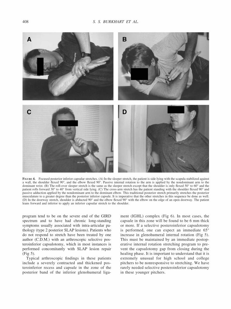
FIGURE 4. Focused posterior inferior capsular stretches. (A) In the sleeper stretch, the patient is side lying with the scapula stabilized againsta wall, the shoulder flexed 90°, and the elbow flexed 90°. Passive internal rotation to the arm is applied by the nondominant arm to thedominant wrist. (B) The roll-over sleeper stretch is the same as the sleeper stretch except that the shoulder is only flexed 50° to 60° and thepatient rolls forward 30° to 40° from vertical side lying. (C) The cross-arm stretch has the patient standing with the shoulder flexed 90° andpassive adduction applied by the nondominant arm to the dominant elbow. This traditional posterior stretch primarily stretches the posteriormusculature to a greater degree than the posterior inferior capsule. It is imperative that the other stretches in this sequence be done as well.(D) In the doorway stretch, shoulder is abducted 90° and the elbow flexed 90° with the elbow on the edge of an open doorway. The patientleans forward and inferior to apply an inferior capsular stretch to the shoulder.
408 S. S. BURKHART ET AL.
The Tethered Shoulder: The Reciprocal CableModel and the Cam Effect
O’Brien et al.31 popularized the concept of theIGHL complex, bounded by an anterior band andposterior band, performing like a hammock to supportthe humeral head when the arm is in abduction (Fig 7).One must remember that throwing is a dynamic ac-tivity, and the position of a given structure will con-tinually shift during the throwing cycle. For example,in full abduction and external rotation (the cockedposition), the posterior band of the IGHL is below thehumeral head. If the posterior band is contracted, itwill exert a posterosuperior force on the humeral head.From a mechanical standpoint, this hammock modelof the IGHL complex can be simplified even further sothat the IGHL complex is represented by 2 dominantstructural components, the anterior band and the pos-terior band, functioning as interdependent cables (Fig 8).
In this model, the primary passive constraints of theglenohumeral joint can be represented simply as asystem composed of 2 cables that develop tensionreciprocally and equally as the shoulder internally andexternally rotates in the 90° abducted position. Theglenoid serves as a tension ring for the cables as theyspan the distance from their humeral attachments tothe glenoid.32 This reciprocal cable model defines theallowable “envelope of motion” of the shoulder in
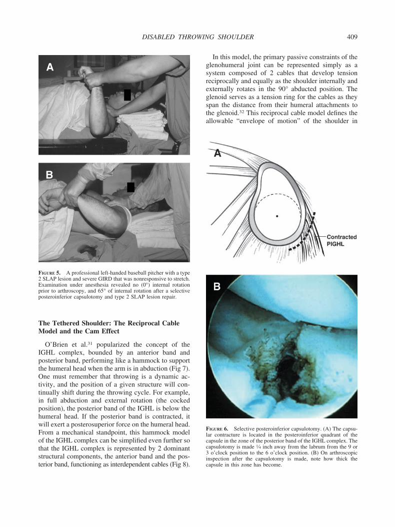
FIGURE 6. Selective posteroinferior capsulotomy. (A) The capsu-lar contracture is located in the posteroinferior quadrant of thecapsule in the zone of the posterior band of the IGHL complex. Thecapsulotomy is made 1⁄4 inch away from the labrum from the 9 or3 o’clock position to the 6 o’clock position. (B) On arthroscopicinspection after the capsulotomy is made, note how thick thecapsule in this zone has become.
FIGURE 5. A professional left-handed baseball pitcher with a type2 SLAP lesion and severe GIRD that was nonresponsive to stretch.Examination under anesthesia revealed no (0°) internal rotationprior to arthroscopy, and 65° of internal rotation after a selectiveposteroinferior capsulotomy and type 2 SLAP lesion repair.
409DISABLED THROWING SHOULDER
much the same way that the 4-bar linkage modeldefines allowable knee motion based on cruciate re-straints.33,34 With external rotation of the humerusabout its central contact point on the glenoid (theglenoid “bare spot”), the cables tighten and developtension equally as they assume an oblique courseacross their allowable envelope of motion (Fig 9).
If the posterior cable is shortened, simulating acontracted posterior band, it acts as a tether, shiftingthe glenohumeral contact point posterosuperiorly dur-ing combined abduction and external rotation (Fig10). This shift occurs because the shortened posteriorcable reaches its maximum elongation with glenohu-meral external rotation before the anterior cable max-
imally elongates so that the anterior band is still per-mitting external rotation anteriorly even though theposterior band is tethering the shoulder from its loca-tion beneath the humeral head, where it also exerts aposterosuperior force on the humerus. Because the arcof motion of the greater tuberosity has now shiftedposterosuperiorly, it no longer abuts against the usualsegment of the posterosuperior glenoid in combinedabduction and external rotation, and additional exter-nal rotation can be obtained.
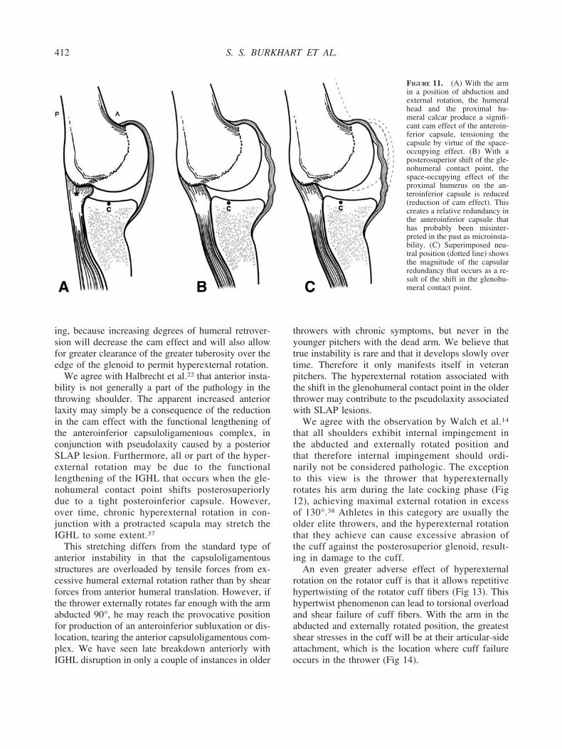
Furthermore, the cam effect of the humeral headand the proximal humeral calcar on the anteroinferiorcapsule is reduced by that shift, because the anteroin-ferior capsule is no longer tightly draped across thecalcar after the shift occurs (Fig 11). In this way, by aposterosuperior shift of the glenohumeral contactpoint, hyperexternal rotation is preserved and even
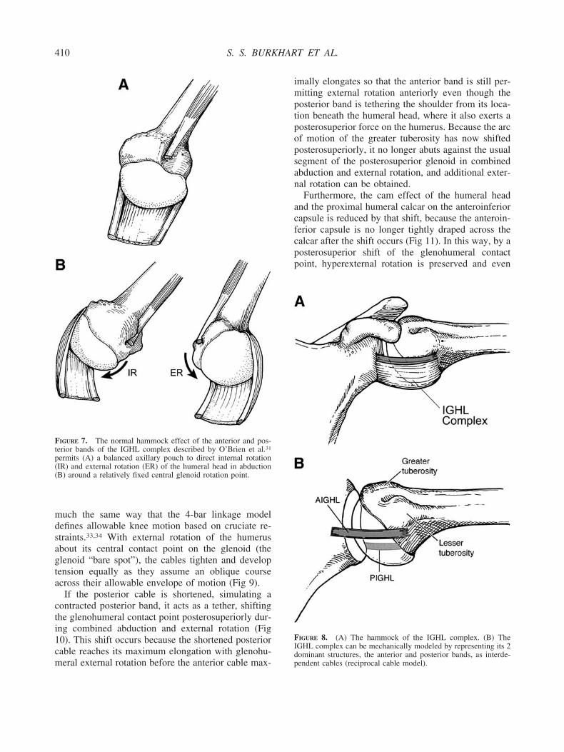
FIGURE 8. (A) The hammock of the IGHL complex. (B) TheIGHL complex can be mechanically modeled by representing its 2dominant structures, the anterior and posterior bands, as interde-pendent cables (reciprocal cable model).
FIGURE 7. The normal hammock effect of the anterior and pos-terior bands of the IGHL complex described by O’Brien et al.31
permits (A) a balanced axillary pouch to direct internal rotation(IR) and external rotation (ER) of the humeral head in abduction(B) around a relatively fixed central glenoid rotation point.
410 S. S. BURKHART ET AL.
potentially increased in the face of a shortened poste-rior band. The cam effect of the humeral head is alsoreduced, creating a relative redundancy in the antero-inferior capsule.
Recently, a study from the biomechanics laboratoryat Temple University used cadaveric shoulderstracked by electromagnetic sensors. They determinedto within 1 mm the relationship of the humeral head tothe articular surface of the glenoid.35 These investiga-tors placed the shoulders in maximum abduction andexternal rotation and evaluated the glenohumeral spa-tial relationships in this position both before and afterposteroinferior capsular plication. Their results clearlydocumented a posterosuperior shift of the humeralhead on the glenoid face of approximately 4.4 mm inthe presence of a posteroinferior capsular plication.
Hyperexternal Rotation of the Humerus:Tuberosity Clearance and Minimizationof the Cam Effect
There are 2 mechanisms by which a tight postero-inferior capsule allows hyperexternal rotation of thehumerus. First, the tethering effect of the shortenedposterior capsule shifts the glenohumeral contact pointposterosuperiorly, allowing the greater tuberosity toclear the glenoid rim through a greater arc of external
rotation before internal impingement occurs (Fig 10).Second, the shift in the glenohumeral contact pointminimizes the cam effect of the proximal humerus onthe anteroinferior capsule to allow greater externalrotation due to the redundancy in the capsule (Fig 11).
To understand this cam effect, one must recognizethat combined abduction and external rotation causesthe anteroinferior capsule to drape tightly across theprotuberant inferior articular surface of the humerus,which is quite prominent due to its location adjacent tothe arc of the humeral calcar. When the contact pointis shifted posterosuperiorly in a thrower’s shoulder,the cam effect of the humeral head is dramaticallydecreased. As a result, the anteroinferior capsule is nolonger tightly draped across a protruding humeralhead (Fig 11). The loosened capsule is, in effect,functionally lengthened by virtue of the change inposition of the contact point, allowing a greater degreeof external rotation. One author (S.S.B.) has con-firmed anatomically the functional lengthening of thecapsule that occurs with a posterosuperior shift of theglenohumeral contact point (unpublished data).
Crockett et al.36 noted increased humeral retrover-sion in the dominant shoulders of professional base-ball pitchers in association with increased externalrotation at 90° abduction. This finding is not surpris-
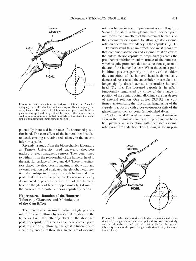
FIGURE 10. When the posterior cable shortens (contracted poste-rior band), the glenohumeral contact point shifts posterosuperiorlyand the allowable arc of external rotation (before the greatertuberosity contacts the posterior glenoid) significantly increases(dotted lines).
FIGURE 9. With abduction and external rotation, the 2 cablesobliquely cross the shoulder as they reciprocally and equally de-velop tension. The center of rotation remains approximately at theglenoid bare spot and the greater tuberosity of the humerus has awell-defined circular arc (dotted line) before it contacts the poste-rior glenoid (internal impingement position).
411DISABLED THROWING SHOULDER
ing, because increasing degrees of humeral retrover-sion will decrease the cam effect and will also allowfor greater clearance of the greater tuberosity over theedge of the glenoid to permit hyperexternal rotation.
We agree with Halbrecht et al.22 that anterior insta-bility is not generally a part of the pathology in thethrowing shoulder. The apparent increased anteriorlaxity may simply be a consequence of the reductionin the cam effect with the functional lengthening ofthe anteroinferior capsuloligamentous complex, inconjunction with pseudolaxity caused by a posteriorSLAP lesion. Furthermore, all or part of the hyper-external rotation may be due to the functionallengthening of the IGHL that occurs when the gle-nohumeral contact point shifts posterosuperiorlydue to a tight posteroinferior capsule. However,over time, chronic hyperexternal rotation in con-junction with a protracted scapula may stretch theIGHL to some extent.37
This stretching differs from the standard type ofanterior instability in that the capsuloligamentousstructures are overloaded by tensile forces from ex-cessive humeral external rotation rather than by shearforces from anterior humeral translation. However, ifthe thrower externally rotates far enough with the armabducted 90°, he may reach the provocative positionfor production of an anteroinferior subluxation or dis-location, tearing the anterior capsuloligamentous com-plex. We have seen late breakdown anteriorly withIGHL disruption in only a couple of instances in older
throwers with chronic symptoms, but never in theyounger pitchers with the dead arm. We believe thattrue instability is rare and that it develops slowly overtime. Therefore it only manifests itself in veteranpitchers. The hyperexternal rotation associated withthe shift in the glenohumeral contact point in the olderthrower may contribute to the pseudolaxity associatedwith SLAP lesions.
We agree with the observation by Walch et al.14
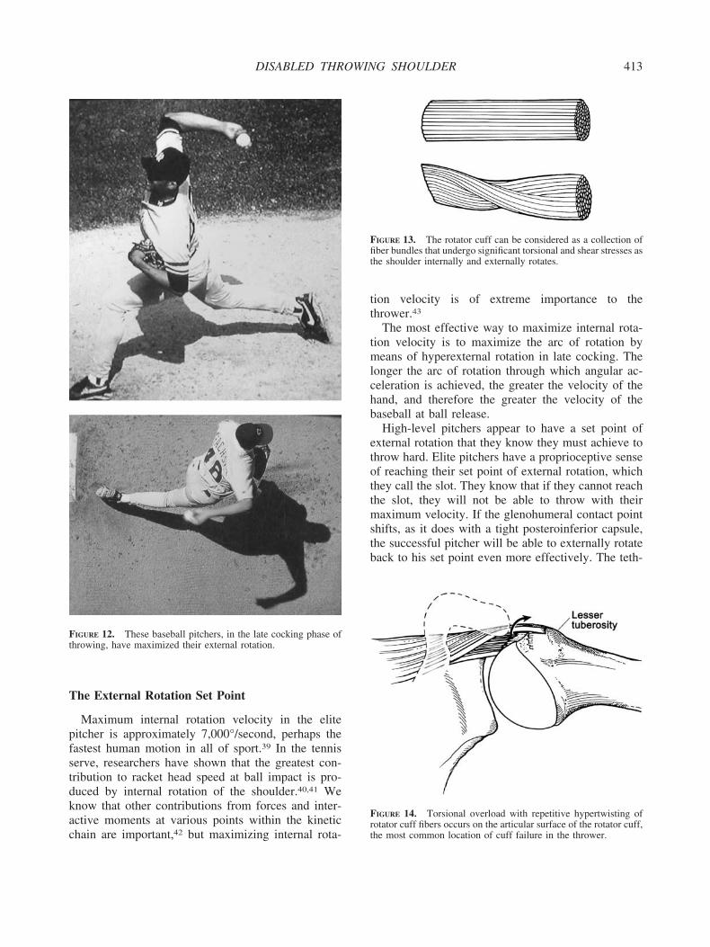
that all shoulders exhibit internal impingement inthe abducted and externally rotated position andthat therefore internal impingement should ordi-narily not be considered pathologic. The exceptionto this view is the thrower that hyperexternallyrotates his arm during the late cocking phase (Fig12), achieving maximal external rotation in excessof 130°.38 Athletes in this category are usually theolder elite throwers, and the hyperexternal rotationthat they achieve can cause excessive abrasion ofthe cuff against the posterosuperior glenoid, result-ing in damage to the cuff.
An even greater adverse effect of hyperexternalrotation on the rotator cuff is that it allows repetitivehypertwisting of the rotator cuff fibers (Fig 13). Thishypertwist phenomenon can lead to torsional overloadand shear failure of cuff fibers. With the arm in theabducted and externally rotated position, the greatestshear stresses in the cuff will be at their articular-sideattachment, which is the location where cuff failureoccurs in the thrower (Fig 14).
FIGURE 11. (A) With the armin a position of abduction andexternal rotation, the humeralhead and the proximal hu-meral calcar produce a signifi-cant cam effect of the anteroin-ferior capsule, tensioning thecapsule by virtue of the space-occupying effect. (B) With aposterosuperior shift of the gle-nohumeral contact point, thespace-occupying effect of theproximal humerus on the an-teroinferior capsule is reduced(reduction of cam effect). Thiscreates a relative redundancy inthe anteroinferior capsule thathas probably been misinter-preted in the past as microinsta-bility. (C) Superimposed neu-tral position (dotted line) showsthe magnitude of the capsularredundancy that occurs as a re-sult of the shift in the glenohu-meral contact point.
412 S. S. BURKHART ET AL.
The External Rotation Set Point
Maximum internal rotation velocity in the elitepitcher is approximately 7,000°/second, perhaps thefastest human motion in all of sport.39 In the tennisserve, researchers have shown that the greatest con-tribution to racket head speed at ball impact is pro-duced by internal rotation of the shoulder.40,41 Weknow that other contributions from forces and inter-active moments at various points within the kineticchain are important,42 but maximizing internal rota-
tion velocity is of extreme importance to thethrower.43
The most effective way to maximize internal rota-tion velocity is to maximize the arc of rotation bymeans of hyperexternal rotation in late cocking. Thelonger the arc of rotation through which angular ac-celeration is achieved, the greater the velocity of thehand, and therefore the greater the velocity of thebaseball at ball release.
High-level pitchers appear to have a set point ofexternal rotation that they know they must achieve tothrow hard. Elite pitchers have a proprioceptive senseof reaching their set point of external rotation, whichthey call the slot. They know that if they cannot reachthe slot, they will not be able to throw with theirmaximum velocity. If the glenohumeral contact pointshifts, as it does with a tight posteroinferior capsule,the successful pitcher will be able to externally rotateback to his set point even more effectively. The teth-
FIGURE 12. These baseball pitchers, in the late cocking phase ofthrowing, have maximized their external rotation.
FIGURE 13. The rotator cuff can be considered as a collection offiber bundles that undergo significant torsional and shear stresses asthe shoulder internally and externally rotates.
FIGURE 14. Torsional overload with repetitive hypertwisting ofrotator cuff fibers occurs on the articular surface of the rotator cuff,the most common location of cuff failure in the thrower.
413DISABLED THROWING SHOULDER
ering effect of the posteroinferior capsule, with theconcomitant shift of the glenohumeral contact point,allows clearance of the greater tuberosity to achieve agreater arc of external rotation. As a result, internalimpingement does not occur until the shoulderachieves a position of hyperexternal rotation. Further-more, posterosuperior shift of the glenohumeral con-tact point lessens the cam effect of the proximal hu-merus in abduction and external rotation, achieving afunctional lengthening of the anteroinferior capsulethat permits greater external rotation.
Pitchers with a tight posteroinferior capsule andGIRD know that they must reach their set point ofexternal rotation, and they will find a way to do it eventhough the deranged mechanics predispose to superiorlabral injury by virtue of increased peel-back forcesand increased shear forces on the labrum. Such pitch-ers are constantly on the brink of injury. In this type ofoverhead athlete, the thinking brain recognizes thatthe arm must be brought back to a certain position (theset point), and the acting brain finds a way to get itthere.
High-level overhead athletes have been shown tocombine ballistic and tracking modes in achievingcontrol with high velocity.44 Ballistic movement is anautomatic movement under preprogrammed neuralcontrol (such as externally rotating to the set point)that can be modified and facilitated by muscle activa-tion in response to feedback from receptors on mus-cles and tendons (tracking mode) to fine-tune thecontrol aspect of throwing a ball to a specific targetpoint. These preprogrammed patterns start from thelegs and trunk, then proceed to the scapular stabilizersand arm positioners for force generation. From there,these patterns are coordinated at the elbow and wristfor pitch control.
Peel-Back Mechanism
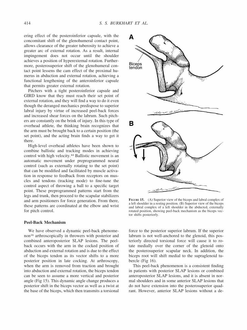
We have observed a dynamic peel-back phenome-non16 arthroscopically in throwers with posterior andcombined anteroposterior SLAP lesions. The peel-back occurs with the arm in the cocked position ofabduction and external rotation and is due to the effectof the biceps tendon as its vector shifts to a moreposterior position in late cocking. At arthroscopy,when the arm is removed from traction and broughtinto abduction and external rotation, the biceps tendoncan be seen to assume a more vertical and posteriorangle (Fig 15). This dynamic angle change produces aposterior shift in the biceps vector as well as a twist atthe base of the biceps, which then transmits a torsional
force to the posterior superior labrum. If the superiorlabrum is not well-anchored to the glenoid, this pos-teriorly directed torsional force will cause it to ro-tate medially over the corner of the glenoid ontothe posterosuperior scapular neck. In addition, thebiceps root will shift medial to the supraglenoid tu-bercle (Fig 16).
This peel-back phenomenon is a consistent findingin patients with posterior SLAP lesions or combinedanteroposterior SLAP lesions, and it is absent in nor-mal shoulders and in some anterior SLAP lesions thatdo not have extension into the posterosuperior quad-rant. However, anterior SLAP lesions without a de-
FIGURE 15. (A) Superior view of the biceps and labral complex ofa left shoulder in a resting position. (B) Superior view of the bicepsand labral complex of a left shoulder in the abducted, externallyrotated position, showing peel-back mechanism as the biceps vec-tor shifts posteriorly.
414 S. S. BURKHART ET AL.
monstrable peel-back sign are not usually seen inthrowers. The typical “thrower’s SLAP” has posteriorextension of the lesion and a positive peel-back sign.
A successful SLAP repair in a throwing athlete
must eliminate the peel-back sign as evidence that thistorsional force has been neutralized. A suture anchorwith a simple suture loop around the labrum developstensile forces within the suture loop to efficiently andeffectively resist the torsional force of the peel-backmechanism. However, translabral tacks are mechani-cally less effective in resisting the torsional peel-backbecause they have only a single point of contact at theperiphery of the labrum.45 Morgan et al.1 reported a97% success rate with suture anchors compared withreported success rates ranging from 71% to 88% withabsorbable translabral tacks.46-49 This higher successrate for the suture anchor technique is not surprising inview of the superior mechanical characteristics ofsuture anchors in resisting torsional forces.
Acceleration Versus Deceleration Injury
Andrews et al.10 postulated a deceleration mecha-nism for labral injuries in throwers as the bicepscontracts to slow down the rapidly extending elbow infollow through. They suggested that this mechanismcreates a high tensile load in the biceps that acts to pullthe biceps and superior labrum complex from thebone. In contrast, we have postulated an accelerationmechanism. In fact, we have found that throwers whorecall the pitch that caused their injury invariablyrelate the severe sudden onset of pain to the abductedand externally rotated position of late cocking, as thearm begins to accelerate forward.
Kuhn et al.50 performed an experimental compari-son of these 2 mechanisms (acceleration and deceler-ation) in a cadaver model. To simulate the decelera-tion mechanism, they applied a tensile force throughthe biceps with the arm in the follow-through position.They were able to produce a superior labral avulsionin only 20% of specimens, and only with a largetensile force (346 � 40 N). To simulate the acceler-ation mechanism, they loaded the biceps of cadaverspecimens in the abducted, externally rotated positionof late cocking and consistently produced a type 2SLAP lesion at a force of 289 � 39 N, 20% less thanthe force required to produce a SLAP lesion by thedeceleration mechanism. Importantly, they were ableto produce type 2 SLAP lesions in 9 of 10 specimensin the abducted, externally rotated position and in only2 of 10 of those in the deceleration position (P �.055). In view of this clinical and experimental evi-dence, we believe that the biceps and superior labrumcomplex is not pulled from bone, but rather is peeledfrom bone.
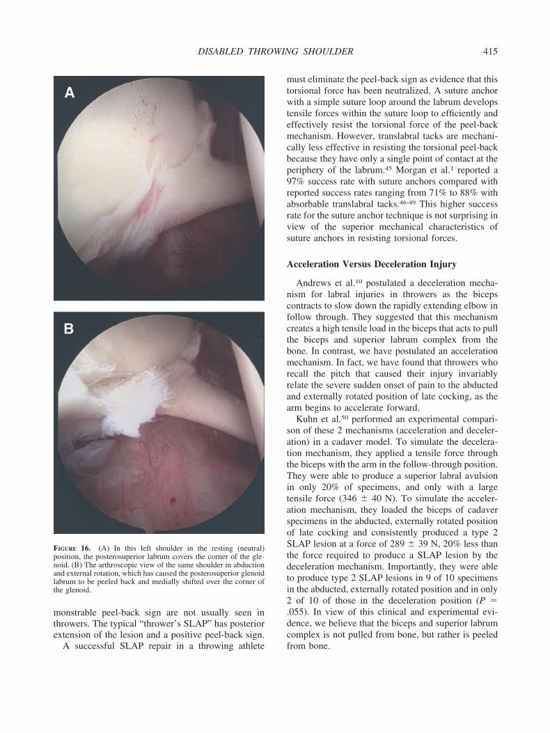
FIGURE 16. (A) In this left shoulder in the resting (neutral)position, the posterosuperior labrum covers the corner of the gle-noid. (B) The arthroscopic view of the same shoulder in abductionand external rotation, which has caused the posterosuperior glenoidlabrum to be peeled back and medially shifted over the corner ofthe glenoid.
415DISABLED THROWING SHOULDER
THE PATHOLOGIC CASCADE
We believe that the acquired posteroinferior capsu-lar contracture is the first and essential abnormalitythat initiates a pathologic cascade that climaxes in thelate cocking phase of throwing. At that point, the shiftin the glenohumeral contact point causes maximumshear stress on the posterosuperior labrum at exactlythe time when the peel-back force and the total forcebeing funneled into the shoulder by the kinetic chainare both at a maximum. This combination of factorsputs the shoulder in a very vulnerable situation.
In the presence of a contracted or shortened poste-rior band of the IGHL complex, the inferior axillarypouch structures are imbalanced and will not allow thenormal cradling or hammock effect described byO’Brien et al.31 This effect normally allows the shoul-der to wind and unwind in abduction around a rela-tively fixed central glenohumeral rotation point lo-cated in the lower half of the glenoid face.Arthroscopists have called this the bare spot. As theshoulder attempts to wind up into the cocked position,the contracted posterior band will not allow the headto fully externally rotate around the normal glenoidrotation point. It acts as a rein or tether that draws thehumeral head posterosuperiorly to a new rotationpoint on the glenoid.35
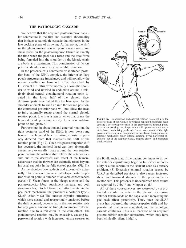
Furthermore, in abduction and external rotation, thetight posterior band of the IGHL is now bowstrungbeneath the humeral head, exerting a posterosuperi-orly directed force that maintains the shift of therotation point (Fig 17). Once this posterosuperior shifthas occurred, the humeral head can then abnormallyexcessively externally rotate around the new rotationpoint because the rotation shift relaxes the anterior cap-sule due to the decreased cam effect of the humeralcalcar such that the thrower can externally rotate beyondhis usual set point in the fully cocked position (Fig 11).
As the shoulder now abducts and excessively exter-nally rotates around this new pathologic posterosupe-rior rotation point, a number of adverse consequencesoccur. (1) Shear forces at the biceps anchor and theposterosuperior labral attachment increase, and bothstructures begin to fail from their attachments via thepeel back mechanism that produces a posterior type 2SLAP lesion.16 (2) The anterior capsular structures,which were normal and appropriately tensioned beforethe shift occurred, become lax in the new rotation axisfor any given amount of true glenohumeral externalrotation due to a reduction in the cam effect. Theglenohumeral rotation may be excessive, causing hy-perexternal rotation with increased tensile stresses on
the IGHL such that, if the patient continues to throw,the anterior capsule may begin to fail either in conti-nuity or at the labrum in the Bankart zone as a tertiaryproblem. (3) Excessive external rotation caused byGIRD as described previously also causes increasedshear and torsional stresses in the posterosuperiorrotator cuff. This presents as undersurface fiber failureas reported by Jobe15 and Morgan et al.1
All of these consequences are worsened by a pro-tracted scapula that antetilts the glenoid, increasinganterior tensile loads on the capsule and increasing thepeel-back effect posteriorly. Thus, once the SLAPevent has occurred, the posterosuperior shift and hy-perexternal rotation are magnified, and the pathologiccascade continues. This is all because of an acquiredposteroinferior capsular contracture, which may havebeen clinically silent initially.
FIGURE 17. In abduction and external rotation (late cocking), theposterior band of the IGHL is bowstrung beneath the humeral head,causing a posterosuperior shift in the glenohumeral rotation point.Also in late cocking, the biceps vector shifts posteriorly and twistsat its base, maximizing peel-back forces. As a result of the tightposteroinferior capsule, this pitcher shows classic derangements ofpitching mechanics: hyper-external rotation, hyper horizontal ab-duction (out of the scapular plane), dropped elbow, and prematuretrunk rotation.
416 S. S. BURKHART ET AL.
Production of the SLAP Lesion: the Coup DeGrace
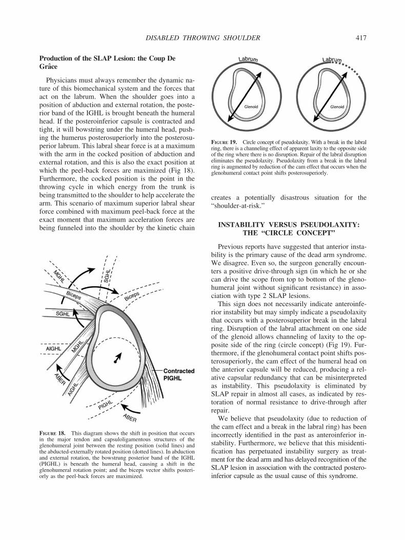
Physicians must always remember the dynamic na-ture of this biomechanical system and the forces thatact on the labrum. When the shoulder goes into aposition of abduction and external rotation, the poste-rior band of the IGHL is brought beneath the humeralhead. If the posteroinferior capsule is contracted andtight, it will bowstring under the humeral head, push-ing the humerus posterosuperiorly into the posterosu-perior labrum. This labral shear force is at a maximumwith the arm in the cocked position of abduction andexternal rotation, and this is also the exact position atwhich the peel-back forces are maximized (Fig 18).Furthermore, the cocked position is the point in thethrowing cycle in which energy from the trunk isbeing transmitted to the shoulder to help accelerate thearm. This scenario of maximum superior labral shearforce combined with maximum peel-back force at theexact moment that maximum acceleration forces arebeing funneled into the shoulder by the kinetic chain
creates a potentially disastrous situation for the“shoulder-at-risk.”
INSTABILITY VERSUS PSEUDOLAXITY:THE “CIRCLE CONCEPT”
Previous reports have suggested that anterior insta-bility is the primary cause of the dead arm syndrome.We disagree. Even so, the surgeon generally encoun-ters a positive drive-through sign (in which he or shecan drive the scope from top to bottom of the gleno-humeral joint without significant resistance) in asso-ciation with type 2 SLAP lesions.
This sign does not necessarily indicate anteroinfe-rior instability but may simply indicate a pseudolaxitythat occurs with a posterosuperior break in the labralring. Disruption of the labral attachment on one sideof the glenoid allows channeling of laxity to the op-posite side of the ring (circle concept) (Fig 19). Fur-thermore, if the glenohumeral contact point shifts pos-terosuperiorly, the cam effect of the humeral head onthe anterior capsule will be reduced, producing a rel-ative capsular redundancy that can be misinterpretedas instability. This pseudolaxity is eliminated bySLAP repair in almost all cases, as indicated by res-toration of normal resistance to drive-through afterrepair.
We believe that pseudolaxity (due to reduction ofthe cam effect and a break in the labral ring) has beenincorrectly identified in the past as anteroinferior in-stability. Furthermore, we believe that this misidenti-fication has perpetuated instability surgery as treat-ment for the dead arm and has delayed recognition of theSLAP lesion in association with the contracted postero-inferior capsule as the usual cause of this syndrome.
FIGURE 18. This diagram shows the shift in position that occursin the major tendon and capsuloligamentous structures of theglenohumeral joint between the resting position (solid lines) andthe abducted-externally rotated position (dotted lines). In abductionand external rotation, the bowstrung posterior band of the IGHL(PIGHL) is beneath the humeral head, causing a shift in theglenohumeral rotation point; and the biceps vector shifts posteri-orly as the peel-back forces are maximized.
FIGURE 19. Circle concept of pseudolaxity. With a break in the labralring, there is a channeling effect of apparent laxity to the opposite sideof the ring where there is no disruption. Repair of the labral disruptioneliminates the pseudolaxity. Pseudolaxity from a break in the labralring is augmented by reduction of the cam effect that occurs when theglenohumeral contact point shifts posterosuperiorly.
417DISABLED THROWING SHOULDER
We suspect that the limited success of the Jobeanterior capsulolabral repair in some throwers hasbeen due to its ability to create an anteroinferiorcapsular contracture that matched the posteroinferiorcapsular contracture, thereby reducing the posterosu-perior shift of the glenohumeral contact point thatbegins the pathologic cascade to the dead arm. Duringpostoperative rehabilitation, patients with these inju-ries would begin with a symmetrically over-tightenedshoulder that could then be symmetrically stretchedduring rehabilitation. Although such an approach canbe successful, the morbidity is significantly greaterand the predictability is much lower than with ourapproach.
Associated Rotator Cuff Tears
We found rotator cuff tears in 31% of throwers withSLAP lesions1; 38% of these tears were full thicknessand 62% were partial-thickness. The full-thicknesstears were located in the midportion of the rotatorcrescent, with varying degrees of anterior and poste-rior extension. The partial-thickness cuff tears were inlesion-specific anatomic locations; that is, the anteriorSLAP lesions were associated with partial-thicknessarticular-surface rotator cuff tears in the anterior por-tion of the rotator crescent, and the posterior SLAPlesions were associated with partial-thickness articularsurface rotator cuff tears in the posterior portion of therotator crescent.
Because of the location specificity of the partial-thickness cuff tears, we believe that repetitive tensileloading of specific areas of the cuff may occur causedby superior subluxation of the humerus in combina-tion with repetitive torsional loading from hyperexter-nal rotation. For posterior type 2 SLAP lesions, webelieve that the humerus subluxes posterosuperiorlybecause of the break in the labral ring, repetitivelyproducing high tensile forces in the posterosuperiorcuff. These forces may ultimately contribute to tearingof the rotator cuff tear. In addition, the hypertwistphenomenon caused by hyperexternal rotation of theshoulder can lead to torsional and shear overload withfatigue failure of cuff fibers in this same area of theposterosuperior cuff (Fig 14).
THE ULTIMATE CULPRIT
We believe that the culprits in development of thedead arm are (1) a tight posteroinferior capsule caus-ing a GIRD and a shift in the glenohumeral rotationpoint; (2) peel-back forces causing the SLAP lesion;
(3) hyper-external rotation of the humerus because ofa reduction in the humeral cam effect on the anteriorcapsule and clearance of the greater tuberosity overthe glenoid rim through a larger arc of external rota-tion before internal impingement occurs; and (4) scap-ular protraction. Of these, the ultimate culprit thatstarts the pathologic cascade is the tight posteroinfe-rior capsule. If we could prevent this from developing,we could prevent the dead arm.
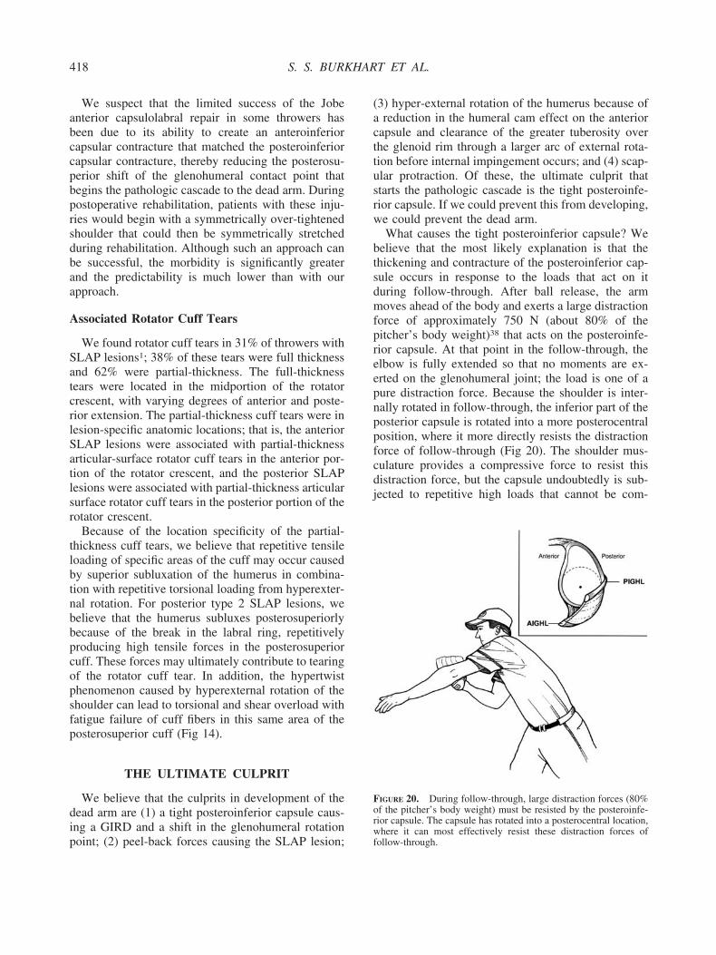
What causes the tight posteroinferior capsule? Webelieve that the most likely explanation is that thethickening and contracture of the posteroinferior cap-sule occurs in response to the loads that act on itduring follow-through. After ball release, the armmoves ahead of the body and exerts a large distractionforce of approximately 750 N (about 80% of thepitcher’s body weight)38 that acts on the posteroinfe-rior capsule. At that point in the follow-through, theelbow is fully extended so that no moments are ex-erted on the glenohumeral joint; the load is one of apure distraction force. Because the shoulder is inter-nally rotated in follow-through, the inferior part of theposterior capsule is rotated into a more posterocentralposition, where it more directly resists the distractionforce of follow-through (Fig 20). The shoulder mus-culature provides a compressive force to resist thisdistraction force, but the capsule undoubtedly is sub-jected to repetitive high loads that cannot be com-
FIGURE 20. During follow-through, large distraction forces (80%of the pitcher’s body weight) must be resisted by the posteroinfe-rior capsule. The capsule has rotated into a posterocentral location,where it can most effectively resist these distraction forces offollow-through.
418 S. S. BURKHART ET AL.
pletely resisted by the muscle forces. This repetitivetensile loading of the posteroinferior capsule couldcause the capsular hypertrophy that is so common inthrowing athletes. If this is the etiology of the thick-ened capsule, there may be nothing we can do tocompletely prevent it. Strengthening the rotator cuffand posterior shoulder musculature to resist the dis-traction force and minimize the load on the capsuleshould be beneficial, but repetitive loading in thefollow-through phase will still probably cause somedegree of adaptive hypertrophy of the posteroinferiorcapsule. Ironically, the inability to accelerate the ballin the dead arm syndrome may ultimately be due tothe inability of the muscles to effectively deceleratethe arm in follow-through.
Editor’s Note: Part II: Evaluation and Treatmentof SLAP Lesions in Throwers will appear in Vol. 19,No. 5, and Part III: The “SICK” Scapula, ScapularDyskinesis, the Kinetic Chain, and Rehabilitation willappear in Vol. 19, No. 6.
Acknowledgment: The authors thank Jeff Cooper, P.T.,A.T.C. (Head Athletic Trainer, Philadelphia Phillies) andPhil Donley, P.T., A.T.C., M.S. (consultant, PhiladelphiaPhillies) for their assistance in the preparation of this paper.
REFERENCES
1. Morgan CD, Burkhart SS, Palmeri M, et al. Type II SLAPlesions: Three subtypes and their relationship to superior in-stability and rotator cuff tears. Arthroscopy 1998;14:553-565.
2. Burkhart SS, Morgan CD. SLAP lesions in the overheadathlete. Orthop Clin North Am 2001;32:431-441.
3. Rowe CR, Pierce DS, Clark DG. Voluntary dislocation of theshoulder. J Bone Joint Surg Am 1973;55:445-410.
4. Bennett GE. Elbow and shoulder dislocations of baseball play-ers. Am J Surg 1959;98:484-488.
5. Lombardo SJ, Jobe FW, Kerlan RK, et al. Posterior shoulderlesions in throwing athletes. Am J Sports Med 1977;5:106-110.
6. Tibone JE, Jobe FW, Kerlan RF, et al. Shoulder impingementsyndrome in athletes treated by anterior acromioplasty. ClinOrthop 1985;188:134-140.
7. Kennedy JC, Hawkins RJ, Krusoff WB. Orthopedic manifes-tations of swimming. Am J Sports Med 1978;6:309-327.
8. Perry J. Anatomy and biomechanics of the shoulder in throwing,swimming, gymnastics, and tennis: Symposium on injuries to theshoulder in the athlete. Clin Sports Med 1983;2:247-270.
9. Tibone JE, Elrod B, Jobe FW, et al. Surgical treatment of tearsof the rotator cuff in athletes. J Bone Joint Surg Am 1986;68:887-891.
10. Andrews JR, Carson W Jr., McLeod W. Glenoid labrum tearsrelated to the long head of the biceps. Am J Sports Med1985;13:337-341.
11. Cook FF, Tibone JE. The Mumford procedure in athletes: Anobjective analysis of function. Am J Sports Med 1988;16:97-100.
12. Garth WP, Allman FL, Armstrong WS. Occult anterior sub-luxation of the shoulder. Am J Sports Med 1987;15:579-585.
13. Jobe FW, Tibone JE, Jobe CM, et al. The shoulder in sports.
In: Rockwood CA Jr., Matsen FA III, eds. The Shoulder.Philadelphia: WB Saunders, 1990;963-967.
14. Walch G, Boileau J, Noel E, et al. Impingement of the deepsurface of the supraspinatus tendon on the posterior superiorglenoid rim: An arthroscopic study. J Shoulder Elbow Surg1992;1:238-243.
15. Jobe CM. Posterior superior glenoid impingement: Expandedspectrum. Arthroscopy 1995;11:530-537.
16. Burkhart SS, Morgan CD. Technical note: The peel-backmechanism. Its role in producing and extending posterior typeII SLAP lesions and its effect on SLAP repair rehabilitation.Arthroscopy 1998;14:637-640.
17. Snyder SJ, Karzel RP, Delpizzo W, et al. SLAP lesions of theshoulder. Arthroscopy 1990;6:274-279.
18. Snyder SJ, Banas MP, Karzel RP. An analysis of 140 injuriesto the superior glenoid labrum. J Shoulder Elbow Surg 1995;4:243-248.
19. Burkhart SS, Morgan CD, Kibler WB. Shoulder injuries inoverhead athletes. Clin Sports Med 2000;19:125-158.
20. Jobe FW, Giangarra CE, Kvitne RS, et al. Anterior capsulo-labral reconstruction of the shoulder in athletes in overheadsports. Am J Sports Med 1991;19:428-434.
21. Rubenstein DL, Jobe FW, Glousman RE, et al. Anterior cap-sulolabral reconstruction of the shoulder in athletes. J ShoulderElbow Surg 1992;1:229-237.
22. Halbrecht JL, Tirman P, Atkin D. Internal impingement of theshoulder: Comparison of findings between the throwing andnonthrowing shoulders of college baseball players. Arthros-copy 1999;15:253-258.
23. Garth WP, Allman FL, Armstrong NS: Occult anterior sub-luxation of the shoulder. Am J Sports Med 1987;15:579-585.
24. Kvitne RS, Jobe FW. The diagnosis and treatment of anteriorinstability in the throwing athlete. Clin Orthop 1993;291:107-123.
25. Jobe CM, Pink MM, Jobe FW, Shaffer B. Anterior shoulderinstability, impingement, and rotator cuff tear: Theories andconcepts. In: Jobe FW, ed. Operative techniques in upperextremity sports injuries. St. Louis: Mosby, 1996;164-176.
26. Glousman RE, Jobe FW. Anterior shoulder instability: Im-pingement and rotator cuff tear. Anterior and multidirectionalinstability. In: Jobe FW, ed. Operative techniques in upperextremity sports injuries. St. Louis: Mosby, 1996;191-209.
27. Verna C. Shoulder flexibility to reduce impingement. Pre-sented at the 3rd Annual PBATS (Professional Baseball Ath-letic Trainer Society) Meeting, Mesa, Arizona, March, 1991.
28. Morgan CD. The thrower’s shoulder. In: McGinty JB, ed.Operative arthroscopy. Ed 3. Philadelphia: Lippincott, 2003;570-584.
29. Kibler WB. SLAP lesions and their relationship to glenohu-meral internal rotation deficit. Am J Sports Med (submitted).
30. Kibler WB. The relationship of glenohumeral internal rotationdeficit to shoulder and elbow injuries in tennis players: Aprospective evaluation of posterior capsular stretching. Pre-sented at the Annual closed meeting of the American Shoulderand Elbow Surgeons, New York, October 1998.
31. O’Brien SJ, Neves MC, Arnoczky SP, et al. The anatomy andhistology of the inferior glenohumeral ligament complex of theshoulder. Am J Sports Med 1990;18:449-456.
32. Leet KM. Fundamentals of structural analysis. New York:MacMillan Publishing, 1988;138.
33. Huson A. Biomechanische probleme des knielgelenks. Ortho-pade 1974;3:119-126.
34. Menschik A. Mechanik des knielgelenks: Teil 1. Z Orthop1974;113:481-495.
35. Koffler KM, Bader D, Eager M, et al. The effect of posteriorcapsule tightness on glenohumeral translation in the late cock-ing phase of pitching: A cadaveric study. The 21st AnnualMeeting of the Arthroscopy Association of North America,Washington, DC, April 25, 2002.
419DISABLED THROWING SHOULDER
36. Crockett HC, Gross LB, Wilk KE, et al. Osseous adaptationand range of motion at the glenohumeral joint in professionalbaseball pitchers. Am J Sports Med 2002;30:20-26.
37. Weiser WM, Lee TQ, McMaster WC. Effects of simulatedscapular protraction on anterior glenohumeral stability. Am JSports Med 1999;27:801-805.
38. Levitz CL, Dugas J, Andrews JR. The use of arthroscopicthermal capsulorraphy to treat internal impingement in base-ball players. Arthroscopy 2001;17:573-577.
39. Fleisig GS, Dillman CJ, Andrews JR. Biomechanics of the shoul-der during throwing. In: Andrews JR, Wilk KE, eds. The athlete’sshoulder. New York: Churchill Livingstone, 1994;360-365.
40. Sprigings E, Marshall R, Elliot B, Jennings L. A three-dimen-sional kinematic method for determining the effectiveness ofarm segment rotations in producing racquet-head speed. J Bio-mech 1994;27:245-254.
41. Marshall RN, Elliott BC. Long-axis rotation: The missing linkin proximal-to-distal segmental sequencing. J Sports Sci 2000;18:247-254.
42. Putnam CA. Sequential motions of body segments in strikingand throwing skills: Descriptions and explanations. J Biomech1993;26:125-135.
43. Fleisig GS, Andrews JR, Dillman CJ, Escamilla RF. Kineticsof baseball pitching with implications about injury mecha-nisms. Am J Sports Med 1995;23:233-239.
44. Ariel GB, Braden VK. Biomechanical analysis of ballistic vs
tracking movements in tennis skills. In: Groppel JL, ed. Pro-ceedings of the National Symposium on Racquet Sports.Champaign, IL: University of Illinois, 1979;105.
45. Burkhart SS, Parten PM. Dead arm syndrome: TorsionalSLAP lesions versus internal impingement. Tech ShoulderElbow Surg 2001;2:74-84.
46. Pagnani MJ, Speer KP, Altchek DW, et al. Arthroscopicfixation of superior labral tears using a biodegradable implant:A preliminary report. Arthroscopy 1995;11:194-198.
47. Segmuller HE, Hayes MG, Saies AD. Arthroscopic repair ofglenolabral injuries with an absorbable fixation device. JShoulder Elbow Surg 1997;6:383-392.
48. Warner JJP, Kann S, Marks P. Arthroscopic repair of com-bined Bankart and superior labral detachment anterior andposterior lesions: Technique and preliminary results. Arthros-copy 1994;10:383-391.
49. Samani JE, Marston SB, Buss DD. Arthroscopic stabilizationof type II SLAP lesions using an absorbable tack. Arthroscopy2001;17:19-24.
50. Kuhn JED, Lindholm SR, Huston LJ, et al. Failure of thebiceps-superior labral complex in the throwing athlete: Acadaveric biomechanical investigation comparing the positionsof late cocking and early deceleration. The Arthroscopy As-sociation of North America Specialty Day, AAOS AnnualMeeting, Anaheim, California, February 7, 1999.
420 S. S. BURKHART ET AL.