Embed Size (px)
Citation preview

REVIEW Open Access
The digestive tract as an essential organ forwater acquisition in marine teleosts: lessonsfrom euryhaline eelsYoshio Takei
Abstract
Adaptation to a hypertonic marine environment is one of the major topics in animal physiology research. Marine teleostslose water osmotically from the gills and compensate for this loss by drinking surrounding seawater and absorbing waterfrom the intestine. This situation is in contrast to that in mammals, which experience a net osmotic loss of water afterdrinking seawater. Water absorption in fishes is made possible by (1) removal of monovalent ions (desalinization) by theesophagus, (2) removal of divalent ions as carbonate (Mg/CaCO3) precipitates promoted by HCO3
− secretion, and (3)facilitation of NaCl and water absorption from diluted seawater by the intestine using a suite of unique transporters. As aresult, 70–85% of ingested seawater is absorbed during its passage through the digestive tract. Thus, the digestive tract isan essential organ for marine teleost survival in the hypertonic seawater environment. The eel is a species that has beenfrequently used for osmoregulation research in laboratories worldwide. The eel possesses many advantages as anexperimental animal for osmoregulation studies, one of which is its outstanding euryhalinity, which enables researchers toexamine changes in the structure and function of the digestive tract after direct transfer from freshwater to seawater. Inrecent years, the molecular mechanisms of ion and water transport across epithelial cells (the transcellular route) andthrough tight junctions (the paracellular route) have been elucidated for the esophagus and intestine. Thanks to the rapidprogress in analytical methods for genome databases on teleosts, including the eel, the molecular identities oftransporters, channels, pumps and junctional proteins have been clarified at the isoform level. As 10 y have passed sincethe previous reviews on this subject, it seems relevant and timely to summarize recent progress in research on themolecular mechanisms of water and ion transport in the digestive tract in eels and to compare the mechanisms withthose of other teleosts and mammals from comparative and evolutionary viewpoints. We also propose future directionsfor this research field to achieve integrative understanding of the role of the digestive tract in adaptation to seawater withregard to pathways/mechanisms including the paracellular route, divalent ion absorption, metabolon formation andcellular trafficking of transporters. Notably, some of these have already attracted practical attention in laboratories.
(Continued on next page)
© The Author(s). 2021 Open Access This article is licensed under a Creative Commons Attribution 4.0 International License,which permits use, sharing, adaptation, distribution and reproduction in any medium or format, as long as you giveappropriate credit to the original author(s) and the source, provide a link to the Creative Commons licence, and indicate ifchanges were made. The images or other third party material in this article are included in the article's Creative Commonslicence, unless indicated otherwise in a credit line to the material. If material is not included in the article's Creative Commonslicence and your intended use is not permitted by statutory regulation or exceeds the permitted use, you will need to obtainpermission directly from the copyright holder. To view a copy of this licence, visit http://creativecommons.org/licenses/by/4.0/.The Creative Commons Public Domain Dedication waiver (http://creativecommons.org/publicdomain/zero/1.0/) applies to thedata made available in this article, unless otherwise stated in a credit line to the data.
Correspondence: [email protected] transporters have both common names and those derived from thesolute carrier (SLC) family. In this review, the SLC family names are shown inaddition to the common names at first appearance to indicate similartransport characteristics and susceptibility to common inhibitors. Thenumber after the common name is not hyphenated, and the acronyms arecapitalized (e.g., PAT1).Laboratory of Physiology, Department of Marine Bioscience, Atmosphere andOcean Research Institute, The University of Tokyo, 5-1-5 Kashiwanoha,Kashiwa, Chiba 277-8564, Japan
Takei Zoological Letters (2021) 7:10 https://doi.org/10.1186/s40851-021-00175-x

(Continued from previous page)
Keywords: Osmoregulation, Seawater adaptation, Anguilla spp., Esophageal desalinization, Biomineralization, Epithelialtransport, Transcellular transport, Paracellular transport, Tight junction protein, Hormonal regulation, Metabolon, Vesicletrafficking
1. Background1.1 Diverse body fluid regulation in marine fishesExtant vertebrates inhabit three different types of habi-tats in terms of osmoregulation: water- and ion-deficientland habitats, water-sufficient and ion-deficient inlandfreshwater (FW) habitats, and water- and ion-sufficientseawater (SW) habitats. Because of the high osmolalityof SW, however, the ocean is generally a desiccative en-vironment for marine teleost fishes. To address thisissue, marine fishes use three different strategies forbody fluid regulation (Fig. 1). The first strategy isadopted by the most primitive extant vertebrate group,the hagfishes, which conform their plasma to environ-mental SW in terms of both ion concentrations andosmolality (acting as iono- and osmoconformers), similarto marine invertebrates (Fig. 1). Thus, they exert littleosmoregulation effort and incur little expense. The ionsinvolved are monovalent ions (Na+ and Cl−), as divalentions (Mg2+ and SO4
2−) are maintained at levels lowerthan those in plasma [63]. The second strategy isemployed by elasmobranchs and a lobe-finned bony fish,the coelacanth, whose plasma ion concentrations arelower than the concentrations in SW but whose plasmaosmolality is maintained at a level similar to that in SW viaaccumulation of urea in the plasma (acting as
ionoregulators and osmoconformers). Thus, they need onlyto excrete excess ions and retain urea at the kidneys, as os-motic forces on water diffusion are nearly abolished. Mar-ine teleosts use a third strategy like that of marinemammals, birds and reptiles, which have plasma ion con-centrations and osmolality approximately one-third that ofSW (making them iono- and osmoregulators) (Fig. 1).Thus, they must counter osmotic water loss and excess iongain. To address this hydromineral challenge, marine tele-osts drink SW and absorb water together with ions in theintestine. Ions also enter the body via the body surfacesdriven by their concentration gradients. Then, excessmonovalent ions are excreted mostly by the gills, and excessdivalent ions are excreted by the kidneys [127, 144].Gill ionocytes (mitochondrion-rich cells) are respon-
sible for active Na+ and Cl− excretion (see [36, 51]). Asacquisition of ionocytes appeared to be one of the cuesfor teleosts to re-enter the marine environment after theteleost-specific whole-genome duplication that occurredca. 300 mya [91], gill ionocytes have been the most in-tensively studied tissue in osmoregulation research. Con-sequently, the molecular mechanisms of transcellularand paracellular ion transport have been elucidated, andthese mechanisms have been summarized in several re-views (e.g., [30, 85, 86, 90]). SW contains high
Fig. 1 Diverse strategies for adaptation of fishes to a hypertonic seawater environment. The ion concentrations are in mM, and osmolality (Osm) is inmOsm/l. The values for eels are for eels acclimated to seawater. The values for seawater are shown in Table 1. All marine mammals, birds and reptilesare iono- and osmoregulators, while the only amphibian that can acclimate to seawater, the crab-eating frog (Fejervarya cancrivora), is an ionoregulatorand osmoconformer, as are elasmobranchs. Illustrated by Mari Kawaguchi of Sophia University (https://www.ginganet.org/mari)
Takei Zoological Letters (2021) 7:10 Page 2 of 34

concentrations of the divalent ions Mg2+ and SO42− in
addition to monovalent ions (Table 1), and the sites ofMgSO4 excretion are the proximal tubules of the kidneys[18, 19]. Transporters involved in transcellular Mg2+
transport have been suggested to exist in the euryhalinepufferfish mefugu, Takifugu obscurus [96, 97], and thosefor SO4
2− transport have been proposed to exist in themefugu [107] and eel [256]. However, the whole picture ofdivalent ion transport in the kidneys has not yet been re-vealed. The urinary bladder also serves as a site for finalwater absorption before excretion in marine teleosts [134].It has been known since the early 1900s that marine
teleosts drink copiously to compensate for water lost os-motically across the body surface [207]. The mechanismsfor eliciting drinking have been investigated in variousteleost species and have been summarized in a few re-views [69, 218, 221, 259]. Relatively recently, the roles ofcerebral mechanisms regulating drinking (thirst) in tele-osts were suggested after the mechanisms were com-pared between fully aquatic eels and semiterrestrialmudskippers [11, 105]. After SW is consumed, it entersthe digestive tract, which is an intracorporeal but exter-nal environment in relation to body fluids. Only after ab-sorption by the intestine does the ingested water join thebody fluids. In this sense, intestinal water absorption iscritical for body fluid regulation and thus for survival ofmarine teleosts in hypertonic SW.
1.2 Role of the digestive tract in adaptation to marineenvironmentsFor terrestrial vertebrates such as mammals, drinkingSW results in severe loss of body fluids. Water is lost byosmosis in the esophagus and stomach when hypertonicSW enters the lumen. NaCl is absorbed significantly bythe intestine and NaCl absorption may be accompaniedby absorption of some water. However, as Mg2+ andSO4
2− in SW are scarcely absorbed by the intestine, theluminal fluid osmolality increases after water absorption,which hinders further water absorption. Thus, severediarrhea occurs after SW drinking in mammals. More-over, as urine NaCl concentrations of terrestrial
mammals are usually lower than SW NaCl concentra-tions, water is further lost in the urine for NaCl excre-tion. Although there is a report showing that whalekidneys can concentrate Cl− to a concentration of 820mM [189], urine NaCl concentrations are usually muchlower than SW concentrations [20]. Marine birds andreptiles possess salt glands that concentrate NaCl aboveSW levels through a mechanism similar to that in teleostionocytes, which are localized in different parts of thebody [189].On the other hand, marine teleosts can absorb 70–
85% of ingested SW across the intestine and excrete ex-cess NaCl from gill ionocytes [127, 144]. This is due tothe excellent ability of the teleost digestive tract toprocess ingested SW along its segments for final absorp-tion in the intestine (see [69, 259]). As detailed below,ingested SW is first diluted in the esophagus by removalof Na+ and Cl− (desalinization) without water loss [83,166]. The ingested SW is further diluted in the anteriorintestine by bicarbonate ion (HCO3
−) secretion into thelumen, which is followed by reductions in Mg2+ andCa2+ concentrations via precipitation of these ions ascarbonates [70, 73, 264]. Finally, water is absorbed inparallel with Na+ and Cl− by the intestine, either trans-cellularly via the suite of transporters unique to marineteleosts [69, 133, 259] or paracellularly via tight junc-tions (TJs).Thanks to the recent development of gene technology
and bioinformatics together with the establishment ofgenome databases for various fish species, it is possibleto identify transport molecules responsible for ion andwater absorption along the digestive tract. The progressmade in this decade is evident: molecular mechanismshave been elucidated at the isoform (paralog) level fortransporters, channels, pumps and TJ proteins. In re-sponse to such progress and because almost 10 y havepassed since the previous reviews on teleost intestinalfunction for osmoregulation were published [69, 213,259], we decided to summarize current knowledge aboutthe molecular mechanisms in order to gain insights intothe future directions. In this review, we focus on the
Table 1 Ion concentrations and osmolality of luminal fluid along the digestive tract of eel. Ion concentrations of eel plasma and ofseawater are also shown
Segment Osmolality (mOsm/l) Na+ (mM) *K+ (mM) Cl- (mM) HCO3- (mM) Mg2+ (mM) Ca2+ (mM) SO4
2- (mM)
Esophagus 449 211 10 225 - 40 8 -
Stomach 460 222 14 255 - 41 10 -
Intestine 295 40 14 68 100 150 12 133
Rectum 300 4 11 59 105 188 14 105
SW 1029 450 10 524 2 50 10 30
Plasma 377 175 4 155 14 3 2 1
-, not measured. The data are based on Tsukada et al. [241]*Data of Pleuronectes platessa [74]
Takei Zoological Letters (2021) 7:10 Page 3 of 34

recently identified transport molecules in relation to thespecific function of each segment of the digestive tract.Although molecular studies have been performed in sev-eral teleost species, we describe the mechanisms mainlyin eels (genus Anguilla) for reasons explained in the nextsection. We include data obtained in mammals whereverappropriate to compare the mechanisms of water-retaining and ion-excreting marine teleosts with those ofwater- and ion-retaining mammals [225].
1.3 Contribution of eels to osmoregulation researchAmong eels in the order Anguilliformes, the eel dis-cussed here is a euryhaline, migratory (catadromous)species that can readily adapt to both hypotonic andhypertonic environments. Eels are SW species in origin,and some eels do not undergo upstream migration butrather stay in coastal SW areas throughout their lives, asshown by the presence of strontium in whole otoliths[242]. Thus, they have an excellent hypo-osmoregulationability and can easily survive in concentrated SW. Ac-cordingly, they can be used to examine how osmoregula-tory mechanisms are altered after direct transfer fromFW to SW seemingly without severe disturbances inother homeostatic mechanisms. For these reasons, theEuropean eel (A. anguilla), Japanese eel (A. japonica)and American eel (A. rostrata) have often been used forosmoregulation researches since the time of Smith[207]. These researches include research on gill func-tion [100, 103, 108, 186], drinking regulation [82, 141,161, 223], and digestive tract function [5, 83, 114,138, 165, 206, 238]. In contrast, another family ofeuryhaline migratory (anadromous) species, thesalmonids, are FW species in origin, and some livetheir whole lives in FW (land-locked subspecies). As aresult, the osmoregulatory mechanisms of eels differconsiderably from those of salmonids and from thoseof stenohaline sedentary marine teleosts [224]. Thus,it is worthwhile to compare the roles of the digestivetract in SW adaptation among the three groups withdifferent osmoregulatory mechanisms.Another advantage of the eel as an experimental fish
for studying intestinal function is that this fish can sur-vive normally for several months without food. We accli-mated eels in FW aquaria for 1 week after purchase andthen transferred them to SW aquaria for 2 weeks to pre-pare SW-acclimated eels. We used them for intestinalexperiments thereafter, and we obtained a full responseto hormones in terms of sensitivity and efficacy for a fewmonths thereafter (see Sect. 5), although in killifish(Fundulus heteroclitus), the water permeability of the in-testine appears to decrease within 24 h after feeding[274]. The major function of the intestine is nutrientabsorption, and ions and water in the food and in the di-gestive juice secreted after feeding significantly influence
salt and water balance in fish [273]. We can excludesuch influences when eels are used as experimental fish.In addition, we can exclude the influences of nutrient-coupled ion absorption on water and ion balance whenunfed eels are used. It is known that ~ 50% of water isabsorbed in parallel with Na+-glucose cotransport in thehuman intestine [132]. Furthermore, whole-genome se-quencing of three Anguilla species has been completed,and the results are publicly available [79, 101, 168].Thus, mining of transporter genes at the paralog level ispossible with the database. In this review, data for eelsare primarily introduced with particular emphasis on themolecular mechanism to serve as a basis for comparisonwith the molecular mechanisms of other teleosts. Thisreview updates the previous reviews on the role of theintestine in osmoregulation [69, 213, 259] and extendsour previous review on the regulation of drinking infishes, i.e., ingestion from the environment into the di-gestive tract [220]. Ion transport by the digestive tractsignificantly affects acid-base balance, which is referredto only when necessary in this review. Readers can findseveral reviews on this topic elsewhere [76, 232, 272]. Adetailed account of osmoregulation in invertebrates andvertebrates can be found in Larsen et al. [127].
2. The esophagus as an organ for desalinizationThe class Teleostei is the most diverse group of verte-brates in terms of ecology and physiology and containsmore than half of the total number of vertebrate species[159]. Unsurprisingly, the morphology of the digestivetract is also diverse among teleost species [261]. Mostteleost digestive tracts include an esophagus, stomach,intestine, and rectum; some lack the stomach, such asthat of medaka (Oryzias spp.), and some have pyloriccaeca at the anterior part of the intestine, as observed insalmonids. The esophagus is the first segment of the di-gestive tract that directly receives ingested SW. Thereare sphincters at the entrance and exit of the esophagusto regulate inflow from the buccal cavity and outflow tothe stomach. Therefore, ingested SW is kept in theesophagus for some time to allow modifications to itscomposition. The eel is an excellent experimental fishwith which to study esophageal function because thisfish has a long and distensible esophagus that is suitablefor use in sac experiments [83, 114, 157, 227]. A mor-phological study has shown that the eel esophageal epi-thelium becomes thinner and that blood vessels developbeneath the epithelium after SW acclimation, supportingthe occurrence of facilitated desalinization [275].As shown in Table 1, ingested SW is processed grad-
ually according to its passage through the digestive tractof eels [113, 207, 241, 257]. Similar results have beenreported in the digestive tract of the winter flounder,Pseudopleuronectes americanus [166]. In both species,
Takei Zoological Letters (2021) 7:10 Page 4 of 34

profound decreases in Na+ and Cl− concentrations (de-salinization) occur in the esophagus. In contrast, the de-creases in Mg2+ and SO4
2− concentrations are small,suggesting that esophageal epithelia are scarcely perme-able to divalent ions and water; thus, osmotic influx ofwater from the serosal side is negligible [83]. In vitrostudies using esophageal sac preparation have shownthat NaCl efflux increases while water influx decreasesdramatically after SW acclimation in eels when SW is onthe luminal side and isotonic Ringer solution is on theserosal side [83, 227]. Unidirectional (mucosa-to-serosa)22Na fluxes have also been examined using esophagealepithelia in Ussing chambers for the winter flounder[166] and the gulf toadfish, Opsanus beta [49]; theresults show that 22Na efflux is elevated in fish kept inhypersaline media.The transporters involved in transepithelial Na+ and Cl−
transport have been examined using various transporter-specific inhibitors (Table 2). Although the efficacy ofinhibition varies among species, consistent effects are ob-tained with mucosal application of DMA/EIPA and DIDS/DNDS and with serosal application of ouabain and DPC,suggesting the involvement of the apical Na+/H+ exchan-ger (NHE) and Cl−/HCO3
− exchanger (anion exchanger,AE) and of basolateral Na+/K+-ATPase (NKA) and Cl−
channels (ClCs) [49, 157, 166, 227]. HCTZ is effective inthe eel [227] but not effective in the toadfish [49]. We ini-tially thought that HCTZ inhibits the SLC12 family oftransporters, such as the Na+-Cl− cotransporter (NCC),found in the intestine (see 4.4.1), but HCTZ also inhibitscarbonic anhydrase (CA), which supplies H+ and HCO3
−
for NHE and AE to facilitate their combined activity [205].
2.1 Molecular mechanisms of desalinizationTransporter molecules involved in desalinization havebeen assessed by transcriptome analysis (RNA-seq) usingthe esophagi of FW- and SW-acclimated eels [227].Among the candidates implied by the inhibitor studies,the candidates were further narrowed down with criteriaof (1) sufficient expression in the esophagus and (2) up-regulation in SW-acclimated eels. Since RNA-seq canhardly distinguish the expressed genes at the isoformlevel, all paralogs were mined from the eel genome
database, and real-time qPCR was performed usingparalog-specific primers after transfer of eels from FWto SW [227]. The final candidate transport proteins thatmet the criteria are shown in Fig. 2 together with thechanges in the transcript levels after SW acclimation.
Mucosal sideFor transcellular NaCl absorption, the major route onthe mucosal side is via coupled NHE3 (SLC9a3) and AEon the apical membranes of epithelial cells in exchangefor H+ and HCO3
− excretion into the lumen (Fig. 2).Several AE genes of the SLC26 and SLC4 families areexpressed in the esophagus, of which apically locatedSLC26a3a/b (also called downregulated in adenoma,DRA) and SLC26a6a (also called putative anion trans-porter 1, PAT1) are candidates, but their expressionlevels are low and not upregulated after SW acclimation.DIDS-sensitive SLC4a2a (AE2a) is another candidate, asits gene is abundantly expressed in the eel esophagus asin the mammalian intestine [244]. Both apical and baso-lateral localization of AE2 have been reported in themammalian intestine [174]. NHE3 and SLC26a3/6 maybe bound together to the scaffolding protein NHERF1(NHE regulatory factor 1) via the PDZ-binding motif toform a metabolon (see 6.3). Their activities may beregulated by NHERF1 through phosphorylation and/orvesicle trafficking of the complex to the apical mem-brane (see 6.4). The NHERF1 gene (slc9a3r1) isexpressed significantly in the eel esophagus [227]. Asvarious second messengers (cAMP, Ca2+ and cGMP)regulate the activity of the metabolon, hormonal regula-tion of esophageal desalinization most likely occurs.The activity of coupled NHE3 and AE should be en-
hanced by the production of H+ and HCO3− from CO2
hydration by cytosolic CAII, which is expressed abun-dantly in the esophagus in SW eels [227]. CAII is alsoknown to bind to NHERF and to form a metabolon forits regulation (see 6.3). Two CAII genes (ca2a and ca2b)are expressed in the eel esophagus, and ca2a is moreabundant and upregulated after SW acclimation (Fig. 2).Thus, CAIIa plays a major role in the production of H+
and HCO3− for activation of NHE3 and AE. This is in
contrast to the eel intestine, where CAIIb is a major
Table 2 Effects of inhibitors on desalinization in the esophagus of teleosts
Species Mucosal side Serosal side Reference
Amiloride(ENaC)
DMA/EIPA(NHE)
DIDS/DNDS(AE, NBC)
Bumet/Furo(NKCC)
HCTZ(NCC)
DPC(ClC)
Ouabain(NKA)
DPC(ClC)
Winter flounder + ND ND – ND ND ++ ND [166]
Japanese eel ND ND + + + ND ++ ND [157]
Gulf toadfish – + ND ND – ND ND ND [49]
Japanese eel ND ++ + – ++ – ++ ++ [227]
+, effective; −, ineffective. Number of + indicates strength of the effect. In parenthesis is target transporter of the inhibitor. For details, see text and abbreviationlist. Bumet bumetanide, Furo furosemide, ND not determined
Takei Zoological Letters (2021) 7:10 Page 5 of 34

cytosolic CAII that produces HCO3− for its secretion
into the lumen and carbonate precipitation (see 4.4.1and 4.4.2).In the gulf toadfish, the NHE1 (SLC9a1), NHE2
(SLC9a2), NHE3, CAII, SLC26a6 and AE2 genes areexpressed in the esophagus, but none of the genes areupregulated after transfer of the fish from SW to con-centrated 60 ppt SW [49]. EIPA inhibits desalinization,and among EIPA-sensitive NHEs, NHE2 is likely
responsible for Na+ uptake given the reduced expressionof the NHE3 gene in concentrated SW. On the otherhand, nhe2 is constitutively expressed in the esophagusin both FW and SW eels. Thus, NHE2 may also functionto take up Na+ irrespective of environmental salinity,while upregulated NHE3 is specifically involved in theenhanced Na+ uptake in the SW eel esophagus. Recruit-ment of NHE3-tagged vesicles to the apical mem-brane has been reported to occur in response to
Fig. 2 Major transporters responsible for desalinization (NaCl absorption) and low water permeability in the esophageal epithelia of SW eels. CAprovides H+ and HCO3
− to facilitate coordinated action of NHE and AE for NaCl absorption. Upregulation of the genes is shown in the insertedfigures. The molecular identity of AE is still unknown because of the lack of upregulated genes in SW. AQP genes are downregulated in SW. Fordetails, see 2.1. For abbreviation definitions, see the list. Modified from Takei et al. [227]
Takei Zoological Letters (2021) 7:10 Page 6 of 34

stimuli via intracellular messengers in mammals (see6.4). The differences in the major transporters usedfor esophageal desalinization may reflect the differ-ences in osmoregulatory mechanisms among euryhal-ine migratory eels and stenohaline, sedentary marinetoadfish [224].
Serosal sideOn the serosal side of epithelial cells, Na+ and Cl− thatenter the cells are extruded into the extracellularinterstitial fluid by the coordinated action of ouabain-sensitive NKA (NKA1c1 and NKA3) and DPC-sensitive Cl− channel 2 (ClC2) on the basolateralmembrane (Fig. 2). NKA1c1 and NKA3 are catalyticα-subunits of NKA, and these genes (atp1a1c1 andatp1a3) are profoundly upregulated after SW acclima-tion (Fig. 2). The NKA1c2 and NKA1c3 genes are alsoexpressed significantly in the esophagus, but no upreg-ulation occurs after SW acclimation [227]. Thus, theseNKAs may play a maintenance role in Na+ absorptionin the esophagus in both FW and SW eels. Eels haveno NKA1a and NKA1b subunits like those found insalmonid gills [85], but the NKA1c subunit is presentand diversified into 3 isoforms in eels [270]. The K+-Cl− cotransporter 1 (KCC1, SLC12a4) gene is alsoexpressed significantly in both the FW and SW eelesophagus; this gene may be responsible for Cl− extru-sion and recycling of K+ accumulated by NKA in thecell on the serosal side. It is also possible that AE2 isinvolved in Cl− extrusion in exchange for HCO3
−, as issuggested to occur in the toadfish [49], as both apicaland basolateral localization has been reported formammalian SLC4-type AEs [174].
2.2 Water transportAnother feature of the SW eel esophagus is low waterpermeability [83]. The low water permeability throughthe epithelial cells is accounted for by the low expres-sion levels of the two aquaporin genes aqp1a andaqp3, which are further downregulated after SW ac-climation (Fig. 2). AQP1 may be on the apical mem-branes of epithelial cells, and AQP3 may be on thebasolateral membrane, as suggested by the localizationof these AQPs in the intestine (see 4.2.1). Theamount of aqp1a transcripts in the esophagus is 1/50that in the intestine. On the other hand, aqp1dup,most likely aqp1b, is expressed weakly in the esopha-gus in European eels and upregulated after cortisoltreatment [149]. Plasma cortisol concentrations in-crease transiently after SW transfer, and cortisol ad-ministration increases intestinal water absorption ineels [84].
2.3 Paracellular pathwayThe TJ protein genes cldn1, cldn3b, cldn5b, cldn7b,cldn11b, cldn11b, cldn12, cldn15 and cldn23a areexpressed in the esophagus, as detected by RNA-seq, ofwhich cldn3b, cldn5b, and cldn15 are upregulated inSW-acclimated eels [227]. TJs are composed of proteinsfrom the claudin (CLDN) family and of TJ-associatedMARVEL proteins such as occludin and tricellulin, butCLDNs are the primary proteins that determine para-cellular ion and water permeability [78]. Among theexpressed CLDN genes, cldn15a is exceptionally highlyexpressed, and its expression is profoundly upregulated(> 80-fold) after SW acclimation, as detected by RNA-seq;this upregulation has been confirmed by qPCR (Fig. 2). AsCLDN15 is known to act as a cation channel in mammals[121], it may serve not only as a barrier for water move-ment but also as a paracellular route for Na+ uptake (Fig.2) if it is also Na+-permeable in teleosts (see 6.1). Thesame sets of CLDN genes are expressed in the esophagusand intestine except that cldn3b is additionally expressedin the esophagus. All CLDNs expressed in the esophagusother than CLDN15 are barrier-forming CLDNs [121],and the amounts of all transcripts are much greater (~ 10-fold) in the esophagus than in the intestine. Thus, theseCLDNs form a barrier and inhibit paracellular watertransport in the SW eel esophagus (Fig. 2). Paracellulartightness can also be confirmed by the much higher trans-epithelial resistance of the esophagus than of the intestinein SW eels.
3.1 Do the stomach and pyloric caeca play rolesin SW acclimation?There is a sphincter at the exit of the stomach thatkeeps SW for further dilution in the stomach forsome time before it is sent out to the anterior intes-tine [261]. The resultant stomach expansion inhibitsfurther drinking in the eel [82]. Measurement of thedrinking rate in the eel by the esophageal fistulamethod with reintroduction of ingested water into thestomach [226] has revealed that SW eels do not drinkconstantly but rather drink in a rhythmic pattern, i.e.,with repeated increases and decreases in intervals of15–30 min. This indicates that the sphincter is relaxedafter the interval, which relieves stomach expansionand restores vigorous drinking in SW.In some species, such as salmonids and sparids, diver-
ticula grow from the anterior intestine to form smallblind-end tubes known as pyloric caeca. The number ofpyloric caeca varies among species, ranging from 5 to 6in sea bream to > 200 in salmonids. The function of pyl-oric caeca has attracted attention since Aristotle’s era,and it was shown that this tissue serves as an exten-sion of the intestine to increase the surface area fornutrient absorption [25]. Vigorous water absorption
Takei Zoological Letters (2021) 7:10 Page 7 of 34

has been demonstrated to occur in the pyloric caeca ofthe chinook salmon, Oncorhynchus tshawytscha, and theuptake capacity is sixfold higher than that of the intestinein SW-acclimated fish [250]. In the gilthead sea bream,Sparus aurata, isolated enterocytes from pyloric caecapossess higher NKA activity than those from the intestine[45]. High NKA activity has also been found in the pyloriccaeca of chinook salmon, and cortisol treatment furtheraugments NKA activity and the capacity for water absorp-tion [248]. In addition to NKA, high expression of aqp8bhas been found in the pyloric caeca of the Atlantic salmon,Salmo salar, supporting the role of water absorption fromimbibed SW in this tissue [235]. Epithelial conductance ishigher in the pyloric caeca than in the anterior intestine inthe rainbow trout Oncorhynchus mykiss [75], suggestingthat precipitation of divalent ions could occur not only inthe intestine but also in the pyloric caeca (see 4.4). Thepyloric caeca may perform both desalinization and waterabsorption, but further studies are required to clarify theosmoregulatory roles of this intriguing tissue.
4. The intestine is an essential organ for wateracquisitionAfter desalinization in the esophagus and subsequentminor processing in the stomach, ingested SW becomesonly slightly hypertonic to body fluids when it enters theintestine. The major task of the intestine for osmoregu-lation is to absorb water together with NaCl using asuite of transporters, of which Na+-K+-2Cl− cotranspor-ter 2 (NKCC2, SLC12a1) is unique to marine teleosts(see 4.1). As SW contains high concentrations of diva-lent ions (Table 1) and as these ions are hardly absorbedby the intestine [166, 207], water absorption results inincreased divalent ion concentrations in the luminalfluid. As 74–85% of water is absorbed by the intestine inSW eels [207], the concentrations of Mg2+ and SO4
2− inthe luminal fluid become 250mM and 150mM, respect-ively, if 80% of the water is absorbed from SW. As a re-sult, the osmolality produced only by these ions almostequals that of plasma, although the osmotic coefficientof MgSO4 (0.58) is lower than that of NaCl (0.93). Thehigh osmolality produced by the divalent ions certainlyinhibits additional water absorption. To overcome thisproblem, the marine teleost intestine secretes HCO3
−
into the lumen and decreases Ca2+ and Mg2+ concentra-tions via precipitation of these ions as carbonates (see4.4). In addition, Mg2+ and SO4
2− appear to be absorbedmeaningfully by the intestine, as discussed in 6.2.
4.1 NaCl absorptionInitial studies have shown that K+ in the luminal fluid isessential for NaCl absorption in the intestine in winterflounder [156] and SW-acclimated eels [6]. The stoichi-ometry of absorbed ions is 1Na+: 1 K+: 2Cl− and thus
electroneutral [164]. Furthermore, NaCl absorption iscompletely blocked by mucosal application of furosem-ide or bumetanide, each of which is an NKCC inhibitor,in the intestines of SW-acclimated eels [7, 12]; red drum,Sciaenops ocellatus [50]; and other marine teleosts (see[69, 133]). These results suggest that the major trans-porter on the apical membrane responsible for NaCl ab-sorption is an NKCC, probably NKCC2. As NKCC2takes up four ions (osmolytes) into the epithelial cell at atime, water efficiently moves in parallel into the cellthrough AQPs (see 4.2). Mucosal application of Ba2+, aK+ channel inhibitor, also blocks NaCl absorption sothat recycling of K+ back into the lumen via a K+ chan-nel is necessary for continuous functioning of NKCC2[57]. A shortage of K+ in the lumen easily develops be-cause of the low concentration of K+ in SW comparedwith the concentrations of Na+ and Cl− (Table 1). K+ ef-flux at the mucosal side and Cl− efflux at the serosal sideproduce a serosa-negative transepithelial potential differ-ence (PD), which is characteristic of the marine teleostintestine (Fig. 3). In mammalian intestines, dominantNa+ influx at the mucosal side by epithelial Na+ chan-nels (ENaCs) produces serosa-positive PD [106]. In thedistal intestine, including the rectum, NCC, more accur-ately NCC1 (SLC12a3), is also involved in NaCl absorp-tion in the eel intestine (see 4.1.1).As observed in the esophagus, Cl− is also taken up by
AE in exchange for HCO3−[68, 69]. This mechanism
is supported by the fact that removal of Cl− from themucosal fluid inhibits NaCl absorption (which also in-hibits NKCC2) and the fact that AE is responsible forHCO3
− secretion into the lumen for carbonate precipi-tation (see 4.4). However, mucosal application ofDIDS, an AE inhibitor, minimally inhibits NaCl ab-sorption in the intestine in SW-acclimated eels [7] andthe goby Gillichthys mirabilis [43]. In addition, morethan 50% of Cl− absorption is accounted for by AEfunction in the toadfish and some other marine tele-osts [68, 73]. To complete NaCl uptake by AE, Na+ istaken up in exchange for H+ by NHE, as observed inthe esophageal epithelium (Fig. 2) and in the intestinesof mammals (see 4.1.1). Collectively, the major apicaltransporters responsible for NaCl absorption areNKCC2 in the intestines of SW eels and other marine(euryhaline) teleosts, while the combination of AE andNHE plays a significant role in some marine species.This variation in responsible transporters also illus-trates the diversity of osmoregulatory mechanismsamong teleost species [224].On the serosal side of the intestinal epithelium, oua-
bain, an NKA inhibitor, has been found to significantlyblock NaCl absorption in all teleost species examinedthus far (see [69, 133]). Thus, a low cytoplasmic Na+
concentration produced by NKA is essential for the
Takei Zoological Letters (2021) 7:10 Page 8 of 34

activity of NKCC2, NCC1 and NHE and important notonly for NaCl absorption but also for HCO3
− secretion[71]. NKA is an electrogenic pump that extrudes 3Na+
in exchange for 2 K+ across the basolateral membrane.As a result, the resting membrane potential of entero-cytes becomes negative, and K+ ia maintained in thecytosol. The accumulated K+ is recycled into the extra-cellular space via a K+ channel for continuous function-ing of NKA, as the K+ concentration in teleost plasma isin the low millimolar range (Fig. 1). Cl− channels havebeen suggested to be present on the basolateral mem-brane for Cl− efflux into the extracellular space [135] tocomplete transcellular NaCl transport with NKA. ThisCl− efflux is the cause of serosa-negative PD in the mar-ine teleost intestine, as mentioned above. The presence
of KCC on the basolateral membrane, which extrucesCl- and K+ to the extracellular fluid at the same time,has also been suggested [208].Na+ and Cl− are also transported via the paracellular
pathway of the intestinal epithelium, which will be dis-cussed in detail in section 6.1. The intestinal epithelia ofmarine teleosts are leaky and exhibit low transepithelialresistance (Rt), and the Rt increases from the anterior toposterior direction and is highest in the rectum in theeel [12] and in other teleosts [133].
4.1.1 Molecular mechanisms of NaCl absorption
Mucosal side Although only one NKCC2 exists inmammals, two isoforms, NKCC2a and NKCC2b, exist in
Fig. 3 Major transporters responsible for Na+ and Cl− absorption (blue rectangles) and water absorption (red rectangle) in the intestinal epitheliaof SW eels. K+ extrusion at the mucosal side and Cl− extrusion at the serosal side (black rectangles) produce the serosa-negative potentialdifference typical of the marine teleost intestine. For the molecular identities of NHE and AE, see Fig. 6. The local increase in osmolality in thelateral interspace (LIS) produced by NKA stimulates water flux into the space through the AQP and tight junction (TJ). The flows of ions andwater within the cell are shown by arrows. The text size for the transporters indicates the relative abundance and upregulation of thetransporters in SW. For details, see 4.1.1. For abbreviation definitions, see the list
Takei Zoological Letters (2021) 7:10 Page 9 of 34

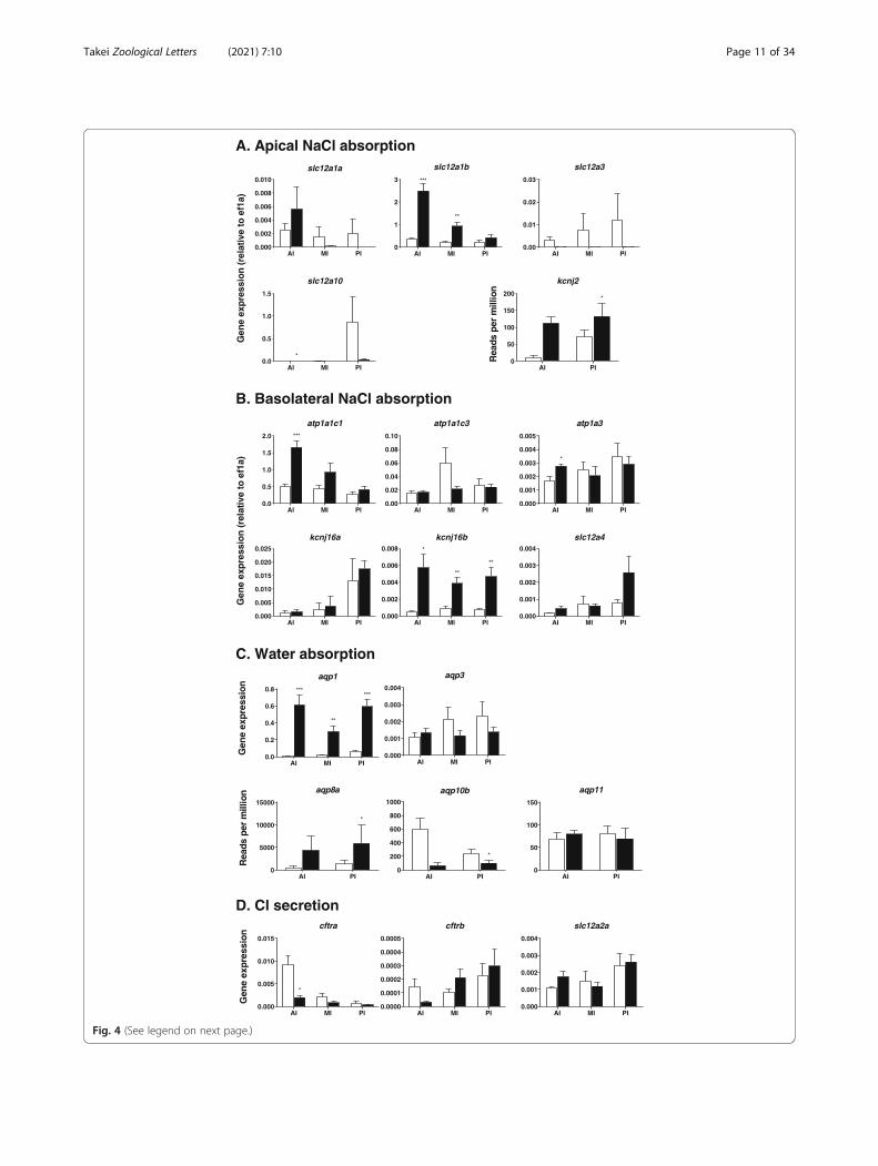
the eel [37, 255], of which NKCC2b is the major isoformin the intestine (Fig. 3). Of the two NKCC2 genes,slc12a1b is expressed at a much higher level thanslc12a1a and is upregulated in all intestinal segments ineels after acclimation to SW (Fig. 4): [12, 268]. The up-regulation of slc12a1b occurs after transfer to SW orhypertonic medium in the intestines of all euryhalineand marine teleost species examined thus far, includingthe Mozambique tilapia, Oreochromis mossambicus[130]; olive flounder, Paralichthys olivaceus [111]; gilt-head sea bream [66]; red drum [50]; and spotted seabass, Lateolabrax maculatus [282]. The slc12a1b expres-sion decreases gradually in the posterior direction in theintestine, while the NCC1 gene slc12a3 expression in-creases gradually and is highest in the rectum in bothEuropean and Japanese eels [38, 255]. Thus, NCC1 mayplay a role in NaCl absorption in the rectum. However,slc12a3 expression is lower in SW eels than in FW eels,and it is much lower than that of slc12a1b even in theposterior intestine (Fig. 4). The expression of slc12a3 isalso much lower than that of slc12a1 in the intestinesof Mozambique tilapia [130] and the three-spinestickleback, Gasterosteus aculeatus [140]. On the otherhand, the NCC2 gene (slc12a10) is significantlyexpressed in the eel intestine, and the expression in-creases in the posterior direction (Fig. 4). NCC2 is in-volved in NaCl absorption in the gills of Mozambiquetilapia and killifish [224] and probably in the NCC cellsof the zebrafish, Danio rerio [90].As mentioned above, active NKCC2 on the apical
membrane causes K+ shortage in the luminal fluid,which inhibits continuous NKCC2 function. To com-pensate for K+ in the luminal fluid, K+ is recycled by aK+ channel, and Kir2.1 is the candidate (Fig. 3). TheKir2.1 gene (kcnj2) is expressed substantially in the eelintestine and upregulated after SW acclimation (Fig. 4).Na+ and Cl− are also absorbed from the lumen intoenterocytes via the coordinated action of AE and NHE.The molecular identities of AE and NHE are discussedin the HCO3
− secretion section (see 4.4).
Serosal side Concerning basolateral transporters forNaCl absorption, NKA1c1 (atp1a1c1), NKA1c3(atp1a1c3) and NKA1c3 (atp1a3) are responsible forNa+ absorption, as they are expressed at considerablelevels in the eel intestine [270]. The atp1a1c1 andatp1a3 expression are upregulated in the anterior intes-tine after SW acclimation (Fig. 4). These NKAs maywork together to extrude Na+ into the interstitial fluid inSW eels (Fig. 3).To ensure the constant activity of NKA, K+ should be
recycled into the extracellular fluid via a basolateral K+
channel, and Kir5.1 is the candidate (Fig. 3). Kir5.1 wasfirst identified as a partner of NKA in the ionocytes of
SW eel gills [216]. In fact, two Kir5.1 genes (kcnj16a andkcnj16b) are expressed in the eel intestine, and kcnj16bis upregulated in all intestinal segments of SW-acclimated eels (Fig. 4). In the small intestines of mam-mals, Kir7.1 is involved in recycling of K+ for continuousactivity of NKA [167], while Kir5.1 and Kir4.1 are re-sponsible for K+ recycling for NKA activity in the distalconvoluted tubules of the kidneys [258].Concerning Cl− efflux on the serosal side, ClC2 and
ClC7 appear to be responsible (Fig. 3) because theirgenes (clcn2 and clcn7) are expressed in the eel intestineand upregulated in the anterior segment after SW accli-mation (unpublished data). The clcn2 expression is par-ticularly substantial and profoundly upregulated in SW.The clcn3 is also expressed in the intestine, but its ex-pression is much lower than that of clcn2 and does notincrease after SW acclimation. In mammals, ClC2 is re-sponsible for Cl− efflux at the serosal side of the intes-tinal epithelium [170], but the cell polarity oflocalization changes depending on various factors (see4.3.1). Notably, ClC2 is localized on the lateral mem-brane close to the TJ and regulates the paracellular per-meability of ions and water in the intestinal epitheliumin mammals [160]. In addition, the KCC1 gene (slc12a4)is expressed in the eel intestine, and its expression tendsto increase after SW acclimation (Fig. 4). It seems likely,therefore, that KCC1 on the basolateral membrane is re-sponsible for K+ recycling for NKA and Cl− efflux intothe interstitial fluid (Fig. 3). Notably, the suite of trans-porters for NaCl absorption develops in the intestineduring smoltification in the Atlantic salmon, when thefish are still in FW but preparing for downstream migra-tion to the sea [215].
Knowledge from mammals In mammals, the majorroutes for NaCl absorption are (1) electroneutral absorp-tion via combined activity of NHE and AE and (2) elec-trogenic absorption via ENaC at the mucosal side of theintestinal epithelium [61, 106, 199]. ENaC transportsonly Na+, which is the major source of serosa-positivePD in the mammalian intestine. The ortholog of ENaCis not identifiable in the teleost genome. Instead, a mem-ber of the acid-sensing ion channel (ASIC) family, a sub-family of the ENaC/degenerin superfamily, exists inteleosts. ASIC, which is localized on the apical mem-branes of ionocytes in FW trout gills, takes up Na+ fromenvironmental FW in exchange for H+ throughvacuolar-type H+-ATPase (VHA) [46]. The transcript ofASIC is not detectable in the intestines of eels even bytranscriptome analysis (unpublished data).Two NHE genes (NHE2 and NHE3) are expressed in
the mammalian intestine, of which NHE3 is dominant,and Nhe3−/− mice exhibit decreased NaCl absorptionand mild diarrhea [192]. In addition, only NHE3, not
Takei Zoological Letters (2021) 7:10 Page 10 of 34
AI MI PI0.000
0.002
0.004
0.006
0.008
0.010
slc12a1a
AI MI PI0
1
2
3
slc12a1b
***
**
AI MI PI0.00
0.01
0.02
0.03
slc12a3
AI MI PI0.0
0.5
1.0
1.5
slc12a10
*
AI PI0
50
100
150
200
kcnj2
*
AI MI PI0.0
0.5
1.0
1.5
2.0
atp1a1c1***
AI MI PI0.00
0.02
0.04
0.06
0.08
0.10
atp1a1c3
AI MI PI0.000
0.001
0.002
0.003
0.004
0.005
atp1a3
*
AI MI PI0.000
0.005
0.010
0.015
0.020
0.025
kcnj16a
AI MI PI0.000
0.002
0.004
0.006
0.008
kcnj16b*
**
**
AI MI PI0.000
0.001
0.002
0.003
0.004
slc12a4
Gen
eex
pre
ssio
n(r
elat
ive
toef
1a)
Rea
dspe
rm
illio
n
A. Apical NaCl absorption
B. Basolateral NaCl absorption
Gen
eex
pre
ssio
n(r
elat
ive
toef
1a)
AI MI PI0.0
0.2
0.4
0.6
0.8
aqp1
******
**
AI MI PI0.000
0.001
0.002
0.003
0.004
aqp3
AI PI0
200
400
600
800
1000
aqp10b
*
AI PI0
5000
10000
15000
aqp8a
*
AI PI0
50
100
150
aqp11
AI MI PI0.000
0.005
0.010
0.015
cftra
*
AI MI PI0.0000
0.0001
0.0002
0.0003
0.0004
0.0005
cftrb
AI MI PI0.000
0.001
0.002
0.003
0.004
slc12a2a
Gen
eex
pre s
sio n
Rea
d spe
rm
i llio
n
C. Water absorption
D. Cl secretion
Gen
eex
pres
sion
Fig. 4 (See legend on next page.)
Takei Zoological Letters (2021) 7:10 Page 11 of 34

NHE2, responds to intracellular messengers such ascAMP for recruitment of transporter-tagged vesicles tothe apical membrane (see 6.4), which demonstrates aregulatory role of NHE3 [44]. The major AE genesexpressed in the intestinal epithelium are Slc26a3 andSlc26a6. Slc26a3 is expressed throughout the wholelength of the intestine, while the Slc26a6 transcript isnot detectable in the distal colon [106]. It has been sug-gested that SLC26a3 exchanges 2Cl−/1HCO3
− and thatSLC26a6 exchanges 1Cl−/2HCO3
− in culture cells withtransient expression, but the stoichiometry is still underdebate [195]. SLC26a3 plays a major role in NaCl ab-sorption, as Slc26a3−/− mice suffer from severe chloride-losing diarrhea [193]. The effect of Slc26a6 knockout onthe intestine is not as strong as the effect on the kidneys[200]. NHE and AE form a metabolon with other trans-porters and enzymes, as discussed in 6.3.The transporters involved in intestinal NaCl absorp-
tion in mammals are similar to those of some marineteleosts but different from those of other teleosts, in-cluding eels, in which NKCC2 plays a dominant role(Fig. 3). Interestingly, NKCC2-based NaCl absorption issimilar to the absorption in the thick ascending limb ofHenle’s loop (TAL) in the mammalian kidneys, in whichNKCC2 and renal outer medullary potassium channel(ROMK) on the apical membrane and NKA, ClC-Kband KCC4 on the basolateral membrane are involved intranscellular NaCl absorption [16]. The advantage of theNKCC2-based method in teleosts is the efficient trans-port of NaCl and water, as mentioned above, butNKCC2 has no regulatory role in acid-base balance, un-like the combination of AE and NHE. It is possible thatteleosts acquired the NKCC2 system in the intestineduring the evolution of their habitat toward the ocean;this is particularly applicable to migratory/euryhalinefishes, which must cope with abrupt changes in environ-mental salinity.
4.2 Water absorptionIn parallel with NaCl transport, water moves from theintestinal lumen into the body fluid through the epithe-lium if the luminal fluid is almost isotonic to the bodyfluid [259]. The transcellular and paracellular routes are
possible routes for water absorption. As the plasmamembrane consists of a lipid bilayer that is almost im-permeable to water, AQPs on the apical and basolateralmembranes greatly facilitate transcellular water absorp-tion [95]. The possibility of water cotransport by trans-porters such as KCC and the Na+-glucose cotransporterhas been suggested [281], but this idea is still under de-bate. Regarding the paracellular route, it is known thatsome TJ proteins have channel-like activity [121], andCLDN2 has been suggested to serve as a water channel(see 6.1). However, paracellular water flux seems to beminor in the intestine of the killifish [274].The marine teleost intestine can absorb water from
slightly hypertonic luminal fluid [62, 68, 194], which isalso true in SW-acclimated eels [206]. However, a highMgSO4 concentration in the luminal fluid certainlylimits water absorption and survival in the hypersalineenvironment in the toadfish [62]. The luminal fluid closeto the apical membrane exists in a microenvironmentcovered by mucus and thus may be made hypotonic tothe body fluid by H2O formation and carbonate precipi-tation catalyzed by extracellular CAIV (see 4.4.2). Onthe basolateral side, NKA extrudes 3Na+ in exchange for2 K+, which generates an osmotic gradient between theintracellular fluid and extracellular fluid. If this occurs inthe lateral space between the adjacent enterocytes justbelow the TJ (called the lateral interspace, LIS), the fluidin the LIS becomes hypertonic to both the luminal andintracellular fluids. Then, water moves from both com-partments into the interstitial fluid passively [42, 126].Although ion fluxes precede that of water to produce adriving force, the fluid transported across the epitheliumis isotonic, which generates the solute (Na+)-recircula-tion model [125]. It is now known that Na+ recirculationis achieved by NHE on the basolateral membrane, notNKCC1, in the eel (see 4.4.1). The mechanisms of waterabsorption across intestinal epithelia have been dis-cussed in detail by Whittamore [259].
4.2.1 Molecular mechanisms of transcellular waterabsorptionTranscellular water movement is achieved throughAQPs. At least four AQPs are expressed in the eel
(See figure on previous page.)Fig. 4 Expression of the genes responsible for NaCl (A and B) and water absorption (C) in the anterior intestine (AI), middle intestine (MI) andposterior intestine (PI) in FW-acclimated (plain column) and SW-acclimated (filled column) eels as determined by real-time qPCR. The expressionof the genes related to Cl− secretion suggested in mammals and other marine teleosts is also shown in (D). The expression levels are correctedby those of elf1a and thus indicate the relative abundance values of the genes. The atp1, kcnj2 and kcnj16 are the genes for NKA, Kir2.1 and Kir5.1,respectively. The figures with ‘Reads per million’ on the ordinate were created from RNA-seq data (n = 5). The figures are depicted based on thedata in Ando et al. [12], Wong et al. [270], and Takei et al. [228] and on unpublished data. The primers for real-time PCR, including those for theunpublished data, are listed in Supplementary Table 1. *p < 0.05, **p < 0.01, ***p < 0.001. For details, see 4.1.1 and 4.2.1. For abbreviationdefinitions, see the list
Takei Zoological Letters (2021) 7:10 Page 12 of 34
intestine, AQP1, AQP3, AQP8, and AQP10 [13, 110,149], of which AQP3 and AQP10 are glyceroaquaporinsthat allow passage not only of water but also of glycerol,urea, etc. [95]. Two isoforms usually exist for AQP1(AQP1a and AQP1b) in the eel and other teleosts. Theexpression of aqp1a has been found to be upregulated inall segments of the eel intestine [13] and in vascularendothelial cells [149]. The tissue distribution of aqp1btranscripts has not yet been examined in eels. The aqp1expression is most abundant in the rectum, where waterabsorption may be enhanced by increased hydrostaticpressure [109]. A valve-like structure exists between theposterior intestine and the rectum in eels, which blocksbackflow when the luminal pressure in the rectum in-creases. AQP1 is localized on the apical membranes ofintestinal epithelial cells, as indicated by immunohisto-chemistry of the intestines of SW-acclimated Japaneseand European eels [13, 149]. Our recent study usingtranscriptome analysis (RNA-seq) showed that aqp1a,aqp3, aqp8a, aqp10b and aqp11 were expressed in theeel intestine, of which aqp1a and aqp8a were expressedat high levels (Fig. 4). Isoforms were identified by areverse-BLAST best hit approach in our eel transcrip-tome data (DDBJ accession number: DRA004258) usingannotations in the zebrafish database (https://zfin.org/).From the RNA-seq data, the expression of aqp1a, aqp3and aqp8a was significantly upregulated after SW accli-mation, but real-time qPCR showed that aqp3 expres-sion did not change after SW acclimation (Fig. 4),suggesting the possible presence of isoforms. On theother hand, our qPCR analysis showed that aqp1a wasexpressed in all intestinal segments and profoundly up-regulated after SW acclimation in the eel (Fig. 4).In the Atlantic salmon [236] and gilthead sea bream
[172], both aqp1a and aqp1b are expressed in the intes-tine, with aqp1a exhibiting higher expression thanaqp1b, and the expression of these genes is higher in fishacclimated to SW than in those acclimated to FW. Im-munoreactive AQP1a is localized on both the apical andlateral membranes of sea bream enterocytes, while it isfound on the basolateral membrane in the Atlanticsalmon. It has been suggested that the direction oftrafficking of transporter-tagged vesicles to either theapical or basolateral membrane is dependent on en-vironmental salinity (see 6.4). Upregulation of aqp1has also been reported in the intestine of the silversea bream, Sparus sarba [40], and the European seabass, Dicentrarchus labrax [64].The aqp3 is most abundantly expressed in the gills of
teleost fishes, but significant expression has also beendetected in the intestine of the eel [149], and the expres-sion level is higher in the rectums of SW-acclimated fishthan in those of FW fish [110]. The aqp3 expression islow in the intestines of Mozambique tilapia [254] and
sea bass [64]. Among AQPs expressed in the eel intes-tine, AQP3 is the only AQP that has been found tolocalize on the basolateral membranes of epithelial cells[39, 254], although AQP3 also localizes to the apicalmembrane in killifish [181]. In mammals, apicalAQP2 and basolateral AQP3 are responsible fortranscellular water absorption at the renal collectingduct [102].AQP8 is another candidate apical membrane AQP
(Fig. 3). The aqp8 is expressed in the eel intestine, andthe transcript levels increase after SW acclimation [110,149]. We have also observed upregulation of aqp8a inthe posterior intestines of SW eels (Fig. 4). Immunohis-tochemical analysis showed that AQP8 exists on the ap-ical membranes of intestinal cells. Three AQP8 genes(aqp8aa, aqp8ab and aqp8b) are expressed in the intes-tine in Atlantic salmon, of which aqp8ab has the highesttranscript levels in the intestine, and aqp8ab is upregu-lated after SW acclimation [236]. The salmon AQP8ab isalso localized on the mucosal (apical) side in enterocytesand appears to play important roles in water absorption[47], as is the case for mammalian AQP8 [240].The aqp10 expression is observed in the intestines of
eels and is upregulated in SW-acclimated fish [110, 149].However, our transcriptome data suggest that aqp10b isdownregulated in the SW eel intestine (Fig. 4).The aqp10 expression has also been reported in theintestines of Atlantic salmon [236] and gilthead seabream [185]. Cellular localization of AQP10 has notyet been examined in fish, but AQP10 is localized onthe apical membranes of enterocytes in humans [154].Expression of aqp3, aqp8 and aqp10, including theirsubtypes, has also been reported in the intestine ofthe three-spine stickleback [140].To summarize the possible transcellular pathway for
water absorption, the major apical AQP may be AQP1ain the eel intestine, as judged by the expression level ofits gene and upregulation after SW acclimation (Fig. 3).In other marine teleosts, AQP8 and AQP10 on the ap-ical membrane are also involved in the uptake of waterinto epithelial cells. The water taken up into the cellsmay be transported into the extracellular fluid via AQP3on the basolateral membrane, although AQP3 gene ex-pression is low compared with that of other AQPs. Itseems that AQP1 also plays a role in water absorption atthe serosal side in the Atlantic salmon [236]. In theeuryhaline medaka, Oryzias latipes, aqp1a, aqp7, aqp8aband aqp10a are downregulated after SW transfer, andimmunoreactive AQP1a and AQP10a move from the ap-ical membrane to the subapical region after transfer toSW, indicating decreased transcellular water permeabil-ity across the intestinal epithelium [139]. Thus, it is hy-pothesized that water is transported mostly via theparacellular route after SW acclimation in medaka,
Takei Zoological Letters (2021) 7:10 Page 13 of 34

although paracellular water permeability appears to besuppressed in salmonids after SW acclimation [213].The water permeability of TJ proteins is discussed in 6.1.
4.3 Cl− secretionAs shown in Table 1, SW contains similar concentra-tions of Na+ (450 mM) and Cl− (524 mM). As esophagealdesalinization removes Na+ and Cl− equally from SW,Na+ and Cl− concentrations in the luminal fluid mayalso be similar when ingested SW enters the intestinefor absorption. In the anterior intestine, NKCC2 takesup 1Na+ and 2Cl− from the luminal fluid, and AE furthertakes up Cl− in exchange for HCO3
− (Fig. 3). Accord-ingly, the amount of Cl− in the luminal fluid decreasesmuch faster than that of Na+ during passage along theintestinal tract. However, the concentration of Cl− in theluminal fluid is higher than the Na+ concentration in theintestine and rectum in eels (Table 1) and other marineteleosts [4, 69, 182]. This implies that Cl− is secretedinto the lumen to maintain the activity of NKCC2 andAE for constant NaCl absorption.In mammals, Cl− secretion has been demonstrated in
crypt cells of the intestine (see [61]). The secretory-typecells were once thought to be restricted to the crypt regionof the intestine, but it was later shown that they are present
more widely along the crypt-villus axis [98]. Accordingly, itis controversial whether the same enterocytes have both ab-sorptive and secretory functions or whether two differentcell types exist. In the eel, guanylin has been shown to in-hibit NKCC2 and stimulate apical Cl− channels at the sametime via a single second messenger, cGMP, resulting in Cl−
secretion [9]. Thus, a single enterocyte seems to be able tochange its function from absorption to secretion (see 5).However, a small population of secretory-type enterocytesappear to exist in the killifish intestine [145].Typical secretory-type cells are characterized by the
presence of cystic fibrosis transmembrane regulatoranion (Cl−) channels (CFTRs) on the apical membraneand NKCC1 (SLC12a3) on the basolateral membrane inmammals (Fig. 5). Low cytosolic Na+ caused by NKApromotes the activity of NKCC1 to take up Cl− from theextracellular fluid into the cell. Then, increased cytosolicCl− is secreted into the intestinal lumen via CFTR, whichis facilitated by the negative intracellular potential pro-duced by NKA. Anion secretion accompanies parallelfluid secretion into the lumen, resulting in secretorydiarrhea in mammals [61]. The suite of transporters forCl− secretion in the intestine is similar to those ofmitochondrion-rich ionocytes of the gills in marine tele-osts [85, 86] and secretory epithelial cells of the rectal
Fig. 5 Transporters involved in transcellular Cl− secretion (blue rectangles) into the lumen by intestinal epithelial cells in mammals (A) andseawater (SW)-acclimated eels (B). Because of the low expression of NKCC1 and CFTR in the intestines of SW eels, alternative molecularmechanisms are suggested. For details, see 4.3.1. For abbreviation definitions, see the list
Takei Zoological Letters (2021) 7:10 Page 14 of 34

gland in marine elasmobranchs [203]. In this sense, therectal gland is similar to the crypt cells of the mamma-lian colon.
4.3.1 Possible molecular mechanismsIn the teleost intestine, Marshall et al. [146] found sub-stantial expression of cftr as high as that in the gills ofSW-acclimated killifish. Apical localization of immuno-reactive CFTR occurs only in the enterocytes of SW fish[145]. The enterocytes with CFTR immunoreactivity atthe brush border membrane compose 20% of the totalenterocyte population, suggesting the presence ofsecretory-type cells (Fig. 5). As immunoreactive CFTRmigrates to the basolateral membrane in FW, thefunction of CFTR may change from Cl− excretion in SWto Cl− absorption in FW (see 6.4). Substantial expressionof cftr has also been detected in the intestines ofMozambique tilapia [130] and spotted sea bass [282],and the expression is enhanced in hypersaline environ-ments in these fishes. The cftr expression has also beenreported in the intestines of gilthead sea bream [66],three-spine stickleback [140], and European sea bass [4].In the sea bream, cftr expression is downregulated in theanterior intestine after transfer from diluted 12-ppt SWto regular SW or to hypertonic 55-ppt SW, but it is up-regulated in the rectum.The partner of apical CFTR for transcellular Cl− secre-
tion is basolateral NKCC1, as mentioned above. Thiscoupling may also be the case in the intestines of someteleost species. The expression of two NKCC1 genes(slc12a2a and slc12a2b) has been demonstrated in theintestine of the spotted sea bass [282]. The expression ofboth NKCC1 genes in the intestine is much lower thanthat in other tissues and much lower than that of theCFTR gene. Furthermore, the expression of the majorisoform, slc12a2a, does not change after acclimation to ahypersaline medium. Two NKCC1 genes are alsoexpressed in the intestine of the gilthead sea bream [66],three-spine stickleback [140], and European sea bass [4].Similar to cftr expression, slc12a2 expression is down-regulated in the anterior intestine of the sea bream butupregulated in the rectum after transfer from an isotonicto a hypertonic medium.In contrast to the roles in these teleost species, the
roles of CFTR and NKCC1 in Cl− secretion seem to beminor in the eel. The expression of two cftr isoforms(cftra and cftrb) is detectable along the intestinal tract inJapanese eels [269]. However, the expression of cftra, adominant isoform in the intestine, is low compared withthat of clcn2 and clcn7, which are upregulated in SW-acclimated eels (Fig. 4). Furthermore, the expression ofcfrta is downregulated in the anterior intestine in SW-acclimated fish (Fig. 4). It is possible that CFTR of theFW eel intestine is localized in the basolateral
membrane for Cl− absorption, as suggested in killifish[145]. Gene expression does not always parallel proteinabundance in cells, and it is possible that CFTR proteinresides on the apical membrane or is stored in vesiclesin the subapical region in enterocytes for recruitmentafter stimulation. In both FW and SW eel intestines,however, immunoreactive CFTR is not found on the ap-ical membrane but rather in cytoplasmic vesicles [269].Thus, it is likely that apical Cl− channels different fromCFTR may exist for Cl− secretion in the eel. The pres-ence of DPC-inhibitable and guanylin-sensitive Cl−
channels has been shown on the apical brush-bordermembrane of the eel intestine (see 5). ClC2 is usually lo-calized to the basolateral membrane for Cl− absorptionin the intestines of mammals [28], as illustrated in Fig. 3.However, guanylin stimulates Cl− secretion in the intes-tines of Cftr−/− mice, where apical ClC2 is suggested tocompensate for CFTR for Cl− secretion (see [56]). As thepolarity of vesicle recruitment changes depending on theenvironmental stimuli, as suggested for CFTR [145], it ispossible that ClC2 is responsible for Cl− secretion stimu-lated by guanylin (unpublished data). Alternatively, it isalso possible that SLC26a6 on the apical membrane ex-cretes Cl− in exchange for SO4
2− driven by its high con-centration (> 100 mM) in the luminal fluid (see 6.2) orthat SLC26a3 excretes Cl− in exchange for HCO3
−
driven by its high concentration (~ 100 mM) in the lu-minal fluid (see 4.4.1). SLC26a6 in the SLC26 family hasa high affinity for SO4
2− [199], and it actively exchangesSO4
2− and Cl− in the renal proximal tubules of eels[256], while SLC26a3 readily exchanges Cl− and HCO3
−
[199].Concerning NKCC1 on the basolateral membrane,
only slc12a2a is expressed in the intestines of bothEuropean eels [37] and Japanese eels [271]. Comparedwith that of the NKCC2 gene (slc12a1b), slc12a2a ex-pression is low, and it does not change after SW accli-mation (Fig. 4). In addition, slc12a2a expression ishigher in silver eels ready for downstream migrationthan in river-dwelling yellow eels [37]. Because of thelow slc12a2 expression in the eel intestine, sulfate aniontransporter 1 (SAT1, SLC26a1) could be a candidate forCl− uptake at the serosal side in exchange for SO4
2−,which is facilitated by uptake of SO4
2− from the luminalfluid by SLC26a6 (Fig. 5, see 6.4). Cl−/SO4
2− exchangeactivity has been demonstrated to occur in the basolat-eral membrane of the rabbit intestine [191].When all the data obtained thus far are taken to-
gether, it seems that secretory-type enterocytes, whichhave CFTR on the apical membrane and NKCC1 onthe basolateral membrane, may exist in the marineteleost intestine for Cl− secretion into the lumen (Fig.5). This Cl− compensation in the luminal fluid allowsconstant activity of NKCC2 and AE/NHE to be
Takei Zoological Letters (2021) 7:10 Page 15 of 34

maintained for NaCl absorption and thus water ab-sorption. This mechanism enables marine teleosts toabsorb > 80% of water from ingested SW and explainswhy the luminal fluid Cl− concentration is higher thanthe Na+ concentration in the luminal fluid of marineteleost intestine. However, knowledge from the eel in-testine suggests that an alternative set of transportersis responsible for Cl− secretion, which requires furtherclarification. Intestine-specific knockdown of a trans-porter gene can be performed evaluate the genes re-sponsible for SW acclimation. The use of membrane-permeable antisense oligonucleotides injected directlyinto the intestinal lumen enables intestine-specificgene knockdown.
4.4 HCO3− secretion and carbonate precipitation
The high CO2 concentration and high pH of the rectalfluid in marine teleosts were recognized 90 years ago byHomer W. Smith [207]. It was shown later that the highpH is caused by active HCO3
− secretion into the lumenby epithelial cells (see [68, 69, 259]). Luminal fluid alkali-zation is more profound after fish are acclimated tohypertonic SW [264], suggesting a role of HCO3
− secre-tion in SW acclimation. As will be discussed in detailbelow (see 4.4.2), the secreted HCO3
− helps precipitatecarbonates of divalent ions (Mg2+ and Ca2+), which arepresent in SW at concentrations higher than those inplasma (Table 1) and further concentrated by water ab-sorption. Precipitate formation decreases luminal fluidosmolality and further enhances water absorption [73].It is important to note that carbonate precipitate forma-tion by marine teleost intestines explains 3–15% of thetotal oceanic carbon cycle and contributes to theamelioration of ocean acidification via fixation of CO2 inthe ocean [265]. Because of its important role in globalsustainability, HCO3
− secretion by the marine teleostintestine is one of the recent topics of interest in fishphysiology [48, 67, 80, 87].The involvement of apical AE in HCO3
− secretion hasbeen suggested by in vitro experiments using Cl−-defi-cient Ringer solution or application of DIDS on the mu-cosal side of the intestinal epithelium. Consistently,mucosal application of DIDS inhibits HCO3
− secretionin the sanddab, Citharichthys sordidus, and the rainbowtrout [74, 75]. However, DIDS is more potent in inhibit-ing HCO3
− secretion when applied to the serosal sidethan to the mucosal side; it is only slightly inhibitorywhen applied to the mucosal side in the eel [7] and otherteleost species [43, 52]. Such species differences may bedue to the differences in major transporters usedamong species [224] and the different sensitivities ofAEs to DIDS among teleost species, as shown inmammals (see below). HCO3
− secretion is significantlyinhibited when Cl−-deficient Ringer solution is on the
mucosal side [73, 263], and the presence of AE onthe apical membrane is now widely recognized [68].Concerning inhibition by serosal DIDS, as removal ofserosal Na+ profoundly decreases HCO3
− secretion inthe eel [7], Na+-HCO3
− cotransporter (NBC), a mem-ber of the DIDS-sensitive SLC4 family of HCO3
−
transporters [174], may be involved in HCO3− uptake
from the serosal side for secretion into the lumen.Two sources of HCO3
− in the cell are conceivable forHCO3
− secretion into the lumen. One is HCO3− taken
up from the serosal side by NBC [231] as mentionedabove, facilitated by NKA-induced low cytosolic Na+ [68,69, 71]. The other is endogenous HCO3
− produced inthe enterocyte by hydration of CO2 catalyzed by cyto-solic CAII [230]. Because of the high metabolic activityof the cells, CO2 production in intestinal epithelial cellsis high. In fact, both sources contribute to the increasein cytosolic HCO3
− in the teleost intestine, while theirrelative contributions differ considerably among species[69]. Since serosal DIDS and removal of HCO3
− in theserosal fluid profoundly decrease HCO3
− secretion inthe goby [43], eel [7] and other marine teleosts [262],the contribution of serosal HCO3
− is significant in thesespecies. On the other hand, almost all HCO3
− is suppliedby CO2 hydration in SW-acclimated rainbow trout [75].CA is one of the enzymes that has the fastest reactionrate and catalyzes the following reaction: CO2 + H2O ⇌HCO3
− +H+. The forward reaction is much faster thanthe reverse reaction, and in high-CO2 environmentssuch as intestinal epithelial cells. Thus, the presence ofcytosolic CAII greatly enhances HCO3
− production.Under high-HCO3
− and high-pH conditions, such as inthe intestinal lumen, however, the reverse reaction oc-curs, which is accelerated by membrane-bound CAIV(see below).
4.4.1 Molecular mechanisms of HCO3− secretion
The molecular mechanisms of HCO3− secretion have
been investigated for more than a decade in several tele-ost species, and ample data have been accumulated [66,69, 122, 231, 259]. In this section, however, we will firstdescribe the data obtained in eels for comparison withthose obtained in preceding studies on other teleostspecies.
Mucosal side Three SLC26a6 genes (slc26a6a, slc26a6b,slc26a6c) are expressed at significant levels in the eel in-testine, as assessed by transcriptomic analyses followedby real-time qPCR for differentiation of the isoforms(Figs. 6 and 7A). Among the isoforms, slc6a6a exhibitsthe most abundant transcripts in the posterior intestine,and the transcript levels decrease in the anterior direc-tion; in contrast, slc26a1b transcripts are most abundantin the anterior intestine, and the transcript levels
Takei Zoological Letters (2021) 7:10 Page 16 of 34

decrease in the posterior direction. The slc26a6c expres-sion is the lowest among these isoforms in all intestinalsegments. The slc6a6a expression is upregulated in allsegments of the intestine during the course of SW accli-mation [228], although the expression returns to the FWlevel after SW acclimation (Fig. 7A). The stoichiometryof SLC26a6 has been suggested to be electrogenic, ex-changing 2HCO3
− with 1Cl−, in the mefugu [107], whilevariable stoichiometry has been reported in mammals [1,106]. The electrogenic nature of SLC26a6 has also beenshown in mefugu [122] and toadfish [76].In addition, two SLC26a3 genes (slc26a3a and
slc26a3b) are expressed abundantly in the eel intestine,and their expression is upregulated profoundly after SWtransfer until the eels are fully acclimated to SW (Fig. 6and 7A). The slc26a3b expression is most abundant inthe anterior intestine and decreases in the posterior dir-ection, while slc26a3a expression is most abundant inthe posterior intestine and decreases in the anterior dir-ection. In this way, two SLC26a3 isoforms and twoSLC26a6 isoforms appear to compensate for each otherin different segments of the intestine. The expressionlevel of slc26a3a/b is several-fold higher than that of
slc26a6a/b (Fig. 7A). The stoichiometry of SLC26a3 isreported to be HCO3
−/2Cl− in mammals (see [106]), butit has not yet been examined in teleosts. As mentionedabove, however, mucosal DIDS fails to decrease HCO3
−
secretion in the eel intestine [7, 228]. Although DIDS isgenerally an effective inhibitor of AE of the SLC26 fam-ily in mammals, it fails to block SLC26a3 in some spe-cies [15, 260], showing species specificity of DIDS forSLC26a3. Among teleosts, DIDS effectively blocksSLC26a6 in the rainbow trout [22], but its effect onSLC26a3 has not been confirmed in any teleost species.Supposing that DIDS is effective for SLC26a6 but inef-fective for SLC26a3 in eels, the major apical AE of theSLC26 family responsible for HCO3
− secretion and Cl−
absorption might be SLC26a3 in eels.Other candidate transporters for HCO3
− secretion areanion channels on the apical membrane, and cftra ex-pression has been detected in the eel intestine (Fig. 4).CFTR is known to readily pass HCO3
− in mammals[204] and couple with SLC26a3/6 to stimulate HCO3
−
secretion [212] (Fig. 8). The polarity (apical or basolat-eral) of CFTR localization seems to be variable depend-ing on the environmental salinity and other conditions
Fig. 6 Major transporters responsible for HCO3− secretion (red rectangles) into the lumen by the intestinal epithelial cells of SW eels. These
transporters also function in NaCl absorption (see Fig. 3) and Mg/CaCO3 precipitation (see Fig. 8). NHEs, SLC26a3/6 s and CFTRs are combined as ametabolon for mutual activity regulation (see 6.3 and Fig. 8). The text size for the transporters is related to the relative abundance andupregulation of the transporters in SW. For details, see 4.4.1. For abbreviation definitions, see the list
Takei Zoological Letters (2021) 7:10 Page 17 of 34

AI MI PI0.00
0.02
0.04
0.06
0.08
0.10
slc26a3a
* *
AI MI PI0.0
0.1
0.2
0.3
slc26a3b
**
*
AI MI PI0.000
0.005
0.010
0.015
slc26a6a
AI MI PI0.00
0.02
0.04
0.06
0.08
slc26a6b
AI MI PI0.0000
0.0005
0.0010
0.0015
slc26a6c
AI MI PI0.000
0.005
0.010
0.015
slc26a1
*
AI MI PI0.0000
0.0005
0.0010
0.0015
slc9a1
AI MI PI0.000
0.001
0.002
0.003
0.004
0.005
slc9a2
AI MI PI0.000
0.002
0.004
0.006
0.008
0.010
slc9a3
AI MI PI0.00000
0.00005
0.00010
0.00015
0.00020
0.00025
slc4a1a
AI MI PI0.000
0.001
0.002
0.003
slc4a2a
*
Gen
eex
pre
s si o
n(r
ela t
i ve
toef
1a)
A. SLC26 family AEs
B. SLC9 family NHEs
Gen
eex
pres
s ion
AI MI PI0.000
0.002
0.004
0.006
0.008
0.010
slc4a2b
AI MI PI0.0
0.2
0.4
0.6
slc4a4
***
*
AI MI PI0.000
0.002
0.004
0.006
0.008
slc4a7
***
AI MI PI0.0000
0.0005
0.0010
0.0015
0.0020
0.0025
slc4a10
**
C. SLC4 family AEs
D. SLC4 family NBCs
Gen
eex
pres
sio n
Gen
eex
pres
sion
AI MI PI0.000
0.001
0.002
0.003
0.004
ca2a
AI MI PI0.00
0.01
0.02
0.03
0.04
ca2b
*
AI MI PI0.00
0.05
0.10
0.15
ca4
**
*
AI MI PI0.00
0.02
0.04
0.06
atp6v1a
***
AI MI PI0.000
0.005
0.010
0.015
atp6v1b1
*
AI MI PI0.0000
0.0005
0.0010
0.0015
atp6v1b2
*
E. Carbonic anhydrases
F. V-ATPases
Gen
eex
pres
sion
Gen
eex
pres
sion
Fig. 7 (See legend on next page.)
Takei Zoological Letters (2021) 7:10 Page 18 of 34

[145]. clcn2 and clcn3 are expressed significantly in theeel intestine (unpublished data). Significant expressionof clcn3 has also been reported in the cod intestine [87].However, the major role of apical ClCs appears to be forCl− secretion in teleost fishes (see 4.3.1).The involvement of SLC26a6 was first suggested in the
euryhaline mefugu, in which slc26a6 expression is pro-foundly upregulated after transfer from FW to SW[122]. Similar results have been reported in the rectumin gulf toadfish [179] and gilthead sea bream [66] trans-ferred from diluted SW to concentrated SW. In contrast,slc26a6 expression in the anterior intestine ofMozambique tilapia is suppressed after transfer fromFW to SW [183]. In addition, slc26a3 expression hasalso been detected in the intestines of several teleostspecies, and upregulation of the transcripts has been
found in sea bream [66] and tilapia [183] after hyperos-motic stimulation. The slc26a3 and slc26a6 expressionhas also been detected in the intestine of the Europeansea bass [4]. It seems that the relative contributions ofSLC26a3 and SLC26a6 to HCO3
− secretion differ amongteleost species.As partners of SLC26a3 and SLC26a6 for coupled up-
take of Na+ and Cl− and excretion of H+ and HCO3−,
three NHE genes, NHE1 (slc9a1), NHE2 (slc9a2) andNHE3 (slc9a3), are expressed in the eel intestine (Fig. 6and 7B). NHE1 and NHE2 are resident-type, whileNHE3 is a mobile-type transporter that is stored in cyto-plasmic vesicles and inserted into the plasma membrane(see 6.4). The expression of NHE genes does not changein the eel intestine after SW transfer (Fig. 7B). AE andNHE are thought to be physically associated in the
Fig. 8 A suite of transporters and enzymes form a metabolon at the apical membrane of enterocytes for H+ and HCO3− secretion and recycling to
form carbonate precipitates in SW eels. VHA may also be involved in H+ secretion into the lumen (see Fig. 7). This idea of metabolon formation wasfirst proposed in the mammalian intestine and may be applicable to the teleost intestine (see 6.3). H+ and HCO3
− secreted into the lumen by apicaltransporters are catalyzed by membrane-bound CAIV to produce CO2 and H2O, which are recycled into the cell via AQP1. Cytosolic CAII then producesH+ and HCO3
− again for continuous supply of the ions into the lumen. NHE3, SLC26a3/6 and CFTR are bound on NHERF for mutual regulation. CAIValso facilitates Mg/CaCO3 production in the microenvironment under the mucus layer, which is maintained by continuous secretion of mucus fromgoblet cells (see 4.5). CAII is known to bind to the NHE/AE complex and to AQP to regulate their activity. CFTR and SLC26a3/6 bind through the Rdomain and the STAS domain of each transporter. For details, see 6.3. For abbreviation definitions, see the list
(See figure on previous page.)Fig. 7 Expression of the genes responsible for HCO3
− secretion in the anterior (AI), middle (MI) and posterior (PI) intestine in FW-acclimated (plaincolumn) and SW-acclimated (filled column) eels as determined by real-time qPCR. The genes are grouped by SLC family and function. The atp6s arethe genes for of VHA subunits. The figures are based on the data in Takei et al. [228] and Wong et al. [269] and on unpublished data. The primers forreal-time qPCR, including those for the unpublished data, are listed in Supplementary Table 1. *p < 0.05, **p < 0.01, ***p < 0.001. For details, see 4.4.1.For abbreviation definitions, see the list
Takei Zoological Letters (2021) 7:10 Page 19 of 34

anchor protein (see 6.3). The gene of the anchor proteinfor NHE3, NHERF1 (slc9a3r1), is expressed significantlyin the intestine, and its expression is upregulated two-fold after SW acclimation, as demonstrated by RNA-seq(unpublished data). NHE3 and AE in the anchor proteinare recruited from the cytoplasmic vesicles to the apicalmembrane upon phosphorylation (see 6.4). NHE3 is anelectroneutral exchanger of 1Na+/1H+. With regard toother teleosts, nhe3 expression has been detected in theintestines of rainbow trout [72] and cod [87].
Serosal side On the basolateral membrane, SLC26a1may be a major AE for HCO3
− uptake into the cell, asits gene expression is consistently upregulated in all seg-ments of the eel intestine after SW transfer [228], al-though the upregulation is not significant in the middleand posterior intestine after full acclimation to SW (Fig.7A). As SLC26a1 exhibits high affinity for SO4
2−, it ispossible that apical SLC26a6 and basolateral SLC26a1work in concert for transcellular secretion of HCO3
− inexchange for SO4
2− absorption in the SW eel intestine(see 6.2). Cooperation of the two sulfate transporters hasbeen observed in the proximal tubules of SW eel kidneys[257]. The SLC4 family of AEs also contains candidatesfor HCO3
− secretion: two AE2 genes (slc4a2a andslc4a2b) are expressed in the eel intestine, and slc4a2atends to be upregulated after SW transfer (Fig. 7C). AE2may be localized on the basolateral membrane, as as-sumed from its location in the intestines of mammals[174] and by the lack of inhibition of HCO3
− secretionby apical application of DIDS in the eel intestinal epithe-lium [228].Among other HCO3
− transporters, three NBC genes,NBCe1a (slc4a4a), NBCn1 (slc4a7) and NBCn2(slc4a10a), are expressed in the SW eel intestine, ofwhich slc4a4a is expressed most profoundly (Fig. 7D).All the NBC genes are upregulated in different segmentsof the eel intestine after SW transfer [228]. Thus,HCO3
− may be taken up with Na+ from the extracellularfluid by electrogenic NBCela and electroneutral NBCn1and NBCn2a. The stoichiometry of NBCe1 is 1Na+/2HCO3
− in mammals [174] and electrogenic in toadfish[231]. However, the external Na+ concentration affectsthe stoichiometry of mefugu NBCe1, and the ratio be-comes 1Na+/4HCO3
− at 120 mM Na+, which is similarto the concentration in the extracellular fluid of teleosts[29]. NBCe1 activity is facilitated by the low levels ofcytosolic Na+ produced by NKAs on the basolateralmembranes of enterocytes [69, 133]. The slc4a4 is alsoexpressed in the intestines of mefugu [122], toadfish[231], and sea bream [66], and its expression is upregu-lated after transfer to hyperosmotic environments. Theinhibition of HCO3
− secretion by serosal DIDS coincideswith the DIDS sensitivity of NBCs [7, 43, 69]. Given all
the results obtained thus far, it seems that transcellularHCO3
− transport occurs principally via apical SLC26a3and SLC26a6 and basolateral NBCe1 and SLC26a1/AE2in marine teleosts (Fig. 6).
Carbonic anhydrases and proton ATPase In additionto uptake from the serosal side, cytosolic HCO3
− is pro-vided by hydration of CO2 in epithelial cells, which iscatalyzed by cytosolic CAII (CAc), as suggested in sev-eral teleost species [66, 75, 187]. CO2 is produced by ac-tive metabolism of enterocytes and is taken up from theluminal fluid (Fig. 8). Two cytosolic CAII genes (ca2aand ca2b) and a membrane-bound CAIV gene (ca4a)are expressed at high levels in the eel intestine, and ca2band ca4a expression is upregulated after SW transfer(Fig. 7E). CAIV has a catalytic site on the luminal sideand may produce H2O and CO2 from H+ and HCO3
−
secreted into the microenvironment between the mucuslayers and the apical membranes of epithelial cells (Fig.8), as observed in rainbow trout [72]. H2O and CO2 pro-duction decrease luminal fluid osmolality to facilitatewater absorption, and the generated CO2 is recycled intothe cell for hydration by CAII (see 4.4.2). It is likely thatCO2 is recycled efficiently via AQP1 on the apical mem-brane, as suggested in zebrafish [31] and mammals[171]. As HCO3
− secretion is inhibited to different de-grees by mucosal application of acetazolamide, whichblocks both CAII and CAIV at the same time, the rela-tive contributions of the CAs to HCO3
− secretion differamong teleost species [69, 259]. The role of CAII inHCO3
− secretion has been documented in the rainbowtrout [72] and other marine teleosts [69].HCO3
− secretion is mediated by mutual activation ofSLC26-family AEs and CFTR on the apical membrane(see 6.3). As SLC26a6 exchanges 1Cl−/n HCO3
− in tele-osts [107], additional H+ secretion into the lumen neu-tralizes the secreted HCO3
− to support the activity ofcoupled SLC26a6 and NHE3, which exchanges 1Na+/1H+ [69, 76]. In fact, H+ secretion facilitates HCO3
− se-cretion and increases the production of CO2 and H2Oby CAIV (see below). H+ secretion also helps protectagainst cellular acidification [76]. The candidate H+
transporter is VHA, which is expressed in the intestinesof various teleost species, such as rainbow trout [72],toadfish [76], sea bream [66], cod [87], tilapia [183],Senegalese sole [182], and sea bass [4]. VHA is a largeprotein complex consisting of two domains, V0 and V1,that consist of six and eight subunits, respectively. Sig-nificant expression of the A subunit and 2B subunits inthe V1 domain (atp6v1a, atp6v1b1, atp6v1b2) has beendetected in the eel intestine. Among these subunits,atp6v1a and atp6v1b1 are highly expressed, and theirexpression is upregulated after SW transfer (Fig. 7F).VHA also appears to be localized on the basolateral
Takei Zoological Letters (2021) 7:10 Page 20 of 34
membrane to help maintain the resting potential andneutral pH of the cell [69]. The CAs and VHA appear tobe physically coupled to the AE/NHE complex to form ametabolon (see 6.3).
4.4.2 Mechanisms of carbonate productionFormation of white precipitates consisting of Ca/Mgcarbonate within the intestinal lumen has been re-ported in various marine teleosts for more than 50years; for example, such precipitates have been ob-served in southern flounder [81], rainbow trout [201],eels [88, 153, 207], toadfish [253], European flounder[262] and mefugu [122]. The solubilities of MgCO3
and CaCO3 in water are 1.65 and 0.13 mmol/l at25 °C, respectively, while luminal fluid Mg2+ and Ca2+
concentrations are 188 and 14 mmol/l, respectively, inthe eel intestine (Table 1). Although solubilitychanges greatly according to pH and the presence ofother ions, concentrated HCO3
− in the luminal fluidcertainly promotes the formation of carbonates [70,265]. The chemical compositions of carbonate precipi-tates have been examined in teleost intestines. Thecarbonate precipitate consists of Ca carbonate and/orcalcite in the flounder [81] and eel [88], and the cal-cite contains one-third (by weight) Mg and a smallamount of Mn, producing a substance called calciankutnohorite, in the gulf toadfish [253]. On the otherhand, the carbonate precipitate contains 22.6 mol%Mg, 76.9 mol% Ca and small amounts of P and S andis called magnesian calcite (Mg calcite) in the eel[153]. Chemical and structural analyses have beenperformed on the crystalline products in the intestinallumens of various tropical fishes and have revealedthat Mg calcite contains 0.5 to more than 40 mol%MgCO3 [184]. The precipitate of the sea bream intes-tine contains 54 mol% Mg2+, and Mg2+ may be re-sponsible for stabilizing the unstable mineral crystals[54]. The mechanisms of Mg calcite formation havebeen investigated in marine organisms, and high-Mgcalcite that contains > 4 mol% MgCO3 is naturallyformed by calcite-coated animals such as foraminifera,echinoids and corals [131]. The ratio of MgCO3 in-creases in proportion to the relative Mg2+ concentra-tion in the medium. Recently, a protein-based matrixwith strong Ca2+-binding properties was found in theteleost intestine; this matrix facilitates carbonate pre-cipitation [188]. The proteins were purified from theCa precipitates in the toadfish intestine and analyzedby proteomics. Many matrix proteins were found tobe highly acidic, which may provide negativelycharged domains for immobilization of Ca2+.Immobilization increases the local Ca2+ concentrationin the microenvironment and facilitates the precipita-tion of divalent ions with secreted HCO3
−.
As mentioned above, the space between the mucuslayer and the apical membrane of enterocytes is amicroenvironment different from the central lumen,which is maintained by constant secretion of mucusby goblet cells (Fig. 8). H+ and HCO3
− are secretedinto the microenvironment, where not only CO2 andH2O production but also carbonate production areefficiently facilitated by membrane-bound CAIV viathe following reaction: Ca2+/Mg2+ + 2HCO3
− ⇌ Ca/MgCO3 + H2O + CO2 [89]. This reaction not onlyremoves divalent ions and produces H2O to decreaseosmolality but also supplies CO2 through AQP1 forhydration to produce HCO3
− via cytosolic CAII forconstant operation of apical AE (Fig. 8).
5. Hormonal controlThe effects of hormones are governed by the presenceof receptors in the tissue. Thus, it is important toexamine the presence of hormone receptors in thedigestive tract using a sensitive detection methodsuch as RNA-seq. It is known that the gut is one ofthe organs that produces the greatest variety ofhormones, and it synthesizes unique hormones suchas guanylin in addition to those produced by othertissues such as brain-gut peptides [151, 222]. Consid-ering the data obtained thus far, it is apparent thathormones that increase cAMP or cGMP as a secondmessenger inhibit NaCl and water absorption [8].
5.1 Hormones that inhibit NaCl transportThe most potent hormone that inhibits NaCl absorptionis the cardiac hormone atrial natriuretic peptide (ANP),which almost nullifies short-circuit current (Isc) in theisolated intestinal epithelium of the eel when it is appliedto the serosal side [10] and in the intestine of winterflounder [163]. The minimum effective dose of homolo-gous eel ANP for inhibition of Isc is as low as 10− 11 Min vitro, which is much lower than those of other inhibi-tory hormones examined thus far [11] and much lowerthan the reported effective doses of ANP in the mamma-lian intestine [219]. The natriuretic peptide (NP) familyis highly diversified in teleost fishes, and at least 7 mem-bers, ANP, B-type NP (BNP), ventricular NP (VNP), and4 types of C-type NPs (CNP1 ~ 4), exist in the eel [93,94]. Biological NP receptors (GC-A and GC-B) aresingle-stranded receptors with a cytosolic guanylyl cy-clase (GC) domain that produces cGMP as an intracellu-lar second messenger consequent to ligand binding [14].Similar to the ligands, NP receptors are diversified infishes compared with mammals [222]. The increasedcGMP after ligand binding activates protein kinase GII(PKGII), which inhibits NKCC2 and decreases Isc byinhibiting K+ efflux on the mucosal side and Cl− effluxon the serosal side (see Fig. 3), resulting in inhibition of
Takei Zoological Letters (2021) 7:10 Page 21 of 34

NaCl absorption [10, 163]. As ANP and VNP are synthe-sized locally in the intestine, these hormones may act ina paracrine fashion as well as in an endocrinefashion [137].Other hormones that potently inhibit NaCl absorption
are the guanylin-family peptides, which are highlyexpressed locally in the intestine. The family members(guanylin, uroguanylin and renoguanylin) and their re-ceptors (GC-C1 and GC-C2) are more diversified in theeel [34, 35, 278, 279] than in mammals, in which theyhave two ligands and one receptor. The guanylin familyand its receptors form the only known hormonal systemwhose genes are all upregulated after transfer to SW.GC-C is another type of single-stranded membrane re-ceptor with intracellular GC domain in addition to theNP receptors GC-A and GC-B. Homologous eel guany-lin profoundly decreases Isc or even reverses it when ap-plied to the mucosal side of the eel intestine [12, 277].The Isc inhibition is due to inhibition of NKCC2, as isthe case for ANP. Similar effects have been observed inthe toadfish using eel renoguanylin, a heterologous hor-mone that is similar to toadfish guanylin [177]. Theminimum effective dose for inhibition is 10− 9 M [12],which is higher than that for ANP [11]. This may be dueto the paracrine action of guanylin from the lumen, intowhich it is secretedwith mucus from goblet cells andwhere it acts on GC-C localized on the apical mem-branes of enterocytes (acting in a luminocrine manner)[278]. Guanylin is more effective on the mucosal sidethan on the serosal side of the intestinal epitheliumin vitro. Thus, guanylin is a local hormone in the intes-tine, although it is also secreted into the circulation.Guanylin and uroguanylin secreted into the circulationmay act directly on the kidneys to inhibit NKCC2 at theTAL and induce diuresis/natriuresis [56].More recent studies have shown that guanylin not only
inhibits NKCC2 but also stimulates Cl− channels on theapical membrane for Cl− secretion into the lumen in theeel [9] and toadfish [177], resulting in reversal of Isc. Inthe eel, this effect is only detectable when fish Ringer so-lution on the mucosal side is replaced with a simulatedluminal fluid in vivo that contains low NaCl and highMgSO4 and when NKCC2 activity is inhibited. The en-hanced Cl− secretion by guanylin occurs via DPC-sensitive Cl− channels, not CFTR, in the apical mem-brane. In the toadfish, on the other hand, guanylin-induced Cl− secretion appears to be mediated by apicalCFTR and basolateral NKCC1, as in mammals [177,180]. In the mammalian intestine, where NKCC2 isscarcely present on the apical membrane, guanylin in-duces Cl− and HCO3
− secretion into the lumen throughactivation of apical CFTR [14, 56]. Guanylin inhibits Na+
absorption in mammals through inhibition of NHE3 andthe scaffolding protein NHERF2, which are the major
players in Na+ absorption in mammals (see 4.1.1). Intoadfish, where AE and NHE play important roles inNaCl absorption, renoguanylin inhibits AE (SLC26a6) todecrease Cl− absorption and HCO3
− secretion [178]. Incontrast, guanylin stimulates HCO3
− secretion in theSW eel intestine, not through direct action on apical AEbut through inhibition of NKCC2 [228]. NKCC2 inhib-ition decreases cytosolic Na+ and Cl−, which stimulateapical AE and/or basolateral NBC, resulting in increasedHCO3
− secretion. Thus, guanylin supplies Cl− to thelumen for continuous NKCC2 activity, increases HCO3
−
secretion for carbonate precipitation, and flushes outcarbonate precipitates from the rectum [12, 177], all ofwhich facilitate SW acclimation, although inhibition ofNKCC2 is certainly disadvantageous for SW acclimation.As reported recently, transfer of rainbow trout to SWstimulates intestinal motility [24]. As guanylin is upregu-lated after SW transfer in the eel and toadfish intestineand activates intestinal motility in mammals [55], the in-creased motility could be due to guanylin.It has been suggested that guanylin-induced Isc inhib-
ition is mediated not by cGMP but by cAMP in mam-mals [14]. This is because the cGMP increased byguanylin inhibits cAMP-specific phosphodiesterase3 (PDE3), resulting in increased cAMP levels in entero-cytes. Transcriptome analysis has shown that the PDE3gene is also expressed in the eel intestine (unpublisheddata). Notably, the effect of renoguanylin on intestinalCl− and HCO3
− secretion is suggested to be mediated byPKA in the toadfish [180]. Thus, hormones that increasecytosolic cAMP inhibit Isc and therefore NaCl/water ab-sorption in teleost fishes. This indicates that hormonesthat bind to G protein-coupled receptors (GPCRs) asso-ciated with the Gαs subunit are inhibitory to transport.Such hormones include vasoactive intestinal peptide,whose inhibitory action has been demonstrated in theintestines of winter flounder [162], Mozambique tilapia[143] and eels [11]. Urotensin I is also inhibitory in thegoby [136], in which it binds to Gαs-associated GPCRs.On the other hand, stimulation of soluble adenylyl cy-
clase (sAC) is suggested to be stimulatory for Isc in thetoadfish intestine [237]. It has been shown that increasedHCO3
− concentrations in Ringer solution on both sidesof an Ussing chamber increase Isc and that this increasedisappears following removal of mucosal Cl− and muco-sal application of bumetanide, suggesting an involvementof NKCC2. The findings also suggest that NKCC2 acti-vation is induced by sAC because sAC inhibition abol-ishes the stimulatory effect of HCO3
− on Isc [237]. Thisidea is interesting and suggests a new role for sAC inthe regulation of intestinal function. However, all hor-mones that increase cytosolic cAMP inhibit Isc. Calvalhoet al. investigated this issue and found that transmem-brane AC activated by hormones and sAC exert opposite
Takei Zoological Letters (2021) 7:10 Page 22 of 34

effects on Isc in the sea bream intestine, although bothACs produce the same effector, cAMP, in the cytoplasm[26]. As mentioned above, NKCC2 governs the wholetransport system in the intestines of most marine tele-osts, and inhibition of NKCC2 profoundly affects othertransporters and electrical parameters involved in intes-tinal transport, as exemplified by guanylin-induced Cl−
secretion [9] and HCO3− secretion [228]. More data may
be required to define the stimulatory effect of sAC onNKCC2, including data on the specificity of sAC inhibi-tors in teleosts.
5.2 Hormones that stimulate NaCl transportIn contrast to inhibitory hormones, hormones that de-crease cAMP appear to increase Isc and NaCl absorptionin the teleost intestine. Examples are somatostatin [245]and neuropeptide Y [246] in eels, whose receptors areGPCRs associated with the Gαi/o subunit that decreasecytosolic cAMP after ligand binding. Urotensin II andAVT also increase Isc in the intestines of tilapia [143]and sea bream [150]. These hormones bind to GPCRsassociated with the Gαq/11 subunit and increase thelevels of the second messengers IP3/Ca
2+ and diacylglyc-erol (DAG). As the seabream intestine used in the aboveexperiment shows low serosa-negative or serosa-positivePD, unlike the high serosa-negative PD in other marineteleosts [133], NKCC2 may be inhibited before AVTstimulation in the seabream intestine. For instance, theIsc of the intestine in SW-acclimated eels is ~ 500 μA/cm2 [12], while it is less than 5 μA/cm2 in the seabreamintestine [150]. AVT does not affect Isc in the intestinesof tilapia [142] or eels (unpublished data), which exhibithigh serosa-negative PD caused by high NKCC2 activity(Fig. 3). It is possible that AVT-induced increases in IP3/DAG levels release the inhibition of NKCC2 in the seab-ream intestine. The different effects of AVT also exem-plify the diversity of osmoregulatory mechanisms amongteleost species [224].The renin-angiotensin system is activated in the SW
environment in teleosts [267], as indicated by elevatedcirculating levels of angiotensin II (Ang II) in the eel[266]. Ang II appears to inhibit NKA activity in the in-testine of the eel to reduce NaCl absorption [148],and causes a reduction in plasma Na+ concentration inconcert with an Ang II-dependent increase in NKA ac-tivity in the gills and kidneys in SW eels. Ang II also in-creases water and Cl− absorption in intestinal sac ofFW-acclimated tilapia but not in those of SW-acclimated fish [142]. It is possible that Ang II, whosereceptor (AT1) is a Gαq/11-coupled GPCR, releases theinhibitory effect of NKCC2 on NaCl absorption, as dis-cussed above for AVT. Cortisol is known to enhanceNKA activity and ion/water absorption in the intestinesof eels, salmonids and other model fish species [84, 151,
197, 249]. Cortisol implantation induces dose-dependentincreases in NKA activity in the posterior intestine andpyloric caeca of chinook salmon [248]. However, the ef-fect of cortisol on water transport is slow, suggestingthat transcriptional activation of transporters occurs[247].
5.3 Hormones that affect HCO3− transport
The effects of calciotropic hormones on HCO3− secre-
tion have been investigated in the teleost intestine in re-lation to CaCO3 precipitation. In teleosts, parathyroidhormone-related peptide (PTHrP) and stanniocalcin 1(STC1) are the major hypercalcemic and hypocalcemichormones, respectively, while calcitonin seems to haveminor roles in Ca2+ metabolism in teleost fishes. Serosalapplication of pufferfish PTHrP increases 45Ca2+ influxin vitro in the seabream intestine [58], whereas salmonSTC1 perfusion into the isolated cod intestine decreasesCa2+ influx [214]. In intestinal epithelia mounted inUssing chambers, PTHrP decreases HCO3
− secretion,while STC1 increases HCO3
− secretion in the sea bream[59]. Interestingly, PTHrP decreases water absorption inintestinal sacs of sea bream [27]. As the in vitro experi-ments revealing these findings used the same fish Ringersolution on both sides of the intestinal epithelium, itwould be intriguing to examine the effects of PTHrPusing simulated in vivo luminal fluid on the mucosalside, in which carbonate precipitation could occur. Asmentioned above, renoguanylin inhibits HCO3
− secre-tion in the toadfish intestine [180], while guanylinstimulates HCO3
− secretion in the eel intestinewhen simulated luminal fluid was used on the mucosalside [228]. In mammals, guanylin stimulates HCO3
− se-cretion from the duodenal epithelium in vitro [77] andin vivo [17] through activation of CFTR. Prolactin de-creases HCO3
− secretion in the seabream intestinein vitro [53]. Interestingly, incubation of anterior in-testinal explants with prolactin for 3 h downregulatesslc4a4 expression but not slc26a6 or slc26a3 expres-sion. Expression of the two prolactin receptor genes(prlr1 and prlr2) has been reported in theMozambique tilapia intestine, and prlr2 expression isupregulated in SW-acclimated fish [198]. A recent re-view has summarized the osmoregulatory action ofprolactin in the gastrointestinal tracts of fishes [23].Research on hormonal regulation of ion and water
transport in the teleost intestine has focused mostly onthe transcellular pathway, but paracellular transportthrough TJ proteins may also be regulated by hormones(see 6.1). ANP has been suggested to affect paracellularion transport in the eel intestine [239]. In addition,several hormones have been shown to affect cldnexpression in the Atlantic salmon [235]. Among thosehormones, cortisol, growth hormone and prolactin
Takei Zoological Letters (2021) 7:10 Page 23 of 34

downregulate the expression of cldn15, the major CLDNin the teleost intestine (see 6.1), in the anterior intestinein FW salmon. This downregulation probably regulatesthe changes in paracellular permeability by reorganizingintestinal tissues in preparation for downstream migra-tion. There have been several studies on the hormonalregulation of CLDNs in the gills [116, 117]. In additionto the intestine, hormonal regulation of esophageal desa-linization is also likely as mentioned above (see 2.1).
6. Future directionsThere are a number of important questions that havenot yet been answered about the role of the intestine inSW acclimation in teleost fishes. Regarding transporters,major transporters essential for NaCl and water absorp-tion may have been identified using contemporary mo-lecular techniques, and more will be identified in thenear future using newly developed bioinformatics toolsthat enable detection of trace expression of genes thatare important for fine-tuning of physiological responses.A more important goal is to confirm the functions ofthe identified genes, probably using genetically modifiedfishes. As discussed above, the molecular mechanisms oftransport are considerably different among species, notonly between euryhaline and stenohaline species but alsowithin euryhaline species such as catadromous eels andanadromous salmonids [224]. In this regard, it is import-ant to thoroughly establish the molecular mechanism inone species for comparison with the mechanisms inother species. The molecules include not onlytransporters but also TJ proteins, the latter of whichconstitutes the first topic of this section. As a modelspecies, the euryhaline medaka (genus Oryzias) may beadvantageous because a reliable genome database is pub-licly available [104], techniques for gene modificationhave been established [112], and kin species with differ-ent salinity tolerances exist [92]. The second topic of thissection is Mg2+ and SO4
2− transport in the intestine.The intestinal epithelium has long been thought to beimpermeable to these divalent ions, but they seem to besignificantly able to pass through the epithelium, as indi-cated in eel studies [257]. The third and fourth topicshave scarcely been investigated in fishes, so these topicswill be discussed based on data obtained in mammals toframe future studies in fishes.
6.1 Paracellular routeThe sheet-like polarized epithelial cells that transportions and water are joined by TJs near the apical part ofthe lateral membrane, which serve as barriers for freemovement of ions and water between cells [243]. Junc-tional proteins consist of several molecules, of whichCLDNs are major constituents and play essential roles inthe sealing mechanism in terms of the structure and
function of TJs [78, 119]. TJs are not simple barriers forions and water; rather, they exhibit selectivity for thesubstances that pass through the junctions according totheir concentration gradients, and CLDNs are the majorcomponents involved in selectivity [60, 121]. The CLDNfamily is composed of 27 members, which are dividedlargely into two groups: barrier-forming CLDNs, such asCLDN1, CLDN3, CLDN4, CLDN5, CLDN6, CLDN8,CLDN12 and CLDN18, and channel-forming CLDNs,such as CLDN2, CLDN10, CLDN15, CLDN16, CLDN17and CLDN19. The channel-forming CLDNs are furthersubdivided into cation-selective channel formers, such asCLDN2, CLDN10b, CLDN15, CLDN16 and CLDN19,and anion-selective channel formers, such as CLDN10aand CLDN17 [121]. CLDN10a and CLDN10b are splicevariants from the same gene. Among channel-formingCLDNs, only CLDN2 serves as a paracellular waterchannel [175]. In leaky epithelia such as those of the in-testine and renal proximal tubules, paracellular fluidtransport plays a significant role in water absorption. Inthe anterior intestines of rodents, CLDN2 and CLDN15are abundantly expressed and serve as routes for Na+ re-cruitment between epithelial cells for continuous activityof Na+-coupled nutrient absorption [229]. CLDN15 isthe most studied CLDN, and its gene knockout resultsin an extraordinarily large intestine in terms of lengthand diameter (megaintestine). Recently, CLDN15 hasbeen suggested to be a paracellular water channel in thesmall intestine in mammals [176].The number of CLDN genes increased by more than
double in teleost fishes via teleost-specific whole-genome duplication and tandem duplication of genes[117]. Duplicated genes are usually nonfunctionalized aspseudogenes or subjected to posttranscriptional genesilencing, but most of the CLDN genes are retained inteleosts. This retention may be related to aquatic life be-cause the body surfaces of teleosts are exposed directlyto environmental water that dissolves various substancesand has an osmolality different from that of body fluids.Thus, the epithelial cells of the body surface must copewith these issues via TJs. As a result, the gills have beenthe major targets of CLDN research in fishes [30]. Forinstance, cation selectivity and salinity-dependent ex-pression has been examined for duplicated channel-forming CLDN10s in killifish gills [147].The expression levels of cldn1, cldn5b, cldn7a,
cldn11a, cldn11b, cldn12, cldn15, and cldn23a have beendetected by RNA-seq in the eel intestine, and cldn7aand cldn15 are upregulated after SW transfer (unpub-lished data). Real-time qPCR has not yet been performedon cldns expressed in the intestine. In the esophagus,similar sets of cldns are expressed, and the number ofexpressed genes is much greater in the esophagus thanin the intestine, illustrating the watertight nature of the
Takei Zoological Letters (2021) 7:10 Page 24 of 34

esophagus (see 2.1). Among the genes, cldn15 isexpressed at the highest level, and its expression is up-regulated after SW transfer in the eel intestine, as in theesophagus [227]. Of the CLDNs expressed in the eelesophagus, only CLDN15 is a cation channel type; theothers are barrier-forming types. Because of the upregu-lation of cldn15 after SW acclimation, paracellular Na+
influx through TJs could be accelerated in the esophagusafter drinking of SW. In the intestine, by contrast, as theNa+ concentration of luminal fluid is much lower thanthat of extracellular fluid (Table 1), absorbed Na+ isrecirculated back into the luminal fluid via CLDN15 tomaintain NKCC2/NCC activity. Recently, CLDN15 hasbeen suggested to be water-permeable in mammals, butNa+ influx blocks water movement through the TJ pro-tein [176]. Thus, even if teleost CLDN15 is also water-permeable, water efflux may be blocked by vigorous Na+
transport caused by luminal SW in the SW eel esopha-gus. Although Na+ may move through TJs, Cl− may notmove in parallel through the paracellular route becauseno candidates for anion-specific CLDNs are expressed inthe eel esophagus. As CLDNs are highly diversified inteleost fishes, the selectivity of CLDNs for ions andwater must be examined in diverse teleost CLDNs in thefuture. Hormonal regulation of paracellular transport isalso an interesting topic for future studies.In other teleost fishes, cldn15 and cldn25b are
expressed significantly, and their expression is upregu-lated after SW transfer in the intestine of the Atlanticsalmon [235]. However, the expression of cldn3 is low inthis species [233]. The barrier-forming CLDN3 geneswere duplicated and are expressed in different intestinalsegments of the euryhaline pufferfish, Tetraodon nigro-viridis; in addition, the expression levels decrease aftertransfer to SW [33]. The cldn15a, cldn15b and cldn25are expressed in the medaka intestine, and cldn15b ex-pression is profoundly downregulated in SW [21]. Asthe aqp genes are downregulated in the medaka intestineafter SW transfer [139], the decreased cldn15 expressionin SW may be related to increased water permeability ofthe paracellular route (see 4.2.1). Recently, Tipsmarket al. [234] suggested that CLDN15 in the medaka intes-tine may be water-permeable because the density of im-munoreactive CLDN15 at TJs did not change in SWdespite downregulation of cldn15 transcripts.
6.2 Absorption of Mg2+ and SO42−
It is generally accepted that Mg2+ and SO42− are hardly
absorbed by the intestine in teleosts [166, 262]; thus,changes in their amounts and concentrations in the lu-minal fluid are sometimes used as markers to estimatethe amounts of ingested SW and subsequent waterabsorption by the intestine, respectively. However, Mg2+
and SO42− may be absorbed significantly by the intestine
because these ions are secreted into the urine at highconcentrations in marine teleosts [18, 173, 257], al-though the fraction of renal excretion of absorbed ionsappears to be small in the toadfish [69]. The Mg2+ trans-porters SLC41a1 and Cyclin M3 (Cnnm3) have beenidentified in the renal proximal tubules of SW-acclimated mefugu, and they have been suggested to beinvolved in Mg2+ secretion into the lumen [96, 97].Mg2+ seems to be concentrated in cytoplasmic vesiclesand secreted into the lumen via exocytotic delivery ofvesicle contents [18]. The SO4
2− transporters SLC26a6(apical) and SLC26a1 (basolateral) exist in renal prox-imal tubule cells of SW eels [256] and SW mefugu [107],and these AEs secrete SO4
2− transcellularly from the ser-osa to the mucosa in exchange for Cl−. As a result, Mg2+
and SO42− concentrations in the urine of SW-acclimated
eels are 116.1 mM and 36.5 mM, respectively, higherthan the concentrations in SW. In contrast, SO4
2− is ac-tively absorbed in exchange for HCO3
− in the proximal tu-bules of FW eels, where apical SLC13a1 and basolateralSLC26a1 transport SO4
2− and HCO3− in the opposite dir-
ection from that in SW eels [158]. Thus, the direction ofSO4
2− transport by SLC26a1 is concentration-dependent.As Mg2+ is precipitated with Ca2+ as carbonates to de-crease its concentration in the luminal fluid, SO4
2− maybe absorbed to a greater extent than Mg2+.Genes involved in Mg2+ transport are significantly
expressed in the intestines of SW mefugu [96, 97]. Inmammals, transient receptor potential melastatin types 6[252] and 7 [190], Cnnm4 [276], and Mg2+ transporter 1[65] are expressed in the intestine [41]. Furthermore,some TJ proteins, such as cation channel-type CLDN16and CLDN19, are known to selectively pass Mg2+
through the paracellular route [118, 202]. As the Mg2+
concentration in the luminal fluid is extremely high (>150 mM), the paracellular pathway needs to be takeninto account. However, expression of cldn16 and cldn19in the SW eel intestine is undetectable by RNA-seq. Al-though research on Mg2+ transport across the intestinalepithelium in teleost fishes is still in its infancy, this iscertainly an intriguing topic for future research.Concerning SO4
2− transport, a pair of SLC26-familytransporters, apical SLC26a3/6 and basolateral SLC26a1,are substantially expressed in the intestinal epithelia ofmarine teleosts (see 4.4.1). These transporters have beensuggested to absorb Cl− and secrete HCO3
−, as discussedabove [69]. We also suggest that SLC26a3/6 may ex-change these monovalent ions in opposing directions,i.e., via Cl− secretion and HCO3
− absorption, as judgedby the high HCO3
− concentration and low Cl− concen-tration in the luminal fluid (see 4.3.1). In addition, wefurther suggest that this pair of SLC26 transporters mayabsorb SO4
2− and secrete HCO3− or Cl− (Figs. 5 and 6).
This exchange is possible because of the high
Takei Zoological Letters (2021) 7:10 Page 25 of 34

concentration of SO42− in the luminal fluid of the distal
intestine (Table 1) and the high affinity of these SLC26AEs for SO4
2− [1]. This anion exchange also increasesluminal HCO3
− and Cl− concentrations to facilitate car-bonate precipitation and to maintain NKCC2 activity forwater and NaCl absorption, respectively. It has beenshown that SO4
2− is absorbed in exchange for Cl− in thewinter flounder intestine in vitro under high SO4
2− andlow Cl− in the mucosal fluid, while SO4
2− secretion oc-curs when both mucosal and serosal fluid contain 1 mMSO4
2− [169]. When 35SO42− is added to the environmen-
tal SW, 15% of the SO42− that enters the blood enters
via intestinal absorption, as demonstrated by esophagealligation in SW eels [257]. 35SO4
2− injected into the bloodis secreted gradually into the environment; the 35SO4
2−
appears in the urine as ions soon after injection, while itis taken up by the liver and gills. Much more 35S than35SO4
2− is secreted slowly from the digestive tract andgills as bile compounds and mucus. Thus, more SO4
2− isabsorbed by the intestine than is indicated by its urineconcentration. Mefugu SLC26a6 can exchange SO4
2−
with Cl−/HCO3−, as demonstrated via transporter ex-
pression in Xenopus oocytes, and the activity isdependent on the SO4
2− concentration in the medium[107]. The ability of teleost SLC26a1 to exchange SO4
2−
and Cl−/HCO3− needs to be verified in a similar expres-
sion system in the future. The rate of SO42− absorption
by the intestine also needs to be calculated using an in-testinal sac preparation containing different concentra-tions of SO4
2− in the lumen.
6.3 Functional coupling of transporters and enzymes(metabolon formation)It has been suggested that in mammals, NHEs, AEs,CAII, and CAIVs form a metabolon on the apicalmembrane in intestinal epithelial cells to facilitate thesecretion of H+ and HCO3
− and the absorption of Na+
and Cl− across the apical membrane [199] (Fig. 8). Phys-ical as well as functional association of NHE3 with CAIIhas been reported on the brush border membranes ofrenal proximal tubules, where > 65% of filtered Na+ isreabsorbed [120]. It has also been shown that CAII bindsto the C-terminal amino acid sequence (DADD) of AE1(SLC4a1) and other SLC4-family HCO3
− transporters(AE2, AE3, and NBCe1) to form a complex called thebicarbonate transport metabolon (Fig. 8), which maxi-mizes transmembrane HCO3
− flux [152, 211]. In an-other group of AEs (the SLC26 family), SLC26a6 hassuch a consensus DADD sequence at the C-terminalregion for CAII binding, and both physical and func-tional associations with the enzyme have been re-ported (Fig. 8). The association is disrupted by AngII-induced PKC activation [3]. SLC26a3 does not pos-sess the consensus sequence, and CAII has been
shown to be functionally coupled but not physicallycoupled with SLC26a3 [210].The physical and functional association of SLC4-family
carbonate transporters with membrane-bound CAIV atthe extracellular domain of transporters to form metabo-lons has also been reported [2, 209]. CAIV is anchored tothe cell surface via a glycosylphosphatidylinositol domainand catalyzes mainly the reverse reaction to generate CO2
and H2O from concentrated HCO3− and H+ in the micro-
environment (Fig. 8). The generated CO2 can diffuseacross the lipid bilayer of the plasma membrane, but thepresence of AQP1 greatly accelerates its transport [171].Water is also transported back into the cells by AQP1,which is activated by CAII bound to the C-terminalDADD motif in the cytoplasmic domain of AQP1 [251].NHE3 and SLC26a3/6 have been suggested to interact
with CFTR for regulation of mutual activity in the mam-malian intestine [106] (Fig. 8). PDZ adaptor proteinssuch as NHERFs are scaffolding proteins that assemblemembrane proteins with PDZ-binding motifs at the C-terminal region [44, 123]. Such proteins include NHE3,SLC26-family transporters and CFTR. As NHERF1 ~NHERF4 have multiple PDZ domains within theirstructures, they assemble the several abovementionedtransporters and anchor them to the plasma membrane(Fig. 8). They also assemble a number of kinases andcytoskeletal molecules to form a multiprotein complexthat regulates transporter activity and recycling (traffick-ing) of endocytic vesicles carrying transporters [123]. Itis also suggested that the NHERF-based complex furtherpolymerizes to form a complex metabolon for more ac-tive interaction among transporters. The C-terminal se-quences, including the PDZ-binding motif of CFTR, arethought to determine its localization on the apical mem-brane [155, 217].In intestinal epithelial cells, CFTR is coupled with
SLC26 transporters through binding between its R-domain and the STAS domains of SLC26 proteins onNHERFs [115, 128] (Fig. 8). The interaction of the twotransporters is usually mutually activated, resulting in in-creased HCO3
− secretion or Cl− secretion dependingupon the tissues examined [106, 115, 212]. After en-dogenous cAMP stimulation via hormones, anion secre-tion is enhanced, while Na+ absorption is suppressed(see 5). This is explained by inhibition of NHE3 byCFTR bound to NHERF [32]. NHE3 has a PDZ-bindingmotif to bind to NHERFs but has no STAS domain tobind with the CFTR R-domain. The interaction of NHE3and SLC26a3 appears to be realized by the subcellularlinkage of two NHERFs to form a complex metabolon[124]. These data regarding the interaction were ob-tained in a heterologous expression system using cul-tured cells. More recently, however, mutual regulation ofCFTR, SLC26a3/6 and NHE3 has been demonstrated
Takei Zoological Letters (2021) 7:10 Page 26 of 34

in vivo in in situ perfused intestines using knockoutmice [204].Based on the data from mammals mentioned above, a
possible scheme for HCO3− secretion and carbonate for-
mation in the teleost intestine can be depicted (Fig. 8);this scheme needs to be confirmed in the future. Ametabolon is proposed to exist on the apical membranesof enterocytes that consists of NHE3, SLC26a3/6, andCFTR assembled on the scaffolding protein NHERF viatheir PDZ-binding motifs. The slc9a3r1 (NHERF1) isexpressed in the eel intestine and upregulated in the an-terior intestine after SW acclimation [228]. CytosolicCAII and membrane-bound CAIV are connected totransporter assembly for efficient conversion of HCO3
− +H+ ⇌ CO2 + H2O. AQP1 also exists near the metabolonfor quick recycling of CO2 and H2O across the apicalmembranes of enterocytes (Fig. 8). Grosell et al. [76]suggested that VHA and SLC26a6 form a metabolon onthe apical membrane of the marine teleost intestinal epi-thelium for active HCO3
− secretion and Cl− absorption.Thus, it is possible that VHA is also involved in thisscheme to increase the efficiency of H+ acquisition andto further accelerate the conversion by CAIV and Mg/CaCO3 formation. The whole scheme needs to be eluci-dated in teleosts in the future.
6.4 Trafficking of transporter-tagged cytoplasmic vesiclesIt is well known that AQP2 is stored on endocytic vesiclesin renal collecting duct cells and inserted into the apicalmembrane upon stimulation by vasopressin-inducedcAMP production in mammals [102]. This recruitmentenables quick regulation of water reabsorption by vaso-pressin to cope with acute dehydration. Conversely, AQP2on the plasma membrane is internalized via endocytic ves-icles that are stored in the subapical epithelial cell layerfor future recruitment. This phenomenon is called vesicletrafficking and is achieved by phosphorylation of trans-porters and activation of cytoskeletal molecules. In thecase of AQP2, PKA is responsible for initial phosphoryl-ation. On the other hand, the scaffolding protein NHERFwith PDZ domains is connected to the actin cytoskeletonby linker proteins in the Ezrin/Radixin/Moesin (ERM)protein family and the protein complex with transportersmediates vesicle trafficking [129, 196]. Therefore, trans-porters with PDF-binding motifs such as CFTR, NHE3,and SLC26a6 are likely to be under the control of vesicletrafficking (Fig. 8).Vesicle trafficking also seems to occur in teleost fishes,
and the direction of trafficking is dependent upon theosmotic environment. For instance, CFTR-tagged vesi-cles are recruited to the basolateral membrane for Cl−
absorption when killifish are in FW, while the vesiclesare recruited to the apical membrane for Cl− secretionafter acclimation to SW, as mentioned above [145]. This
suggests that the direction of trafficking is relatively flex-ible, but such flexibility for CFTR trafificking has beendemonstrated only in fishes thus far. A small populationof secretory-type cells exists among intestinal epithelialcells in mammals; these cells express high levels of apicalmembrane CFTR [99]. Prominent CFTR-tagged vesiclesare also observed beneath the apical membrane. It seemsthat cAMP stimulation recruits vesicles to the apicalmembrane and causes robust Cl− and water secretion,resulting in diarrhea. In contrast, the sorting of CFTR-tagged vesicles seems to be more complex, being accom-plished not by cAMP alone but by cooperation withother messengers in the killifish intestine [145].NHEs are a group of transporters involved in intestinal
Na+ absorption and H+ secretion [174]. Among theNHEs, NHE1 and NHE2 are resident-type (nontraffick-ing) transporters localized on the basolateral and apicalmembranes, respectively, in intestinal epithelial cells inmammals [280]. Thus, vesicles containing these NHEsare tagged for trafficking and fusion with the plasmamembrane soon after protein synthesis. On the otherhand, NHE3-containing cytoplasmic vesicles are taggedand inserted into the apical plasma membrane uponstimulation by intracellular messengers [280]. Sincenhe2 and nhe3 are expressed in significant amountsin the esophagus and intestine in eels and otherteleosts, it would be interesting to investigate whethera similar difference in the regulation of traffickingalso exists among NHEs in teleost fishes.
Conclusions
1. Anguillid eels are extraordinarily euryhaline teleostsand offer many advantages for research on the roleof the digestive tract in SW acclimation. Althougheels are now endangered species, research incultured eels will enable great progress to be madetoward understanding the mechanisms of SWacclimation in the future.
2. The key processes in eels (and marine teleosts) thatenable water gain after SW drinking aredesalinization by the esophagus without osmoticwater loss, bicarbonate secretion for Mg/CaCO3
precipitation in the intestine, and efficient water/NaCl absorption by the intestine using uniquetransporters.
3. The major transporters responsible for such processeshave been illuminated in eels and other marineteleosts. As transcriptomic data on the digestive tractsof eels after transfer from FW to SW are available tothe public (DDBJ accession number: DRA004258),genes that are involved in fine-tuning of transporterfunction will be elucidated in the near future.
Takei Zoological Letters (2021) 7:10 Page 27 of 34

4. Several biological processes in the digestive tractthat have not received sufficient attention but needto be investigated are suggested for future studies.
AbbreviationsAC: Adenylyl cyclase; AE: Anion exchanger; ANP: Atrial natriuretic peptide;AQP: Aquaporin; BNP: B-type natriuretic peptide; CA: Carbonic anhydrase;CFTR: Cystic fibrosis transmembrane conductance regulator; ClC: Cl− channel;Cnnm: Cyclin M; CNP: C-type natriuretic peptide; DIDS: 4,4′-diisothiocyano-2,2′-disulfonic acid; DMA: Dimethyl amiloride; DMSO: Dimethyl sulfoxide;DNDS: 4,4′-dinitrostilbene-2,2′-disulfonic acid; DPC: Diphenylamine-2-carboxylic acid; DRA: Downregulated in adenoma; EIPA: Ethylisopropylamiloride; ENaC: Epithelial Na+ channel; FW: Freshwater; GC: Guanylyl cyclase;HCTZ: Hydrochlorothiazide; Isc: Short-circuit current; KCC: K+-Cl−
cotransporter; LIS: Lateral interspace; NBC: Na+-HCO3− cotransporter;
NCC: Na+-Cl− cotransporter; NHE: Na+/H+ exchanger; NHERF: NHE regulatoryfactor; NKA: Na+/K+-ATPase; NKCC: Na+-K+-2Cl− cotransporter; NPPB: 5-nitro-2-(3-phenylpropylamino)-benzoate; PAT1: Putative anion transporter 1;PD: Potential difference; PDZ: Postsynaptic density-95/discs large/zonulaoccludens-1; ROMK: Renal outer medullary potassium channel; SAT1: Sulfateanion transporter 1; SLC: Solute carrier; SW: Seawater; STAS: Sulfatetransporter antisigma; TAL: Thick ascending limb of Henle’s loop;VHA: Vacuolar-type H+-ATPase; VNP: Ventricular natriuretic peptide
Supplementary InformationThe online version contains supplementary material available at https://doi.org/10.1186/s40851-021-00175-x.
Additional file 1: Supplementary Table 1. Primer sequences forquantitative real time PCR
AcknowledgmentsThe data described herein for the eel are the fruits of collaborations with Dr.Masaaki Ando (Professor Emeritus at Hiroshima University) and Dr.Christopher A. Loretz (University at Buffalo) for the physiological studies andwith Dr. Marty K. S. Wong (Atmosphere and Ocean Research Institute) for themolecular biological studies. The author is grateful to Dr. Loretz and Dr.Martin Grosell of the University of Miami for their valuable comments on themanuscript and for editing the English. Dr. Wong also helped the authormake the figures and commented on the manuscript.
Author detailsProfessor Emeritus, Atmosphere and Ocean Research Institute, The Universityof Tokyo, 5–1-5 Kashiwanoha, Kashiwa, Chiba 277–8564, Japan.
Author’s contributionsYT is responsible for all contents of this review. The authors read andapproved the final manuscript.
FundingThe author’s works introduced herein were supported by Grants-in-Aid forScientific Research (A) from the Japan Society for the Promotion of Science(23247010) and for Scientific Research on Innovation Areas ‘Genome Science’from the Ministry of Education, Culture, Sports, Science and Technology ofJapan (221S0002) to YT.
Availability of data and materialsThe RNA-seq datasets used in this review are available at the DDBJhomepage (https://www.ddbj.nig.ac.jp/index-e.html) under accessionnumber DRA004258.
Declarations
Ethics approval and consent to participateNot applicable.
Consent for publicationNot applicable.
Competing interestsThe author declares that he has no competing interests.
Received: 2 September 2020 Accepted: 16 April 2021
References1. Alper SL, Sharma AK. The SLC26 gene family of anion transporters and
channels. Mol Asp Med. 2013;34:494–515.2. Alvarez BV, Loiselle FB, Supuran CT, Schwartz GJ, Casey JR. Direct
extracellular interaction between carbonic anhydrase IV and the humanNBC1 sodium/bicarbonate co-transporter. Biochemistry. 2003;42:12321–9.
3. Alvarez B, Vilas GL, Casey JR. Metabolon disruption: a mechanism thatregulates bicarbonate transport. EMBO J. 2005;24:2499–511.
4. Alves A, Gregorio SF, Egger RC, Fuentes J. Molecular and functionalregionalization of bicarbonate secretion cascade in the intestine of theEuropean sea bass (Dicentrarchus labras). Comp Biochem Physiol 2019. 2019;233A:53–64.
5. Ando M. Intestinal water transport and chloride pump in relation to sea-water adaptation of the eel, Anguilla japonica. Comp Biochem Physiol. 1975;52:229–33.
6. Ando M. Potassium-dependent chloride and water transport across theseawater eel intestine. J Membr Biol. 1983;73:125–30.
7. Ando M, Subramanyam MVV. Bicarbonate transport systems in the intestineof the seawater eel. J Exp Biol. 1990;150:381–94.
8. Ando M, Hara I. Alteration of sensitivity to various regulators in the intestineof the eel following seawater acclimation. Comp Biochem Physiol. 1994;109A:447–53.
9. Ando M, Takei Y. Guanylin activates cl− secretion into the lumen ofseawater eel intestine via apical cl− channel under simulated in vivoconditions. Am J Phys. 2015;308:R400–10.
10. Ando M, Kondo K, Takei Y. Effects of eel atrial natriuretic peptide on NaCland water transport across the intestine of the seawater eel. J CompPhysiol. 1992;162B:436–9.
11. Ando M, Mukuda T, Kozaka T. Water metabolism in the eel acclimated to seawater: from mouth to intestine. Comp Biochem Physiol. 2003;136B:621–33.
12. Ando M, Wong MKS, Takei Y. Mechanisms of guanylin action on water andion absorption at different regions of seawater eel intestine. Am J Phys.2014;307:R653–63.
13. Aoki M, Kaneko T, Katoh F, Hasegawa S, Tsutsui N, Aida K. Intestinal waterabsorption through aquaporin 1 expressed in the apical membrane ofmucosal epithelial cells in seawater-adapted Japanese eel. J Exp Biol. 2003;206:3495–505.
14. Arshad N, Visweswariah SS. The multiple and enigmatic roles of guanylylcyclase C in intestinal homeostasis. FEBS Lett. 2012;586:2835–40.
15. Barmeyer C, Ye JH, Sidani S, Geibel J, Binder HJ, Rajendran VM.Characteristics of rat downregulated in adenoma (rDRA) expressed in HEK293 cells. Pflü Arch. 2007;454:441–50.
16. Bazúa-Valenti S, Castaňeda-Bueno M, Gamba G. Physiological role of SLC12family members in the kidney. Am J Phys. 2016;311:F131–44.
17. Bengtsson MW, Jedstedt G, Flemström G. Duodenal bicarbonate secretionin rats: stimulation by intra-arterial and luminal guanylin and uroguanylin.Acta Physiol. 2007;191:309–17.
18. Beyenbach KW. Renal handling of magnesium in fish: from whole animal tobrush border membrane vesicles. Front Biosci. 2000;5:712–9.
19. Beyenbach KW, Petzel DH, Cliff WH. Renal proximal tubule of flounder. IPhysiol Propert Am J Physiol. 1986;250:R608–15.
20. Birukawa N, Ando H, Goto M, Kanda N, Pastene LA, Nakatsuji H, et al.Plasma and urine levels of electrolytes, urea and steroid hormones involvedin osmoregulation in cetaceans. Zoological Science. 2005;22:1245–57.
21. Bossus MC, Madsen SS, Tipsmark CK. Functional dynamics of claudinexpression in Japanese medaka (Oryzias latipes): response to environmentalsalinity. Comp Biochem Physiol. 2015;187A:74–85.
22. Boyle D, Clifford AM, Orr E, Chamot D, Goss GG. Mechanisms of cl− uptakein rainbow trout: cloning and expression of slc26a6, a prospective cl−/HCO3
− exchanger. Comp Biochem Physiol. 2015;180A:43–50.23. Breves JP, Popp EE, Rothenberg EF, Rosenstein CW, Maffett KM, Guertin RR.
Osmoregulatory actions of prolactin in the gastrointestinal tract of fishes.Gen Comp Endocrinol. 2020;298:113589.
Takei Zoological Letters (2021) 7:10 Page 28 of 34

24. Brijs J, Hennig GW, Grans A, Denkens E, Axelsson M, Olsson C. Exposure toseawater increases intestinal motility in euryhaline rainbow trout(Oncorhynchus mykiss). J Exp Biol. 2017;220:2397–408.
25. Buddington RK, Diamond JM. Aristotle revisited: the function of pyloriccaeca in fish. Proc Natl Acad Sci U S A. 1986;83:8012–4.
26. Carvalho ASM, Gregório SF, Power DM, Canário AVM, Fuentes J. Waterabsorption and bicarbonate secretion in the intestine of the sea bream areregulated by transmembrane and soluble adenylyl cyclase stimulation. JComp Physiol B. 2012;182:1069–80.
27. Carvalho ASM, Gregório SF, Canário AVM, Power DM, Fuentes J. PTHrPregulates water absorption and aquaporin expression in the intestine of themarine sea bream (Sparus aurata, L.). gen. Comp. Endocrinol. 2015;213:24–31.
28. Catalán M, Cornejo I, Figueroa CD, Niemeyer MI, Sepulveda FV, Cid LP. ClC-2in Guinea pig colon: mRNA, immunolabeling, and functional evidence forsurface epithelium localization. Am J Phys. 2002;283:G1004–13.
29. Chang MH, Plata C, Kurita Y, Kato A, Hirose S, Romero MF. Euryhalinepufferfish NBCe1 differs from nonmarine species NBCe1 physiology. Am JPhys. 2012;302:C1083–95.
30. Chasiotis H, Kolosov D, Bui P, Kelly SP. Tight junctions, tight junctionproteins and paracellular permeability across the gill epithelium of fishes: areview. Respir Physiol Neurobiol. 2012;184:269–81.
31. Chen L-M, Zhao J, Musa-Aziz R, Pelletier MF, Drummond IA, Boron WF.Cloning and characterization of a zebrafish homologue of human AQP1: abifunctional water and gas channel. Am J Phys. 2010;299:R1163–74.
32. Clarke LL, Harline MC (1996). CFTR is required for cAMP inhibition ofintestinal Na+ absorption in a cystic fibrosis mouse model. Am J Physiol.1996;270:G259-67.
33. Clelland ES, Bui P, Bagherie-Lachidan M, Kelly SP. Spatial and salinity-inducedalterations in claudin-3 isoform mRNA along the gastrointestinal tract of thepufferfish Tetraodon nigroviridis. Comp Biochem Physiol. 2010;155A:154–63.
34. Comrie MM, Cutler CP, Cramb G (2001a). Cloning and expression ofguanylin from the European eel (Anguilla anguilla). Biochem Biophys ResComm 2001a;281:1078–1085.
35. Comrie MM, Cutler CP, Cramb G. Cloning and expression of two isoforms ofguanylate cyclase c (GC-C) from the European eel (Anguilla anguilla). Comp.Physiol. Biochem. 2001b;129B:575–86.
36. Conte FP. Origin and differentiation of ionocytes in gill epithelium of teleostfish. Int Rev Cell Mol Biol. 2012;299:1–25.
37. Cutler CP, Cramb G. Two isoforms of the Na+/K+/2Cl− cotransporter areexpressed in the European eel (Anguilla anguilla). Biochim Biophys Acta.2002;1566:92–103.
38. Cutler CP, Cramb G. Differential expression of absorptive cation-chloride-cotransporters in the intestinal and renal tissues of the European eel(Anguilla anguilla). Comp Biochem Physiol. 2008;149B:63–73.
39. Cutler CP, Martinez A-S, Cramb G. The role of aquaporin 3 in teleost fish.Comp Biochem Physiol. 2007;148A:82–91.
40. Deane EE, Luk JCY, Woo NYS. Aquaporin 1a expression in gill, intestine, andkidney of the euryhaline silver sea bream. Front Physiol. 2011;2:39.
41. de Baaij JHF, Hoenderop JGJ, Bindels RJM. Magnesium in man: implicationsfor health and disease. Physiol Rev. 2015;95:1–46.
42. Diamond JM, Bossert WH. Standing-gradient osmotic flow – a mechanismfor coupling of water and solute transport in epithelia. J Gen Physiol. 1967;50:2061–83.
43. Dixon JM, Loretz CA. Luminal alkalinization in the intestine of the goby. JComp Physiol. 1986;156B:803–11.
44. Donowitz M, Li X. Regulatory binding partners and complexes of NHE3.Physiol Rev. 2007;87:825–72.
45. Dópido R, Rodríguez C, Gómez T, Acosta NG, Díaz M. Isolation andcharacterization of enterocytes along the intestinal tract of the giltheadseabream (Sparus aurata L.). Comp Biochem Physiol. 2004;139:21–31.
46. Dymowska AK, Schultz AG, Blair SD, Chamot D, Goss GG. Acid-sensing ionchannels (ASICs) are involved in epithelial Na+ uptake in rainbow trout(Oncorhynchus mykiss). Am J Phys. 2014;307:C255–65.
47. Englund MB, Chauvigne F, Christensen BM, Finn RN, Cerda J, Madsen SS.Differential expression and novel permeability properties of three aquaporin8 paralogs from seawater-challenged Atlantic salmon smolts. J Exp Biol.2013;216:3873–85.
48. Esbaugh AJ. Physiological implications of ocean acidification for marine fish:emerging patterns and new insights. J Comp Physiol. 2018;188B:1–13.
49. Esbaugh AJ, Grosell M. Esophageal desalination is mediated by Na+, H+
exchanger-2 in the gulf toadfish (Opsanus tau). Comp Biochem Physiol.2014;171A:57–63.
50. Esbaugh AJ, Cutler B. Intestinal Na+, K+, 2Cl− cotransporter 2 plays a crucialrole in hyperosmotic transitions of a euryhaline teleost. Physiol Rep. 2016;4:e13028.
51. Evans DH, Piermarini PM, Choe KP. The multifunctional fish gill: dominantsite of gas exchange, osmoregulation, acid-base regulation, and excretion ofnitrogenous waste. Physiol Rev. 2005;85:97–177.
52. Faggio C, Tirre A, Lando G, Sabatino G, Trischitta F. Carbonate precipitatesand bicarbonate secretion in the intestine of sea bass, Dicentrarchus labrax.J Comp Physiol. 2011;181B:517–25.
53. Ferlazzo A, Carvalho ES, Gregorio SF, Power DM, Canario AVM, Trischitta F,et al. Prolactin regulates luminal bicarbonate secretion in the intestine ofthe sea bream (Sparus aurata L.). J Exp Biol. 2012;215:3836–44.
54. Foran E, Weiner S, Fine M. Biogenic fish-gut calcium carbonate is astable amorphous phase in the gilt-head seabream, Sparus aurata. SciRep. 2013;3:1700.
55. Forte LR. Uroguanylin and guanylin peptides: pharmacology andexperimental therapeutics. Pharm Therap. 2004;104:137–67.
56. Forte LR, London RM, Krause WJ, Freeman RH. Mechanisms of guanylinaction via cyclic GMP in the kidney. Annu Rev Physiol. 2000;62:673–95.
57. Frizzell RA, Halm DR, Musch MW, Steward CP, Field M. Potassium transportby flounder intestinal mucosa. Am J Phys. 1984;246:F946–51.
58. Fuentes J, Figueiredo J, Power DM, Canario AVM. Parathyroid hormone-related protein regulates intestinal calcium transport in sea bream (Sparusauratus). Am J Phys. 2006;291:R1499–506.
59. Fuentes J, Power DM, Canario AVM. Parathyroid hormone-relatedprotein-stanniocalcin antagonism in regulation of bicarbonate secretionand calcium precipitation in a marine fish intestine. Am J Phys. 2010;299:R150–8.
60. Garcia-Hernandez V, Quiros M, Nusrat A. Intestinal epithelial claudins:expression and regulation in homeostasis and inflammation. Ann N Y AcadSci. 2017;1397:66–79.
61. Geibel JP. Secretion and absorption by colonic crypt. Annu Rev Physiol.2005;67:471–90.
62. Genz J, McDonald MD, Grosell M. Concentration of MgSO4 in the intestinellumen of Opsanus beta limits osmoregulation in response to acutehypersalinity stress. Am J Phys. 2011;300:R895–909.
63. Giacomin M, Dal Point G, Eom J, Schulte PM, Wood CM. The effects ofsalinity and hypoxia on oxygen consumption, ventilation, diffusive waterexchange and ionoregulation in the Pacific hagfish (Eptatretus stoutii). CompBiochem Physiol A. 2019;232:47–59.
64. Giffard-Mena I, Boulo V, Aujoulat F, Fowden H, Castille R, Charmantier G,et al. Aquaporins molecular characterization in the sea-bass (Dicentrachuslabrax): the effect of salinity on AQP1 and AQP3 expression. Comp BiochemPhysiol. 2007;148A:430–44.
65. Goytain A, Quamme GA. Identification and characterization of a novelmammalian Mg2+ transporter with channel-like properties. BMC Genomics.2005;6:48.
66. Gregório SF, Carvalho ESM, Encarnação S, Wilson JM, Power DM, CanárioAVM, et al. Adaptation to different salinities exposes functionalspecialization in the intestine of the sea bream (Sparus aurata L.). J Exp Biol.2013;216:470–9.
67. Gregório SF, Luiz-Jarabo I, Carvalho EM, Fuentes J. Increased intestinalcarbonate precipitate abundance in the sea bream (Sparus aurata L.) inresponse to ocean acidification. PLoS One. 2019;14:e021843.
68. Grosell M. Intestinal anion exchange in marine fish osmoregulation. J ExpBiol. 2006;209:2813–27.
69. Grosell M. The role of the gastrointestinal tract in salt and waterbalance. In: Grosell M, Farrell AP, Brauner CJ, editors. Themultifunctional Gut of Fish Fish Physiology Vol. 30. San Diego:Academic Press; 2011. p. 135–164.
70. Grosell M. CO2 and calcification processes in fish. In: Grosell M, Munday P,Farrell AP, Brauner CJ, editors. Carbon Dioxide. Fish Physiology vol. 37. SanDiego: Academic Press; 2019. p. 133–59.
71. Grosell M, Genz J. Ouabain sensitive bicarbonate secretion and acidabsorption by the marine teleost fish intestine play a role inosmoregulation. Am J Phys. 2006;291:R1145–56.
Takei Zoological Letters (2021) 7:10 Page 29 of 34

72. Grosell M, Gilmour KM, Perry SF. Intestinal carbonic anhydrase, bicarbonate,and proton carriers play a role in the acclimation of rainbow trout toseawater. Am J Phys. 2007;293:R2099–111.
73. Grosell M, Wood CM, Wilson RW, Burry NR, Hogstrand C, Rankin C, et al.Bicarbonate secretion plays a role in chloride and water absorption of theEuropean flounder intestine. Am J Phys. 2005;288:R936–46.
74. Grosell M, Laliberte CN, Wood S, Jensen FB, Wood CM. Intestinal HCO3−
secretion in marine teleost fish: evidence for an apical rather thanbasolateral cl−/HCO3
− exchanger. Fish Physiol Biochem. 2001;24:81–95.75. Grosell M, Genz J, Taylor JR, Perry SF, Gilmour KM. The involvement of H+-
ATPase and carbonic anhydrase in intestinal HCO3− secretion in seawater-
acclimated rainbow trout. J Exp Biol. 2009a;212:1940–8.76. Grosell M, Mager EM, Williams C, Taylor JR. High rates of HCO3
− secretionand cl− absorption against adverse gradients in the marine teleost intestine:the involvement of an electrogenic anion exchanger and H+-pumpmetabolon? J Exp Biol. 2009b;212:1684–96.
77. Guba M, Kuhn M, Forssmann WG, Classen M, Gregor M, Seidler U. Guanylinstrongly stimulates rat duodenal HCO3
− secretion: proposed mechanism andcomparison with other secretagogues. Gastroenterology. 1996;111:1558–68.
78. Günzel D, Yu ASL. Claudins and the modulation of tight junctionpermeability. Physiol Rev. 2013;93:525–69.
79. Henkel CV, Dirks RP, de Wijze DL, Minegishi Y, Aoyama J, Jansen HJ, et al.First draft genome sequence of the Japanese eel, Anguilla japonica. Gene.2012;511:195–201.
80. Heuer RM, Grosell M. Physiological impacts of elevated carbon dioxide andocean acidification on fish. Am J Phys. 2014;307:R1061–84.
81. Hickman CP Jr. Ingestion, intestinal absorption, and elimination of seawaterand salts in the southern flounder, Paralichthys lethostigma. Can J Zool.1968;46:457–66.
82. Hirano T. Some factors regulating water intake by the eel, Anguilla japonica.J Exp Biol. 1974;61:737–47.
83. Hirano T, Mayer-Gostan N. Eel esophagus as an osmoregulatory organ. ProcNatl Acad Sci U S A. 1976;73:1348–50.
84. Hirano T, Utida S. Effects of ACTH and cortisol on water movement in isolatedintestine of the eel, Anguilla japonica. Gen Comp Endocrinol. 1968;11:373–80.
85. Hiroi J, McCormick SD. New insights into gill ionocyte and ion transporterfunction in euryhaline and diadromous fish. Resp Physiol Neurobiol. 2012;184:257–68.
86. Hirose S, Kaneko T, Naito N, Takei Y. Molecular biology of majorcomponents of chloride cells. Comp Biochem Physiol. 2003;136B:593–620.
87. Hu MY, Michael K, Kreiss CM, Stumpp M, Dupont S, Tseng Y-C, et al.Temperature modulates the effects of ocean acidification on intestinal iontransport in Atlantic cod, Gadus morhua. Front Physiol. 2016;7:198.
88. Humbert W, Kirsch R, Simonneaux V. Is mucus involved in biocrystallization?Study of the intestinal mucus of the seawater eel Anguilla anguilla L. CellTissue Res. 1986;245:599–604.
89. Humbert W, Voegel JC, Kirsch R, Simonneaux V. Role of intestinal mucus incrystal biogenesis—an electron-microscopical, diffraction and x-raymicroanalytical study. Cell Tissue Res. 1989;255:575–83.
90. Hwang P-P, Lee T-H, Lin L-Y. Ion regulation in fish gills: recent progress inthe cellular and molecular mechanisms. Am J Phys. 2011;301:R28–47.
91. Inoue J, Sato Y, Sinclair R, Tsukamoto K, Nishida M. Rapid genome reshaping bymultiple-gene loss after whole-genome duplication in teleost fish suggested bymathematical modeling. Proc Natl Acad Sci U S A. 2015;112:14918–23.
92. Inoue K, Takei Y. Asian medaka fishes offer new models for studyingseawater adaptation. Comp Biochem Physiol. 2003;136B:635–45.
93. Inoue K, Naruse K, Yamagami S, Mitani H, Suzuki N, Takei Y. Fourfunctionally distinct C-type natriuretic peptides found in fish revealevolutionary history of the natriuretic peptide system. Proc Natl Acad Sci US A. 2003;100:10079–84.
94. Inoue K, Sakamoto T, Yuge S, Iwatani H, Yamagami S, Tsutsumi M, et al.Structural and functional evolution of three cardiac natriuretic peptides. MolBiol Evol. 2005;22:2428–34.
95. Ishibashi K, Kondo S, Hara S, Morishita Y. The evolutionary aspects ofaquaporin family. Am J Phys. 2011;300:R566–76.
96. Islam Z, Hayashi N, Yamamoto Y, Doi H, Romero MF, Hirose S, et al.Identification and proximal tubular localization of the Mg2+ transporter,Slc41a1, in a seawater fish. Am J Phys. 2013;305:R385–96.
97. Islam Z, Hayashi N, Inoue H, Umezawa T, Kimura Y, Doi H, et al. Identificationand lateral membrane localization of cyclin M3, likely to be involved in renalMg2+ handling in seawater fish. Am J Phys. 2014;307:R525–37.
98. Jakab RL, Collaco AM, Ameen NA. Physiological relevance of cell specificdistribution patterns of CFTR, NKCC1, NBCe1, and NHE3 along the crypt-villus axis in the intestine. Am J Phys. 2011;300:G82–98.
99. Jakab RL, Collaco AM, Ameen NA. Characterization of CFTR high expressercells in the intestine. Am J Phys. 2013;305:G453–65.
100. Jampol LM, Epstein FH. Sodium-potassium activated adenosine triphosphataseand osmotic regulation by fishes. Am J Phys. 1970;218:607–11.
101. Jansen HJ, Liem M, Jong-Raadsen SA, Dufour S, Welzien F-A, Swinckls W,et al. Rapid de novo assembly of the European eel genome from nanoporesequencing reads. Sci Rep. 2017;7:7213.
102. Jing HJ, Kwon T-H. Molecular mechanisms regulating aquaporin-2 in kidneycollecting duct. Am J Phys. 2016;311:F1318–28.
103. Kamiya M, Utida S. Changes in activity of Na-K activated adenosinetriphosphatase in gills during adaptation of the Japanese eel to sea water.Comp Biochem Physiol. 1968;26:675–85.
104. Kasahara M, et al. The medaka draft genome and insights into vertebrategenome evolution. Nature. 2007;447:714–9.
105. Katayama Y, Sakamoto T, Saito K, Tsuchimori H, Kaiya H, Watanabe T, et al.Drinking by amphibious fish: convergent evolution of thirst mechanismsduring vertebrate terrestrialization. Sci Rep. 2018;8:625.
106. Kato A, Romero MF. Regulation of electroneutral NaCl absorption by thesmall intestine. Annu Rev Physiol. 2011;73:261–81.
107. Kato A, Chang MH, Kurita Y, Nakada T, Ogoshi M, Nakazato T, et al.Identification of renal transporters involved in sulfate excretion in marineteleost fish. Am J Phys. 2009;297:R1647–59.
108. Keys A, Willmer E. Chloride secreting cells in the gills of fishes with specialreference to the common eel. J Physiol (London). 1932;76:368–78.
109. Kim YK, Ideuchi H, Watanabe S, Park SI, Huh MD, Kaneko T. Rectal waterabsorption in seawater-adapted Japanese eel Anguilla japonica. CompBiochem Physiol. 2008;151A:533–41.
110. Kim YK, Watanabe S, Kaneko T, Huh MD, Park SI. Expression of aquaporin 3,8, 10 in the intestines of freshwater- and seawater-acclimated Japanese eelsAnguilla japonica. Fish Sci. 2010;76:695–702.
111. Kim YK, Watanabe S, Park SI, Jeong JB, Kaneko T, Park MA, et al. Molecularcharacterization and gene expression of Na+-K+-2Cl− cotransporter 2(NKCC2) in the gastrointestinal tract of olive flounder (Paralichythyesolivaceus) during four days after infection with Streptococcus parauberis.Marine Freshwater. Behav Physiol. 2013;46:145–57.
112. Kinoshita M, Murata K, Naruse K, Tanaka M. Transgenesis. In: Murata K, KinoshitaM, Naruse K, Tanaka M, Kamei Y, editors. Medaka: biology, management, andexperimental protocols. Chichester: Wiley; 2012. p. 277–96.
113. Kirsch R, Meister MF. Progressive processing of ingested water in the gut ofseawater teleost. J Exp Biol. 1982;98:67–81.
114. Kirsch R, Guinier D, Meens R. Water balance in the European eel (Anguillaanguilla L.). role of the esophagus in the utilization of drinking water and astudy of the osmotic permeability of the gills (in French). J Physiol Paris.1975;70:605–26.
115. Ko SB, Zeng W, Dorwart MR, Luo X, Kim KH, Millen L, et al. Gating of CFTRby the STAS domain of SLC26 transporters. Nat Cell Biol. 2004;6:343–50.
116. Kolosov D, Kelly SP. C-type natriuretic peptide regulates the molecularcomponents of the rainbow trout gill epithelium tight junction complex.Peptides. 2010;124:170211.
117. Kolosov D, Bui P, Chasiotis H, Kelly SP. Claudins in teleost fishes. Tiss Barriers.2013;1:e25391.
118. Konrad M, Schaller A, Seelow D, Pandey AV, Waldegger S, Lesslauer A, et al.Mutations in the tight-junction gene claudin 19 (CLDN19) are associatedwith renal magnesium wasting, renal failure, and sever ocular involvement.Am J Human Genet. 2006;79:949–57.
119. Krause G, Winkler L, Mueller SL, Haseloff RF, Piontek J, Blasig IE. Structureand function of claudins. Biochim Biophys Acta. 2008;1778:631–45.
120. Krishnan D, Liu L, Wiebe SA, Casey JR, Cordat E, Alexander RT. Carbonicanhydrase II binds to and increases the activity of the epithelial sodium-proton exchanger, NHE3. Am J Phys. 2015;309:F383–92.
121. Krug SM, Schulzke JD, Fromm M. Tight junction, selective permeability, andrelated disease. Semin Cell Develop Biol. 2014;36:166–76.
122. Kurita Y, Nakada T, Kato A, Doi H, Mistry AC, Chang M-H, et al. Identificationof intestinal bicarbonate transporters involved in formation of carbonateprecipitates to stimulate water absorption in marine teleost fish. Am J Phys.2008;294:R1402–12.
123. Lamprecht G, Seidler U. The emerging role of PDZ adapter proteins forregulation of intestinal ion transport. Am J Phys. 2006;291:G766–77.
Takei Zoological Letters (2021) 7:10 Page 30 of 34

124. Lamprecht G, Heil A, Baisch S, Lin-Wu E, Yun CC, et al. The downregulated in adenoma (dra) gene product binds to the second PDZdomain of the NHE3 kinase a regulatory protein (E3KARP), potentiallylinking intestinal cl−/HCO3
− exchange to Na+/H+ exchange. Biochemistry.2002;41:12336–42.
125. Larsen EH, Mobjerg N. Na+ recirculation and isosmotic transport. J MembrBiol. 2006;212:1–15.
126. Larsen EH, Willumsen NJ, Mobjerg N, Sorensen JN. The lateral intercellularspace as osmotic coupling in isotonic transport. Acta Physiol. 2009;195:171–86.
127. Larsen EH, Deaton LE, Onken H, O’Donnell M, Grosell M, Danzler WH, et al.Osmoregulation and excretion. Compr Physiol. 2014;4:405–573.
128. Lee MG, Wigley WC, Zeng W, Noel LE, Marino CR, Thomas PJ, et al.Regulation of cl−/HCO3
− exchange by cystic fibrosis transmembraneconductance regulator expressed in NIH 3T3 and HEK 293 cells. J BiolChem. 1999;274:3414–21.
129. Li J, Dai Z, Jana D, Callaway DJE, Bu Z. Ezrin controls the macromolecularcomplexes formed between an adapter protein Na+/H+ exchangerregulatory factor and the cystic fibrosis transmembrane conductanceregulator. J Biol Chem. 2005;280:37634–43.
130. Li Z, Lui EY, Wilson JM, Ip YK, Lin Q, Lam TJ, et al. Expression of key iontransporters in the fill and esophageal-gastrointestinal tract of euryhalineMozambique tilapia Oreochromis mossambicus acclimated to fresh water,seawater and hypersaline water. PLoS One. 2014;9:e87591.
131. Long X, Ma Y, Qi L. Biogenic and synthetic high magnesium calcite – areview. J Struct Biol. 2014;185:1–14.
132. Loo DDF, Wright EM, Zeuthen T. Water pumps. J Physiol (London). 2002;542:53–60.133. Loretz CA. Electrophysiology of ion transport in teleost intestinal cells. In: Wood
CM, Shuttleworth TJ, editors. Cellular and molecular approaches to fish ionicregulation, fish Physiol, vol. 14. New York: Academic Press; 1995. p. 25–56.
134. Loretz CA, Bern HA. Ion transport by the urinary bladder of the gobiidteleost, Gillichthys mirabilis. Am J Physiol. 1980;239:R415–23.
135. Loretz CA, Fourtner CR. Functional characterization of a voltage-gated anionchannel from teleost fish intestinal epithelium. J Exp Biol. 1988;136:383–403.
136. Loretz CA, Bern HA, Foskett JK, Mainoya JR. The caudal neurosecretory systemand osmoregulation in fish. In: Farner DS, Lederis K, editors. Neurosecretion:molecules, cells, systems. New York: Plenum; 1981. p. 319–28.
137. Loretz CA, Pollina C, Kaiya H, Sakaguchi H, Takei Y. Local synthesis ofnatriuretic peptides in the eel intestine. Biochem Biophys Res Commun.1977;238:817–22.
138. MacKay WC, Janicki R. Changes in the eel intestine during seawateradaptation. Comp Biochem Physiol. 1979;62A:757–61.
139. Madsen SS, Bujak J, Tipsmark CK. Aquaporin expression in the Japanesemedaka (Oryzias latipes) in freshwater and seawater: challenging paradigmof intestinal water transport? J Exp Biol. 2014;217:3108–21.
140. Madsen SS, Weber C, Nielsen AM, Mohiseni M, Bossus MC, Tipsmark CK,et al. Sexual maturation and changes in water and salt transportcomponents in the kidney and intestine of three-spine stickleback(Gasterosteus aculeatus L.). Comp Biochem Physiol. 2015;188A:107–19.
141. Maetz J, Skadhauge E. Drinking rates and gill ionic turn-over in relation toexternal salinities in the eel. Nature. 1968;217:371–3.
142. Mainoya JR. Effect of peptide hormones on intestinal transport in fish. In:Lofts B, Holmes WN, editors. Current trends in comparative endocrinology,vol. 2. Hong Kong: Hong Kong University Press; 1985. p. 910–3.
143. Mainoya JR, Bern HA. Influence of vasoactive intestinal peptide andurotensin II on the absorption of water and NaCl by the anterior intestine ofthe tilapia, Sarotherondon mossambicus. Zool Sci. 1984;1:100–5.
144. Marshall WS, Grosell M. Ion transport, osmoregulation, and acid–basebalance. In: Evans DH, Claiborne JB, editors. The physiology of fishes. BocaRaton: CRC Press; 2005. p. 177–230.
145. Marshall WS, Singer TD. Cystic fibrosis transmembrane conductanceregulator in teleost fish. Biochim Biophys Acta. 2002;1566:16–27.
146. Marshall WS, Howard JA, Cozzi JRRF, Lynch EM. NaCl and fluid secretion bythe intestine of the teleost Fundulus heteroclitus: involvement of CFTR. J ExpBiol. 2002;205:745–58.
147. Marshall WS, Breves JP, Doohan EM, Tipsmark CK, Kelly SP, Robertson GN, et al.Claudin-10 isoform expression and cation selectivity change with salinity insalt-secreting epithelia of Fundlus heteroclitus. J Exp Biol. 2018;221:jeb168906.
148. Marsigliante S, Muscella A, Greco S, Elia MG, Vilella S, Storelli C. Na+/K+ATPase activity inhibition and isoform-specific translocation of proteinkinase C following angiotensin II administration in isolated eel enterocytes. JEndocrinol. 2001;168:339–46.
149. Martinez A-S, Cutler CP, Wilson G, Phillips C, Hazon N, Cramb G. Regulationof expression of two aquaporin homologues in the intestine of theEuropean eel: effects of seawater acclimation and cortisol treatment. Am JPhys. 2005;288:R1733–43.
150. Martos-Sitcha JA, Gregório SF, Carvalho ESM, Canario AVM, Power DM, ManceraJM, et al. AVT is involved in the regulation of ion transport in the intestine of thesea bream (Sparus aurata). Gen Comp Endocrinol. 2013;193:221–8.
151. McCormick SD. Endocrine control of osmoregulation in teleost fish. AmZoo. 2001;41:781–94.
152. McMurtrie HL, Cleary HJ, Alvarez BV, Loiselle FB, Sterling D, Morgan PE, et al.The bicarbonate transport metabolon. J Enzy Inhib Med Chem. 2004;19:231–6.
153. Mekuchi M, Hatta T, Kaneko T. Mg-calcite, a carbonate mineral, constitutesCa precipitates produced as a byproduct of osmoregulation in the intestineof seawater-acclimated Japanese eel, Anguilla japonica. Fish Sci. 2010;76:199–205.
154. Mobasheri A, Shakibaei M, Marples D. Immunohistochemical localization ofaquaporin 10 in the apical membrane of the human ileum: a potentialpathway for luminal water and small solute absorption. Histochem Cell Biol.2004;121:463–71.
155. Moyer BD, Duhaime M, Shaw C, Denton J, Reynolds D, Karlson KH, et al. ThePDZ-interacting domain of cystic fibrosis transmembrane conductanceregulator is required for functional expression in the apical plasmamembrane. J Biol Chem. 2000;275:27069–74.
156. Musch MW, Orellana SA, Kimberg LS, Field M, Halm DR, Krasny EJ Jr, et al. Na+-K+-cl− co-transport in the intestine of a marine teleost. Nature. 1982;300:351–3.
157. Nagashima K, Ando M. Characterization of esophageal desalination in theseawater eel, Anguilla japonica. J Comp Physiol. 1994;164:47–54.
158. Nakada T, Zandi-Nejad K, Kurita Y, Kudo H, Broumand V, Kwon CY, et al. Rolesof Slc13a1 and Slc26a1 sulfate transporters of eel kidney in sulfate homeostasisand osmoregulation in freshwater. Am J Phys. 2005;289:R575–85.
159. Nelson JS, Grande T, Wilson MKV. Fishes of the world. 5th ed. Chichester:Wiley; 2016.
160. Nighot PK, Blikslager AT. ClC-2 regulates mucosal barrier function associatedwith structural changes to the villus and epithelial tight junction. Am J Phys.2010;299:G449–56.
161. Nobata S, Ando M, Takei Y. Hormonal control of drinking behavior in fishes:insights from studies using eels. Gen Comp Endocrinol. 2013;192:214–21.
162. O'Grady SM. Cyclic nucleotide-mediated effects of ANF and VIP on flounderintestinal ion transport. Am J Phys. 1989;256:C142–6.
163. O’Grady SM, Field M, Nash NT, Rao MC. Atrial natriuretic factor inhibits Na-K-cl cotransport in teleost intestine. Am J Phys. 1985;249:C531–4.
164. O’Grady SM, Musch MW, Field M. Stoichimetry and ion affinities of the Na-K-cl cotransport system in the intestine of the winter flounder(Pseudopleuronectes amaricanus). J Memb Biol. 1986;91:33–41.
165. Oide M, Utida S. Changes in water and ion transport in isolated intestines ofthe eel during salt adaptation and migration. Mar Biol. 1967;1:102–6.
166. Parmelee JT, Renfro JL. Esophageal desalination of seawater in flounder: roleof active sodium transport. Am J Phys. 1983;245:R888–93.
167. Partiseti M, Collura V, Agnel M, Culouscou JM, Graham D. Cloning andcharacterization of a novel human inwardly rectifying potassiumchannel predominantly expressed in small intestine. FEBS Lett. 1998;434:171–6.
168. Pavey SA, Laporte M, Normandeau E, Gaudin J, Letourneau L, Boisvert S,et al. Draft genome of the American eel (Anguilla rostrata). Mol Ecol Resor.2017;17:806–11.
169. Pelis RM, Renfro JL. Active sulfate secretion by the intestine of winter frounderis through exchange for luminal chloride. Am J Phys. 2003;284:R380–8.
170. Pena-Munzenmayer G, Catalan M, Cornejo I, Figueroa CD, Melvin JE, et al.Basolateral localization of native ClC-2 chloride channels in absorptiveintestinal epithelial cells and basolateral sorting encoded by a CBS-2domain di-leucine motif. J Cell Sci. 2005;118:4243–52.
171. Prasad GV, Coury LA, Finn F, Zeidel ML. Reconstituted aquaporin 1 waterchannels transport CO2 across membranes. J Biol Chem. 1998;273:33123–6.
172. Raldúa D, Otero D, Fabra M, Cerdà J. Differential localization and regulationof two aquaporin-1 homologs in the intestinal epithelia of the marineteleost Sparus aurata. Am J Phys. 2008;294:R993–1003.
173. Renfro JL. Recent developments in teleost renal transport. J Exp Zool. 1999;283:653–61.
174. Romero MF, Chen A-P, Parker MD, Boron WF. The SLC4 family ofbicarbonate (HCO3
−) transporters. Mol Asp Med. 2013;34:159–82.
Takei Zoological Letters (2021) 7:10 Page 31 of 34

175. Rosenthal R, Milatz S, Krug SM, Oelrich B, Schulzke J-D, Amasheh S, et al.Claudin-2, a component of the tight junction, forms a paracellular waterchannel. J Cell Sci. 2010;123:1913–21.
176. Rosenthal R, Gunzel D, Piontek J, Krug SM, Ayala-Torres C, Hempel C,et al. Claudin-15 forms a water channel through the tight junctionwith distinct function compared to claudin-2. Acta Physiol. 2020;228:e13334.
177. Ruhr IM, Bodinier C, Mager EM, Esbaugh AJ, Williams C, Takei Y, et al.Guanylin peptides regulate electrolyte and fluid transport in the Gulftoadfish (Opsanus beta) posterior intestine. Am J Phys. 2014;307:R1167–79.
178. Ruhr IM, Mager EM, Takei Y, Grosell M. The differential role ofrenoguanylin in osmoregulation and apical cl−/HCO3
− exchange activityin the posterior intestine of the Gulf toadfish (Opsanus beta). Am J Phys.2015;309:R399–409.
179. Ruhr I, Takei Y, Grosell M. The role of the rectum in osmoregulation and thepotential effect of renoguanylin on SLC26a6 transport activity in the Gulftoadfish (Opsanus beta). Am J Phys. 2016;311:R179–91.
180. Ruhr IM, Schauer KL, Takei Y, Grosell M. Renoguanylin stimulates apical CFTRtranslocation and decreases HCO3
− secretion through PKA activity in theGulf toadfish (Opsanus beta). J Exp Biol. 2018;221:jeb173948.
181. Ruhr IM, Wood CM, Schauer KL, Wang Y, Mager EM, Stanton B, et al. Isaquaporin-3 involved in water-permeability changes in the killifish duringhypoxia and normoxic recovery, in freshwater or seawater? J Exp Zool A.2020;333:511–25.
182. Ruiz-Jarabo I, Barany-Ruiz A, Jerez-Cepa I, Mancera JM, Fuentes J. Intestinalresponse to salinity challenge in the Senegalese sole (Solea senegalensis).Comp Biochem Physiol. 2017a;204A:57–64.
183. Ruiz-Jarabo I, Gregório SF, Gaetano P, Trischitta F, Fuentes J. High rates ofintestinal bicarbonate secretion in seawater tilapia (Oreochromismossambicus). Comp Biochem Physiol. 2017b;207A:57–64.
184. Salter MA, Perry CT, Wilson RW. Production of mud-grade carbonates bymarine fish: crystalline products and their sedimentary significance.Sedimentology. 2012;59:2172–98.
185. Santos CRA, Estêvão MD, Fuentes J, Cardoso JCR, Fabra M, Passos AL, et al.Isolation of a novel aquaglyceroporin from a marine teleost (Sparus auratus):function and tissue distribution. J Exp Biol. 2004;207:1217–27.
186. Sardet C, Pisam M, Maetz J. The surface epithelium of teleostean fish gills:cellular and junctional adaptations of the chloride cell in relation to saltadaptation. J Cell Biol. 1979;80:96–117.
187. Sattin G, Mager EM, Grosell M. Cytosolic carbonic anhydrase in the Gulftoadfish is important for tolerance to hypersalinity. Comp Biochem PhysiolA. 2010;156:169–75.
188. Schauer KL, LeMoine CMR, Pelin A, Corradi N, Warren WC, Grosell M, et al. AProteinaceous organic matrix regulates carbonate mineral production in themarine teleost intestine. Sci Rep. 2016;6:34494.
189. Schmidt-Nielsen K. Animal Physiology - Adaptation and Environment. 5thed. Cambridge: Cambridge University Press; 1997. p. 348–9.
190. Schmitz C, Perraud A-L, Johnson CO, Inaba K, Smith MK, Penner R, et al. Regulationof vertebrate cellular Mg2+ homeostasis by TRPM7. Cell. 2003;114:191–200.
191. Schron CM, Knickelbein RG, Aronson PS, Dobbins JW. Evidence for carrier-mediated cl-SO4 exchange in rabbit ileal basolateral membrane vesicles. AmJ Phys. 1987;253:G404–10.
192. Schultheis PJ, Clarke LL, Meneton P, Miller ML, Soleimani M, et al. Renal andintestinal absorptive defects in mice lacking the NHE3 Na+/H+ exchanger.Nat Genet. 1998;19:282–5.
193. Schweinfest CW, Spyropoulos DD, Henderson KW, Kim JH, Chapman JM,et al. Slc26a3 (dra)-deficient mice display chloride-losing diarrhea, enhancedcolonic proliferation, and distinct up-regulation of ion transporters in thecolon. J Biol Chem. 2006;281:37962–71.
194. Scott GR, Baker DW, Schulte PM, Wood CM. Physiological and molecularmechanisms of osmoregulatory plasticity in killifish after seawater. J ExpBiol. 2008a;211:2450–9.
195. Shcheynikov N, Wang Y, Park M, Ko SB, Dorwart M, et al. Coupling modesand stoichiometry of cl−/HCO3
− exchange by Slc26a3 and Slc26a6. J GenPhysiol. 2006;127:511–24.
196. Short DB, Trotter KW, Reczek D, Kreda SM, Bretscher A, Boucher RC, et al. Anapical PDZ protein anchors the cystic fibrosis transmembrane conductanceregulator to the cytoskeleton. J Biol Chem. 1998;273:19797–801.
197. Scott GR, Baker DW, Schulte PM, Wood CM. Physiological and molecularmechanisms of osmoregulatory plasticity in killifish after seawater transfer. JExp Biol. 2008b;211:2450–9.
198. Seale AP, Stagg JJ, Yamaguchi Y, Breves JP, Soma S, Watanabe S, et al.Effects of salinity and prolactin on gene transcript levels of ion transporters,ion pumps and prolactin receptors in Mozambique tilapia intestine. GenComp Endocrinol. 2014;206:146–54.
199. Seidler U, Nikolovska K. Slc26 family of anion transporters in thegastrointestinal tract: expression, function, regulation, and role in disease.Compr Physiol. 2019;9:839–72.
200. Seidler U, Rottinghaus I, Hillesheim J, Chen M, Riederer B, et al. Sodium andchloride absorptive defects in the small intestine in Slc26a6 null mice. PflügArch. 2008;455:757–66.
201. Shedadeh ZH, Gordon MS. The role of intestine salinity adaptation of therainbow trout, Salmo gairdneri. Comp Biochem Physiol. 1969;30:397–418.
202. Simon DB, Lu Y, Choate KA, Velazques H, Al-Sabban E, Praga M, et al.Paracellin-1, a renal tight junction protein required for paracellular Mg2+
resorption. Science. 1999;285:103–6.203. Silva P, Solomon RJ, Epstein FH. The rectal gland of Squalus acanthias: a
model for the transport of chloride. Kid Intern. 1996;49:1552–6.204. Singh AK, Riederer B, Chen M, Xiao F, Krabbenhoft A, Engelhardt R, et al.
The switch of intestinal SLC26 exchangers from anion absorptive to HCO3−
secretory mode is dependent on CFTR anion channel function. Am J Phys.2010;298:C1057–65.
205. Sinke AP, Kortenoeven MLA, de Groot T, Baumgarten R, Devuyst O, WetzelsJFM, et al. Hydrochlorothiazide attenuates lithium-induced nephrogenicdiabetes insipidus independently of the sodium-chloride cotransporter. AmJ Phys. 2014;306:F525–33.
206. Skadhauge E. Mechanism of salt and water absorption in intestine of eel(Anguilla anguilla) adapted to waters of various salinities. J Physiol (London).1969;204:135–58.
207. Smith HW. The absorption and excretion of water and salts by marineteleosts. Am J Phys. 1930;93:480–505.
208. Smith CP, Smith PL, Welsh MJ, Frezzell RA, Orellana SA, Field M. Potassiumtransport by the intestine of the winter flounder Pseudopleuronectesamericanus: evidence for KCl cotransport. Bull Mt Desert Island Biol Lab.1980;20:92–6.
209. Sterling D, Alvarez BV, Casey JR. The extracellular component of a transportmetabolon. Extracellular loop 4 of the human AE1 cl−/HCO3
− exchangerbinds carbonic anhydrase IV. J Biol Chem. 2002a;277:25239–46.
210. Sterling D, Brown NJD, Supuran CT, Casey JR. The functional and physicalrelationship between the DRA bicarbonate transporter and carbonicanhydrase II. Am J Phys. 2002b;283:C1522–9.
211. Sterling D, Reithmeier RA, Casey JR. A transport metabolon. Functionalinteraction of carbonic anhydrase II and chloride/bicarbonate exchangers. JBiol Chem. 2001;276:47886–94.
212. Stewart AK, Yamamoto A, Nakakuki M, Kondo T, Alper SL, Ishiguro H.Functional coupling of apical cl−/HCO3
− exchange with CFTR in stimulatedHCO3
− secretion by Guinea pig interlobular pancreatic duct. Am J Phys.2009;296:G1307–17.
213. Sundell KS, Sundh H. Intestinal fluid absorption in anadromoussalmonids: importance of tight junctions and aquaporins. Front Physiol.2012;3:e00388.
214. Sundell KS, Bjornsson BT, Itoh H, Kawauchi H. Chum salmon (Oncorhynchusketa) stanniocalcin inhibits in vitro intestinal calcium uptake in Atlantic cod(Gadus morhua). J Comp Physiol. 1992;162:489–95.
215. Sundh H, Nilsen TO, Lindstrom J, Hasselberg-Frank L, Stefansson SO,McCormick SD, et al. Development of intestinal ion-transportingmechanisms during smoltification and seawater acclimation in Atlanticsalmon Salmo salar. J Fish Biol. 2014;85:1227–52.
216. Suzuki Y, Itakura M, Kashiwagi M, Nakamura N, Matsuki T, Sakuta H, et al.Identification by differential display of a hypertonicity-inducible inwardrectifier potassium channel highly expressed in chloride cells. J Biol Chem.1999;274:11376–82.
217. Swiatecka-Urban A, Duhaime M, Coutermarsh B, Karlson KH, Collawn J,Milewski M, et al. PDZ domain interaction controls the endocytic recyclingof the cystic fibrosis transmembrane conductance regulator. J Biol Chem.2002;277:40099–105.
218. Takei Y. Comparative physiology of body fluid regulation in vertebrates withspecial reference to thirst regulation. Jpn J Physiol. 2000;50:171–86.
219. Takei Y. Exploring novel hormones essential for seawater adaptation inteleost fish. Gen Comp Endocrinol. 2008;157:3–13.
220. Takei Y. From aquatic to terrestrial life: evolution of the mechanisms forwater acquisition. Zool Sci. 2015;32:1–7.
Takei Zoological Letters (2021) 7:10 Page 32 of 34

221. Takei Y, Balment RJ. The neuroendocrine regulation of fluid intake and fluidbalance. In: Bernier N, Van Der Kraak G, Farrell AP, Brauner CJ, editors. FishNeuroendocrinology. Fish Physiology Vol. 28. San Diego: Academic Press;2009. p. 365–419.
222. Takei Y, Loretz CA. The gastrointestinal tract as an endocrine, paracrine andautocrine organ. In: Grosell M, Farrell AP, Brauner CJ, editors. TheMultifunctional Gut of Fish. Fish Physiology Vol. 30. San Diego: AcademicPress; 2010. p. 261–317.
223. Takei Y, Hirano T, Kobayashi H. Angiotensin and water intake in theJapanese eel, Anguilla japonica. Gen Comp Endocrinol. 1979;38:466–75.
224. Takei Y, Hiroi J, Takahashi T, Sakamoto T. Diverse mechanisms of body fluidregulation in teleost fishes. Am J Phys. 2014;307:R778–92.
225. Takei Y, Ogoshi M, Inoue K. A ‘reverse’ phylogenetic approach foridentification of novel osmoregulatory and cardiovascular hormones invertebrates. Front Neuroendocrinol. 2007;28:143–60.
226. Takei Y, Tsuchida T, Tanakadate A. Evaluation of water intake in seawateradaptation in eels using a synchronized drop counter and pulse injectorsystem. Zool Sci. 1998;15:677–82.
227. Takei Y, Wong MKS, Pipil S, Ozaki H, Iwasaki W, Suzuki Y, et al. Molecularmechanisms underlying active desalination in the esophagus of seawatereels. Am J Phys. 2017;312:R231–44.
228. Takei Y, Wong MKS, Ando M. Molecular mechanisms for intestinal HCO3−
secretion and its regulation by guanylin in seawater-acclimated eels. J ExpBiol. 2019;222:jeb203539.
229. Tanaka H, Tamura A, Suzuki K, Tuskita S. Site-specific distribution of claudin-based paracellular channels with roles in biological fluid flow andmetabolism. Ann N Y Acad Sci. 2017;1405:44–52.
230. Taylor JR, Grosell M. The intestinal response to feeding in seawater gulftoadfish, Opsanus beta, includes elevated base secretion and increasedoxygen consumption. J Exp Biol. 2009;212:3873–81.
231. Taylor JR, Mager EM, Grosell M. Basolateral NBCe1 plays a rate-limiting rolein transepithelial intestinal HCO3
− secretion, contributing to marine fishosmoregulation. J Exp Biol. 2010;213:459–68.
232. Taylor JR, Cooper CA, Mommsen TP. Implication of GI function for gasexchange, acid-base balance and nitrogen metabolism. In: Grosell M, FarrellAP, Brauner CJ, editors. The Multifunctional Gut of Fish. Fish Physiology Vol.30. San Diego: Academic Press; 2011. p. 213–59.
233. Tipsmark CK, Madsen SS. Tricellulin, occludin and claudin-3 expression insalmon intestine and kidney during salinity adaptation. Comp BiochemPhysiol. 2012;162A:378–85.
234. Tipsmark CK, Nielsen AM, Bossus MC, Ellis LV, Baun C, Andersen TL, et al.Drinking and water handling in the medaka intestine: a possible role ofclaudin-15 in paracellular absorption. Int J Mol Sci. 2020;21:1853.
235. Tipsmark CK, Sørensen KJ, Hulgard K, Madsen SS. Claudin-15 and -25bexpression in the intestinal tract of Atlantic salmon in response to seawateracclimation, smoltification and hormone treatment. Comp Biochem Physiol.2010a;155A:361–70.
236. Tipsmark CK, Sørensen KJ, Madsen SS. Aquaporin expression dynamics inosmoregulatory tissues of Atlantic salmon during smoltification andseawater acclimation. J Exp Biol. 2010b;213:368–79.
237. Tresguerres M, Levin LR, Buck J, Grosell M. Modulation of NaCl absorptionby [HCO3
−] in the marine teleost intestine mediated by soluble adenylylcyclase. Am J Phys. 2010;299:R62–71.
238. Trischitta F, Denaro MG, Faggio C, Schettino T. Comparison of cl− absorption inthe intestine of the seawater-adapted and freshwater-adapted eel, Anguillaanguilla—evidence for the presence of a Na-K-cl cotransport system on theluminal membrane of the enterocyte. J Exp Zool. 1992;263:245–53.
239. Trischitta F, Denaro MG, Faggio C, Mandolfino M, Schettino T. Differenteffects of cGMP and cAMP in the intestine of the European eel, Anguillaanguilla. J Comp Physiol. 1996;166B:30–6.
240. Tritto S, Gastaldi G, Zelenin S, Grazioli M, Orsenigo MN, Ventura U, et al.Osmotic water permeability of rat intestinal brush border membranevesicles: involvement of aquaporin-7 and aquaporin-8 and effect of metalions. Biochem Cell Biol. 2007;85:675–84.
241. Tsukada T, Rankin JC, Takei Y. Mechanisms underlying hyponatremic effectof atrial natriuretic peptide in seawater eels: physiological significance ofdrinking and intestinal absorption. Zool Sci. 2005;22:77–85.
242. Tsukamoto K, Nakai I, Tesch WV. Do all freshwater eels migrate? Nature.1998;396:635–6.
243. Tsukita S, Furuse M, Itoh M. Multifunctional strands in tight junctions. NatRev Mol Cell Biol. 2001;2:285–93.
244. Uchiyama H, Hayashi H, Suzuki Y. Functional characterization of cl−/HCO3−
exchange in villous cells of the mouse ileum. Biomed Res. 2006;27:265–74.245. Uesaka T, Yano K, Yamasaki M, Nagashima K, Ando M. Somatostatin-related
peptides isolated from the eel gut: effects on ion and water absorptionacross the intestine of the seawater eel. J Exp Biol. 1994;188:205–16.
246. Uesaka T, Yano K, Sugimoto S, Ando M. Effects of eel neuropeptide Y onion transport across the seawater eel intestine. Zool Sci. 1996;13:341–6.
247. Usher ML, Talbott C, Eddy FB. Intestinal water transport in juvenile Atlanticsalmon (Salmo salar L.) during smolting and following transfer to seawater.Comp Biochem Physiol A. 1991;100:813–8.
248. Veillette PA, Young G. Temporal changes in intestinal Na+, K+-ATPaseactivity and in vitro responsiveness to cortisol in juvenile Chinook salmon.Comp Biochem Physiol A. 2004;138:297–303.
249. Veillette PA, Sundell K, Specker JL. Cortisol mediates the increase inintestinal fluid absorption in Atlantic salmon during parr-smolttransformation. Gen Comp Endocrinol. 1995;97:250–8.
250. Veillette PA, White RJ, Specker JL, Young G. Osmoregulatory physiology ofpyloric ceca: regulated and adaptive changes in Chinook salmon. J Exp ZoolA. 2005;303:608–13.
251. Vilas G, Krishnan D, Lognathan SK, Malhotra D, Liu L, Beggs MR, et al.Increased water flux induced by an aquaporin-1/carbonic anhydrase IIinteraction. Mol Biol Cell. 2015;26:1106–18.
252. Walder RY, Landau D, Meyer P, Shalev H, Tsolia M, Borochowits Z, et al.Mutation of TRPM6 causes familial hypomagnesemia with secondaryhypocalcemia. Nat Genet. 2002;31:171–4.
253. Walsh PJ, Blackwelder PK, Gill A, Danulat E, Mommsen TP. Carbonatedeposits in marine fish intestines: a new source of biomineralization. LimnolOceanogr. 1991;36:1227–32.
254. Watanabe S, Kaneko T, Aida K. Aquaporin-3 expressed in the basolateralmembrane of gill chloride cells in Mozambique tilapia Oreochromismossambicus adapted to freshwater and seawater. J Exp Biol. 2005;208:2673–82.
255. Watanabe S, Mekuchi M, Ideuchi H, Kim YK, Kaneko T. Electroneutral cation-cl− transporters NKCCβ and NCCβ expressed in the intestinal tract ofJapanese eel Anguilla japonica. Comp Biochem Physiol. 2011;159A:427–35.
256. Watanabe T, Takei Y. Molecular physiology and functional morphology ofSO4
2− excretion by the kidney of seawater-adapted eels. J Exp Biol. 2011;214:1783–90.
257. Watanabe T, Takei Y. Vigorous SO42− influx via the gills is balanced by
enhanced SO42− excretion by the kidney in eels after seawater adaptation. J
Exp Biol. 2012;215:1775–81.258. Welling PA. Roles and regulation of renal K channels. Annu Rev Physiol.
2016;78:415–35.259. Whittamore JM. Osmoregulation and epithelial water transport: lessons from
the intestine of marine teleost fish. J Comp Physiol. 2012;182B:1–39.260. Whittamore JM, Hatch M. Loss of the anion exchanger DRA (Slc26a3), or
PAT1 (Slc26a6), alters sulfate transport by the distal ileum and overall sulfatehomeostasis. Am J Phys. 2017;313:G166–79.
261. Wilson JM, LFC C. Morphological diversity of the gastrointestinal tract infishes. In: Grosell M, Farrell AP, Brauner CJ, editors. The Multifunctional Gutof Fish. Fish Physiology Vol. 30. San Diego: Academic Press; 2011. p. 1–55.
262. Wilson RW, Grosell M. Intestinal bicarbonate secretion in marineteleost fish –source of bicarbonate, pH sensitivity, and consequencesfor whole animal acid-base and calcium homeostasis. BiochimBiophys Acta. 1618;2003:163–74.
263. Wilson RW, Gilmour K, Henry R, Wood CM. Intestinal base excretion in theseawater-adapted rainbow trout: a role in acid–base balance? J Exp Biol.1996;199:2331–43.
264. Wilson RW, Wilson JM, Grosell M. Intestinal bicarbonate secretion by marineteleost fish–why and how? Biochim Biophys Acta. 2002;1566:182–93.
265. Wilson RW, Millero FJ, Taylor JR, Walsh PJ, Christensen V, Jennings S, et al.Contribution of fish to the marine inorganic carbon cycle. Science. 2009;323:359–62.
266. Wong MKS, Takei Y. Changes in plasma angiotensin subtypes in Japaneseeel acclimated to various salinities from deionized water to double-strengthseawater. Gen Comp Endocrinol. 2012;178:250–8.
267. Wong MKS, Takei Y, Woo NYS. Differential status of the renin-angiotensinsystem of silver sea bream (Sparus sarba) in different salinities. Gen CompEndocrinol. 2006;149:81–9.
268. Wong MKS, Ozaki H, Suzuki Y, Iwasaki W, Takei Y. Discovery of osmoticsensitive transcription factors in fish intestine via a transcriptomic approach.BMC Genomics. 2014;15:1134.
Takei Zoological Letters (2021) 7:10 Page 33 of 34

269. Wong MKS, Pipil S, Kato A, Takei Y. Duplicated CFTR isoforms in eelsdiverged in regulatory structures and osmoregulatory functions. CompBiochem Physiol. 2016a;199A:130–41.
270. Wong MKS, Pipil S, Ozaki H, Iwasaki W, Suzuki Y, Takei Y. Flexible selection ofdiversified Na+/K+-ATPase α-subunit isoforms for osmoregulation in teleosts.Zool Lett. 2016b;2:15.
271. Wong MKS, Tsukada T, Ogawa N, Pipil S, Ozaki H, Suzuki Y, et al. A sodiumbinding system alleviates acute salt stress during seawater acclimation ineels. Zool Lett. 2017;3:22.
272. Wood CM. Internal special and temporal CO2 dynamics: fasting, feeding,drinking, and alkaline tide. In: Grosell M, Munday P, Farrell AP, Brauner CJ,editors. Carbon Dioxide. Fish Physiology Vol. 37. San Diego: Academic Press;2019. p. 245–86.
273. Wood CM, Bucking C. The role of feeding in salt and water balance. In:Grosell M, Farrell AP, Brauner CJ, editors. The Multifunctional Gut of Fish.Fish Physiology, vol. 30. San Diego: Academic Press; 2011. p. 165–212.
274. Wood CM, Grosell M. Independence of net water flux from paracellularpermeability in the intestine of Fundulus heteroclitus, a euryhaline teleost. JExp Biol. 2012;215:508–17.
275. Yamamoto M, Hirano T. Morphological changes in the esophagealepithelium of the eel, Anguilla japonica, during adaptation to seawater. CellTiss Res. 1978;192:25–38.
276. Yamazaki D, Funato Y, Miura J, Sato S, Toyasawa S, Furutani K, et al.Basolateral Mg2+ extrusion via CNNM4 mediates transcellular Mg2+ transportacross epithelia: a mouse model. PLoS Genet. 2013;9:e1003983.
277. Yuge S, Takei Y. Regulation of ion transport in eel intestine by thehomologous guanylin family of peptides. Zool Sci. 2007;24:1222–30.
278. Yuge S, Inoue K, Hyodo S, Takei Y. A novel guanylin family (guanylin,uroguanylin and renoguanylin) in eels: possible osmoregulatory hormonesin intestine and kidney. J Biol Chem. 2003;278:22726–33.
279. Yuge S, Yamagami S, Inoue K, Suzuki N, Takei Y. Identification of two functionalguanylin receptors in eel: multiple hormone-receptor system for osmoregulationin fish intestine and kidney. Gen Comp Endocrinol. 2006;149:10–20.
280. Zachos NC, Tse M, Donowitz M. Molecular physiology of intestinal Na+/H+
exchange. Annu Rev Physiol. 2005;67:411–43.281. Zeuthen T, Belhage B, Zeuthen E. Water transport by Na+-coupled cotransporters
of glucose (SGLT1) and of iodide (NIS). The dependence of substrate size studiedat high resolution. J Physiol (London). 2006;570:485–99.
282. Zhang K, Zhang X, Wen H, Xin Q, Fan H, Tian Y, et al. Spotted sea bass(Lateolabrax maculatus) cftr, nkcc1a, nkcc1b and nkcc2: genome-wideidentification, characterization and expression analysis under salinity stress. JOcean Univ China. 2019;18:1470–80.
Publisher’s NoteSpringer Nature remains neutral with regard to jurisdictional claims inpublished maps and institutional affiliations.
Takei Zoological Letters (2021) 7:10 Page 34 of 34


![Gastrointestinal System Chapter 23. GI: Overview: Organ systems Gastrointestinal (GI) tract [Alimentary canal] a continuous muscular digestive tube](https://img.dokumen.tips/doc/110x75/56649dc75503460f94abc510/gastrointestinal-system-chapter-23-gi-overview-organ-systems-gastrointestinal.jpg)