Embed Size (px)
Citation preview
The Diagnostic Workup for Multiple Myeloma: Guidelines and Recommendations
Philip L. McCarthy, MDProfessor of Oncology
Director, Blood and Marrow Transplant ProgramRoswell Park Cancer Institute
Buffalo, New York
Hello, my name is Dr. Philip McCarthy, welcome to Managing Myeloma. Today I will review guidelines and recommendations for the diagnostic workup for multiple myeloma. During the course of this presentation, I will define guideline‐based testing strategies for multiple myeloma diagnostic workup, outline disease definitions in asymptomatic and symptomatic multiple myeloma, and list myeloma‐defining events (MDEs) and their impact on determining when to initiate treatment.
1
The Diagnostic Workup for Multiple Myeloma: Guidelines and Recommendations
©2018 MediCom Worldwide, Inc.

Disclosures for Philip McCarthy, MD
• The presentation will include off‐label use of drugs for multiple myeloma treatment
• Research Support/Institute: Celgene
• Consultant: Bristol‐Myers Squibb, Celgene, Gamida Cell, Janssen, Karyopharm, Millennium/Takeda, Sanofi, The Binding Site
These are my disclosures, and this presentation will include off‐label use of drugs for multiple myeloma treatment.
2
The Diagnostic Workup for Multiple Myeloma: Guidelines and Recommendations
©2018 MediCom Worldwide, Inc.

Multiple Myeloma Presentations
• CRAB criteria
– Bone lesions
Bone pain/back pain
– Anemia
– Renal failure
Rising creatinine
– Calcium (hypercalcemia)
Fatigue and somnolence
• Myeloma‐defining events
• Age
– Not always over 65 years old
• Family history
• Race
– Greater incidence in African Americans
• History of MGUS (monoclonal gammopathy of undetermined significance)
• Other diseases
– Amyloidosis, unexplained neuropathies
• Asymptomatic
– Laboratory abnormalities
Multiple myeloma traditionally has been thought of as presenting with CRAB criteria. This stands for bone lesions (bone pain, back pain), anemia, renal failure manifested by a rising creatinine, and hypercalcemia which often is manifested by fatigue and somnolence. These are pretty significant events and oftentimes are accompanied by irreversible organ damage such as cord compression or renal failure. We then developed what are called myeloma‐defining events which call for earlier treatment for patients who are at very high risk for developing end‐organ damage. Another consideration is that, in the past, people thought of myeloma as something that occurs in an older age population, but patients are not always above the age of 65. There is also occasionally a family history of multiple myeloma and related disorders. There is a greater incidence in African Americans, many patients have a history of monoclonal gammopathy of undetermined significance (MGUS), and there are other diseases that present along with multiple myeloma such as amyloidosis and unexplained neuropathies. There are some patients who are completely asymptomatic and who present only with laboratory abnormalities that are picked up on a routine physical exam and lab testing.
3
The Diagnostic Workup for Multiple Myeloma: Guidelines and Recommendations
©2018 MediCom Worldwide, Inc.

Initial Diagnostic Work‐up Laboratory/Radiographic Select Review
• General tests– Serum total protein
Not elevated in light chain disease
– Urine protein
– Creatinine
– Hemoglobin/hematocrit
– Calcium
– Albumin
– LDH
• Specific tests– Immunoglobulin levels
– Serum protein electrophoresis (SPEP)
– Urine protein electrophoresis (UPEP)
Random versus 24‐hour urine
– Serum and urine immunoelectrophoresis
– Serum‐free light chains (not total light chains!)
IgD if light chains only
– Beta‐2 microglobulin
– Bone marrow test with CD 138 selected FISH
– Skeletal survey/MRI for back pain/PET CT scan
– Whole body low dose CT
– Bone density scan?
– Gene expression profiling?
These are just some of the general laboratory tests. On the left‐hand side are things that you may see in the results, such as total serum protein elevation. Remember that it’s not elevated in light chain disease because it won’t be reflected in the total protein, but this may be one of the first signs in the workup of a monoclonal gammopathy or multiple myeloma. The other thing would be urine proteinuria, in that a patient will have a urine dipstick that will show an excess amount of protein that needs to be evaluated. Then, as mentioned earlier, kidney function, hemoglobin and hematocrit, and the chemistries. One thing that is important is the LDH. The LDH is definitely part of the myeloma workup and I will go over that in detail in subsequent slides.
One of the first tests we’ll do is the serum protein electrophoresis. This tells us if there’s a monoclonal protein, usually within the gamma region (but not always). Sometimes a serum‐ free light chain is done. These are now very easy to be done, and you do not have to do a 24‐hour urine as often because this will often at least substitute for this for follow‐up. We do still recommend a 24‐hour urine if it can be done because it’s a useful test as opposed to just a random test. One thing that’s very important is that serum free light chains are different from a test called total light chains which are not useful with regards to multiple myeloma evaluation. Another little tidbit of information is that if somebody does have light chain only disease, then check an IgD, which is not always done. It is usually IgA, IgG, and IgM, but if someone has light chain only disease, they may also have a monoclonal IgD. Then part of the workup in terms of imaging traditionally has been the skeletal survey; an MRI for back pain; and now PET/CT is becoming more utilized, as well as whole‐body low‐dose CT. This is really directed towards the marrow, as opposed to a whole body with all the organs being tested, and we are looking for lesions within the bones and bone marrow cavity.
4
The Diagnostic Workup for Multiple Myeloma: Guidelines and Recommendations
©2018 MediCom Worldwide, Inc.
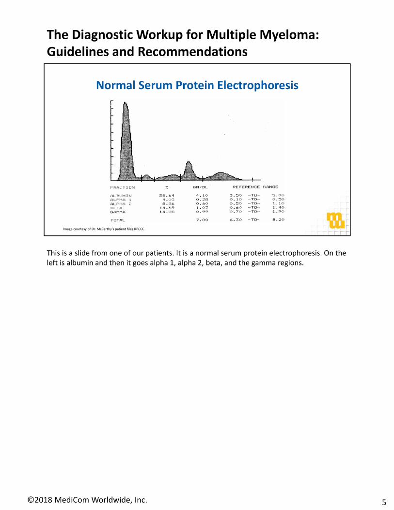
Normal Serum Protein Electrophoresis
Image courtesy of Dr. McCarthy’s patient files RPCCC
This is a slide from one of our patients. It is a normal serum protein electrophoresis. On the left is albumin and then it goes alpha 1, alpha 2, beta, and the gamma regions.
5
The Diagnostic Workup for Multiple Myeloma: Guidelines and Recommendations
©2018 MediCom Worldwide, Inc.

IgG Monoclonal Gammopathy (Myeloma)
Image courtesy of Dr. McCarthy’s patient files RPCCC
Here is an abnormal SPEP showing, on the right‐hand side, the M‐spike. The one thing to remember is that this is measured as area under the curve. The radiologist will set the calipers and will measure on either side of this M‐spike, so there’s often a variation by 10% to 15% from read to read. That’s something that’s important to remember as you are working up a patient and following them later on, that there is some element of qualitative measurement of the M‐spike.
6
The Diagnostic Workup for Multiple Myeloma: Guidelines and Recommendations
©2018 MediCom Worldwide, Inc.
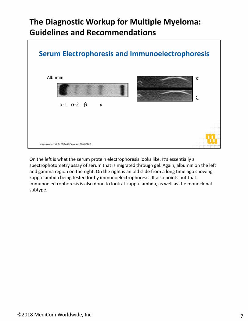
Albumin
α‐1 α‐2 β γ
Serum Electrophoresis and Immunoelectrophoresis
Image courtesy of Dr. McCarthy’s patient files RPCCC
On the left is what the serum protein electrophoresis looks like. It’s essentially a spectrophotometry assay of serum that is migrated through gel. Again, albumin on the left and gamma region on the right. On the right is an old slide from a long time ago showing kappa‐lambda being tested for by immunoelectrophoresis. It also points out that immunoelectrophoresis is also done to look at kappa‐lambda, as well as the monoclonal subtype.
7
The Diagnostic Workup for Multiple Myeloma: Guidelines and Recommendations
©2018 MediCom Worldwide, Inc.

8
Normal Urine Electrophoresis
Image courtesy of Dr. McCarthy’s patient files RPCCC
This is a normal urine electrophoresis showing that sometimes there is a tiny bit of albumin there on the far left.
The Diagnostic Workup for Multiple Myeloma: Guidelines and Recommendations
©2018 MediCom Worldwide, Inc.

9
Light Chain Disease Urine Electrophoresis
Image courtesy of Dr. McCarthy’s patient files RPCCC
Here is an abnormal urine collection showing light chains in aggregate there on the right, which is often seen with light chain disease. Obviously, if somebody has nephrotic syndrome, they’ll be putting out a lot more albumin which will be heavily present on the left side of the electrophoretic curve.
The Diagnostic Workup for Multiple Myeloma: Guidelines and Recommendations
©2018 MediCom Worldwide, Inc.

Rouleaux Formation
Image courtesy of Dr. McCarthy’s patient files RPCCC
This slide shows a rouleaux formation, which essentially looks like a stack of coins that have been pushed over. Aggregated there in the middle, you can see the red cells that are stuck together, and this is a manifestation in particular of a high immunoglobulin protein. You often won’t see this rouleaux formation in light chain disease only, but you will see it with IgG, IgA, and IgM monoclonal proteins in high amount in the peripheral blood.
10
The Diagnostic Workup for Multiple Myeloma: Guidelines and Recommendations
©2018 MediCom Worldwide, Inc.

Image courtesy of Dr. McCarthy’s patient files RPCCC
On this slide you can see on the left some small lytic lesions with the little arrows pointing to them. On the right there are also holes in these long bones and oftentimes it is the long bones that will have lytic lesions.
11
The Diagnostic Workup for Multiple Myeloma: Guidelines and Recommendations
©2018 MediCom Worldwide, Inc.

Image courtesy of Dr. McCarthy’s patient files RPCCC
30
39mm
mm
On the next slide, you’ll see on the right‐hand side, it says 30 mm where there’s a large lytic lesion in the skull. Then on the left‐hand side you’ll see where the arrow is pointing is mottled, and this is where there are multiple small lesions, so you can see a discrete hole as well as the mottling. I don’t have any MRI pictures to show, but MRI has given us now a lot more sensitivity with regard to picking up lesions even before they are readily apparent on the skeletal survey.
12
The Diagnostic Workup for Multiple Myeloma: Guidelines and Recommendations
©2018 MediCom Worldwide, Inc.

Useful Under Some Circumstances• Whole body low‐dose CT scan• Whole body or skeletal MRI or whole body
PET/CT scan• Tissue biopsy to diagnose a solitary
osseous or extraosseous plasmacytoma• Bone densitometry• Plasma cell proliferation• Staining of marrow and fat pad for amyloid• Serum viscosity• HLA typing• Echocardiogram• Evaluation for light chain amyloidosis,
if appropriate
Initial Diagnostic Workup• History and physical exam• CBC, differential, platelet count• Exam of peripheral blood smear• Serum BUN/creatinine, electrolytes, albumin, and calcium• Creatinine clearance (calculated or measured directly)• Serum uric acid• Serum LDH and beta‐2 macroglobulin• Serum quantitative immunoglobulins, serum protein electrophoresis
(SPEP), serum immunofixation electrophoresis (SIFE)• 24‐hour urine for total protein, urine protein electrophoresis (UPEP),
urine immunofixation electrophoresis (UIFE)• Serum free light chain (FLC) assay• Skeletal survey or whole body low‐dose CT scan• Unilateral bone marrow aspirate + biopsy, including bone marrow
immunohistochemistry and/or bone marrow flow cytometry• Metaphase cytogenetics on bone marrow• Plasma cell FISH [del 13, del 17p13, t(4;14), t(11;14), t(14;16), t(14;20),
1q21 amplification], 1p abnormality
For complete details including clinical presentation and all footnotes, refer to https://www.nccn.org/professionals/physician_gls/pdf/myeloma.pdf
NCCN Guidelines v4.2018 Deletions from previous versions shown in red strikethrough, additions in pink boxes
This slide the updated NCCN Guidelines. The pink boxes outline what’s new: examination of the peripheral smear, evaluation of the creatinine clearance, whole body low‐dose CT, and they’ve added new chromosome abnormalities to look for when assessing risk. There are other things as part of the workup that could be done including skeletal MRI, echocardiogram (especially in cases where carfilzomib may be considered early on in the therapy), and evaluating for light chain amyloidosis as appropriate.
13
The Diagnostic Workup for Multiple Myeloma: Guidelines and Recommendations
©2018 MediCom Worldwide, Inc.

Follow‐up/Surveillance• CBC, differential, platelet count• Serum BUN, creatinine, corrected calcium• Serum quantitative immunoglobulins, SPEP, SIFE• 24‐hour urine for total protein, UPEP, UIFE• Serum FLC assay as clinically indicated• Skeletal survey or whole body low‐dose CT scan as clinically indicated• Bone marrow aspirate and biopsy at relapse with FISH as clinically
indicated• Whole body or skeletal MRI as clinically indicated• PET/CT scan as clinically indicated• Multi‐parameter flow cytometry as clinically indicated
Primary TreatmentObserve at 3‐ to 6‐mo intervals (category 1)ORClinical trial
NCCN Guidelines v4.2018Smoldering (Asymptomatic) Myeloma
Deletions from previous versions shown in red strikethrough, additions in pink boxes
For complete details including clinical presentation and all footnotes, refer to https://www.nccn.org/professionals/physician_gls/pdf/myeloma.pdf
This is the latest, showing again now the new additions which are whole body low‐dose CT as part of the follow up in surveillance (as clinically indicated), and skeletal MRI. This is for smoldering asymptomatic myeloma before it progresses to symptomatic myeloma.
14
The Diagnostic Workup for Multiple Myeloma: Guidelines and Recommendations
©2018 MediCom Worldwide, Inc.

Follow‐up/Surveillance• CBC, differential, platelet count• Serum BUN, creatinine, corrected calcium• Serum quantitative immunoglobulins, SPEP, SIFE as clinically indicated• 24‐hour urine for total protein, UPEP, UIFE• Serum FLC assay as clinically indicated• Skeletal survey or whole body low‐dose CT scan as clinically indicated• Bone marrow aspirate and biopsy at relapse with FISH as clinically
indicated• Whole body or skeletal MRI as clinically indicated• PET/CT scan as clinically indicated• Assess for stem cell transplant candidacy:
• Refer for evaluation at a stem cell transplant center• Harvest stem cells (adequate consider for 2 transplants)
NCCN Guidelines v4.2018Active (Symptomatic) Myeloma
Deletions from previous versions shown in red strikethrough, additions in pink boxes
For complete details including clinical presentation and all footnotes, refer to https://www.nccn.org/professionals/physician_gls/pdf/myeloma.pdf
Primary TreatmentMyeloma therapy,bisphosphonates or denosumab+ adjunctive treatment as indicated
Venetoclax is not FDA approved for use in patients with multiple myeloma
The next slide shows followup in the surveillance of somebody who is getting active therapy. The big thing to note now is the addition of denosumab – which now has a label for preventing skeletal‐related events – in addition to the bisphosphonates. Also in follow‐up and surveillance: use of the serum free light chain assay if required to follow up disease response. Again, whole body low‐dose CT is indicated or skeletal survey, and in particular now a bone marrow aspirate at relapse with FISH analysis. This had not been done as often in the past, where if patients had an elevated monoclonal protein it was obvious that they were having disease progression. Sometimes a bone marrow test would not be done but we find that it is now useful because there are certain druggable cytogenetic abnormalities: in particular, 11;14 which may not be present at diagnosis but may be present at relapse. Also, venetoclax in an off‐label way has been utilized with bortezomib‐dexamethasone for those patients with good response. It is just something to be thinking about because multiple myeloma is a heterogeneous disease and it can behave differently at relapse when you compare it to what the abnormalities were at diagnosis. Again there are issues with regard to stem cell transplant candidacy. We are seeing still that’s an indication for those who are transplant illegible. There are some patients who may be considered for two transplants, but that’s more for another discussion.
15
The Diagnostic Workup for Multiple Myeloma: Guidelines and Recommendations
©2018 MediCom Worldwide, Inc.

NCCN Guidelines v4.2018Active (Symptomatic) Myeloma
Deletions from previous versions shown in red strikethrough, additions in pink boxes
For complete details including clinical presentation and all footnotes, refer to https://www.nccn.org/professionals/physician_gls/pdf/myeloma.pdf
Follow‐up/Surveillance• CBC, differential, platelet count• Serum quantitative immunoglobulins, SPEP, SIFE• 24‐hour urine for total protein, UPEP, UIFE• Serum BUN, Creatinine, calcium• Serum FLC assay as clinically indicated• Skeletal survey or whole body low‐dose CT scan as
clinically indicated• Bone marrow aspirate and biopsy as clinically indicated• Whole body MRI as clinically indicated• PET/CT scan as clinically indicated• Assess minimal residual disease (MRD) as indicated
Response after Primary TherapyAutologous stem cell transplant
OR
Allogeneic stem cell transplant
OR
• Monitor as aboveContinuous myeloma therapy and/or maintenance therapy
This is also the old guidelines and the new guidelines. Again, the big one for symptomatic myeloma is the follow up with whole‐body low‐dose CT. This is after transplant. Most patients who are transplant eligible will be considered for autologous transplant. Allogeneic transplant (in the middle) is still more of a protocol situation; it's not considered standard as yet. Then there’s a whole idea of continuous therapy or maintenance therapy which now has a label for those who have received a stem cell transplant.
16
The Diagnostic Workup for Multiple Myeloma: Guidelines and Recommendations
©2018 MediCom Worldwide, Inc.

Hyperdiploidy: Odd numbered chromosomes are often duplicated. Probes detecting numerical changes involving #5 (green), #9 (aqua) and #15(red). 3 copies instead of the expected 2 copies.
IGH/CCND1 rearrangement. Dual color, dual fusion probesCCND1 at 11q13: red and IGH: green.Normal: 1 red (normal #11) & 1 green (normal #14).2 “fusion” red/green signals: abnormal der(11) & der(14)
Slide courtesy of AM Block RPCCC
This slide shows hyperdiploidy on the left, and interestingly it is almost the odd chromosomes: chromosome 5 in green, chromosome 9 in blue, and chromosome 15 in red showing hyperdiploidy or trisomies. Now this is another FISH analysis. This one is showing the 11;14: normal is where you see two signals separate (a red and a green signal) and when they fuse, this means there’s a fusion between chromosome 11 and 14 and thus a translocation. As mentioned earlier, this is a chromosome abnormality which now has a drug that fits for treatment in the relapse/refractory setting. I would like to thank Dr. AnneMarie Block for providing me both the slides.
17
The Diagnostic Workup for Multiple Myeloma: Guidelines and Recommendations
©2018 MediCom Worldwide, Inc.

Green probe: chromosome #17 centromere, red probe: P53. Normal chromosome: one green/one red signal pattern. Deletion: loss of a red signal from one 17.
Green probe: CDKN2C; short arm at 1p32.3 & red probe: CKS1B; long arm at 1q21.3. Gains of the long arm, show3‐4 red signals
Slide courtesy of AM Block RPCCC
Here’s another one showing a very important abnormality, the chromosome 17 abnormality. Normally, what you would see is two red dots, two green dots per cell, and if there’s a loss of a signal. What you are seeing is now a deletion of 17 and this is important prognostically because chromosome 17 carries a less good prognosis. This is one looks at chromosome 1 and you can see here there is a gain of a red signal of the long arm showing that you now have a 1q reduplication. In some cases different probes will tell you there is a 1p deletion, also a higher risk cytogenetic abnormality.
18
The Diagnostic Workup for Multiple Myeloma: Guidelines and Recommendations
©2018 MediCom Worldwide, Inc.

Multiple Myeloma Workup and Risk Factors
• Performance status/comorbidities, not age– PS matters more than age
– Renal failure [bortezomib‐containing regimen (BCR)]1
– Supportive care: anticoagulation with IMiDs, zoster prophylaxis, bisphosphonates
• International Scoring System (check LDH and B2‐microglobulin)– Stage II or III2
• Cytogenetics/molecular testing– CD138 selection of marrow aspirate– Metaphase karyotyping: del(13) (BCR)3
– FISH: t(4: 14), (14:16) del(1p), +(1q), del(17p) (BCR)4
– Molecular: GEP 70, EMC‐92 (validation and what to do with high risk patients)5,6
• Other disease features– Extra‐medullary disease, plasma cell leukemia, high LDH
Adapted from Ludwig H, et al. Oncologist. 2012;17:592‐606.; 1Sonneveld P, et al. J Clin Oncol. 2012;30(24):2946‐2955. 2Greipp PR, et al. J Clin Oncol. 2005;23(15):3412‐3420. 3Jagannath S, et al. Leukemia. 2007;21(1):151‐167. 4Munshi NC, et al. Blood. 2011;117(18):4696‐4700. 5Shaughnessy JD, et al. Br J Haematol. 2007;137(6):530‐536. 6Kuiper R, et al. Leukemia. 2012;26(11):2406‐2413.
Higher risk in orange
These are the things that you’re thinking about with the workup and risk factors, performance status mattered most with regard to what regimens are chosen. Renal failure is very important because certain IMiDs such as lenalidomide cannot be used in renal failure, and bortezomib‐based regimens are emphasized. Supportive care, if we can emphasize that more, that patients who are on IMiDs need anticoagulation, those receiving proteasome inhibitors needs zoster prophylaxis and bisphosphonates to prevent skeletal‐related events. The scoring system I’ll go over in a little bit. It’s very important to have a CD138 selected marrow aspirate for FISH because metaphase karyotyping really is not very helpful. Occasionally, it picks up the deletion 13 but often the metaphase karyotyping will be normal and the FISH will be abnormal and that’s what allows you to risk stratify. There are molecular tests, GEP70 and EMC92 have been used to identify high‐risk patients; and then high‐risk features such as extramedullary disease, plasma cell leukemia, and high LDH.
19
The Diagnostic Workup for Multiple Myeloma: Guidelines and Recommendations
©2018 MediCom Worldwide, Inc.

Diagnostic Evaluation of Suspected Myeloma
Myeloma Requiring Therapy
• Any urine or serum M‐protein
• Clonal BM plasma cells ≥10% or biopsy proven plasmacytoma and
• Any CRAB criteria or
• Any myeloma‐defining event (MDE):
Clonal BM % ≥60%
Abnormal FLC ratio
≥100 (involved kappa) or
<0.01 (involved lambda)
Two or more focal lesions on MRI, ≥5 mm
Adapted from Ghobrial IM, et al. Blood. 2014;124:3380‐3388.; Rajkumar SV, et al. Lancet Oncol. 2014;15:e538‐548.;Rajkumar SV, et al. Blood. 2015;125:3069‐3075.
Smoldering MM • Serum M‐protein ≥3g/dL or• Urinary monoclonal protein
>500 mg per 24 hours and/or• Light‐chain restricted bone
marrow plasma cells ≥10% and up to 60%
• No CRAB criteria or MDE (end organ damage)
MGUS • Serum M‐protein <3g/dL• Light‐chain restricted
bone marrow plasma cells <10%
• No end organ damage (CRAB Criteria or MDE)
CBC, creatinine, calcium, serum protein electrophoresis (SPEP), serum immunofixation (IFE), 24‐hour urine for UPEP/IFE, Ig levels, SFLC, bone marrow test, CD 138 selected FISH, whole body MRI, especially with back pain, PET scan. skeletal survey being replaced by WBMRI and PET scan
CRAB Criteria: Hypercalcemia, Renal insufficiency/Failure, Anemia, Bone lesions
This is the overall diagnostic evaluation of a suspected myeloma patient. The upper box tells you the types of test that you’ll need to get, which have been described in the previous slides. On the right are patients with MGUS who can be followed: low levels of protein and no end organ damage. In the middle are smoldering myeloma patients: those with higher levels of proteins in the blood and urine, a small amount of plasma cells (more than in MGUS but less than true myeloma), and no CRAB or myeloma‐defining events. On the left are higher levels of plasma cells in the marrow, CRAB criteria, myeloma‐defining events (which are clonal bone marrow greater than 60% plasma cells, abnormal free light chain ratio of greater than 100 or less than 0.01 depending on the light chain involved, and two or more focal lesions on MRI).
20
The Diagnostic Workup for Multiple Myeloma: Guidelines and Recommendations
©2018 MediCom Worldwide, Inc.

For complete details including clinical presentation and all footnotes, refer to https://www.nccn.org/professionals/physician_gls/pdf/myeloma.pdf
NCCN Guidelines v4.2018 Definition of Active and Smoldering Myeloma
Smoldering (Asymptomatic) Myeloma
• Serum monoclonal protein ≥3 g/dL;
OR
• Bence‐Jones protein ≥500 mg/24 h
AND/OR
• Clonal bone marrow plasma cells 10%–60%
AND
• Absence of myeloma‐defining events or amyloidosis
– If skeletal survey negative, assess for bone disease with whole body MRI or PET/CT
Active (Symptomatic) Myeloma• Clonal bone marrow plasma cells ≥10% or biopsy‐
proven bony or extramedullary plasmacytomaAND• Any one or more of the following myeloma‐defining
events:– Calcium >0.25 mmol/L (>1 mg/dL) higher than the
upper limit of normal or >2.75 mmol/L (>11 mg/dL)– Renal insufficiency (creatinine >2 mg/dL) [>177
μmol/L] or creatinine clearance <40 mL/min– Anemia (hemoglobin <10 g/dL or hemoglobin >2 g/dL
below the lower limit of normal)– One or more osteolytic bone lesions on skeletal
radiography, CT, or PET/CT– Clonal bone marrow plasma cells ≥60%– Abnormal serum FLC ratio ≥100 (involved kappa) or
≤0.01 (involved lambda)– >1 focal lesions on MRI studies ≥5 mm
These are definitions of smoldering and active myeloma, and this reiterates what I said before in terms of the CRAB criteria as well as MDEs, which are the clonally abnormal plasma cells of greater than 60%, severely abnormal free light chain ratio and greater than one focal lesion on the MRI that are greater than 5 mm.
21
The Diagnostic Workup for Multiple Myeloma: Guidelines and Recommendations
©2018 MediCom Worldwide, Inc.

Revised International Staging System for MM: A Report from the International Myeloma Working Group
Stg Factor Pt N (%) 5 yr PFS 5 yr OS
I Absence of adverse factors (no high LDH, ISS 2 or 3, t(4;14) and/or t(14;16) and/or del(17p))
871 (28) 55% 82%
II Not R‐ISS I or III 1,894 (62) 36% 62%
III ISS 3 and high‐risk CA by iFISH or high LDH 295 (10) 24% 40%
From: GIEMEMA. PETHEMA/GEM, HOVON/GMMG, IFM
Palumbo A, et al. J Clin Oncol. 2015;33:2863.; Moreau P, et al. J Clin Oncol. 2014;32:2173.
β2‐M=beta‐2 microglobulin; CA=chromosomal abnormalities; iFISH=interphase fluorescent in‐situ hybridization; ISS=International Staging System; LDH=lactate dehydrogenase; L=liter; mg=milligrams; MM=multiple myeloma; Pts=patients; R‐ISS=Revised International Staging System. 3,060 evaluable patients
Original ISS Stage Criteria
I Serum β2‐M <3.5 mg/L, serum albumin ≥3.5 g/dL
II Not ISS stage I or III
III Serum β2‐M ≥5.5 mg/L
This is why it’s important to risk stratify. The old ISS is on the top which was based on beta‐2 microglobulin and albumin levels, and there are sometimes patients referred into us who will not have had a beta‐2 done, which was important for original ISS staging. Now in the revised ISS what we’re seeing is the number of patients who have the different stages. Stage I are those who have no risk factors. They don’t have a high LDH, high‐burden disease or 4;14, 14;16, or deletion 17. Stage II is sort of in between, neither I nor II. In stage III, most importantly, are those who have high‐burden disease, high‐risk cytogenetics by FISH, or high LDH. It comprises about 10% of the patient population. As you can see here next to it in the progression free survival (PFS), you will see that the five‐year PFS is 24% versus those who are stage I at 55% and the overall survival is inferior. This is a patient population we need to know about so that they can be considered for protocol therapies or monitored very aggressively for recurrence of disease.
22
The Diagnostic Workup for Multiple Myeloma: Guidelines and Recommendations
©2018 MediCom Worldwide, Inc.

Tackling Early Morbidity and Mortality in Myeloma (TEAMM): Assessing the Benefit of Antibiotic Prophylaxis and Its Effect on Healthcare
Associated Infections in 977 Patients
• Phase III trial of newly diagnosed multiple myeloma (NDMM) patients requiring therapy, randomized to levofloxacin (500 mg) or placebo for 12 weeks
• Primary endpoint
– Febrile episodes (oral temp of ≥38°C, Rx with anti‐infectives) and/or death due to any cause in the first 12 weeks
• Results:
– Placebo: 134 of 488 patients (27%) (112 febrile episodes; 15 deaths; 7 febrile episodes and death)
– Levofloxacin: 95 of 489 patients (19%) (87 febrile episodes; 4 deaths; 4 febrile episodes and death)
– HR 1.52 (95%CI 1.17‐1.97) P = .002
– HR 1.80 (95% CI 1.26‐2.43) P = .0008; greater benefit with levofloxacin and trimethoprim sulfamethoxazole1
Levofloxacin is not FDA approved for this use in patients with multiple myeloma.1Drayson M, et al. Blood. 2017;130:903.
On this slide, we see the results of a British study from the last ASH meeting in 2017, tackling early morbidity and mortality in myeloma, assessing the benefit of antibiotic prophylaxis and its effect on healthcare‐associated infections in 977 patients. What the British group did was they had a phase 3 randomized trial where they looked at newly diagnosed patients who required therapy. They were randomized to either levofloxacin for placebo for 12 weeks. The primary endpoint was febrile episodes, as well as treatment with anti‐infectives, and/or death due to any cause in the first 12 weeks of treatment. The results are, of the placebo patients, 134 of 488 patients (27%) developed febrile episodes or died, whereas in the levofloxacin group, 95 of 489 (19%) developed febrile episodes or died. The hazard ratio is 1.52. This is highly significant at equal 0.002. When you account for the fact that some of these patients also receive trimethoprim/sulfamethoxazole antimicrobial prophylaxis, the hazard ratio is even higher at 1.8. We think that this may be a new standard, we would like to be able to study this a little bit more, and we await the publication. We think that this may now become a new recommendation for the newly diagnosed myeloma patient, and we are beginning to integrate this type of approach into our patients who are requiring induction therapy.
23
The Diagnostic Workup for Multiple Myeloma: Guidelines and Recommendations
©2018 MediCom Worldwide, Inc.

Tackling Early Morbidity and Mortality in Myeloma (TEAMM)
Drayson M, et al. Blood. 2017;130:903.
Unadjusted treatment effect n=977, 229 events
Treatment adjusted for baseline factors and use of septrin n=783, 192 events
Unadjusted treatment effect n=977, 229 events
Treatment adjusted for baseline factors and use of septrin n=783, 192 events
This just shows you the Kaplan‐Meier curve in terms of the incidence of febrile episodes or deaths. Placebo is in blue, the levofloxacin group is in red, and this on the right goes through all the different factors that they adjusted for: the performance status, fractures, neutrophil count, and then the prophylactic trimethoprim/sulfamethoxazole use as well as the staging.
24
The Diagnostic Workup for Multiple Myeloma: Guidelines and Recommendations
©2018 MediCom Worldwide, Inc.

Algorithms for Myeloma Therapy in US 2018
Adapted from Reece D, et al. Lancet. 2015;385:2197‐2208.
ASCTMaintenance until PD vs fixed duration
Chemotherapyuntil PD vs fixed duration
Fit, age limit?
ASCT >12‐18 mos; ASCT <12‐18 mos ProgressionASCT >12‐18 mos; ASCT <12‐18 mos Progression
2, 3 or 4 drug combo
Early vs Late ASCT Pt/MD choice
??
“Less aggressive relapse” Lenalidomide + dex +/‐ elotuzumab, ixazomib or carfilzomib or daratumumab
Bortezomib + dex +/ daratumumab, panobinostat; pomalidomide + dex +/‐ other drug
“Less aggressive relapse” Lenalidomide + dex +/‐ elotuzumab, ixazomib or carfilzomib or daratumumab
Bortezomib + dex +/ daratumumab, panobinostat; pomalidomide + dex +/‐ other drug
PS, Age, Comorbidities
Reinduction and 2nd ASCT
Reinduction and 2nd ASCT
Frail, age (>80 years?)
Preferably Combination
Regimen or Trial
Preferably Combination
Regimen or Trial
< 6‐9 mos,
Change Regimen
< 6‐9 mos,
Change Regimen
> 6‐9 mos,
Could Repeat Regimen
> 6‐9 mos,
Could Repeat Regimen
“Advanced relapse”(Pomalidomide + dex +/‐ other, daratumumab, carfilzomib, panobinostat+bortezomib/dex
bendamustine, clinical trial or re‐use previous drugs)
“Advanced relapse”(Pomalidomide + dex +/‐ other, daratumumab, carfilzomib, panobinostat+bortezomib/dex
bendamustine, clinical trial or re‐use previous drugs)
This is the algorithm for therapy in 2018, I won’t go over it in great detail. It used to be that we would be thinking about just age, but we really need to think about fitness. You have very fit patients who might be considered early on for transplant. There are some patients who are delayed, and we recommend that they have their stem cells collected and then transplant at recurrence. In the past we commonly used two agents, now we are thinking of three, and there are some trials now looking at four‐drug combinations which will perhaps in the future be the standard, as opposed to three‐drug combinations for the frail. It may now be three or four‐drug combinations depending on what is chosen and how the patient is monitored and treated over time, and then there is a variety of different combinations that can be utilized for less or more advanced relapses. That is getting outside the scope of this presentation, but it brings up the good problem for our patients that we have multiple choices for managing the disease.
25
The Diagnostic Workup for Multiple Myeloma: Guidelines and Recommendations
©2018 MediCom Worldwide, Inc.

Continued Therapy to Maintain Response Induction
Transplant Ineligible
Supportive Care: Infectious disease prophylaxis, antivirals, DVT prophylaxis, bisphosphonates, mitigation of steroid effects
Slide Adapted from Andrew Spencer.
I adapted this slide from Andrew Spencer from Australia. For the transplant‐ineligible, I think the standard can be induction and then continued therapy to maintain response. As mentioned earlier, supportive care for IV prophylaxis, DVT prophylaxis, bisphosphonates, as well as mitigation of steroid effects.
26
The Diagnostic Workup for Multiple Myeloma: Guidelines and Recommendations
©2018 MediCom Worldwide, Inc.

Maintenance until PDASCTInduction
ConsolidationMaintenance
ASCTInduction
Transplant Eligible
Supportive Care: Infectious disease prophylaxis, antivirals, DVT prophylaxis, bisphosphonates, mitigation of steroid effects
For the transplant eligible, I think the standard now would be, after induction and transplant, maintenance until progression. In Europe, there’s a bit more interesting consolidation and for high‐risk patients, we will consider consolidation. Again, there are the same issues with supportive care.
27
The Diagnostic Workup for Multiple Myeloma: Guidelines and Recommendations
©2018 MediCom Worldwide, Inc.

Key Points
• It is important to do a full workup for the suspected and newly diagnosed multiple myeloma patient
• This includes key laboratory tests such as serum LDH and beta‐2 microglobulin, CD‐138 selected FISH from the bone marrow
• Consider anti‐microbial prophylaxis during induction therapy
• Myeloma‐defining events have been expanded to include: clonal BM % >60%, abnormal FLC ratio >100 (involved kappa) or <0.01 (involved lambda) and 2 or more focal lesions on MRI ≥5 mm
• Follow patients every 3 months for by SFLC/SPEP, check a bone marrow test when progression is suspected
These are the key points that I would like to leave you with at the end of this talk. It is important to do a full workup if suspecting a newly diagnosed multiple myeloma patient. This includes laboratory testing such as serum LDH and beta‐2 microglobulin, as well as CD138 selected FISH from the bone marrow to adequately risk‐stratify the patient and stage them. I would consider what is currently sort of an off‐label use of antimicrobial prophylaxis for at least three months during induction therapies. The British group did show a decrease in infections and deaths. We must also consider the use of myeloma‐defining events in addition to CRAB criteria for initiation of therapy (greater than 60% plasma cells, an abnormal free light chain ratio, and two or more focal lesions on the MRI greater than 5 mm. We did not discuss this in great detail, but I mentioned it in the guidelines that the patient should be followed every three months by serum free light chains as well as SPEP, and check a bone marrow test when disease progression is suspected.
28
The Diagnostic Workup for Multiple Myeloma: Guidelines and Recommendations
©2018 MediCom Worldwide, Inc.

People and Services Who Make the BMT Program Possible
• S Balderman• G Chen• C Ho• M Ross• M Aungst• J Bertolo‐Kurtzner• M Burgess• M Everett• A Koeppel• J Lex‐Sikinoff• A Nemmer• S Myszka• A Phillips ‐Hall• P Paplham• R Russell• A Beck• S Flavin• D Oliansky• F Zhang• A Kariapper• H Jacobson• R McKenzie• S Oakley
• T Hahn• S Schinnagel• L Privitere• K West• J Pleskow• M Cimino• M Steward• D Swinnich• K Stawicki• D Cipolla• K Dubel• P Lipka• S Siconolfi• C Warren• L Yoerg• S Pry• R De Wald• L Markel• R Kumpf• K Dunn• A Kader• J NIchols• H Bashaw• D Manfredi
• Managed Care and Finance Svc• S Randolph• M Budd• Medical Oncology Fellows• Leukemia, Lymphoma and Myeloma Services• 5 East, 5 North and 6 North Nursing and Secretarial Staff• Hospitalist Staff• F Hernandez• E Wang• M Ernstoff• J Lau• E Repasky and Lab• J Hillengass• P Torka• P Wallace• J Tario• Y Zhang• S Johnson• C Johnson• V Filadora• T Schwaab• B Kuvshinoff• Support by RPCI Alliance, RPCI Core
Grant, NCI, NHLBI and B and E McCarthy
• T Chodon• R Koya• C Choi• J Becker • E Duman • L Vesneske• Rad Onc Service• Radiology Svc• Surgery Svc• Pathology Svc• Lab Medicine• Stem Cell Lab• Apheresis Unit• S Szeglowski• L Regan• S Segal• J Maxick‐Jason• J Kapinos• A Singh• ID service• B Segal
These are all the people I work with at Roswell Park and actually, this is just part of the people I work with. It is always a team effort to take care of these patients, because they can be complex in nature and require a multidisciplinary approach.
29
The Diagnostic Workup for Multiple Myeloma: Guidelines and Recommendations
©2018 MediCom Worldwide, Inc.

I’d like to thank you very much for viewing this activity. This is the happy neutrophil Thank you again .
30
The Diagnostic Workup for Multiple Myeloma: Guidelines and Recommendations
©2018 MediCom Worldwide, Inc.