Embed Size (px)
Citation preview
1994, 176(17):5474. J. Bacteriol.
P Chen, D I Andersson and J R Roth Salmonella typhimurium.The control region of the pdu/cob regulon in
http://jb.asm.org/content/176/17/5474Updated information and services can be found at:
These include:
CONTENT ALERTS more»cite this article),
Receive: RSS Feeds, eTOCs, free email alerts (when new articles
http://journals.asm.org/site/misc/reprints.xhtmlInformation about commercial reprint orders: http://journals.asm.org/site/subscriptions/To subscribe to to another ASM Journal go to:
on July 9, 2012 by UC
DA
VIS
SH
IELD
S LIB
RA
RY
http://jb.asm.org/
Dow
nloaded from
Vol. 176, No. 17JOURNAL OF BACEERIOLOGY, Sept. 1994, p. 5474-54820021-9193/94/$04.00+0Copyright X 1994, American Society for Microbiology
The Control Region of the pdu/cob Regulonin Salmonella typhimurium
PING CHEN,' DAN I. ANDERSSON,2 AND JOHN R. ROTH`*Department of Biology, University of Utah, Salt Lake City, Utah 84112,1 and Department of
Microbiology, Biomedical Center, Uppsala University, Uppsala S-75123, Sweden2
Received 14 April 1994/Accepted 28 June 1994
The pdu operon encodes proteins for the catabolism of 1,2-propanediol; the nearby cob operon encodesenzymes for the biosynthesis of adenosyl-cobalamin (vitamin B12), a cofactor required for the use of propanediol.These operons are transcribed divergently from distinct promoters separated by several kilobases. Theregulation of the two operons is tightly integrated in that both require the positive activator protein PocR andboth are subject to global control by the Crp and ArcA proteins. We have determined the DNA nucleotidesequences of the promoter-proximal portion of thepdu operon and the region between thepdu and cob operons.Four open reading frames have been identified, pduB, pduA, pduF, and pocR. The pduA and pduB genes are thefirst two genes of the pdu operon (transcribed clockwise). The pduA gene encodes a hydrophobic protein with56% amino acid identity to a 10.9-kDa protein which serves as a component of the carboxysomes of severalphotosynthetic bacteria. The pduF gene encodes a hydrophobic protein with a strong similarity to the GIpFprotein of Escherichia coli, which facilitates the diffusion of glycerol. The N-terminal end of the PduF proteinincludes a motif for a membrane lipoprotein-lipid attachment site as well as a motif characteristic of the MIP(major intrinsic protein) family of transmembrane channel proteins. We presume that the PduF proteinfacilitates the diffusion of propanediol. ThepocR gene encodes the positive regulatory protein of the cob andpduoperons and shares the helix-turn-helix DNA binding motif of the AraC family of regulatory proteins. Themutations cobR4 and cobR58 cause constitutive, pocR-independent expression of the cob operon under bothaerobic and anaerobic conditions. Evidence that each mutation is a deletion creating a new promoter near thenormal promoter site of the cob operon is presented.
Under anaerobic conditions, the bacterium Salmonella typhi-murium is capable of full synthesis of the cofactor adenosyl-cobalamin (vitamin B,2) (19). Genetic studies have shown thatmost of the B12 synthetic genes are located in the cob operonat minute 41 of the genetic map (13, 20). The phenotypicdefect of these mutants can be corrected by supplying exoge-nous cyanocobalamin (CN-B12). Most of the cob operon hasbeen cloned, and its nucleotide sequence has been determined.It has been possible to assign functions to many of the cobproteins of S. typhimurium by comparing their inferred aminoacid sequences to those of cobalamin synthetic genes inPseudomonas denitnificans (34).
Only four B,2-dependent enzymes in S. typhimurium havebeen identified. Homocysteine methyl transferase (metH)transfers a methyl group from N-5-methyltetrahydrofolate toconvert L-homocysteine to L-methionine (36). Ethanolamineammonia-lyase cleaves ethanolamine to acetaldehyde and am-monia (32, 33). Queuosine synthetase reduces epoxyqueuosineto queuosine, a modified base found in four tRNAs (17).Propanediol dehydratase rearranges 1,2-propanediol to yieldpropionaldehyde and H20 (18, 38).Enzymes for the use of propanediol are encoded by the pdu
operon, which is located adjacent to the cob operon at minute41 (19, 20). The pdu and cob operons are divergently tran-scribed and map together at minute 41 of the Salmonellachromosome. Recent work shows that both operons are in-duced by propanediol under the same conditions and that thisinduction is mediated by a single positive regulatory protein,PocR (7, 31). Furthermore, both thepdu and cob operons andthe gene for the positive activator (pocR) are subject to positive
* Corresponding author.
control by the global regulatory proteins Crp and ArcA (1, 3).The regulatory integration of thepdu and cob operons suggeststhat the most important use of B12 in S. typhimurium is thebreakdown of 1,2-propanediol. The produced propionalde-hyde can be used as a hydrogen sink when it is reduced topropanol and excreted (27). Propionaldehyde can also providea carbon and energy source aerobically when it is converted topropionyl-coenzyme A and ultimately to pyruvate (16, 17a, 18).The nucleotide sequence of the region between the diver-
gent pdu and cob operons is reported here. These datacomplete the sequence of the regulatory gene pocR, providethe sequence of two pdu genes, and reveal a new gene locatedbetween the operons, pduF, which we infer encodes a trans-porter of propanediol. We also report that two cob regulatorymutations (cobR4 and cobR58) are deletions that extend fromthe normal cob promoter into the region between the pdu andcob operons.
MATERUILS AND METHODS
Bacterial strains and plasmids. The bacterial strains used inthis study (Table 1) are all derived from S. typhimurium LT2except for strains MC1061, TT17507, and Tr17509, which arederived from Escherichia coli K-12. Plasmid pDA2979 is apUC19 plasmid vector carrying a 3.7-kb EcoRI DNA fragmentderived from the Salmonella chromosome. The insert includesthe control region of the pdulcob regulon. Plasmid pPC3 is apUC118 vector carrying a 1.4-kb Eco47III subfragment fromthe middle of the original 3.7-kb S. typhimurium fragment.Growth media and 13-galactosidase assay. Rich medium was
Difco nutrient broth with NaCl added (15 g/liter). Minimalmedium was the E medium of Vogel and Bonner (41) withglucose (0.2%) as the carbon source. For P-galactosidase
5474
on July 9, 2012 by UC
DA
VIS
SH
IELD
S LIB
RA
RY
http://jb.asm.org/
Dow
nloaded from
JOURNAL OF BACTERIOLOGY, Sept. 1994, p. 5474-5482 0021-9193/94/$04.00+0 Copyright © 1994, American Society for Microbiology
Vol. 176, No. 17
The Control Region of the pdu/cob Regulon in Salmonella typhimurium
PING CHEN,! DAN I. ANDERSSON,2 AND JOHN R. ROTHh
Department of Biology, University of Utah, Salt Lake City, Utah 84112,1 and Department of Microbiology, Biomedical Center, Uppsala University, Uppsala S-75123, Sweden2
Received 14 April 1994/Accepted 28 June 1994
The pdu operon encodes proteins for the catabolism of l,2-propanediol; the nearby cob operon encodes enzymes for the biosynthesis of adenosyl-cobalamin (vitamin BU>, a cofactor required for the use of propanediol. These operons are transcribed divergently from distinct promoters separated by several kilobases. The regulation of the two operons is tightly integrated in that both require the positive activator protein PocR and both are subject to global control by the Crp and AreA proteins. We have determined the DNA nucleotide sequences of the promoter-proximal portion of the pdu operon and the region between the pdu and cob operons. Four open reading frames have been ide .. tified, pduB, pduA, pduF, and pocR. The pduA and pduB genes are the first two genes of the pdu operon (transcribed clockwise). The pduA gene encodes a hydrophobic protein with 56% amino acid identity to a lO.9-kDa protein which serves as a component of the carboxysomes of several photosynthetic bacteria. The pduF gene encodes a hydrophobic protein with a strong similarity to the GlpF protein of Escherichia coli, which facUitates the dimlsion of glycerol. The N-terminal end of the PduF protein includes a motif for a membrane lipoprotein-lipid attachment site as well as a motif characteristic of the MIP (major intrinsic protein) family of transmembrane channel proteins. We presume that the PduF protein facilitates the diJFusion of prop,anediol. The pocR gene encodes the positive regulatory protein of the cob and pdu operons and shares the helix-turn-helix DNA binding motif of the AraC family of regulatory proteins. The mutations cobR4 and cobR58 cause constitutive, pocR-independent expression of the cob operon under both aerobic and anaerobic conditions. Evidence that each mutation is a deletion creating a new promoter near the normal promoter site of the cob operon is presented.
Under anaerobic conditions, the bacterium Salmonella typhimurium is capable of full synthesis of the cofactor adenosylcobalamin (vitamin Bl2) (19). Genetic studies have shown that most of the B12 synthetic genes are located in the cob operon at minute 41 of the genetic map (13, 20). The phenotypic defect of these mutants can be corrected by supplying exogenous cyanocobalamin (CN-BI2). Most of the cob operon has been cloned, and its nucleotide sequence has been determined. It has been possible to assign functions to many of the cob proteins of S. typhimurium by comparing their inferred amino acid sequences to those of cobalamin synthetic genes in Pseudomonas denitrificans (34).
Only four Bl2-dependent enzymes in S. typhimurium have been identified. Homocysteine methyl transferase (metH) transfers a methyl group from N-5-methyltetrahydrofolate to convert L-homocysteine to L-methionine (36). Ethanolamine ammonia-lyase cleaves ethanolamine to acetaldehyde and ammonia (32, 33). Queuosine synthetase reduces epoxyqueuosine to queuosine, a modified base found in four tRNAs (17). Propanediol dehydratase rearranges 1,2-propanediol to yield propionaldehyde and H20 (18, 38).
Enzymes for the use of propanediol are encoded by the pdu operon, which is located adjacent to the cob operon at minute 41 (19, 20). The pdu and cob operons are divergently transcribed and map together at minute 41 of the Salmonella chromosome. Recent work shows that both operons are induced by propanediol under the same conditions and that this induction is mediated by a single positive regulatory protein, PocR (7, 31). Furthermore, both the pdu and cob operons and the gene for the positive activator (pocR) are subject to positive
• Corresponding author.
5474
control by the global regulatory proteins Crp and AreA (1, 3). The regulatory integration of the pdu and cob operons suggests that the most important use of Bl2 in S. typhimurium is the breakdown of 1,2-propanediol. The produced propionaldehyde can be used as a hydrogen sink when it is reduced to propanol and excreted (27). Propionaldehyde can also provide a carbon and energy source aerobically when it is converted to propionyl-coenzyme A and ultimately to pyruvate (16, 17a, 18).
The nucleotide sequence of the region between the divergent pdu and cob operons is reported here. These data complete the sequence of the regulatory gene pocR, provide the sequence of two pdu genes, and reveal a new gene located between the operons, pduF, which we infer encodes a transporter of propanediol. We also report that two cob regulatory mutations (cobR4 and cobR58) are deletions that extend from the normal cob promoter into the region between the pdu and cob operons.
MATERIALS AND METHODS
Bacterial strains and plasmids. The bacterial strains used in this study (Table 1) are all derived from S. typhimurium L T2 except for strains MC1061, TT17507, and TT17509, which are derived from Escherichia coli K-12. Plasmid pDA2979 is a pUC19 plasmid vector carrying a 3.7-kb EcoRI DNA fragment derived from the Salmonella chromosome. The insert includes the control region of the pdu/cob regulon. Plasmid pPC3 is a pUC118 vector carrying a l.4-kb Ec047III subfragment from the middle of the original 3.7-kb S. typhimurium fragment.
Growth media and p-galactosidase assay. Rich medium was Difco nutrient broth with NaCI added (15 g/liter). Minimal medium was the E medium of Vogel and Bonner (41) with glucose (0.2%) as the carbon source. For ~-galactosidase
CONTROL REGION OF THE SALMONELLA pdu/cob REGULON 5475
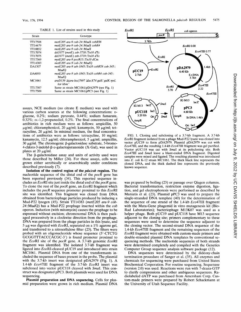
TABLE 1. List of strains used in this studyStrain Genotype
Tf17508 metE205 ara-9 cob-24::MudJ cobR58TT14679 metE205 ara-9 cob-24::MudJ cobR41T10852 metE205 ara-9 cob-24::MudJ1T17074 dellO77 (metE) zeb-3718::TnlO dTcTT17075 dell077 (metE) zeb-3719::TnlO dTcTT17569 metE205 ara-9 pocRl5::TnlO dCmFT14383 metE205 ara-9 cob-24::MudQDA1307 metE205 ara-9 zeb-1845::TnlO cobR58 cob-345::
MudQDA4095 metE205 ara-9 zeb-1845::TnlO cobR4 cob-345::
MudQMC1061 araD139 A(ara-leu)7697 AlacX74 galU galK rpsL
hsr hbm+FT17507 Same as strain MC1061/pDA2979 (see Fig. 1)FT17509 Same as strain MC1061/pPC3 (see Fig. 1)
assays, NCE medium (no citrate E medium) was used withvarious carbon sources at the following concentrations: D-
glucose, 0.2%; sodium pyruvate, 0.44%; sodium fumarate,0.32%; DL-1,2-propanediol, 0.2%. The final concentrations ofantibiotics in rich medium were as follows: ampicillin, 50,ug/ml; chloramphenicol, 20 ,ug/ml; kanamycin, 50 ,ug/ml; tet-racycline, 20 pLg/ml. In minimal medium, the final concentra-tions of antibiotics were as follows: tetracycline, 10 mg/ml;kanamycin, 12.5 ,ug/ml; chloramphenicol, 5 ,ug/ml; ampicillin,30 ,ug/ml. The chromogenic ,-galactosidase substrate, 5-bromo-4-chloro-3-indolyl-3-D-galactopyranoside (X-Gal), was used inplates at 20 ,ug/ml.The f-galactosidase assay and units of activity used were
those described by Miller (24). For these assays, cells weregrown either aerobically or anaerobically under conditionsdescribed previously (7).
Isolation of the control region of the pdulcob regulon. Thenucleotide sequence of the distal end of the pocR gene hasbeen reported previously (34). This reported sequence in-cludes an EcoRI site just inside the distal end of thepocR gene.To clone the rest of the pocR gene, an EcoRI fragment whichincludes the pocR sequence promoter proximal to this EcoRIsite was identified. This fragment was cloned from DNAobtained from bacteriophage particles produced by inducing aMud-P22 lysogen (45). Strain 1T14383 (metE205 ara-9 cob-24::MudQ) has a Mud-P22 prophage inserted within the coboperon. Induction (with mitomycin) causes the prophage to beexpressed without excision; chromosomal DNA is then pack-aged processively in a clockwise direction from the prophage.DNA was prepared from the phage particles in this lysate, and5 pLg was digested with EcoRI, separated on a 1% agarose gel,and transferred to a nitrocellulose filter (23). The filters wereprobed with an oligonucleotide whose sequence (5'-CTCTGGCGGTlTTACCCACGC-3') is found promoter proximal tothe EcoRI site of the pocR gene. A 3.7-kb genomic EcoRIfragment was identified. The isolated 3.7-kb fragment wasligated into EcoRI-cleaved pUC19 and introduced into strainMC1061. Plasmid DNA from one of the transformants in-cluded the sequence of bases present in the probe. The plasmidwith the 3.7-kb insert was designated pDA2979 (Fig. 1). A1.4-kb Eco47III fragment of the 3.7-kb EcoRI insert wassubcloned into vector pUC118 cleaved with SmaI. This con-struct was designated pPC3. Both plasmids were used for DNAsequencing.
Plasmid preparation and DNA sequencing. Cells for plas-mid preparation were grown in rich medium. Plasmid DNA
FIG. 1. Cloning and subcloning of a 3.7-kb fragment. A 3.7-kbEcoRI fragment isolated from a phage Mud-P22 lysate was cloned intovector pUC19 to form pDA2979. Plasmid pDA2979 was cut withEco47III, and the resulting 1.4-kb Eco47III fragment was gel purified.Vector pUC118 was cut with SmaI at its polycloning site. BothEco47III and SmaI leave a blunt-ended DNA fragment. Digestedsamples were mixed and ligated. The resulting plasmid was introducedinto E. coli K-12 strain MC1061. The thick black line represents thecloned DNA, and the thick dashed line represents the previouslyknown sequence.
was prepared by boiling (23) or passage over Qiagen columns.Bacterial transformation, restriction enzyme digestion, liga-tion, and gel electrophoresis were performed as described byManiatis et al. (23). Plasmid pPC3 was used to prepare thesingle-stranded DNA template (40) for the determination ofthe sequence of one strand of the 1.4-kb Eco47III fragmentwith the Muta-Gene phagemid in vitro mutagenesis kit (Bio-Rad Laboratories); bacteriophage M13K07 was used as a
helper phage. Both pUC19 and pUC118 have M13 sequenceadjacent to the cloning site; primers complementary to thesesequences were used to determine the sequence adjacent tothe cloning vector. The second-strand DNA sequence of the1.4-kb Eco47III fragment and the remaining sequences of theEcoRI fragment were obtained with custom-made primers anddouble-stranded plasmid DNA templates by conventional se-quencing methods. The nucleotide sequences of both strandswere determined completely and compiled with the GeneticsComputer Group sequence analysis software package (12).DNA sequences were determined by the dideoxy-chain
termination procedure of Sanger et al. (35). All enzymes andchemicals for sequencing were purchased from United StatesBiochemical Corporation. For routine sequencing, Sequenase(version 2.0) was used. Reactions were run with 7-deaza-GTPto clarify compressions and other ambiguous sequences. Ra-diolabeled dATP was purchased from Amersham Corp. Cus-tom-made primers were prepared by Robert Schackmann atthe University of Utah Sequence Facility.
EcoRI EcoRI
IpocR h rcob operon
I I r--I
VOL. 176, 1994
on July 9, 2012 by UC
DA
VIS
SH
IELD
S LIB
RA
RY
http://jb.asm.org/
Dow
nloaded from
VOL. 176, 1994 CONTROL REGION OF THE SALMONELLA pdu/cob REGULON 5475
TABLE 1. List of strains used in this study
Strain
TI17508 TI14679 TI10852 TI17074 TI17075 TI17569 TI14383 DA1307
DA4095
MC1061
TI17507 TI17509
Genotype
metE205 ara-9 cob-24::MudJ cobR58 metE205 ara-9 cob-24::MudJ cobR4 metE205 ara-9 cob-24::MudJ del1077 (metE) zeb-3718::TnlO dTc delI077 (metE) zeb-3719::TnlO dTc metE205 ara-9 pocR15::TnlO dCm ~~tE205 ara-9 cob-24::MudQ metE205 ara-9 zeb-1845::TnlO cobR58 cob-345::
MudQ metE205 ara-9 zeb-1845::TnlO cobR4 cob-345::
MudQ araD139 !l(ara-leu)7697 !llacX74 gaLU galK rpsL
hsrhbm+ Same as strain MC1061/pDA2979 (see Fig. 1) Same as strain MC1061/pPC3 (see Fig. 1)
assays, NCE medium (no citrate E medium) was used with various carbon sources at the following concentrations: 0-
glucose, 0.2%; sodium pyruvate, 0.44%; sodium fumarate, 0.32%; oL-1,2-propanediol, 0.2%. The final concentrations of antibiotics in rich medium were as follows: ampicillin, 50 f-Lg/ml; chloramphenicol, 20 f-Lg/ml; kanamycin, 50 f-Lg/ml; tetracycline, 20 f-Lg/ml. In minimal medium, the final concentrations of antibiotics were as follows: tetracycline, 10 mg/ml; kanamycin, 12.5 f-Lg/ml; chloramphenicol, 5 f-Lg/ml; ampicillin, 30 f-Lg/ml. The chromogenic j3-galactosidase substrate, 5-bromo-4-chloro-3-indolyl-j3-o-galactopyranoside (X-Gal), was used in plates at 20 f-Lg/ml.
The j3-galactosidase assay and units of activity used were those described by Miller (24). For these assays, cells were grown either aerobically or anaerobically under conditions described previously (7).
Isolation of the control region of the pdu/cob regulon. The nucleotide sequence of the distal end of the poeR gene has been reported previously (34). This reported sequence includes an EeoRI site just inside the distal end of the poeR gene. To clone the rest of the poeR gene, an EeoRI fragment which includes the poeR sequence promoter proximal to this EeoRI site was identified. This fragment was cloned from DNA obtained from bacteriophage particles produced by inducing a Mud-P22 lysogen (45). Strain TT14383 (metE205 ara-9 eob-24::MudQ) has a Mud-P22 prophage inserted within the cob operon. Induction (with mitomycin) causes the prophage to be expressed without excision; chromosomal DNA is then packaged processively in a clockwise direction from the prophage. DNA was prepared from the phage particles in this lysate, and 5 f-Lg was digested with EeoRI, separated on a 1 % agarose gel, and transferred to a nitrocellulose filter (23). The filters were probed with an oligonucleotide whose sequence (5' -CTCTG GCGGTTTACCCACGC-3') is found promoter proximal to the EeoRI site of the poeR gene. A 3.7-kb genomic EeoRI fragment was identified. The isolated 3.7-kb fragment was ligated into EeoRI-cleaved pUC19 and introduced into strain MC1061. Plasmid DNA from one of the transformants included the sequence of bases present in the probe. The plasmid with the 3.7-kb insert was designated pDA2979 (Fig. 1). A 1.4-kb Eeo47III fragment of the 3.7-kb EeoRI insert was subcloned into vector pUC118 cleaved with Sma!. This construct was designated pPC3. Both plasmids were used for DNA sequencing.
Plasmid preparation and DNA sequencing. Cells for plasmid preparation were grown in rich medium. Plasmid DNA
EeoRI EcoRi cob operon I "'-1 p-oc-=R-+oh I ~""""~J ........ ~J)////.v/////////////////////////////////////. '--_____ ~3~.7~K~b ____ ~-
EcoRIO.OO
)
FIG. 1. Cloning and subcloning of a 3.7-kb fragment. A 3.7-kb EcoRI fragment isolated from a phage Mud-P22 lysate was cloned into vector pUC19 to form pDA2979. Plasmid pDA2979 was cut with Eco47III, and the resulting 1.4-kb Eco47III fragment was gel purified. Vector pUC118 was cut with SmaI at its polycloning site. Both Eco47III and SmaI leave a blunt-ended DNA fragment. Digested samples were mixed and ligated. The resulting plasmid was introduced into E. coli K-12 strain MC1061. The thick black line represents the cloned DNA, and the thick dashed line represents the previously known sequence.
was prepared by boiling (23) or passage over Qiagen columns. Bacterial transformation, restriction enzyme digestion, ligation, and gel electrophoresis were performed as described by Maniatis et al. (23). Plasmid pPC3 was used to prepare the single-stranded DNA template (40) for the determination of the sequence of one strand of the 1.4-kb Eeo47111 fragment with the Muta-Gene phagemid in vitro mutagenesis kit (BioRad Laboratories); bacteriophage M13K07 was used as a helper phage. Both pUC19 and pUC118 have M13 sequence adjacent to the cloning site; primers complementary to these sequences were used to determine the sequence adjacent to the cloning vector. The second-strand DNA sequence of the 1.4-kb Eeo47III fragment and the remaining sequences of the EeoRI fragment were obtained with custom-made primers and double-stranded plasmid DNA templates by conventional sequencing methods. The nucleotide sequences of both strands were determined completely and compiled with the Genetics Computer Group sequence analysis software package (12).
DNA sequences were determined by the dideoxy-chain termination procedure of Sanger et al. (35). All enzymes and chemicals for sequencing were purchased from United States Biochemical Corporation. For routine sequencing, Sequenase (version 2.0) was used. Reactions were run with 7-deaza-GTP to clarify compressions and other ambiguous sequences. Radiolabeled dATP was purchased from Amersham Corp. Custom-made primers were prepared by Robert Schackmann at the University of Utah Sequence Facility.

5476 CHEN ET AL.
0/100
A
3
B2
-l
-2
-3
102
2000
3
2
1
-l
-2
-3
3000
EcoRIpocR
I~~~~~~~~~cob operon
FiAFI
5712
FIG. 2. Open reading frames in the determined sequence. (A) Genetic map of the Salmonella chromosome. (B) Translation (six frames) of the3.7-kb DNA sequence with DNA Strider (version 1.2) software. The long vertical lines indicate the positions of termination codons; the shortvertical lines indicate the positions of AUG codons (potential initiation codons). (C) Genetic map of the pdulcob control region. The thin linerepresents the new sequence, and the thick line represents the known sequence from previous studies (28). The rectangles are open reading frames.The stem-loop structure is the putative pduF terminator. The arrows show the directions of gene transcription. The numbers indicate positions inthe sequence, numbered from the left end of the determined sequence.
Characterization of cobR4 and cobRS8 mutations. ThecobR4 and cobR58 mutations cause constitutive high expres-sion of the cob operon (4). Each mutation was transduced(with a linked TnlO element) into a strain carrying a nearbyMud-P22 prophage (strains DA1307 and DA4095). These twostrains were induced to yield a phage lysate enriched for thechromosome region including the cobR4 and cobR58 muta-tions. DNA was isolated from phage particles by the, method ofYouderian et al. (45). DNA obtained from the phage lysate
was sequenced directly to determine the sequence alteration ofthe two mutations. An oligonucleotide of sequence 5'-CTTACCGGTATATTGACG-3' was used as the primer for se-
quencing. This oligonucleotide corresponds to the complementof bases +72 to +89 in the cob leader sequence as describedpreviously (30). The cobR4 and cobR58 mutations are dele-tions known from genetic data to remove thepocR gene and toextend clockwise from the cob promoter region.Computer analysis. Nucleotide sequence data were assem-
TABLE 2. Genes in the control region of the pdulcob regulon
Codon Position Peptide length Mol wtGene Upstream region' a) kaStart Stop First base Last base (aa)b (kDa)
pocR CTGAGGGGTTTTATC ATG TGA 2823 3735 304 34.4pduF CTCAGAGGTGTCAC ATG TAA 1813 2607 265 27.7pduA CATGCGAGGGTCTTT ATG TGA 1287 994 98 10pduB ATACGAGAGACGGCT ATG TGA 897 196 234 24pduC CCACGAGGCTGATTC ATG Unknown 177 Unknown >59 >6.9
a Region immediately before the start codon. Putative ribosome binding sites are underlined.b aa, amino acids.
EcoRI
C
pdu operon
elF a I
96
rAnpduF
I I-
177
%-11 I I- I
897 it 1287994
1813 2607 2823 3054
2657
3735 3871 4333
ApduF pocR
c pduB
J. BACTERIOL.
on July 9, 2012 by UC
DA
VIS
SH
IELD
S LIB
RA
RY
http://jb.asm.org/
Dow
nloaded from
5476 CHEN ET AL. J. BACTERIOL.
01100
A
B
3 1111 I 1111 II II ..wlYrI~.+---'r+I' ~llf&IIU ~J~] []] m 2 Lt I II II~ I II I Jl III I II II I I II
• I • pduFI I I I II ~R I I I ~,-~~~-L~~~~~~~~~~~~~~~~~~ -IS I pduB I I pd, i II I I III .1 I
3
2
-2 I I I I J I I I I II I -2
-3 I 1111 I I II I,J I I I • Im.1 II... I I -3 II' I •
1000 2000 3000
£coRI pduF pocR
£CoRl pdaoperon cob operon
c c B ciliA
2fH1 2823 3OS4 373S 3871 4333 S712 177
26S7
FIG. 2. Open reading frames in the determined sequence. (A) Genetic map of the Salmonella chromosome. (B) Translation (six frames) of the 3.7-kb DNA sequence with DNA Strider (version 1.2) software. The long vertical lines indicate the positions of termination codons; the short vertical lines indicate the positions of AUG codons (potential initiation codons). (C) Genetic map of the pdu/cob control region. The thin line represents the new sequence, and the thick line represents the known sequence from previous studies (28). The rectangles are open reading frames. The stem-loop structure is the putative pduF terminator. The arrows show the directions of gene transcription. The numbers indicate positions in the sequence, numbered from the left end of the determined sequence.
Characterization of cobR4 and cobRS8 mutations. The cobR4 and cobR58 mutations cause constitutive high expression of the cob operon (4), Each mutation was transduced (with a linked TnlO element) into a strain carrying a nearby Mud-P22 prophage (strains DA1307 and DA4095). These two strains were induced to yield a phage lysate enriched for the chromosome region including the cobR4 and cobR58 mutations. DNA was isolated from phage particles by the. method of Youderian et al. (45). DNA obtained from the phage lysate
was sequenced directly to determine the sequence alteration of the two mutations. An oligonucleotide of sequence 5'-CT TACCGGTATATTGACG-3' was used as the primer for sequencing. This oligonucleotide corresponds to the complement of bases + 72 to + 89 in the cob leader sequence as described previously (30). The cobR4 and cobR58 mutations are deletions known from genetic data to remove the pocR gene and to extend clockwise from the cob promoter region.
Computer analysis. Nucleotide sequence data were assem-
TABLE 2. Genes in the control region of the pdu/cob regulon
Codon Gene Upstream region"
Start Stop First base
pocR C~TTTTATC ATG TGA 2823 pduF CTC~TGTCAC ATG TAA 1813 pduA CATGC~TCTTT ATG TGA 1287 pduB ATACl3AGAGACGGCT ATG TGA 897 pduC CC~CTGATTC ATG Unknown 177
" Region immediately before the start codon. Putative ribosome binding sites are underlined. baa, amino acids.
Position Peptide length Molwt (aa)b (kOa) Last base
3735 304 34.4 2607 265 27.7 994 98 10 196 234 24 Unknown >59 >6.9

CONTROL REGION OF THE SALMONELLA pdu/cob REGULON 5477
S. typhimuriumE. coZ iB.subtilisE.coliP. aeruginosaY. enterocoZ i ticaE.coZi
Sequence
ARELLCHSDW RNL SQTS K QTYQ AYRAKLLLSTTRM RNV DDQL SR KCTGA EFRAKLLLSTTRM RNV DDQL R KCTGA EFRAKLLLSTTRM RNV DDQL R KCTGA EFRAHQLLLNSDM EA SSQS Q RRFGC RSRAHQLLLNGKM EA SSQS Q RRFG QARVRALLSDTDK I LTA RSSSIS KYVGM QQYR
I I I YY V--V----GF -----F---F-------P
yFIG. 3. Helix-turn-helix motif of the PocR protein and its alignment with those of other members of the AraC family. The following sequences
are compared: arabinose operon regulatory protein of E. coli (AraC; GenBank accession number J01041), methylphosphotriester-DNAalkyltransferase of Bacillus subtilis (AdaA; GenBank accession number X53399), L-rhamnose operon regulatory protein of E. coli (RhaS; GenBankaccession number X06058), exoenzyme S regulatory protein of Pseudomonas aeruginosa (ExsA; GenBank accession number M64975), virulenceregulon transcriptional activator of Yersinia enterocolitica (VirF; GenBank accession number M22781), and melibiose operon regulatory proteinof E. coli (MelR; GenBank accession number M18425). The helix-turn-helix motif is highlighted by gray boxes. The consensus motif is shown belowthe aligned sequences.
bled and analyzed with the sequence analysis software packageof Genetics Computer Group, Madison, Wis. (12). By usingthe TFASTA program (28), which translates each databasesequence in all six frames, the entire GenBank and EMBLdatabases were searched for proteins similar to those inferredfrom the sequence data. Gene Works version 2.1 (17b) wasused for GC content analysis. The DNA Strider program
version 1.2 (23a) was used for hydrophobicity plots accordingto the method of Kyte and Doolittle (21). OLIGO version 4primer analysis software (34a) was used for the analysis andselection of primers for sequencing. Loopviewer (17a) wasused to help draw RNA secondary structures.
Nucleotide sequence accession number. The nucleotide se-quence of the 3.7-kb EcoRI fragment reported here has been
APercent Similarity: 83% Percent Identity: 65%
PduF 1 ..MNDSLKAQCGAEFLGTGLFLFFGIGCLSALKVAGASLGLWEICIIWGL
GIpF 1 MSQTSTLKGQCIAEFLGTGLLIFFGVGCVAALKVAGASFGQWEISVIWGL
49 GISLAVYLTAGISGGHLNPAVTIALWLFACFPKQRVLPYIIAQFAGAFGG1 111:1 1 111111 1111111111.11 1:1:1 1 1 I111
51 GVAMAIYLTAGVSGAHLNPAVTIALWLFACFDKRKVIPFIVSQVAGAFCA
99 ALLAYVLYSSLFTEFETAHHMVAGSVESLQLASIFSTYPAAALNVWQAAL
101 AALVYGLYYNLFFDFEQTHHIVRGSVESVDLAGTFSTYPNPHINFVQAFA
149 VEVVITSILMGMIMALTDDGNGIPKGPLRLCLLGILVAVIGASTGPLTGF
151 VEMVITAILMGLILALTVDGNGVPRGPLAPLLIGLLIAVIGASMGPLTGF
199 AMNPARDFGPKLFTWLAGWGNMAMSGGREIPYFIVPIVAPVIGACAGAAI
201 AMNPARDFGPKVFAWLAGWGNVAFTGGRDIPYFLVPLFGPIVGAIVGAFA
249 YRYFIGKNLPCNRCELZ.............. 265 PduF
251 YRKLIGRHLPCDICVVEEKETTTPSEQKASL 281 GIpF
B
432
LA.1
-1
23
0 0
0.
Q.
-1
-2-3
100 200I IIIIUIII.III I IIIIIIII II I
- Lipo. MWP
_ v m
f V vyr
MM, ~WV Vy 'v'p--,a P -1 - - . -lo - V-iwv v y
1 III aI III IIIIII I.. . .. 1 .III100
Amino acid Residues200
C DProkaryotic-lipo (n)K(n)(D,E,R,K)(6)(F,G)(2)(l)(G)C
protein motifPduF 1-MNDSLAQCGAEFLGTGLFLIFG=-28GIpF 1-MSQTSTLdGQCIAEFLGTGLLILFGVF-3 0
MIP family Motif:
PduFGIpF
SGGHLNPAVT
61-SGGHLNPAVT-7563-SGAHLNPAVT-77
FIG. 4. Comparison of the PduF and GlpF proteins. (A) Alignment of PduF and GlpF amino acid sequences. Identical residues are indicatedby vertical lines, and similar residues are indicated by dots. One dot indicates a similarity value between 0.0 and 0.5 on the Dayhoff scale; two dotsindicate a value between 0.5 and 1.5 on that scale (35a). The two proteins share 65% overall identity. (B) Hydrophobicity profiles of PduF andGlpF. Hydropathy values were calculated by the method of Kyte and Doolittle (21). Black bars show the positions of the prokaryotic lipoproteinmotif and MIP family motif. (C) Prokaryotic lipoprotein motifs of PduF and GlpF. n, variable base number. Conserved residues are underlined.The only mismatch with the consensus is indicated by a dot. (D) The MIP motifs of PduF and GlpF.
Protein Organic.
PocRAraCAdaARhaSExsAVirFMeIR
HTH-notif of AraC famil1
VOL. 176, 1994
-
I I I I I I I I I I I I I I I a I I I I I I I I I II I I I I I I I I I I I I I I I I I I I I I I I I I
-
on July 9, 2012 by UC
DA
VIS
SH
IELD
S LIB
RA
RY
http://jb.asm.org/
Dow
nloaded from
VOL. 176, 1994 CONTROL REGION OF THE SALMONELLA pdu/cob REGULON 5477
Protein Organi ..
PocR S.typhimurium AraC E.coli AdaA B. subtil is RhaS E.coli ExsA P.aeruginosa VirF Y.enterocolitica MelR E.coli
RTR-motif of AraC family
Sequence
SQTS'YIICIMIIR DDQL DDQL DDQL SSQS SSQS
LTA RSSS I I I Y V--V----GF-----F---F-------P
Y
FIG. 3. Helix-tum-helix motif of the PocR protein and its alignment with those of other members of the AraC family. The following sequences are compared: arabinose operon regulatory protein of E. coli (AraC; GenBank accession number J01041), methylphosphotriester-DNA alkyltransferase of Bacillus subtilis (AdaA; GenBank accession number X53399), L-rhamnose operon regulatory protein of E. coli (RhaS; GenBank accession number X06058), exoenzyme S regulatory protein of Pseudomonas aemginosa (ExsA; GenBank accession number M64975), virulence regulon transcriptional activator of Yersinia enterocolitica (VirF; GenBank accession number M22781), and melibiose operon regulatory protein of E. coli (MeIR; GenBank accession number MI8425). The helix-tum-helix motif is highlighted by gray boxes. The consensus motif is shown below the aligned sequences.
bled and analyzed with the sequence analysis software package of Genetics Computer Group, Madison, Wis. (12). By using the TFASTA program (28), which translates each database sequence in all six frames, the entire GenBank and EMBL databases were searched for proteins similar to those inferred from the sequence data. Gene Works version 2.1 (17b) was used for GC content analysis. The DNA Strider program
A Percent Similarity: 83% Percent Identity: 65%
PdoF 1 .. MNDSLKAQCGAEFLGTGLFLFFGIGCLSALKVAGASLGLWEICI IWGL ... II :" 11111111: :III : II : . 11111111 : I III:: 1111
~pF 1 MSQTSTLKGQCIAEFLGTGLLIFFGVGCVAALKVAGASFGQWEISVIWGL
49 GISLAVYLTAGISGGHLNPAVTIALWLFACFPKQKVLPYIIAQFAGAFGG I : • : I : 11111 : II :1111111111111111 . I . II : I : I : . I . 1111 . :
51 GVAMAIYLTAGVSGAHLNPAVTIALWLFACFDKRKVIPFIVSQVAGAFCA
99 ALLAYVLYSSLFTEFETAHHMVAGSVESLQLASIFSTYPAAALNVWQAAL I 1·1·11 ·11 : II ·11: I 11111:: II: ·11111·: : I· II
101 AALVYGLYYNLFFDFEQTHHIVRGSVESVDLAGTFSTYPNPHINFVQAFA
149 VEVVITSILMGMIMALTDDGNGIPKGPLRLCLLGILVAVIGASTGPLTGF II : III . 1111 :I : III 1111: I : III I :I : I : 111111 111111
151 VEMVITAILMGLILALTVDGNGVPRGPLAPLLIGLLIAVIGASMGPLTGF
199 AMNPARDFGPKLFTWLAGWGNMAMSGGREIPYFIVPIVAPVIGACAGAAI 11111111111: 1·1111111: I: ·111: 1111: II :.: I:: 11··11
201 AMNPARDFGPKVFAWLAGWGNVAFTGGRDIPYFLVPLFGPIVGAIVGAFA . . .
249 YRYFIGKNLPCNRCELZ .............. 265 PdoF II : II:: III: I :
251 YRKLIGRHLPCDICVVEEKETTTPSEQKASL 281 GlpF
C
(n )K( n)( D,E,R,K) (6) (F ,G) (2) (1)( G)C Prokaryotic-lipo protein motif
PdaF I-MNDSLXAQCGAEF!-GTGlPLl:FG~-2 8 I-MSQTST~GQClAEF~TGLLI~F~-30 GlpF
o
B
version 1.2 (23a) was used for hydrophobicity plots according to the method of Kyte and Doolittle (21). OLIGO version 4 primer analysis software (34a) was used for the analysis and selection of primers for sequencing. Loopviewer (17a) was used to help draw RNA secondary structures.
Nucleotide sequence accession number. The nucleotide sequence of the 3.7-kb EcoRI fragment reported here has been
4
3
2
.... 1 ::J ~ 0 D..
-1
-2
4 3
2
.... ~o \.?
-1
-2 -3
MIP family Motif:
PdaF ~pF
100
Amino acid Residues
SGGID..NPAVT
61-SGGID..NP A VT -75 63-SGAlD..NPAVT-77
FIG. 4. Comparison of the PduF and GlpF proteins. (A) Alignment of PduF and GlpF amino acid sequences. Identical residues are indicated by vertical lines, and similar residues are indicated by dots. One dot indicates a similarity value between 0.0 and 0.5 on the Dayhoff scale; two dots indicate a value between 0.5 and 1.5 on that scale (35a). The two proteins share 65% overall identity. (B) Hydrophobicity profiles of PduF and GlpF. Hydropathy values were calculated by the method of Kyte and Doolittle (21). Black bars show the positions of the prokaryotic lipoprotein motif and MIP family motif. (C) Prokaryotic lipoprotein motifs of PduF and GlpF. n, variable base number. Conserved residues are underlined. The only mismatch with the consensus is indicated by a dot. (D) The MIP motifs of PduF and GlpF.

5478 CHEN ET AL.
A
Proteins Alignment of PduA with related proteins Residuenumbers
PduA MQQEALGMVETKGLTAAIEAADAMVKSANVMLVGYEKIGSGLVTVIVRGDVGAVKA 1-56CcmK MPIAVGMIETLGFPAVVEAADAMVKAARVTLVGYEKIGSGRVTVIVRGDVSEVQA 1-55AnaI MATRSHQNVGAIGLIETNGFPALVGAADAMLKSANVKLICYEKTGSGLCTAIVQGTVSNVTV 1-63SyoI MPIALGMVEVLGHPPALAVADVMVKAARVTLVGYEVVSGARLTIIVRGDVSEVQI 1-55
* * * * ..*** * * * * ** * ** * * *
ATDAGAAAARNV--GEVK------AVHVIPRPHTDVEKILPKELANEQQZ 57-98SVSAGLDSAKRVAGGEVL------SHHIIARPHENLEYVLPIRYTEAVEQFRMZ 55-102AVEAGMYAAERI--GQLN------AIMVIPRPLDDLMDSLPEPQSDSEAAQPLQLPLR-- 64-112 (160)AVAAGVEAAKKIPAQSPKEKTLYLSSTVIPRPHENLEAVFPKMRFQYGDGWERFLVZ 56-112
Identity Similarityto PduA to PduA
(over the entire98 residues)
55% 70%46% 64%43% 61%
BPduA CcmK
98 100
2 2
0~~~~~~~~~~~~~0
-1
-2 -2
98 100
Amino acid residues
FIG. 5. Comparison of the PduA and CcmK proteins. (A) Sequence alignment of PduA and related proteins. These sequences were alignedwith the new protein alignment program of Gene Works version 2.1. The CcmK sequence is from Synechococcus sp. strain PCC7942 (26), AnaIis from Anacystis nidulans 6301 (33), and SyoI is an open reading frame from Synechococcus elongatus (GenBank accession number D16540).Symbols: *, invariant residue; ., conserved residue; -, gap. (B) Hydrophobicity profiles of the PduA and CcmK sequences. Hydropathy values werecalculated by the method of Kyte and Doolittle (21).
submitted to GenBank and been assigned the locus STYPDUC with accession number L31414.
RESULTS AND DISCUSSION
Cloning and subcloning the control region of the pdu/cobregulon. The previously reported sequence of the S. typhi-munum cob operon (34) includes the promoter-distal end of agene inferred to encode the regulatory gene pocR. An oligo-nucleotide from the known end of the pocR gene was used asa probe to identify a 3.7-kb EcoRI fragment of the S. typhi-murium genome. The cloned fragment overlaps the previouslyknown sequence by less than 690 bp and thus includes 3 kb ofpreviously uncharacterized DNA derived from the chromo-somal region just clockwise of the cob operon. The DNAnucleotide sequence of this fragment is reported here. Thesubcloning and sequencing procedures used are described inMaterials and Methods.
Detected open reading frames. Figure 2 depicts the openreading frames (four complete and one partial) found in the3.7-kb DNA fragment. The inferred genes are designatedpduC(partial), pduB, pduA4, pduF, and pocR (left to right). ThepduABC genes are read clockwise, which is consistent with theorientation of thepdu operon as determined by genetic studieswhich suggest that the entire pdu operon includes six or sevengenes (18, 43). An open reading frame (designatedpduF) (seebelow) between the pdu operon and thepocR gene was found.Multiple insertion mutations in this region showed no strikingphenotype (7). Several less precisely mapped insertions in thisgeneral region resulted in slightly impaired induction of the
cob operon by propanediol, consistent with either a regulatoryor transport role for this region (31). The pduF open readingframe has several possible start codons. However, several linesof evidence suggest that translation starts with the ATG codonat position 1813. First, there is a good Shine-Dalgamo ribo-some binding site near this start codon; none are found nearthe other potential start codons. Second, nucleotide contentanalysis (see below) (see Fig. 7) shows that the GC percentagerises sharply at bp 1813 and is high across the entire subse-quent open reading frame, suggesting the existence of anAT-rich intergenic sequence to the left of that position. Finally,alignment of the PduF amino acid sequence with that of theGlpF protein starts at base 1813.The pocR gene was initially identified genetically (7, 31).
Part of this gene was sequenced previously (34). The newsequence extends and completes the DNA sequence of thepocR gene. We will present evidence elsewhere that pocRregulatory mutations alter the sequence that includes this openreading frame (10). The 3.7-kb EcoRI fragment whose cloningand sequencing are described here includes all but the last 21bp of thepocR gene; this explains the failure of this plasmid tocomplement pocR mutations. The pduF and pocR genes areboth transcribed counterclockwise.
All open reading frames described here have an ATG startcodon which is preceded by a reasonable match to the Shine-Dalgarno ribosome binding site (Table 2). ThepduF gene hasa TAA stop codon; all the other genes terminate with a TGAstop codon (Table 2).The pocR gene appears to encode a member of AraC family
of regulatory proteins. Genetic studies identified a trans-acting
PduACcmKAnaISyoI
J. BAC-1ERIOL.
** *. . . . .
* ** *. . . .. . . on July 9, 2012 by U
C D
AV
IS S
HIE
LDS
LIBR
AR
Yhttp://jb.asm
.org/D
ownloaded from
5478 CHEN ET AL. J. BACfERlOL.
A
Proteins Alignment of PduA with related proteins Residue numbers
Identity Similarity to PduA to PduA
PduA MQQEALGMVETKGLTAAlEAADAMVKSANVMLVGYEKIGSGLVTVIVRGDVGAVKA 1-56 1-55 1-63 1-55
CcmK MPIAVGMIETLGFPAVVEAADAMVKAARVTLVGYEKIGSGRVTVIVRGDVSEVQA (over the entire 98 residues) Anal MATRSHQNVGAIGLIETNGFPALVGAADAMLKSANVKLICYEKTGSGLCTAIVQGTVSNVTV
SyoI MPIALGMVEVLGHPPALAVADVMVKAARVTLVGYEWSGARLTIIVRGDVSEVQI
* * * • ** * * * * * ** *.**.* * *
PduA CcmK Anal SyoI
A~--GEVK------AVHVIPRPHTDVEKILPKELANEQQZ 57-98 SVSAGLDSAKRVAGGEVL------SHHIIARPHENLEYVLPIRYTEAVEQFRMZ 55-102 55%
46% 43%
70% 64% 61%
AVEAGMYAAERI--GQLN------AIMVIPRPLDDLMDSLPEPQSDSEAAQPLQLPLR-- 64-112(160) AVAAGVEAAKKIPAQSPKEKTLYLSSTVIPRPHENLEAVFPKMRFQYGDGWERFLVZ 56-112
** * • .* *
B PduA
98 CcmK
100
~ .... u ....
.Q 0
.<: 8'
2
0
2 1
+dFLI~....,II;.¥fIh-l "'--'~--.F ... 't-IIII-+ 0 -1
i -2 -1
-2
98 100
Amino acid residues
FIG. 5. Comparison of the PduA and CcmK proteins. (A) Sequence alignment of PduA and related proteins. These sequences were aligned with the new protein alignment program of Gene Works version 2.1. The CcmK sequence is from Synechococcus sp. strain PCC7942 (26), Anal is from Anacystis nidulans 6301 (33), and Syol is an open reading frame from Synechococcus elongatus (GenBank accession number 016540). Symbols: *, invariant residue; ., conserved residue; -, gap. (B) Hydrophobicity profiles of the PduA and CcmK sequences. Hydropathy values were calculated by the method of Kyte and Doolittle (21).
submitted to GenBank and been assigned the locus STYP DUe with accession number L31414.
RESULTS AND DISCUSSION
Cloning and subcloning the control region of the pdulcob regulon. The previously reported sequence of the S. typhimurium cob operon (34) includes the promoter-distal end of a gene inferred to encode the regulatory gene pocR. An oligonucleotide from the known end of the pocR gene was used as a probe to identify a 3.7-kb EcoRI fragment of the S. typhimurium genome. The cloned fragment overlaps the previously known sequence by less than 690 bp and thus includes 3 kb of previously uncharacterized DNA derived from the chromosomal region just clockwise of the cob operon. The DNA nucleotide sequence of this fragment is reported here. The subcloning and sequencing procedures used are described in Materials and Methods.
Detected open reading frames. Figure 2 depicts the open reading frames (four complete and one partial) found in the 3.7-kb DNA fragment. The inferred genes are designatedpduC (partial), pduB, pduA, pduF, and pocR (left to right). The pduABC genes are read clockwise, which is consistent with the orientation of the pdu operon as determined by genetic studies which suggest that the entire pdu operon includes six or seven genes (18, 43). An open reading frame (designatedpduF) (see below) between the pdu operon and the pocR gene was found. Multiple insertion mutations in this region showed no striking phenotype (7). Several less precisely mapped insertions in this general region resulted in slightly impaired, induction of the
cob operon by propanediol, consistent with either a regulatory or transport role for this region (31). The pduF open reading frame has several possible start codons. However, several lines of evidence suggest that translation starts with the ATG codon at position 1813. First, there is a good Shine-Dalgamo ribosome binding site near this start codon; none are found near the other potential start codons. Second, nucleotide content analysis (see below) (see Fig. 7) shows that the GC percentage rises sharply at bp 1813 and is high across the entire subsequent open reading frame, suggesting the existence of an AT-rich intergenic sequence to the left of that position. Finally, alignment of the PduF amino acid sequence with that of the GlpF protein starts at base 1813.
The pocR gene was initially identified genetically (7, 31). Part of this gene was sequenced previously (34). The new sequence extends and completes the DNA sequence of the pocR gene. We will present evidence elsewhere that pocR regulatory mutations alter the sequence that includes this open reading frame (10). The 3.7-kb EcoRI fragment whose cloning and sequencing are described here includes all but the last 21 bp of the pocR gene; this explains the failure of this plasmid to complement pocR mutations. The pduF and pocR genes are both transcribed counterclockwise.
All open reading frames described here have an ATG start codon which is preceded by a reasonable match to the ShineDalgamo ribosome binding site (Table 2). The pduF gene has a TAA stop codon; all the other genes terminate with a TGA stop codon (Table 2).
The pocR gene appears to encode a member of AraC family of regulatory proteins. Genetic studies identified a trans-acting

CONTROL REGION OF THE SALMONELLA pdu/cob REGULON 5479
u GcU U
c U
A-UConformation IG-C
C-G G CA
U-A G A
UUA C- GA-U C-G
A G C-G
B U-A U-A
B2 B' C-G 2226.52 A-U D U-A D' 2725GUGAG-CAAAACG-CU UUU GA
C A
G A
C A-U C
Conformation 11 C - GC-G
uGc U-A
UC-G
Uc uU-A
U cG-CG-C
AU
AU-AA A-UAG-C ADA-UD'CG
A-U
C-G
2652 B U-A B'U-AGUGAGAUAA U-ACU GA -U AU
2731
UG
FIG. 6. Putative terminator of the pduF gene. The putative termi-nator shown in conformation I includes a stretch of uridine residuesfollowing the stem-loop. This sequence may also adopt the alternative,conformation II.
regulatory gene located between thepdu and cob operons. Thisgene,pocR, coregulates the expression of both operons (7, 31).Deletions that fail to recombine with pocR mutations removethe sequences that we have designated pocR (4, 7). The pocRgene encodes a 34.4-kDa protein with 304 amino acids. Theinferred amino acid sequence at the C-terminal end of thePocR protein (amino acids 191 to 295) is similar to thesequences of transcriptional regulators of the AraC family(55% similarity and 33% identity compared with AraC); thereis no notable similarity between the N-terminal portion of thePocR sequence (from amino acid 1 to 190) and any otherprotein in the database. The region of similarity between thePocR protein and proteins of the AraC family includes thehelix-turn-helix motif shown in Fig. 3. This similarity suggeststhat PocR protein could bind specific DNA sites to activatetranscription and is consistent with the regulatory effects ofmutations in this region (7).The pduF gene may encode a propanediol diffusion facilita-
tion protein. The inferred amino acid sequence of the PduFprotein shows 65% identity to the GlpF protein of E. coli (Fig.4A) (25). The glpF gene has been extensively characterized andencodes a protein that facilitates the diffusion of glycerolacross the cytoplasmic membrane (37, 46). The similarity ofthe PduF and GlpF proteins spans the entire protein sequencewith no gaps. The similarity of the PduF and GlpF proteins(and the chemical similarity of propanediol and glycerol)suggests that the PduF protein may facilitate the diffusion ofpropanediol. This function for the gene would account for theobservation that mutants in this region show impaired induc-tion of the cob operon by propanediol (31). The existence of anexit facilitator for propanediol in E. coli has been predicted(46). Hydrophobicity plots of the PduF protein (Fig. 4B)indicate that it is a very hydrophobic protein with multiplemembrane-spanning domains at positions similar to those ofthe GlpF protein. The PduF and GlpF proteins both contain
good candidates for a prokaryotic membrane lipoprotein-lipidattachment site (42). The PduF and GlyF sequences match thismotif with only one mismatched amino acid (marked by a dotin Fig. 4C). In the GlpF and PduF sequences, as in otherexamples, the motif is found within the first 30 amino acids ofthe N terminus (Fig. 4C). A lysine (K) residue should bepresent between this motif and the N terminus. The cysteine(C) residue of the consensus is the lipid attachment site. Thiscysteine residue is amino acid 28 of the PduF sequence andresidue 30 of the GlpF sequence.Another motif, called the major intrinsic protein (MIP)
family signature, is found between positions 61 and 72 of thePduF protein and between residues 63 and 77 of the GlpFsequence (Fig. 4D), a position similar to that for the motif inother proteins. While the function of this motif is unclear, allproteins with this motif seem to be transmembrane channelproteins and contain six transmembrane segments. This motifis found in the mammalian MIP, a major component of thelens fiber gap junction which mediates the direct exchange ofions and small molecules from one cell to another; it is alsofound in the soybean nodulin-26 protein (6). It has beensuggested that the MIP, soybean nodulin-26 protein, and GlpFprotein all have a common ancestor, although these proteinsare distributed in widely different organisms (6, 44). In theGlpF and PduF proteins, the MIP motif is located in aninferred cytoplasmic loop between the second and third trans-membrane regions (Fig. 4B).The pduA, pduB, and pduC genes are the most promoter-
proximal genes in the pdu operon. Genetic studies have shownthat the cob operon is transcribed counterclockwise and thatthe pdu operon is transcribed clockwise. The pdu region islikely to represent a single operon since regulatory mutantsand insertions between thepduA gene and its promoter affectthe expression of lac fusions throughout the region (7, 10, 43).Nucleotide sequence analysis showed that thepduA,pduB, andpduC genes are transcribed clockwise and thus are likely torepresent the upstream end of thepdu operon. We will presentevidence elsewhere that pdu mutations alter this sequence(10).A TFASTA search of the GenBank and EMBL databases
showed that the 98-amino-acid PduA protein is similar toseveral proteins found in photoautotrophic cyanobacteria (Fig.5). Among these proteins, the PduA protein is most similar tothe CcmK protein (carbon dioxide concentrating mechanismprotein) of Synechococcus sp. strain PCC7942, with 55%identity and 70% similarity (29). The CcmK protein has 102amino acids and is involved in the assembly and function of thecarboxysome of synechococcus. Carboxysomes are small poly-hedral subcellular structures enclosed by a thin protein shellcomposed of at least 12 structural proteins. The structurecontains most of the cell's ribulose-1,5-bisphosphate carbox-ylase/oxygenase activity and serves as a microcompartment inwhich HCO3- is concentrated and converted to CO2 bycarbonic anhydrase (29). The CcmK and PduA proteins arecompared in Fig. 5. Propanediol dehydratase from Kiebsiellapneumoniae (ATCC 8724) has five subunits (39). We proposethat PduA protein may assemble a similar complex of Pduproteins in S. typhimurium and that this complex could serve toprotect the Pdu enzymes from oxygen.The PduB and PduC proteins showed no significant matches
to any sequences in the database. There is a 98-bp noncodingsequence between the end of the pduA gene and the start ofthe pduB gene. No promoter-like sequence could be found inthis region. The space between thepduB andpduC genes is 18bp.
Features of the intergenic regions. Two intergenic regions
VOL. 176, 1994
on July 9, 2012 by UC
DA
VIS
SH
IELD
S LIB
RA
RY
http://jb.asm.org/
Dow
nloaded from
VOL. 176, 1994 CONTROL REGION OF THE SALMONELLA pdu/cob REGUWN 5479
UGC U U C U ~:g Conformation
A U-A A' G-C c-G CA U-A G A
UU-AC C ~:g C' A-U C-G
A G C-G
B U-A B' U-A C-G
26~2 A-U 0 U-A 0' 27}S GUGAG-CAAAACG-CUUUUGA
CA G A
C ~:g C' Conformation II g : g
U Gc U-A C-G
U U U-A CA_Uu cG-c G-C A-U
A-U A u-AA'DA-UD'
G-C A-U
2652 B C-G B,C-G 2731 • U-A U-A • GUGAGAUAA U-ACUGA -U AU UG
FIG. 6. Putative terminator of the pduF gene. The putative terminator shown in conformation I includes a stretch of uridine residues following the stem-loop. This sequence may also adopt the alternative, conformation II.
regulatory gene located between the pdu and cob operons. This gene,pocR, coregulates the expression of both operons (7, 31). Deletions that fail to recombine with pocR mutations remove the sequences that we have designated pocR (4, 7). The pocR gene encodes a 34.4-kDa protein with 304 amino acids. The inferred amino acid sequence at the C-terminal end of the PocR protein (amino acids 191 to 295) is similar to the sequences of transcriptional regulators of the AraC family (55% similarity and 33% identity compared with AraC); there is no notable similarity between the N-terminal portion of the PocR sequence (from amino acid 1 to 190) and any other protein in the database. The region of similarity between the PocR protein and proteins of the AraC family includes the helix-turn-helix motif shown in Fig. 3. This similarity suggests that PocR protein could bind specific DNA sites to activate transcription and is consistent with the regulatory effects of mutations in this region (7).
The pduF gene may encode a propanediol ditfusion facilitation protein. The inferred amino acid sequence of the PduF protein shows 65% identity to the GlpF protein of E. coli (Fig. 4A) (25). The g/pF gene has been extensively characterized and encodes a protein that facilitates the diffusion of glycerol across the cytoplasmic membrane (37, 46). The similarity of the PduF and GlpF proteins spans the entire protein sequence with no gaps. The similarity of the PduF and GlpF proteins (and the chemical similarity of propanediol and glycerol) suggests that the PduF protein may facilitate the diffusion of propanediol. This function for the gene would account for the observation that mutants in this region show impaired induction of the cob operon by propanediol (31). The existence of an exit facilitator for propanediol in E. coli has been predicted (46). Hydrophobicity plots of the PduF protein (Fig. 4B) indicate that it is a very hydrophobic protein with multiple membrane-spanning domains at positions similar to those of the GlpF protein. The PduF and GlpF proteins both contain
good candidates for a prokaryotic membrane lipoprotein-lipid attachment site (42). The PduF and GlyF sequences match this motif with only one mismatched amino acid (marked by a dot in Fig. 4C). In the GlpF and PduF sequences, as in other examples, the motif is found within the first 30 amino acids of the N terminus (Fig. 4C). A lysine (K) residue should be present between this motif and the N terminus. The cysteine (C) residue of the consensus is the lipid attachment site. This cysteine residue is amino acid 28 of the PduF sequence and residue 30 of the GlpF sequence.
Another motif, called the major intrinsic protein (MIP) family signature, is found between positions 61 and 72 of the PduF protein and between residues 63 and 77 of the GlpF sequence (Fig. 4D), a position similar to that for the motif in other proteins. While the function of this motif is unclear, all proteins with this motif seem to be transmembrane channel proteins and contain six transmembrane segments. This motif is found in the mammalian MIP, a major component of the lens fiber gap junction which mediates the direct exchange of ions and small molecules from one cell to another; it is also found in the soybean nodulin-26 protein (6). It has been suggested that the MIP, soybean nodulin-26 protein, and GlpF protein all have a common ancestor, although these proteins are distributed in widely different organisms (6, 44). In the GlpF and PduF proteins, the MIP motif is located in an inferred cytoplasmic loop between the second and third transmembrane regions (Fig. 4B).
The pduA, pduB, and pduC genes are the most promoterproximal genes in the pdu operon. Genetic studies have shown that the cob operon is transcribed counterclockwise and that the pdu operon is transcribed clockwise. The pdu region is likely to represent a single operon since regulatory mutants and insertions between the pduA gene and its promoter affect the expression of lac fusions throughout the region (7, 10, 43). Nucleotide sequence analysis showed that the pduA,pduB, and pduC genes are transcribed clockwise and thus are likely to represent the upstream end of the pdu operon. We will present evidence elsewhere that pdu mutations alter this sequence (10).
A TF ASTA search of the GenBank and EMBL databases showed that the 98-amino-acid PduA protein is similar to several proteins found in photoautotrophic cyanobacteria (Fig. 5). Among these proteins, the PduA protein is most similar to the CcmK protein (carbon dioxide concentrating mechanism protein) of Synechococcus sp. strain PCC7942, with 55% identity and 70% similarity (29). The CcmK protein has 102 amino acids and is involved in the assembly and function of the carboxysome of synechococcus. Carboxysomes are small polyhedral subcellular structures enclosed by a thin protein shell composed of at least 12 structural proteins. The structure contains most of the cell's ribulose-l,5-bisphosphate carboxylase/oxygenase activity and serves as a microcompartment in which HC03 - is concentrated and converted to CO2 by carbonic anhydrase (29). The CcmK and PduA proteins are compared in Fig. 5. Propanediol dehydratase from Klebsiella pneumoniae (ATCC 8724) has five subunits (39). We propose that PduA protein may assemble a similar complex of Pdu proteins in S. typhimurium and that this complex could serve to protect the Pdu enzymes from oxygen.
The PduB and PduC proteins showed no significant matches to any sequences in the database. There is a 98-bp noncoding sequence between the end of the pduA gene and the start of the pduB gene. No promoter-like sequence could be found in this region. The space between the pduB and pduC genes is 18 bp.
Features of the intergenic regions. Two intergenic regions

5480 CHEN ET AL.
40
v 200
8
80
60
40
20
1 1288 1812 2607 2823 3735 4334 5711
Residues ( base pairs )
FIG. 7. GC content of the control region. The GC content was calculated by using 25-bp windows. Rectangles represent open reading frames.The arrows indicate the directions of gene transcription. Lines across the GC content curve indicate the average GC contents of the coveredregions, with the corresponding percentages indicated underneath.
are described here. One lies between thepocR andpduF genes
(bp 2608 to 2822); the other is between the pduA and pduFgenes (bp 1288 to 1812). Both regions are AT rich, 66 and64%, respectively. One stretch of 120 bp between thepduF andpduA genes is 73% AT. While AT richness is common forintergenic regions, it contrasts sharply in this case with theadjacent GC-rich coding sequences described below.
Outside the promoter-distal end of the pduF gene is apotential stem-loop structure which may adopt two conforma-tions (Fig. 6). Conformation I has a hairpin structure with fiveGC pairs followed by a stretch of U residues. This hairpinstructure fits the criteria for a terminator (8, 11); the energy ofthe structure for conformation I is about -20 kcal (1 cal =
4.184 J). The conformation I structure may terminate pduFtranscription. The alternate mRNA structure (conformationII; -26 kcal) could play a regulatory role, allowing transcrip-tion to proceed frompduF into the immediately adjacentpocRgene under some conditions.GC content of the control region. The GC content of the
3.7-kb EcoRI fragment, the cob leader sequence, and theentire cbiA gene (first gene of the cob operon) is shown in Fig.7. The GC content of the pocR gene is 46%, and that of thepduF gene is 49%; the entire known sequence of the pduoperon (pduA,pduB, and part ofpduC) is about 57% GC. TheGC content of the cbiA gene and the rest of the cob operon isalso high (58% GC) (34). The typical GC content for the genesof S. typhimurium and E. coli is 53% (26). Since neither thepduoperon nor the cob operon is present in E. coli, this unusualGC content suggests that S. typhimurium may have acquiredthe entire coblpdu region by horizontal transfer from anotherorganism (22, 34). The difference in GC content between thecoding sequences and the spacers fits well with the positions ofinferred open reading frames (Fig. 7).The regulatory mutants cobR4 and cobR58 are deletions that
may create new constitutively expressed promoters. The coboperon is maximally induced by propanediol under anaerobicconditions. During aerobic growth, induction occurs only on avery poor carbon source when cyclic AMP levels are high (7,14). Thus, the expression of the normal cob operon in cellsgrown aerobically on glucose is minimal. Regulatory mutantsthat express the cob operon at high levels under these condi-tions were isolated (4, 5). Two of these mutations, cobR4 andcobR58, are described here.The cobR4 mutation was previously shown to be a dominant,
cis-acting mutation which (in the absence of B12) increasesexpression of the cob operon about 90-fold (compared withthat of wild-type cells) during aerobic growth on either glucoseor glycerol (4). The cobR58 mutation was isolated in the sameway and is phenotypically similar to the cobR4 mutation asshown in Table 3. Both mutants express the cob operonregardless of the global growth conditions, with or withoutpropanediol; however, both mutants retain a sensitivity torepression by B12. Repressibility by B12 appears to be con-ferred by the long cob leader sequence immediately upstreamof the first cob open reading frame (30). It should be noted thatthese deletion mutants show better repression by B12 than doesthe fully induced wild-type operon. This has been notedpreviously and appears to be due to the fact that the removalof the pocR gene prevents the expression of the pdu operon,which includes genes that antagonize repression by B12 (2).The cobR4 and cobR58 mutations are deletions (Fig. 8A).
The cobR58 mutation removes bp 2734 to 3885, and the cobR4mutation removes bp 2340 to 3837. Both deletions retain theintact cob operon leader sequence required for B12 repression(30); this explains why cobR4 and cobR58 mutants retain a B12repression phenotype (Table 3). Both deletions remove theentirepocR gene, so their expression of the cob operon must beindependent of the normal activator protein. The upstream
TABLE 3. Expression of the cob operon in cobR4 and cobR58 mutants
,B-Galactosidase activity in cells grown under the indicated conditions?
Aerobic AnaerobicStrain Genotype
Glucose Pyruvate Glucose Pyruvate-fumarate
None PD B12 None PD None PD B12 None B12 PD PD + B12
1T10852 cob-24::MudJ 3 10 2 10 110 8 25 3 60 5 550 270T1'4679 cob-24::MudJ cobR4 300 310 5 380 350 340 330 6 410 16 420 15TT17508 cob-24::MudJ cobR58 320 350 7 370 390 370 380 8 450 20 440 24
a Enzyme activities are expressed in Miller units (17). Cells were grown at 37°C in NCE medium with methionine and the indicated sources of carbon and energy.PD, propanediol. Cobalamin (B12) was added to a final concentration of 1.5 x 10⁻⁷ M.
J. BACTERIOL.
on July 9, 2012 by UC
DA
VIS
SH
IELD
S LIB
RA
RY
http://jb.asm.org/
Dow
nloaded from
5480 CHEN ET AL.
80
£ 60 ""-+I~.,.,.-tIW~1-
i 40
80
60
40
20
J. BACfERIOL.
~ 20
g -r~~~~~~~~-+-4~~~--~--~----4-
1288 1812 2607 2823 3735 4334 5711
Residues ( base pairs )
FIG. 7. GC content of the control region. The GC content was calculated by using 25-bp windows. Rectangles represent open reading frames. The arrows indicate the directions of gene transcription. Lines across the GC content curve indicate the average GC contents of the covered regions, with the corresponding percentages indicated underneath.
are described here. One lies between the pocR and pduF genes (bp 2608 to 2822); the other is between the pduA and pduF genes (bp 1288 to 1812). Both regions are AT rich, 66 and 64%, respectively. One stretch of 120 bp between the pduF and pduA genes is 73% AT. While AT richness is common for intergenic regions, it contrasts sharply in this case with the adjacent GC-rich coding sequences described below.
Outside the promoter-distal end of the pduF gene is a potential stem-loop structure which may adopt two conformations (Fig. 6). Conformation I has a hairpin structure with five GC pairs followed by a stretch of U residues. This hairpin structure fits the criteria for a terminator (8, 11); the energy of the structure for conformation I is about -20 kcal (1 cal = 4.184 J). The conformation I structure may terminate pduF transcription. The alternate mRNA structure (conformation II; -26 kcal) could playa regulatory role, allowing transcription to proceed from pduF into the immediately adjacent pocR gene under some conditions.
GC content of the control region. The GC content of the 3.7-kb EcoRI fragment, the cob leader sequence, and the entire cbiA gene (first gene of the cob operon) is shown in Fig. 7. The GC content of the pocR gene is 46%, and that of the pduF gene is 49%; the entire known sequence of the pdu operon (pduA,pduB, and part ofpduC) is about 57% GC. The GC content of the cbiA gene and the rest of the cob operon is also high (58% GC) (34). The typical GC content for the genes of S. typhimurium and E. coli is 53% (26). Since neither the pdu operon nor the cob operon is present in E. coli, this unusual GC content suggests that S. typhimurium may have acquired the entire cob/pdu region by horizontal transfer from another organism (22, 34). The difference in GC content between the coding sequences and the spacers fits well with the positions of inferred open reading frames (Fig. 7).
The regulatory mutants cobR4 and cobR58 are deletions that
may create new constitutively expressed promoters. The cob operon is maximally induced by propanediol under anaerobic conditions. During aerobic growth, induction occurs only on a very poor carbon source when cyclic AMP levels are high (7, 14). Thus, the expression of the normal cob operon in cells grown aerobically on glucose is minimal. Regulatory mutants that express the cob operon at high levels under these conditions were isolated (4, 5). Two of these mutations, cobR4 and cobR58, are described here.
The cobR4 mutation was previously shown to be a dominant, cis-acting mutation which (in the absence of B12) increases expression of the cob operon about 90-fold (compared with that of wild-type cells) during aerobic growth on either glucose or glycerol (4). The cobR58 mutation was isolated in the same way and is phenotypically similar to the cobR4 mutation as shown in Table 3. Both mutants express the cob operon regardless of the global growth conditions, with or without propanediol; however, both mutants retain a sensitivity to repression by B12. Repressibility by B12 appears to be conferred by the long cob leader sequence immediately upstream of the first cob open reading frame (30). It should be noted that these deletion mutants show better repression by B12 than does the fully induced wild-type operon. This has been noted previously and appears to be due to the fact that the removal of the pocR gene prevents the expression of the pdu operon, which includes genes that antagonize repression by B12 (2).
The cobR4 and cobR58 mutations are deletions (Fig. 8A). The cobR58 mutation removes bp 2734 to 3885, and the cobR4 mutation removes bp 2340 to 3837. Both deletions retain the intact cob operon leader sequence required for B12 repression (30); this explains why cobR4 and cobR58 mutants retain a B12 repression phenotype (Table 3). Both deletions remove the entire pocR gene, so their expression of the cob operon must be independent of the normal activator protein. The upstream
TABLE 3. Expression of the cob operon in cobR4 and cobR58 mutants
~·Galactosidase activity in cells grown under the indicated conditions"
Aerobic Anaerobic Strain Genotype
Glucose Pyruvate Glucose Pyruvate-fumarate
None PD BI2 None PD None PD BI2 None BI2 PD PD + BI2
TT10852 cob-24::MudJ 3 10 2 10 110 8 25 3 60 5 550 270 TT14679 cob-24::MudJ cobR4 300 310 5 380 350 340 330 6 410 16 420 15 TT17508 cob-24::MudJ cobR58 320 350 7 370 390 370 380 8 450 20 440 24
• Enzyme activities are expressed in Miller units (17). Cells were grown at 37"C in NCE medium with methionine and the indicated sources of carbon and energy. PD, propanediol. Cobalamin (B12) was added to a final concentration of 1.5 x 10⁻⁷ M.

CONTROL REGION OF THE SALMONELLA pdulcob REGULON 5481
ApduF
L1813
pocR 3871
1 3837 IITcob operon
4333
Formation of deletions
2340l
CCTAAAGGGCC?TTG GCCTTTGCTTATTGGGGATACTGGTTG
CCTAAAGGGCG
-10ATTGTCATAG CE TAATAAAATTTAGGGCA ACT
3837 -1 02734
TTTTGATATTGTCATTgZAC Ii ¶TIA$AGGGCCAGGATA
CComparison of promoters
Promoterconsensus
Original promoterof cob operon
Putative cobR4new promoter
-35 -10TTGACA-(15-21)-TATAAT
CAAA--(17)-- LCTMGCACCAGMT2TTIl
cobR4 break point cobR58 break point
TTGCAA--(19)--TAAACT
?bR58 TGTCATTQ2AC-35 IIATJ~ATGAGGTAC
Parental -10right end GGC31
-10 3885
Putative cobR58 TTGATA--(16)--TTTTGTnew promoter
FIG. 8. Molecular characterization of the cobR4 and cobR58 deletions. (A) Extent of cobR4 and cobR58 deletions. Rectangles represent openreading frames. The arrows indicate the directions of gene transcription. The numbers are base pair positions. Heavy black bars indicate the deletedsequences. (B) Ends of the cobR4 and cobR58 deletions. The left and right break sites are indicated, and the original and putative new promotersare boxed. Repeated bases that may have contributed to deletion formation are underlined and in boldface type. (C) Comparison of the originaland putative new promoters with the conserved consensus promoter. The -10 and -35 sequences are underlined.
end of the cobR4 deletion lies within the 3' end of the pduFgene; the upstream end of the cobR58 deletion lies down-stream of the putative terminator of the pduF gene. Thedownstream end of the cobR4 deletion falls within the -35region of the normal cob promoter, and the cobRS8 deletionends 11 bp downstream of the -10 TATA box of the normalcob promoter (Fig. 8B). In Fig. 8B, the base pairs that mayhave contributed to the formation of these deletions are inboldface type and underlined (15).To ensure that the expression of the cob operon seen in
these deletion mutants is not due to transcription from thepduF promoter, two TnlO dTc insertions in thepduF gene werecrossed into cobR58 and cobR4 deletion strains (FT17074 andTF17075). Although these TnlO dTc insertions should haveterminated the transcripts from thepduF promoter(s), they didnot reduce the high expression of the cob operon caused by thecobR4 and cobRS8 deletions (data not shown).The normal cob promoter matches the consensus -35 box
very poorly (Fig. 8C), and its expression depends on activationby the PocR protein. The two new promoters are PocRindependent, and both appear to have acquired a better -35region. For the cobR4 deletion, the new -35 sequence isTTGCAA; for the cobR58 deletion, the new -35 sequence is1TGATA. Both new promoters match the -35 consensussequence better than the wild-type -35 sequence does (Fig.8C).
Apparently, the cobR4 and cobR58 deletions each create anew independent promoter rather than fusing cob operon
expression to the preexistingpduF promoter. Since repressionby B12 is retained by strains with new constitutive promoters,the structures of these deletions provide strong support for theidea that B12 "repression" is exerted at a level other than thevariation of the frequency of promoter starts (30).
ACKNOWLEDGMENTS
This work was supported in part by NIH grant GM34804.We thank Tom Bobik, Jeffrey Lawrence, and Michael Ailion for
helpful suggestions and technical advice.
REFERENCES1. Ailion, M., T. A. Bobik, and J. R. Roth. 1993. Two global
regulatory systems (Crp and Arc) control the cobalamin/pro-panediol regulon of Salmonella typhimurium. J. Bacteriol. 175:7200-7208.
2. Ailion, M., and J. R. Roth. Unpublished data.3. Andersson, D. I. 1992. Involvement of the Arc system in redox
regulation of the Cob operon in Salmonella typhimuiium. Mol.Microbiol. 6:1491-1494.
4. Andersson, D. I., and J. R. Roth. 1989. Mutations affectingregulation of cobinamide biosynthesis in Salmonella typhimurium.J. Bacteriol. 171:6726-6733.
5. Andersson, D. I., and J. R. Roth. 1989. Redox regulation of thegenes for cobinamide biosynthesis in Salmonella typhimurium. J.Bacteriol. 171:6734-6739.
6. Baker, M. E., and M. H. J. Saier. 1990. A common ancestor forbovine lens fiber major intrinsic protein, soybean nodulin-26protein and E. coli glycerol facilitator. Cell 60:185-186.
2340 1 2734 12607 2823 3735
cobR58
cobR4
B
Parentalleft end
cobR4
Parentalright end
Parentalleft end
co
VOL. 176, 1994
on July 9, 2012 by UC
DA
VIS
SH
IELD
S LIB
RA
RY
http://jb.asm.org/
Dow
nloaded from
VOL 176, 1994 CONTROL REGION OF THE SALMONELLA pdu!cob REGULON 5481
A
I 813
B
Parental left end
cobR4
Parental right end
Parental left end
cobR58
Parental right end
pduF • 2340 I 2734
2fJ)7
Formation of deletions
2340 I
pocR
I 2823
cobR58
cobR4
CCTAAAGGGCgzrTG ~ GCCTTTGCTTATTGGGGATACTGGTTG
CCTAAA~ •
-35 ~TAATAAAATTTAGGGC~ ~-:nr
ATTGTCAT~ ~ ¥TAATAAAATTTAGGGCM'AAACTI
3837 -10
2p4 TTTTGATATTGTCATTlllAC n: ~GGGCCAGGATA
~TCA'I'Tg'l.AC T_ - -~~TGAGGTAC
-10
GGC~ACCAgfTT n: A,ft'I7'l'GTGATGAGGTAC
-10 3~8S
C
3871 cob operon .. • ,I I 3837 3885 I I 3735 4333
Comparison of promoters
Promoter consensus
Original promoter of cob operon
Putative cobR4 new promoter
Putative cobR58 new promoter
-35 -10 ~-(15-21)-~
~--(17)--~CCAG~
I I cobR4 break point cobR58 break point
~--(19)--~
~--(16)--~
FIG. 8. Molecular characterization of the cobR4 and cobR58 deletions. (A) Extent of cobR4 and cobR58 deletions. Rectangles represent open reading frames. The arrows indicate the directions of gene transcription. The numbers are base pair positions. Heavy black bars indicate the deleted sequences. (B) Ends of the cobR4 and cobR58 deletions. The left and right break sites are indicated, and the original and putative new promoters are boxed. Repeated bases that may have contributed to deletion formation are underlined and in boldface type. (C) Comparison of the original and putative new promoters with the conserved consensus promoter. The -10 and -35 sequences are underlined.
end of the cobR4 deletion lies within the 3' end of the pduF gene; the upstream end of the cobR58 deletion lies downstream of the putative terminator of the pduF gene. The downstream end of the cobR4 deletion falls within the -35 region of the normal cob promoter, and the cobR58 deletion ends 11 bp downstream of the -10 TATA box of the normal cob promoter (Fig. 8B). In Fig. 8B, the base pairs that may have contributed to the formation of these deletions are in boldface type and underlined (15).
To ensure that the expression of the cob operon seen in these deletion mutants is not due to transcription from the pduF promoter, two TolO dTc insertions in the pduF gene were crossed into cobR58 and cobR4 deletion strains (TT17074 and TT17075). Although these TolO dTc insertions should have terminated the transcripts from the pduF promoter( s), they did not reduce the high expression of the cob operon caused by the cobR4 and cobR58 deletions (data not shown).
The normal cob promoter matches the consensus -35 box very poorly (Fig. 8e), and its expression depends on activation by the PocR protein. The two new promoters are PocR independent, and both appear to have acquired a better - 35 region. For the cobR4 deletion, the new -35 sequence is TTGCAA; for the cobR58 deletion, the new -35 sequence is TTGATA. Both new promoters match the -35 consensus sequence better than the wild-type -35 sequence does (Fig. 8e).
Apparently, the cobR4 and cobR58 deletions each create a new independent promoter rather than fusing cob operon
expression to the preexistingpduF promoter. Since repression by B12 is retained by strains with new constitutive promoters, the structures of these deletions provide strong support for the idea that B12 "repression" is exerted at a level other than the variation of the frequency of promoter starts (30).
ACKNOWLEDGMENTS
This work was supported in part by NIH grant GM34804. We thank Tom Bobik, JeJIrey Lawrence, and Michael Ailion for
helpful suggestions and technical advice.
REFERENCES 1. Ailion, M., T. A. Bobik, and J. R. Roth. 1993. Two global
regulatory systems (Crp and Arc) control the cobalamin/propanediol regulon of Salmonella typhimurium. J. Bacteriol. 175: 7200-7208.
2. Ailion, M., and J. R. Roth. Unpublished data. 3. Andersson, D. I. 1992. Involvement of the Arc system in redox
regulation of the Cob operon in Salmonella typhimurium. Mol. Microbiol. 6:1491-1494.
4. Andersson, D. I., and J. R. Roth. 1989. Mutations affecting regulation of cobinamide biosynthesis in Salmonella typhimurium. J. Bacteriol. 171:6726--6733.
5. Andersson, D. I., and J. R. Roth. 1989. Redox regulation of the genes for cobinamide biosynthesis in Salmonella typhimurium. J. Bacteriol. 171:6734-6739.
6. Baker, M. E., and M. H. J. Saier. 1990. A common ancestor for bovine lens fiber major intrinsic protein, soybean nodulin-26 protein and E. coli glycerol facilitator. Cell 60:185-186.
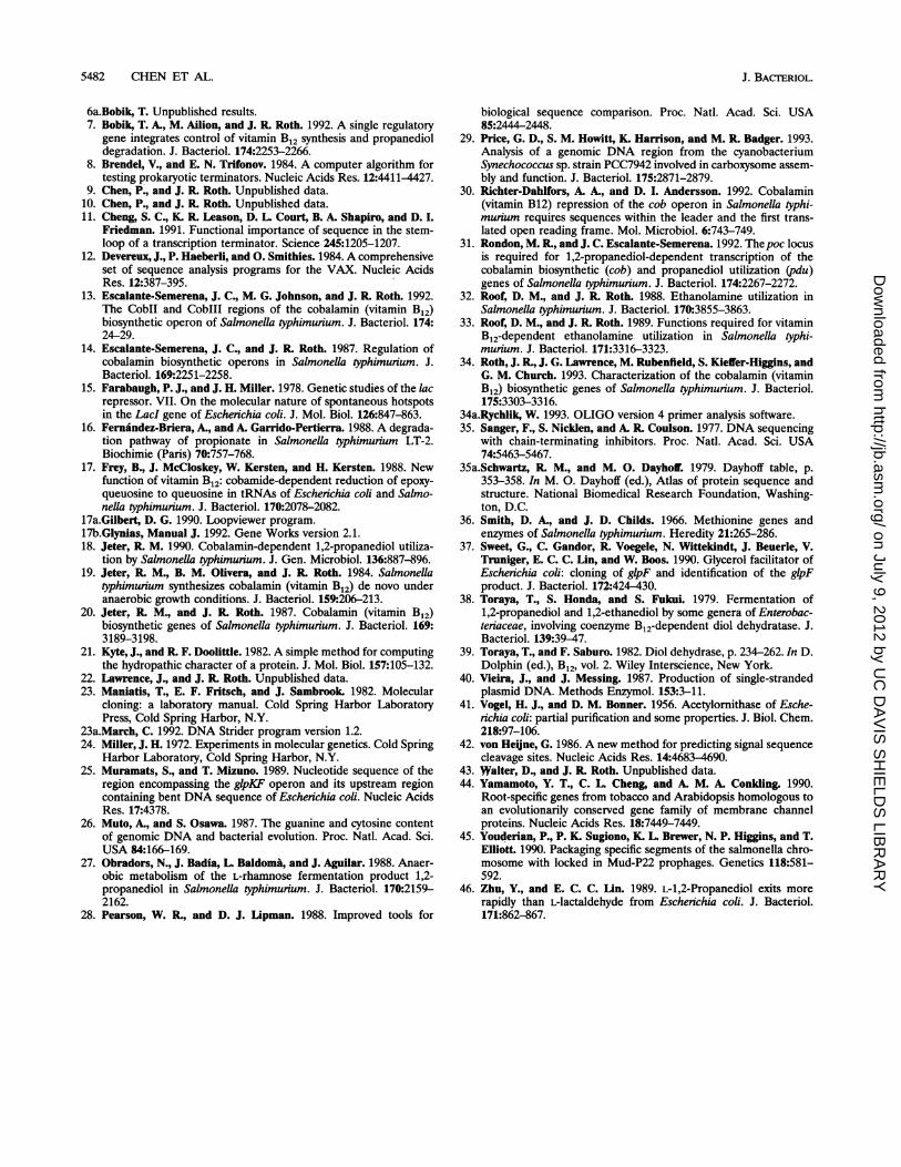
5482 CHEN ET AL.
6a.Bobik, T. Unpublished results.7. Bobik, T. A., M. Ailion, and J. R Roth. 1992. A single regulatory
gene integrates control of vitamin B12 synthesis and propanedioldegradation. J. Bacteriol. 174:2253-2266.
8. Brendel, V., and E. N. Trifonov. 1984. A computer algorithm fortesting prokaryotic terminators. Nucleic Acids Res. 12:4411-4427.
9. Chen, P., and J. R Roth. Unpublished data.10. Chen, P., and J. R Roth. Unpublished data.11. Cheng, S. C., K. R. Leason, D. L. Court, B. A. Shapiro, and D. I.
Friedman. 1991. Functional importance of sequence in the stem-loop of a transcription terminator. Science 245:1205-1207.
12. Devereux, J., P. Haeberli, and 0. Smithies. 1984. A comprehensiveset of sequence analysis programs for the VAX. Nucleic AcidsRes. 12:387-395.
13. Escalante-Semerena, J. C., M. G. Johnson, and J. R Roth. 1992.The CoblI and CoblIl regions of the cobalamin (vitamin B12)biosynthetic operon of Salmonella typhimurium. J. Bacteriol. 174:24-29.
14. Escalante-Semerena, J. C., and J. R Roth. 1987. Regulation ofcobalamin biosynthetic operons in Salmonella typhimurium. J.Bacteriol. 169:2251-2258.
15. Farabaugh, P. J., and J. H. Miller. 1978. Genetic studies of the lacrepressor. VII. On the molecular nature of spontaneous hotspotsin the LacI gene of Escherichia coli. J. Mol. Biol. 126:847-863.
16. Fernandez-Briera, A., and A. Garrido-Pertierra. 1988. A degrada-tion pathway of propionate in Salmonella typhimurium LT-2.Biochimie (Paris) 70:757-768.
17. Frey, B., J. McCloskey, W. Kersten, and H. Kersten. 1988. Newfunction of vitamin B12: cobamide-dependent reduction of epoxy-queuosine to queuosine in tRNAs of Escherichia coli and Salmo-nella typhimuium. J. Bacteriol. 170:2078-2082.
17a.Gilbert, D. G. 1990. Loopviewer program.17b.Glynias, Manual J. 1992. Gene Works version 2.1.18. Jeter, R M. 1990. Cobalamin-dependent 1,2-propanediol utiliza-
tion by Salmonella typhimurium. J. Gen. Microbiol. 136:887-896.19. Jeter, R M., B. M. Olivera, and J. R Roth. 1984. Salmonella
typhimunrum synthesizes cobalamin (vitamin B12) de novo underanaerobic growth conditions. J. Bacteriol. 159:206-213.
20. Jeter, R M., and J. R Roth. 1987. Cobalamin (vitamin B12)biosynthetic genes of Salmonella typhimunum. J. Bacteriol. 169:3189-3198.
21. Kyte, J., and R F. Doolittle. 1982. A simple method for computingthe hydropathic character of a protein. J. Mol. Biol. 157:105-132.
22. Lawrence, J., and J. R Roth. Unpublished data.23. Maniatis, T., E. F. Fritsch, and J. Sambrook 1982. Molecular
cloning: a laboratory manual. Cold Spring Harbor LaboratoryPress, Cold Spring Harbor, N.Y.
23a.March, C. 1992. DNA Strider program version 1.2.24. Miller, J. H. 1972. Experiments in molecular genetics. Cold Spring
Harbor Laboratory, Cold Spring Harbor, N.Y.25. Muramats, S., and T. Mizuno. 1989. Nucleotide sequence of the
region encompassing the glpKF operon and its upstream regioncontaining bent DNA sequence of Escherichia coli. Nucleic AcidsRes. 17:4378.
26. Muto, A., and S. Osawa. 1987. The guanine and cytosine contentof genomic DNA and bacterial evolution. Proc. Natl. Acad. Sci.USA 84:166-169.
27. Obradors, N., J. Badia, L Baldoma, and J. Aguilar. 1988. Anaer-obic metabolism of the L-rhamnose fermentation product 1,2-propanediol in Salmonella typhimurium. J. Bacteriol. 170:2159-2162.
28. Pearson, W. R., and D. J. Lipman. 1988. Improved tools for
biological sequence comparison. Proc. Natl. Acad. Sci. USA85:2444-2448.
29. Price, G. D., S. M. Howitt, K. Harrison, and M. R Badger. 1993.Analysis of a genomic DNA region from the cyanobacteriumSynechococcus sp. strain PCC7942 involved in carboxysome assem-bly and function. J. Bacteriol. 175:2871-2879.
30. Richter-Dahlfors, A. A., and D. I. Andersson. 1992. Cobalamin(vitamin B12) repression of the cob operon in Salmonella typhi-murium requires sequences within the leader and the first trans-lated open reading frame. Mol. Microbiol. 6:743-749.
31. Rondon, M. R., and J. C. Escalante-Semerena. 1992. Thepoc locusis required for 1,2-propanediol-dependent transcription of thecobalamin biosynthetic (cob) and propanediol utilization (pdu)genes of Salmonella typhimurium. J. Bacteriol. 174:2267-2272.
32. Roof, D. M., and J. R Roth. 1988. Ethanolamine utilization inSalmonella typhimurium. J. Bacteriol. 170:3855-3863.
33. Roof, D. M., and J. R Roth. 1989. Functions required for vitaminB12-dependent ethanolamine utilization in Salmonella typhi-murium. J. Bacteriol. 171:3316-3323.
34. Roth, J. R, J. G. Lawrence, M. Rubenfield, S. Kieffer-Higgins, andG. M. Church. 1993. Characterization of the cobalamin (vitaminB12) biosynthetic genes of Salmonella typhimunum. J. Bacteriol.175:3303-3316.
34a.Rychlik, W. 1993. OLIGO version 4 primer analysis software.35. Sanger, F., S. Nicklen, and A. R Coulson. 1977. DNA sequencing
with chain-terminating inhibitors. Proc. Natl. Acad. Sci. USA74:5463-5467.
35a.Schwartz, R M., and M. 0. Dayhoff. 1979. Dayhoff table, p.353-358. In M. 0. Dayhoff (ed.), Atlas of protein sequence andstructure. National Biomedical Research Foundation, Washing-ton, D.C.
36. Smith, D. A., and J. D. Childs. 1966. Methionine genes andenzymes of Salmonella typhimurium. Heredity 21:265-286.
37. Sweet, G., C. Gandor, K Voegele, N. Wittekindt, J. Beuerle, V.Truniger, E. C. C. Lin, and W. Boos. 1990. Glycerol facilitator ofEscherichia coli: cloning of glpF and identification of the glpFproduct. J. Bacteriol. 172:424-430.
38. Toraya, T., S. Honda, and S. Fukui. 1979. Fermentation of1,2-propanediol and 1,2-ethanediol by some genera of Enterobac-teriaceae, involving coenzyme B12-dependent diol dehydratase. J.Bacteriol. 139:39-47.
39. Toraya, T., and F. Saburo. 1982. Diol dehydrase, p. 234-262. In D.Dolphin (ed.), B12, vol. 2. Wiley Interscience, New York.
40. Vieira, J., and J. Messing. 1987. Production of single-strandedplasmid DNA. Methods Enzymol. 153:3-11.
41. Vogel, H. J., and D. M. Bonner. 1956. Acetylornithase of Esche-richia coli: partial purification and some properties. J. Biol. Chem.218:97-106.
42. von Heine, G. 1986. A new method for predicting signal sequencecleavage sites. Nucleic Acids Res. 14:4683-4690.
43. Walter, D., and J. R Roth. Unpublished data.44. Yamamoto, Y. T., C. L Cheng, and A. M. A. Conkling. 1990.
Root-specific genes from tobacco and Arabidopsis homologous toan evolutionarily conserved gene family of membrane channelproteins. Nucleic Acids Res. 18:7449-7449.
45. Youderian, P., P. K. Sugiono, K. L Brewer, N. P. Higgins, and T.Elliott. 1990. Packaging specific segments of the salmonella chro-mosome with locked in Mud-P22 prophages. Genetics 118:581-592.
46. Zhu, Y., and E. C. C. Lin. 1989. L-1,2-Propanediol exits morerapidly than L-lactaldehyde from Escherichia coli. J. Bacteriol.171:862-867.
J. BAC-1ERIOL.
on July 9, 2012 by UC
DA
VIS
SH
IELD
S LIB
RA
RY
http://jb.asm.org/
Dow
nloaded from
5482 CHEN ET AL.
6a.Bobik, T. Unpublished results. 7. Bobik, T. A., M. Allion, and J. R. Roth. 1992. A single regulatory
gene integrates control of vitamin B12 synthesis and propanediol degradation. J. Bacteriol. 174:2253-2266.
8. Brendel, V., and E. N. Trifonov. 1984. A computer algorithm for testing prokaryotic terminators. Nucleic Acids Res. 12:4411-4427.
9. Chen, P:, and J. R. Roth. Unpublished data. 10. Chen, P., and J. R. Roth. Unpublished data. 11. Cheng, S. C., K. R. Leason, D. L. Court, B. A. Shapiro, and D. I.
Friedman. 1991. Functional importance of sequence in the stemloop of a transcription terminator. Science 245:1205-1207.
12. Deverenx, J., P. Haeberli, and O. Smithies. 1984. A comprehensive set of sequence analysis programs for the VAX. Nucleic Acids Res. 12:387-395. .
13. Es~ante-Semerena, J. C., M. G. Johnson, and J. R. Roth. 1992. The CobII and CobIII regions of the cobalamin (vitamin Bd biosynthetic operon of Salmonella typhimurium. J. Bacteriol. 174: 24-29.
14. Esc:alante-Semerena, J. C., and J. R. Roth. 1987. Regulation of cobalamin biosynthetic operons in Salmonella typhimurium. J. Bacteriol. 169:2251-2258.
15. Farabaugh, P. J., and J. H. Miller. 1978. Genetic studies of the lac repressOr. VII. On the molecular nature of spontaneous hotspots in the LacI gene of Escherichia coli. J. Mol. BioI. 126:847-863.
16. Fermindez-Briera, A., and A. Garrido-Pertierra. 1988. A degradation pathway of propionate in Salmonella typhimurium LT-2. Biochimie (Paris) 7&:757-768.
17. Frey, B., J. McCloskey, W. Kersten, and H. Kersten. 1988. New function of vitamin B12: cobamide-dependent reduction of epoxyqueuosine to queuosine in tRNAs of Escherichia coli and Salmonella typhimurium. J. Bacteriol. 17&:2078-2082.
17a.Gilberl; D. G. 1990. Loopviewer program. 17b.Glynias, Manual J. 1992. Gene Works version 2.1. 18. Jeter, R. M. 1990. Cobalamin-dependent 1,2-propanediol utiliza
tion by Salmonella typhimurium. J. Gen. Microbiol. 136:887-896. 19. Jeter, R. M., B. M. Olivera, and J. R. Roth. 1984. Salmonella
typhimurium synthesizes cobalamin (vitamin Bd de novo under anaerobic growth conditions. J. Bacteriol. 159:206-213.
20. Jeter, R. M., and J. R. Roth. 1987. Cobalamin (vitamin Bd biosynthetic genes of Salmonella typhimurium.· J. Bacteriol. 169: 3189-3198.
21. Kyte, J., and R. F. Doolittle. 1982. A simple method for computing the hydropathic character of a protein. J. Mol. BioI. 157:105-132.
22. Lawrence, J., and J. R. Roth. Unpublished data. 23. Maniatis, T., E. F. Fritsch, and J. Sambrook. 1982. Molecular
cloning: a laboratory manual. Cold Spring Harbor Laboratory Press, Cold Spring Harbor, N.Y. .
23a.March, C. 1992. DNA Strider program version 1.2. 24. Miller, J. H. 1972. Experiments in molecular genetics. Cold Spring
Hlirbor Laboratory, Cold Spring Harbor, N.Y. 25. Muramats, S., and T. Miznno. 1989. Nucleotide sequence of the
region encompassing the glpKF operon and its upstream region containing bent DNA sequence of Escherichia coli. Nucleic Acids Res. 17:4378.
26. Muto, A., and S. Osawa. 1987. The guanine and cytosine content of genomic DNA and bacterial evolution. Proc. Natl. Acad. Sci. USA 84:166-169.
27. Obradors, N., J. Badia, L. Baldoma, and J. Aguilar. 1988. Anaerobic metabolism of the L-rhamnose fermentation product 1,2-propanediol in Salmonella typhimurium. J. Bacteriol. 17&:2159-2162.
28. Pearson, W. R., and D. J. Lipman. 1988. Improved tools for
J. BACI'ERIOL.
biological sequence comparison. Proc. Natl. Acad. Sci. USA 85:2444-2448.
29. Price, G. D., S. M. Howitt, K. Harrison, and M. R. Badger. 1993. Analysis of a genomic DNA region from the cyanobacterium Synechococcus sp. strain PCC7942 involved in carboxysome assembly and function. J. Bacteriol. 175:2871-2879.
30. Richter-Dahlfors, A. A., and D. I. Andersson. 1992. Cobalamin (vitamin B12) repression of the cob operon in Salmonella typhimurium requires sequences within the leader and the first translated open reading frame. Mol. Microbiol. 6:743-749.
31. Rondon, M. R., and J. C. Escalante-Semerena. 1992. The poe locus is required for 1,2-propanediol-dependent transcription of the cobalamin biosynthetic (cob) and propanediol utilization (pdu) genes of Salmonella typhimurium. J. Bacteriol. 174:2267-2272.
32. Roof, D. M., and J. R. Roth. 1988. Ethanolamine utilization in Salmonella typhimurium. J. Bacteriol. 17&:3855-3863.
33. Roof, D. M., and J. R. Roth. 1989. Functions required for vitamin B12-dependent ethanolamine utilization in Salmonella typhimurium. J. Bacteriol. 171:3316-3323.
34. Roth, J. R., J. G. Lawrence, M. Rubenfteld, S. Kietrer-Higgins, and G. M. Church. 1993. Characterization of the cobalamin (vitamin Bd biosynthetic genes of Salmonella typhimurium. J. Bacteriol. 175:3303-3316.
34a.Rychlik, W. 1993. OLIGO version 4 primer analysis software. 35. Sanger, F., S. Niclden, and A. R. Coulson. 1977. DNA sequencing
with chain-terminating inhibitors. Proc. Natl. Acad. Sci. USA 74:5463-5467.
35a.Schwartz, R. M., and M. O. DayhoW. 1979. Dayhoff table, p. 353-358. In M. O. Dayhoff (ed.), Atlas of protein sequence and structure. National Biomedical Research Foundation, Washington, D.C.
36. Smith, D. A., and J. D. Childs. 1966. Methionine genes and enzymes of Salmonella typhimurium. Heredity 21:265-286.
37. Sweet, G., C. Gandor, R. Voegele, N. Wittekindt, J. Beuerle, V. Truniger, E. C. C. Lin, and W. Boos. 1990. Glycerol facilitator of Escherichia coli: cloning of glpF and identification of the glpF product. J. Bacteriol. 172:424-430.
38. Toray&, T., S. Qonda, and S. Fnkui. 1979. Fermentation of 1,2-propanediol and 1,2-ethanediol by some genera of Enterobacteriaceae, involving coenzyme B12-dependent diol dehydratase. J. Bacteriol. 139:39-47.
39. Toraya, T., and F. Saburo. 1982. Diol dehydrase, p. 234-262. In D. Dolphin (ed.), B12, vol. 2. Wiley Interscience, New York.
40. Vieira, J., and J. Messing. 1987. Production of single-stranded plasmid DNA. Methods Enzymol. 153:3-11.
41. Vogel, H. J., and D. M. Bonner. 1956. Acetylomithase of Escherichia coli: partial purification and some properties. J. BioI. Chem. 218:97-106.
42. von Heijne, G. 1986. A new method for predicting signal sequence cleavage sites. Nucleic Acids Res. 14:4683-4690.
43. Walter, D., and J. R. Roth. Unpublished data. 44. Yamamoto, Y. T., C. L. Cheng, and A. M. A. Conkling. 1990.
Root-specific genes from tobaCco and Arabidopsis homologous to an evolutionarily conserved gene family of membrane channel proteins. Nucleic Acids Res. 18:7449-7449.
45. You4erian, P., P. K. Sugiono, K. L. Brewer, N. P. Higgins, and T. Elliott. 1990. Packaging specific segments of the salmonella chromosome with locked in Mud-P22 prophages. Genetics 118:581-592.
46. Zhn, Y., and E. C. C. Lin. 1989. L-l,2-Propanediol exits more rapidly than L-lactaldehyde from Escherichia coli. J. Bacteriol. 171:862-867.