Embed Size (px)
Citation preview

8/4/2019 The Chest Ptx
http://slidepdf.com/reader/full/the-chest-ptx 1/46
The Chest:
Pneumothorax, Hemothorax,
Effusions, & Empyema
Bradley J. Phillips, M.DBurn-Trauma-ICU
Adults & Pediatric

8/4/2019 The Chest Ptx
http://slidepdf.com/reader/full/the-chest-ptx 2/46
Pneumothorax
definition, classification,
& management

8/4/2019 The Chest Ptx
http://slidepdf.com/reader/full/the-chest-ptx 3/46
Pneumothorax (1)
collection of air within the pleural space
• transforms the potential space into a real one
• may lead to various degrees of respiratory compromise
• with progression, the intrapleural pressure may exceed
atmospheric pressure creating a tension-scenario
• impairs respiratory function
• decreases venous return to the right-side of the heart
8/4/2019 The Chest Ptx
http://slidepdf.com/reader/full/the-chest-ptx 4/46
Pneumothorax (2)
• General Management
– First: evacuate the air
– Second: address the underlying source
– Third: promote pleural symphysis

8/4/2019 The Chest Ptx
http://slidepdf.com/reader/full/the-chest-ptx 5/46
Pneumothorax (3)
Classification System
• Spontaneous Pneumothorax
– Primary
– Secondary
• Traumatic Pneumothorax
– Pulmonary source
– Tracheobronchial source – Esophageal source
8/4/2019 The Chest Ptx
http://slidepdf.com/reader/full/the-chest-ptx 6/46
Pneumothorax (4)
• Primary Spontaneous Ptx
– a disease of younger individuals (15 - 35 yrs of age)
– males > females
– tall, slim body habitus
– cigarette smoking implicated
– usual cause: parenchymal blebs
• apex of the upper lobe
• superior segment of the lower lobe

8/4/2019 The Chest Ptx
http://slidepdf.com/reader/full/the-chest-ptx 7/46
Pneumothorax (5)
• Primary Spontaneous Ptx:
“in most instances, the treatment
of a first-occurrence consists of hospitalization,tube-thoracostomy to closed drainage,
lung-re-expansion against the chest wall,
and
control of any persistent air-leak”
[Graeber „98]

8/4/2019 The Chest Ptx
http://slidepdf.com/reader/full/the-chest-ptx 8/46
Pneumothorax (6)
when do you
operate on
a primary spontaneous
pneumothorax ?

8/4/2019 The Chest Ptx
http://slidepdf.com/reader/full/the-chest-ptx 9/46
Pneumothorax (7)
• Secondary Ptx: due to underlying pulmonary disease
– COPD / Asthma / Cystic Fibrosis
– Immunocompromised Infections
• Tb & Cocci• PCP (becoming more common)
– Treatment: Closed Thoracostomy
• Water-seal
• Heimlich-Flutter Valve
• V.A.T.S.

8/4/2019 The Chest Ptx
http://slidepdf.com/reader/full/the-chest-ptx 10/46
Pneumothorax (8)
Traumatic Ptx• Parenchymal Injury vs. Tracheobronchial vs. Esophageal
– Blunt or Penetrating
– Iatrogenic
• central lines / thoracentesis / biopsy
• endotracheal tube placement (esp. dual-lumen tubes !)
• endoscopy / dilational techniques
– Barotrauma
• Ventilation / blast injury / Boerhave‟s syndrome
– Operative

8/4/2019 The Chest Ptx
http://slidepdf.com/reader/full/the-chest-ptx 11/46
Pneumothorax (9)
• The Tension Ptx
– “path of least resistance”
– life-threatening emergency…how do you treat a tension ptx ??
• The Open Ptx: sucking-chest wound
– intrinsic lung compliance creates complete collapse
– 3-sided dressing
– thoracostomy away from the traumatic wound

8/4/2019 The Chest Ptx
http://slidepdf.com/reader/full/the-chest-ptx 12/46
Pneumothorax (10)
• Treatment Options
– Observation: Inpatient vs. Outpatient
– Thoracostomy Drainage
• 3rd Interspace / 5th Interspace
• Negative Suction / Water-seal
– V.A.T.S. (becoming the “standard”)
– Muscle-sparing Thoracotomy
– Posterolateral & Anterolateral Thoracotomy

8/4/2019 The Chest Ptx
http://slidepdf.com/reader/full/the-chest-ptx 13/46
Pneumothorax (11)
Questions ?

8/4/2019 The Chest Ptx
http://slidepdf.com/reader/full/the-chest-ptx 14/46
Pneumothorax (12)
Questions…well, I have some -
1. What is the best diagnostic study ?
2. What is the role of “100 % Oxygen” & “Conservative-mgmt” ?
3. How would YOU treat a small Ptx (1 cm) in acute trauma ?
4. What is the predicted recurrence rate for a spontaneous Ptx ?
5. What is a “deep sulcus sign” ?

8/4/2019 The Chest Ptx
http://slidepdf.com/reader/full/the-chest-ptx 15/46
Pleural Effusions
what are they ?
where do they come from ?
& how do you treat them ?

8/4/2019 The Chest Ptx
http://slidepdf.com/reader/full/the-chest-ptx 16/46
Definition
the accumulation of excess fluid within the
pleural space in response to injury,
inflammation, or both
may represent a local response to diseaseor may just be a manifestation of a systemic illness

8/4/2019 The Chest Ptx
http://slidepdf.com/reader/full/the-chest-ptx 17/46
Pathogenesis of Effusions
Rate of Fluid Rate of Fluid
Accumulation Removal
1. Altered Pleural Membrane Permeability
2. Decreased Intravascular Oncotic Pressure
3. Increased Capillary Hydrostatic Pressure
4. Lymphatic Obstruction5. Abnormal Sites of Entry

8/4/2019 The Chest Ptx
http://slidepdf.com/reader/full/the-chest-ptx 18/46
Clinical Manifestations
• Pain
• Cough
• Dyspnea
• Dullness to Percussion
• Diminished or Absent Vocal Resonance
• Diminished or Absent Tactile Vocal Fremitus
• Friction Rub

8/4/2019 The Chest Ptx
http://slidepdf.com/reader/full/the-chest-ptx 19/46
Clinical: A Few Points
Large Effusions that prevent contact between the
Visceral & Parietal Pleura during respiration are seldom
associated with pleuritic chest pain.
• Tumors involving the parietal pleura generally produce constan
dull pain (Remember Ben Daly, M.D.)
• Large effusions interfere with expansion of the lung and
produce dyspnea, shortness of breath, and atelectasis
8/4/2019 The Chest Ptx
http://slidepdf.com/reader/full/the-chest-ptx 20/46
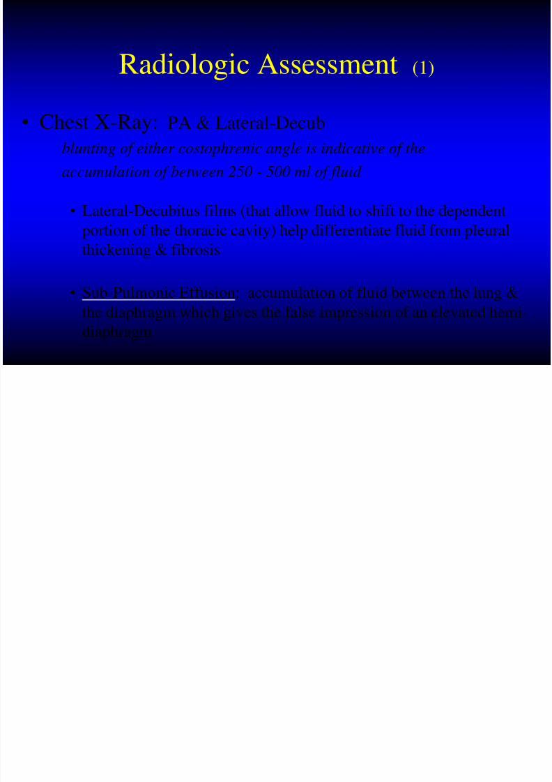
Radiologic Assessment (1)
• Chest X-Ray: PA & Lateral-Decub
blunting of either costophrenic angle is indicative of the
accumulation of between 250 - 500 ml of fluid
• Lateral-Decubitus films (that allow fluid to shift to the dependent
portion of the thoracic cavity) help differentiate fluid from pleural
thickening & fibrosis
• Sub-Pulmonic Effusion: accumulation of fluid between the lung &the diaphragm which gives the false impression of an elevated hemi
diaphragm

8/4/2019 The Chest Ptx
http://slidepdf.com/reader/full/the-chest-ptx 21/46
Radiologic Assessment (2)
• Ultrasound: Helpful in Confirming the Presence of a
Small Pleural Effusion & Identifying Loculations
• C.T. : Extremely Sensitive !!
• also helps to view the underlying lung (which may be
obscured by pleural disease)
• can distinguish between Lung Abscess & Empyema
8/4/2019 The Chest Ptx
http://slidepdf.com/reader/full/the-chest-ptx 22/46
Pleural Fluid Analysis
Thoracentesis = Pneumothorax

8/4/2019 The Chest Ptx
http://slidepdf.com/reader/full/the-chest-ptx 23/46
Pleural Fluid Analysis
Thoracentesis: Transudate vs. Exudate
1. Gross Appearance
2. Cell Count & Differential3. Gm Stain, C & S
4. Cytology
5. LDH
6. Protein
7. Glucose, Amylase

8/4/2019 The Chest Ptx
http://slidepdf.com/reader/full/the-chest-ptx 24/46
Transudate
straw-colored, clear, odorless fluid with a
WBC less than 1000 / ul
• Pleural Membranes are Intact
• Secondary to Altered Starling Forces
• Low in Protein & other Large Molecules
CHF, Cirrhosis, Nephrotic SyndromeHypoalbuminemia, Constrictive
Pericarditis, SVC Obstruction, PE

8/4/2019 The Chest Ptx
http://slidepdf.com/reader/full/the-chest-ptx 25/46
Exudate
• Characterized by Increased Protein & LDH
[Pleural Fluid vs. Serum Levels]
• Secondary to Disruption of Pleural Membrane or Obstruction of Lymphatic Drainage
Parapneumonic, Infections, Malignancy,
Vasculitic Disease, GI Disease, TB, PE

8/4/2019 The Chest Ptx
http://slidepdf.com/reader/full/the-chest-ptx 26/46
Criteria for “Exudative Effusion”
criteria value
1. Pleural Protein : Serum Protein > 0.5
2. Pleural LDH : Serum LDH > 0.6
3. Pleural LDH > 200
only need 1 critical value to establish the diagnosis of exudate

8/4/2019 The Chest Ptx
http://slidepdf.com/reader/full/the-chest-ptx 27/46
a bloody pleural effusion
occurring in a patient without a history of trauma or
pulmonary infarctionis
Indicative of Neoplasm
in 90 % of cases!
Because a RBC count as low as 5000 - 10,000 /ul, can cause a pleural effusion
to turn red, the finding of blood-tinged fluid per se has little diagnostic
value (usually from needle trauma)
A True Hemothorax is when the Pleural Fluid Hct exceeds 50 %
of the Peripheral Blood Hct !

8/4/2019 The Chest Ptx
http://slidepdf.com/reader/full/the-chest-ptx 28/46
Treatment
• Transudative Effusion: focus on the systemic cause
• Exudative Effusion: dependent on the exact sub-type
• Consider Chest Thoracostomy
• Gross Pus / Empyema
• pH < 7.2
• Hemothorax• Complicated Parapneumonic Processes
• Malignant Effusions…but remember the role of pleurodesis!

8/4/2019 The Chest Ptx
http://slidepdf.com/reader/full/the-chest-ptx 29/46
although pleural disease
itself is rarely fatal, it may be a
significant cause of patient morbidity
appropriate treatment may produce
dramatic symptomatic relief !

8/4/2019 The Chest Ptx
http://slidepdf.com/reader/full/the-chest-ptx 30/46
Pleural Effusions
Questions ?

8/4/2019 The Chest Ptx
http://slidepdf.com/reader/full/the-chest-ptx 31/46
Hemothorax
“ the collection of blood between the
visceral and parietal pleura…”

8/4/2019 The Chest Ptx
http://slidepdf.com/reader/full/the-chest-ptx 32/46
Hemothorax (1)
• Causes of a Spontaneous Hemothorax
– Pulmonary: bullous emphysema, PE, infarction, Tb, AVM‟s
– Pleural: torn adhesions, endometriosis
– Neoplastic: primary, metastatic (melanoma)
– Blood Dyscrasias: thrombocytopenia, hemophilia, anticoagulation
– Thoracic Pathology: ruptured aorta, dissection
– Abdominal Pathology: pancreatic pseudocyst, hemoperitoneum
8/4/2019 The Chest Ptx
http://slidepdf.com/reader/full/the-chest-ptx 33/46
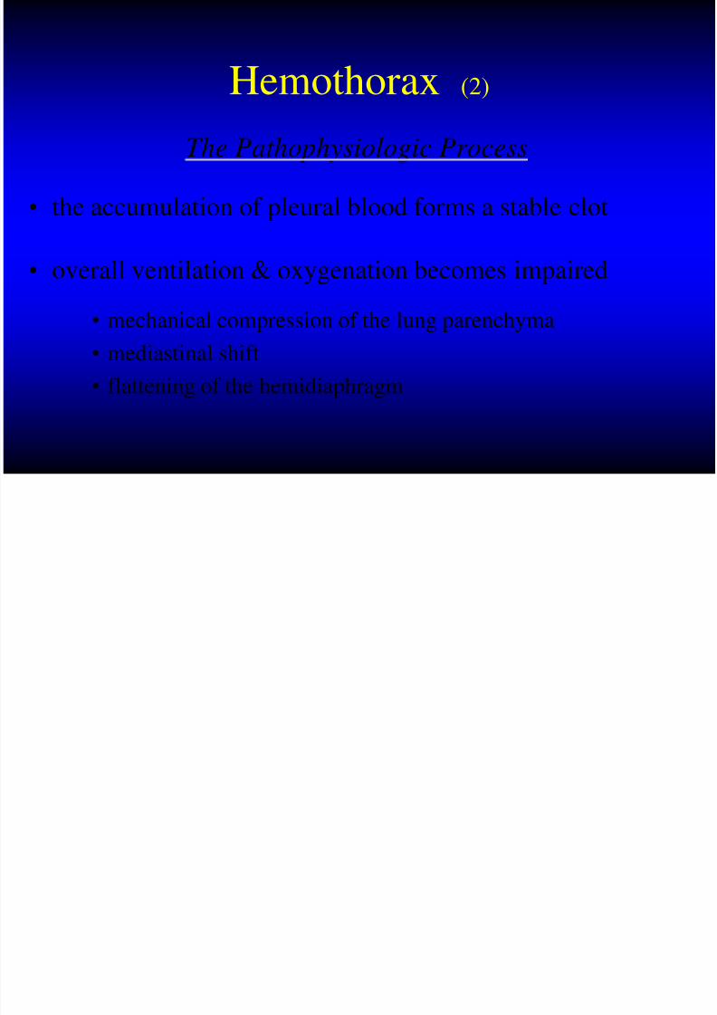
Hemothorax (2)
The Pathophysiologic Process
• the accumulation of pleural blood forms a stable clot
• overall ventilation & oxygenation becomes impaired
• mechanical compression of the lung parenchyma
• mediastinal shift
• flattening of the hemidiaphragm

8/4/2019 The Chest Ptx
http://slidepdf.com/reader/full/the-chest-ptx 34/46
Hemothorax (3)
The Pathophysiologic Process
• over time, the clot is partially-absorbed, leaving behind
loculated fluid and fibrinous septations
• macro-fibrin deposition begins to provide a structural
framework
• this “peel” slowly contracts to entrap the underlying lung

8/4/2019 The Chest Ptx
http://slidepdf.com/reader/full/the-chest-ptx 35/46
Hemothorax (5)
Goal of Treatment
to remove the pleural blood
and allow for
complete lung re-expansion

8/4/2019 The Chest Ptx
http://slidepdf.com/reader/full/the-chest-ptx 36/46
Hemothorax (4)
• General Management Options
– thoracentesis: bedside / ultrasound-guided / C.T.-guided
– thoracostomy drainage: the mainstay
– thorascopic surgery: less than 2 wks. & use a 30-degree scope
– thoracotomy: massive hemothorax / instability / chronic hemothorax
– local fibrinolytic therapy: urokinase (1000 IU/ml) in 150cc solution

8/4/2019 The Chest Ptx
http://slidepdf.com/reader/full/the-chest-ptx 37/46
Hemothorax (6)
• Often, there is an accompanying pneumothorax
– Dual Chest Tube Management
• Superior-Apical: Ptx
• Diaphragmatic-posterior: Htx
• Consider targeted-drainage into a loculated collection
– All tubes to negative suction with protective water-seal
– Prophylactic antibiotics may be indicated while the tubesare in (controversial!!)
– Chest tubes removed: 100 -150 cc‟s / day

8/4/2019 The Chest Ptx
http://slidepdf.com/reader/full/the-chest-ptx 38/46
Hemothorax (6)
Undrained hemothorax increases the risk
of empyema & fibrothorax
• Large collections should be drained slowly to minimize
the development of re-expansion-pulmonary-edema
[“R.E.E.P.”] (stop after 2 liters…wait 6 -8 hrs, then drain out another 1-2 liters, etc)
• Computed tomography is the diagnostic of choice

8/4/2019 The Chest Ptx
http://slidepdf.com/reader/full/the-chest-ptx 39/46
Hemothorax
Questions ?

8/4/2019 The Chest Ptx
http://slidepdf.com/reader/full/the-chest-ptx 40/46
Hemothorax
Questions…well, I have some –
1. When do YOU operate on a “Traumatic Hemothorax” ?
2. What options exist in trying to drain a hemothorax (chest tube placement) ?
3. What are the reported complications of chest tube placement ?

8/4/2019 The Chest Ptx
http://slidepdf.com/reader/full/the-chest-ptx 41/46
What is an Empyema ?

8/4/2019 The Chest Ptx
http://slidepdf.com/reader/full/the-chest-ptx 42/46
Empyema Thoracis
An Accumulation of Pus in the Pleural Cavity
• 1-2 % incidence in the pediatric population
• Up to 18 % in immunocompromised adults• General Management
– Appropriate Antibiotic Coverage
– Thoracostomy Drainage
– Streptokinase / Urokinase
– Surgical Intervention - Decortication

8/4/2019 The Chest Ptx
http://slidepdf.com/reader/full/the-chest-ptx 43/46
The Stages of Empyema
• Stage I - “Exudative”• sterile pleural fluid develops secondary to inflammation without
fusion of the pleura
• Stage II - “Fibrinopurulent” • a fibrinous peel develops on both pleural surfaces limiting lung
expansion
• Stage III - “Organizing”
• in-growth of capillaries & fibroblasts into the fibrinous peel

8/4/2019 The Chest Ptx
http://slidepdf.com/reader/full/the-chest-ptx 44/46
Empyema: A Pediatric Review
# of Cases
# of Positive Cultures
Staph aureusStrep pneumo
0
500
# of Cases
# of PositiveCultures
Staph aureus
Strep pneumo

8/4/2019 The Chest Ptx
http://slidepdf.com/reader/full/the-chest-ptx 45/46
Empyema...
Questions ?
“don’t let it happen !!!”

8/4/2019 The Chest Ptx
http://slidepdf.com/reader/full/the-chest-ptx 46/46
The Chest:
Pneumothorax, Hemothorax,
Effusions, & Empyema
Any Questions…?