Embed Size (px)
Citation preview
www.cellsignal.com/cellsimple
AP
PLI
CAT
ION
NO
TE
The CellSimple Cell Analyzer: Application for multiplex bead-based assay to measure the activation state of key proteins.
Introduction to the CellSimple Cell Analyzer
Complex cell and bead-based assays are made simple and fast with the CellSimple™ Cell Analyzer. This instru-ment combines a 488 nm laser, dual photomultiplier tubes (PMT), Coulter Principle-based cell measurements and on-board software to provide easy-to-run applications and data analysis. Moreover, the instrument relies on disposable cassettes for sample handling, which allevi-ates the need for flow cell cleaning and fluidics mainte-nance. And, the instrument is small enough to be portable between the lab bench and the hood.
When paired with highly validated kits and reagents from Cell Signaling Technology (CST), the CellSimple Cell Analyzer enables powerful plug-and-play assays, such as measuring independent changes in pathway activation.
Background and Experimental Design:Multiplexed bead-based assays can be used to simultaneously measure the activation state of multiple analytes in a single sample. In this application note, the activation state of multiple targets was measured in order to understand how signaling cascades function.
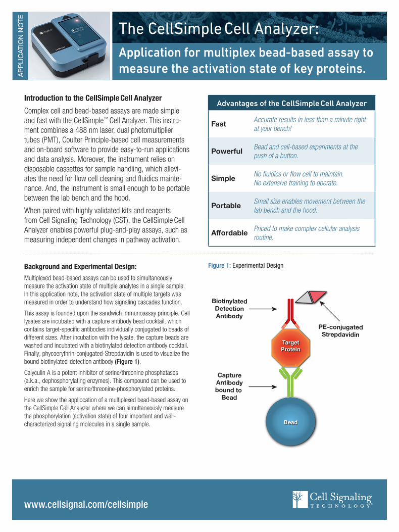
This assay is founded upon the sandwich immunoassay principle. Cell lysates are incubated with a capture antibody bead cocktail, which contains target-specific antibodies individually conjugated to beads of different sizes. After incubation with the lysate, the capture beads are washed and incubated with a biotinylated detection antibody cocktail. Finally, phycoerythrin-conjugated-Strepdavidin is used to visualize the bound biotinylated-detection antibody (Figure 1).
Calyculin A is a potent inhibitor of serine/threonine phosphatases (a.k.a., dephosphorylating enzymes). This compound can be used to enrich the sample for serine/threonine-phosphorylated proteins.
Here we show the appliocation of a multiplexed bead-based assay on the CellSimple Cell Analyzer where we can simultaneously measure the phosphorylation (activation state) of four important and well-characterized signaling molecules in a single sample.
Figure 1: Experimental Design
Bead
TargetProtein
PE-conjugatedStrepdavidin
BiotinylatedDetectionAntibody
CaptureAntibodybound to
Bead
Advantages of the CellSimple Cell Analyzer
Fast Accurate results in less than a minute right at your bench!
Powerful Bead and cell-based experiments at the push of a button.
Simple No fluidics or flow cell to maintain. No extensive training to operate.
Portable Small size enables movement between the lab bench and the hood.
Affordable Priced to make complex cellular analysis routine.

www.cellsignal.com/cellsimple
The CellSimple Cell Analyzer
Results:
The Open Flow Application on the CellSimple Cell Analyzer was used to resolve four populations of beads based on size in both the untreated and Calyculin A treated cell lysates (Figure 2). The plot on the left shows an overlay of the data from the untreated cell lysates (below the red dashed line) and the treated cell lysates (above the red dashed line). These data are plotted in the graph on the right and indicate that phosphorylation of each of the target proteins is stimulated in response to Calyculin A treatment.
Conclusion:These data demonstrate that the CellSimple Cell Analyzer can support multiplexed, bead-based analysis of signaling pathways.
Importantly, the CellSimple Cell Analyzer can be paired with bead-bound antibodies antibodies, in single-plex or multiplex, allowing investigators to measure the activation state of individual proteins in response to treatment. This provides investigators with the flexibility to develop powerful assays for unraveling the interplay between components of a single signaling cascade or between the components of interconnected pathways. Together, these assays run on the CellSimple Cell Analyzer enable powerful results that are rapid, accurate, affordable, and performed right at your lab bench.
Methods:Culture Conditions: HeLa cells were grown to 80% confluency in DMEM with 10% fetal bovine serum and penicillin/streptomycin at 37°C in 5% CO2.
Treatments: Cells were serum-starved overnight and then either left untreated or treated with Calyculin A #9902 (100 nM, 60 min).
Sample Preparation and Analysis: Cell lysates were incubated with the 4-plex capture antibody bead cocktail followed by a biotinylated detection antibody cocktail. Capture beads were washed and incubated with a biotinylated detection antibody cocktail (see table below). Streptavidin, R-phycoerythrin conjugate (SAPE) (#S866, Thermo Fisher Scientific Inc.) was then used to visualize the bound detection antibody. Mean fluorescent intensities (MFI) were quantified using the Open Flow Application on the CellSimple™ Cell Analyzer followed by further analysis and plotting using Microsoft® Excel®.
16APNCSCA0028ENG
© 2016 Cell Signaling Technology, Inc. Cell Signaling Technology, CST, CellSimple, and XP are all trademarks of Cell Signaling Technology, Inc. Microsoft and Excel are registered trademarks of Microsoft.
For Research Use Only. Not For Use In Diagnostic Procedures.
0
150
50
100
250
350
200
PRAS40 (Thr246) S6 (Ser235/236) Akt (Ser473) ERK (Thr202/Tyr204)
300
Untreated+Cal A
MFI
sFigure 2: Instrument screen shot showing four separate populations of beads (representing separate targets) in the presence (above the red line) or absence (below the red line) of Calyculin A (+Cal A) treatment in the panel on the left. Data was exported from the instrument and plotted in the panel on the right.
561
nm L
P (M
FI)
Diameter (μm)
+Cal AUntreated