Embed Size (px)
Citation preview
The Care and Keeping of Vascular Access for Home Hemodialysis PatientsRose Faratro, RN, BHScN, CNeph(C)1
Janine Jeffries, RN2
Gihad E Nesrallah, MD, FRCPC, FACP3
Jennifer M MacRae, MSc , MD, FRCPC4
1University Health Network, Toronto, Ontario, Canada; 2Princess Alexandria Hospital, Brisbane, Australia; 3Humber River Regional Hospital, Toronto, Ontario, Canada; 4University of Calgary, Calgary, Alberta, Canada
7

Vascular Access inHome Hemodialysis
2
C O N T E N T S3
4
5
6
7
8
9
9
13
13
14
16
16
17

Vascular Access inHome Hemodialysis
3
AbstractCreating and maintaining a healthy vascular access is a critical
factor in successful home hemodialysis (HD). This module aims
to serve as a “how-to manual” regarding vascular access issues
for both patients and healthcare providers in a home HD program.
This module outlines cannulation options for patients with
arteriovenous access and describes troubleshooting techniques
for potential complications; strategies are suggested to help
patients overcome fear of cannulation and address problems
associated with difficult cannulation. Technical aspects of central
venous catheter care, as well as a guide to troubleshooting
catheter complications, are covered in detail. Monitoring
for access-related complications of stenosis, infection, and
thrombosis is a key part of every home HD program. Key
performance and quality indicators are important mechanisms
to ensure patient safety in home HD and should be used during
routine clinic visits.
Arteriovenous Fistula
Arteriovenous Fistula Cannulation OptionsCannulation of the arteriovenous fistula (AVF), even when done
properly, causes pain and local trauma; repeated cannulation can
weaken blood vessel walls and promote wall dilation and the
formation of aneurysms.1, 2 Unsuccessful cannulation can result
in needle infiltration (swelling that happens when the needle goes
through the fistula wall), which in turn causes localized bruising
and increases the risk of thrombosis and loss of AVF patency.3
Two methods of needling are commonly used: rotating sites/
rope ladder (RL) technique, and buttonhole (BH) technique. The
standard is RL, wherein the needling site is alternated along the
length of the AVF, resulting in minimal scar tissue formation.
Many patients are trained on this method of cannulation when
beginning home dialysis. While discouraged, some patients
prefer particular sites (ie, use the “area wall technique”), which
increases the potential for damage to the AVF wall and dilation
of the fistula, and can result in the development of an aneurysm.4-6
The BH technique, also known as constant site needling, is a
cannulation method that uses the same location, angle, and depth
repeatedly.1, 2 Sharp needles are used to form a tract of scar tissue
for entry into the fistula over time. Once this tract is formed,
the patient can begin cannulating using a blunt needle, which
is theoretically less traumatizing to the vascular structure and
should improve survival of the access.1
To date, there are no high-quality clinical trials comparing AVF
outcomes with RL vs BH cannulation in home HD patients or other
self-needler patients (Table 1). The majority of the evidence
supporting the use of the BH technique was generated through
observational studies, and the generalizability of the existing
observational and clinical trial data to the self-needling patient
is unknown.
Useful Resources
» La Société Française de l’Abord Vasculaire. History of Buttonhole Technique
» End Stage Renal Disease Network, Cannulation of the AV Fistula
Table 1. Advantages and Disadvantages of BH Cannulation Technique
Advantages for the Patient
• Reduced needling attempts1, 2
• Fewer hematomas7
• Reduced number of infiltrations5, 7, 8
• Reduced number of aneurysms and aneurysm size8, 9
• Prevents “area” cannulation • Reduced pain (shown in observational
studies only)5, 6, 10-13
• Beneficial for individuals with needle phobia (opinion only)
Disadvantages for the Patient
• Increased risk of infection7-14
• Need for meticulous hygiene• Possibility of introducing sharps into
tunnel when confronted with difficult needling
• If tract moves, BH may require re-siting

Vascular Access inHome Hemodialysis
4
Buttonhole Cannulation TechniqueIn each unit, specialized, highly trained clinic staff are responsible
for teaching BH tract creation. Ideally, there should only be 1
cannulator for the BH, and it is best if that cannulator is the
patient himself; however, there are cases where a dedicated
helper can be taught to cannulate. It is very important that the
angle, position of the arm, and tourniquet placement are kept
constant with each cannulation in order to create and maintain
the BH tract. Previous teachings have suggested that the angle
of entry should be 45 degrees for all BH cannulations, but in fact,
the angle of entry depends on the depth and the anatomy of the
fistula, and, thus, varies with each patient. To provide consistency
for the angle of needle insertion, the touch cannulation technique
can be taught. This technique refers to the placement of the
thumb and forefingers on the needle tubing (and not the wings)
while the other fingers rest upon the arm to provide stabilization
(for more information and cannulation images, see End Stage
Renal Disease Network, Cannulation of the AV Fistula).
Many clinics recommend that 2 separate BH sites (ie, 2 arterial
and 2 venous) be created, each 6 to 8 cm apart. Ideally, patients
should alternate between these sites and if there is ongoing
difficulty with accessing a site or if it becomes infected (see
section Increased Risk of Infection with Buttonhole Cannulation),
that site should be abandoned.
BH tract creation requires repeated cannulation with a sharp
fistula needle, an intravenous (IV) needle, or placement
of a polycarbonate peg (eg, BioHole™ Plug, Nipro Corporation,
Belgium). With each of these methods (except for the peg, which
has no scab formation), the scab on the BH tract is removed
before cannulation to allow the access site to be viewed and
permit accurate insertion of the needles. The needles are inserted
using the exact location, angle, and depth for each HD treatment.
Canadian guidelines suggest that topical antimicrobial
prophylaxis be applied to the BH site after the dialysis treatment
is completed.15
The most common way to create a BH tract is with any type
of sharp standard HD needle. The BH is initially created after
approximately 8 to 12 cannulations using this approach. Once the
BH tract is developed, the needles are switched to a dull/ blunt
BH needle (eg, Medisystems) or a dull/blunt IV needle with
a plastic cannula (eg, Nipro BioHoleTM Cath) to cannulate the
BH sites.
Intravenous needles with plastic cannulas (eg, the SupercathTM
Clampcath or angiocatheter) have also been used to create
BH tracts with repeated needling. As described above, these
needles are also inserted into the exact spot, using the same
angle and depth for each HD treatment; the scab on the BH is
removed before cannulation. Once the BH tract is developed,
the blunt version of these needles can be used to cannulate the
BH sites. These types of catheters have a large enough cannula
to sustain dialysis blood flow, and the plastic (instead of steel)
cannula limits the potential for needle infiltration.
Of note, there are descriptions in the literature using these
IV needles with plastic cannulas to create a BH by leaving
the catheter indwelling for periods of time.16 Readers should
be warned that there are possible complications with these
indwelling catheters, namely the chance of infection, needle
dislodgement, and cannula breakage with migration into
a vessel.17 The authors have personally treated catheter breakage
in patients who have used this technique and we do not
recommend this approach.18
Polycarbonate pegs are emerging as preferred tools with which
to create BHs. The peg is a small, sterile, thumbtack-shaped plug
used to maintain the needle tract between cannulations. Scar
tissue forms around the peg, which facilitates the development
of the BH tract. The use of a polycarbonate peg may lead
to improved tract creation, which may in turn improve AVF
survival. A randomized trial by Vaux et al8 used polycarbonate
pegs to create BHs, and they found improved AVF survival with
BH cannulation at 1 year, whereas with conventional BH tract
formation, there was no difference in AVF survival in a comparison
with RL needling.19 (See “Buttonhole Tract Formation Using
Polycarbonate Pegs” in the Appendix) Note that some, but not all,
BH protocols include antibiotic prophylaxis.20, 21

Vascular Access inHome Hemodialysis
5
The BH technique is not recommended for all patients and
is contraindicated in patients with arteriovenous grafts (AVGs).
In North America and Australasia, the BH technique is considered
a relative contraindication for newly created AVFs because
the fistula is undergoing dynamic changes that influence the
BH tracts; however, this practice is not consistent globally.
In Europe, BH cannulation is performed in patients with newly
created fistulae.
Indications for and against BH cannulation are summarized
in Table 2, and a checklist for assisting clinicians in choosing the
best cannulation method for patients can be found in “Criteria for
Determining Type of Self-Cannulation” in the Appendix. Choosing
a cannulation method is discussed further in the section “Tools
to Determine the Best Type of Needling”. Patients with limited
vision should use prescription lenses or a magnifying glass during
the self-cannulation evaluation.
Complications of Buttonhole Cannulation
Indentation/HubbingOver time BH sites can develop a widening and an indentation
at the entry to the skin. This is commonly known as “hubbing”.
Hubbing occurs when the hub of the needle is buried into the
skin, which can result in incomplete scab removal, patient’s
inability to clean the puncture site, and breakdown of the lining
of the tunnel tract.22 Hubbing can be prevented by leaving space
between the hub of the needle and the puncture site.
Trampoline EffectThe trampoline effect describes the motion of a blunt needle
meeting resistance and bouncing back toward the cannulator. This
occurs because of a thickening of the tunnel tract or poor tract
development. When this occurs, the patient’s needling technique
should be reassessed.Useful Resources
» Big D and Me. Dialysis Buttons for your Buttonholes.
» Home Dialysis Central. The Art of Teaching Buttonhole Self-Cannulation.
Table 2. Indications For or Against BH Cannulation Technique
Indications For Its Use
• AVF is short in length or has short usable segments
• AVF with tortuous anatomy• AVF with aneurysmal dilatation6
• AVF is difficult to cannulate. The patient is unable to self-cannulate using the RL technique
• AVF is mature• Patient preference. Risk factors
discussed and understood by patient• Needle phobia. Patient expresses
considerable fear related to self-cannulation
Indications Against Its Use
• AVF is relatively straight• Patient experiences hand tremors.
Unsuitable placement of needle on the BH may lead to the creation of multiple tracts within the BH
• Patient reports or demonstrates difficulty visualizing the BH site. Poor vision and improper placement of needle on the BH may lead to the creation of multiple tracts within the BH
• Patient has bioprosthesis (eg, mechanical heart valve, artificial joint)
Vascular Access inHome Hemodialysis
6
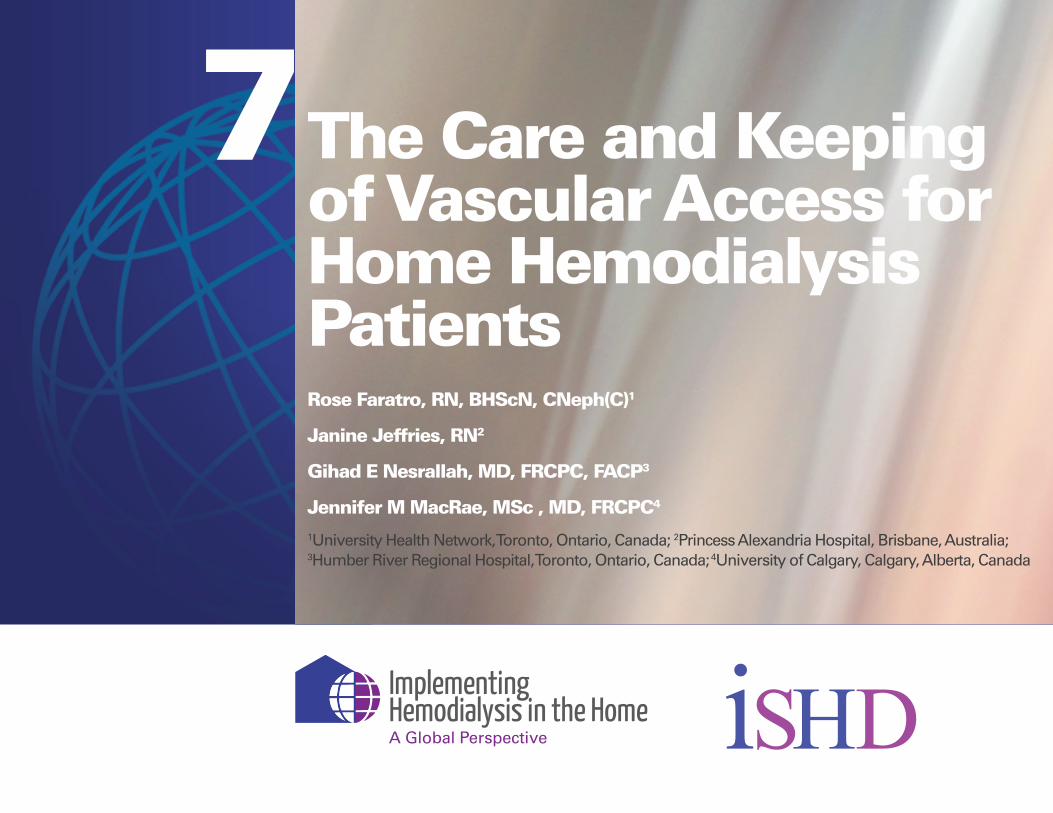
Increased Risk of Infection with Buttonhole CannulationSeveral clinical studies have demonstrated an increased risk
of infection with the use of BH cannulation.5-7, 21-26
The incidence of localized infections is increased with BH and
other infectious complications have been reported. These include
septic arthritis, bacterial endocarditis, and bacteremia; however,
these conditions may not appear until long after the BH technique
is initiated.5, 6, 24-26 While incidence of infection varies between
studies (and by patient population and locality), 1 retrospective
study reported a rate of bacteremia of 0.073 per 1000 AVF days
for BH patients, compared with no bacteremia for RL patients.26
One systematic review of observational and randomized studies
reported an increased risk of AVF-related infections using
BH cannulation, with relative risk ranging from 3.15 to 3.34
comparing before and after changes and with RL cannulation,
respectively.27
Patients should be informed of the increased risk of infection
and receive specialized training and frequent evaluations of their
cannulation techniques.15 Strict adherence to aseptic technique
in performing cannulations is essential; additional measures
of infection prevention are also recommended for BH patients (Table
3).15, 23, 28, 29 Each clinic should track and regularly review infection
rates (see section Key Performance and Quality Indicators)
Table 3. Summary of Measures to Reduce Infection Risk
Adhere strictly to aseptic technique for access skin preparation and BH scab removal
Use hand disinfectant prior to decannulation
Perform routine audit of patient cannulation technique on a quarterly basis (recommended) at clinic visits (see the “Arteriovenous
Fistula/Graft Audit Tool” and the “Central Venous Catheter Audit Tool” in the Appendix)
Use face masks on the patient (and staff/ helper if applicable) to lower the theoretical risk of nasal transmission of Staphylococcus aureus during cannulation28
Discuss and provide topical prophylaxis: Patients considering BH technique require counseling regarding the increased risk of infection and the potential for devastating consequences resulting from infection. Topical prophylaxis is strongly recommended for the prevention of infection15, 24
• Options for topical agents:
» Polysporin triple ointment: A formulation of polymyxin B sulfate, bacitracin zinc, and gramicidin used for the treatment of infections caused by bacteria
» Povidone-iodine ointment: A broad-spectrum antiseptic for the treatment and prevention of infection
» Mupirocin ointment: Utilized to treat staphylococcal infections or attempt to decrease the incidence of subsequent staphylococcal infections. Note: continued use may result in antimicrobial resistance
» Polyhexamethylene biguanide (PHMB): A dressing infused with a broad-spectrum antimicrobial for the prevention of infection and promotion of wound healing
Perform routine screening of the nares for S aureus and, when present, pursue an eradication program (see the “Mupirocin
Protocol” in the Appendix)

Vascular Access inHome Hemodialysis
7
Management of BH InfectionsThe optimal duration and choice of antibiotic therapy to treat
BH-related infections has not been directly studied. The following
suggestions are based on the authors’ opinion. Empiric treatment
should begin with a first-generation cephalosporin (eg, cefazolin)
or vancomycin, depending on local methicillin-resistant S aureus
(MRSA) colonization rates. Subsequent choice of antimicrobials
should be based on culture and susceptibility results.
• Infection of BH with fever and/or bacteremia: This infection should be treated with appropriate antibiotics for a minimum of 4 weeks. Treatment should be extended to 6 weeks in the case of S aureus bacteremia and/or if there is a metastatic complication. If further complications exist, abort use of infected BH site and re-site the BH
• Cellulitis or exit site infection: Local infection without fever and bacteremia should be treated with appropriate antibiotics for a minimum of 2 weeks. Abort use of BH located in the vicinity of cellulitis and re-site the BH. Reevaluate the need to change the dressing type and cleansing agent
• Abscess: Abscess of BH, especially with fever, should be treated with appropriate antibiotics for a minimum of 4 weeks and extended to 6 weeks if bacteremia is present. Treatment may be extended if there is progression of serious metastatic complications. Abort use of infected AVF. The abscess may require surgical intervention30
Tools to Determine the Best Type of NeedlingFor patients with AVF, the RL method of cannulation is the
preferred type of needling.15 In general, RL cannulation is used
with an AVF that is of adequate length and superficial depth.
In addition, the RL method is favored among patients who
have poorer vision or those who have a slight tremor. However,
in patients who have an AVF that is of short length, consists
of tortuous anatomy, or involves aneurysmal sections,
BH cannulation should be considered (see Table 2). Patients
with a needle phobia can often overcome this phobia with
the BH cannulation technique (see “Fear of Needles” in the
Appendix). Due to the increased risk of infections with BH, this
technique is not recommended for patients with a history of AVF
infections, mechanical heart valves, or other prostheses.
A downloadable tool to assist clinicians in choosing the most
appropriate type of self-cannulation can be found in “Criteria for
Determining Type of Self-Cannulation” in the Appendix.
Additional Cannulation References
» End Stage Renal Disease Network, Cannulation of the AV Fistula
» BC Renal. Vascular Access Guideline, Rope Ladder Cannulation of AV Fistulas and Grafts
» Home Dialysis Central. The Art of Teaching Buttonhole Self-Cannulation. Step-by-step PDF booklet with color photos

Vascular Access inHome Hemodialysis
8
Fistula Cannulation Methods
Indications for Use of Standard Sharp Fistula NeedlesStandard sharp fistula needles are used if the patient is unable
to cannulate using the IV needle with cannula or with the dull/
blunt needle at a BH tract. If the sharp fistula needle is used, then
the patient is encouraged to cannulate at a new site rather than
using the established BH tract. The use of sharp fistula needles
for nocturnal dialysis is not preferred due to the potential for
needle infiltration during the treatment. However, if sharp fistula
needles are used for nocturnal dialysis, it is imperative to ensure
that these needles are secured well. See “Taping Methods for
Hemodialysis Needle” and “Taping Method of Intravenous Needle
with Cannula” protocols in the Appendix. For more information
on nocturnal dialysis, see “Prescriptions for Home Hemodialysis”.
Indications for Use of IV Needle with Cannula (Examples: Supercath Clampcath Needles, Nipro Biohole Cath)For nocturnal dialysis, the flexible cannula is used for comfort and
to prevent needle infiltration during treatment. The use of this
needle system should be considered in:
• Patients who have an allergy to metals
• Restless patients who may be at risk of needle infiltration (ideal use)
• RL technique for the nocturnal HD patient
Protocols on use of IV needle with cannula can be found at:
• “Buttonhole Cannulation for Creation and Maintenance of Tract with Intravenous Needle and Cannula” in the Appendix
• BC Renal, Vascular Access Guideline, Self-Cannulation of Buttonholes on AV Fistulas
Indications for Use of Dull/Blunt NeedleFor nocturnal dialysis the dull/blunt needle is used to prevent
needle infiltration during treatment. When used with the
BH technique, once the BH tract is created, a dull/ blunt needle
can be inserted into that tract for the dialysis treatment. Protocols
on use of dull/blunt needles can be found at:
• “Buttonhole Cannulation Technique with Dull (Blunt) Bevel” in the Appendix
• BC Renal, Vascular Access Guideline, Self-Cannulation of Buttonholes on AV Fistulas
• BC Renal, Patient Teaching Tool, Self-Needling Your Fistula Using the Buttonhole Method
• BC Renal, Buttonhole Cannulation
Troubleshooting Arteriovenous Fistula ComplicationsCenters should conduct technique review of self-needling
patients every 3 months, with the patient being examined while
cannulating in the clinic or during a home visit (see “Arteriovenous
Fistula/Graft Audit Tool” in the Appendix). The major focus here
is on prevention of infections.
Pain with Needling: StrategiesFor patients who experience painful needling, a warm compress
should be applied to the access site 5 to 10 minutes before
needling. A topical anesthetic (preferred to a subcutaneous
injection of lidocaine) should be used to numb the skin surface.
Topical lidocaine preparations can be applied to the skin at the
desired cannulation sites in a thick layer and then covered with
an occlusive dressing or plastic wrap for 60-120 minutes prior
to cannulation. Of note, the anesthetic needs to be thoroughly
washed off the skin prior to cannulation. It is best to avoid
injection of lidocaine into BH sites to minimize the chance for
vessel and BH tract movement and potential for vasoconstriction
of the blood vessel.

Vascular Access inHome Hemodialysis
9
Cannulation Dependency IssuesSome BH patients may become fearful of cannulating using
sharp needles at sites other than at the BH site. As a result,
these patients can become dependent on the home HD clinic
to troubleshoot access issues and reestablish the BH site.
Fear of NeedlesFear of needling can be a barrier to the uptake of home dialysis.31
Needle fear should not be a contraindication to teaching self-
cannulation. In fact, this fear can be overcome if a stepwise
approach is followed in which the patient slowly increases his
or her comfort level with needles. Patients should start off simply
watching the insertion of another patient’s needles, followed
by watching the insertion of his or her own needles. Becoming
familiar with simply holding the needles and holding needle
sites after needle removal is also an important step. For more
information, please see this website or “Fear of Needles” in the
Appendix.
Strategies for Addressing Difficult CannulationPatients who experience difficulty with cannulation should
be scheduled to return to the home HD unit as soon as possible
for a review of their cannulation technique and an access
assessment for a possible complication of stenosis or thrombosis
(Table 4). Thus, in addition to a physical examination (see,
“Physical Examination of the Fistula” approach below), an access
flow assessment should be made with a subsequent plan for
intervention, if needed.
Regardless of the type of cannulation, patients should
be instructed to avoid flipping needles. Flipping a sharp needle
can actually damage the vessel, while flipping a blunt needle may
be indicative of an underlying access problem.
Some home HD programs will use heparin locks or flushes
to ensure patency of the access when patients experience
temporary cannulation difficulties (eg, if needling is difficult due
to onset of stenosis) or in those who need extra guidance for
needling. Locking involves instilling a diluted heparin solution into
the cannulas (needles and tubing) of the arteriovenous access and
allowing it to dwell for a specified period of time (ie, “locking” the
heparin in the lumens); flushing involves passing diluted heparin
through the cannula before initiating dialysis. Some programs may
substitute citrate 4% for the heparin. Before using this approach,
patients should be informed of the potential risk of needle
dislodgement and possible sequelae, such as bleeding and
infection. More information can be found in the “Heparin Flushing
of Cannulas” protocol in the Appendix.
Table 4. Troubleshooting for Buttonhole Cannulation Difficulties
Monitor established sites frequently
Avoid sharp needles in an established BH site. The use of sharp needles in a BH site may lead to excessive scarring
Review AVF for possible underlying access dysfunction and/or re-site BH if patient presents with cannulation difficulties
Reposition the arm, change the angle, and slightly rotate the cannula when resistance is felt during the cannulation of BH
Check for an inflamed or infected BH site. If infection is present, do not cannulate. Cannulation in this instance increases the risk of severe hemorrhage with possible exsanguination
Check for keloid formation at the BH cannulation site
Ensure that the BH needle site aligns with the tract entrance into the AVF. A cannulation failure could occur if the BH needle site does not align properly. A new BH site may need to be created to correct the problem
Check for an enlarged BH needle site that may lead to prolonged bleeding

Vascular Access inHome Hemodialysis
10
Appropriate Blood Pump SpeedsThe ideal blood pump speed for HD is unknown. However, there
are potential deleterious effects of high blood pump speeds
on fistula integrity. Needle turbulence is the intense flow that
is created by a needle in an arteriovenous access, and has been
shown to cause endothelial dysfunction with decreased nitrous
oxide formation and loss of endothelial integrity.32, 33 The effect
of higher pump speeds has not been proven, but injury to the
endothelial wall from altered flow mechanics of high pump
speeds is likely to occur.34 Expert opinion recommends that lower
pump speeds should be used to promote vessel integrity and
maintain fistula longevity. More information on pump speeds used
for different HD modalities can be found in the “Prescriptions
for Home Hemodialysis” module. Regardless of the blood pump
speeds utilized, most programs aim to maintain the arterial and
venous pressures below -250 and 250 mm Hg, respectively;
however, these pressures are not strongly evidence-based.
A recent observational study of patients on in-center HD reported
an increased risk of access failure with venous pressures outside
the range of 100–150 mm Hg.35
Physical Examination of the FistulaThe access arm must be examined regularly by the patient.
Routine evaluation of the arm using a “look, listen, and feel”
approach may help detect access complications and subsequent
intervention before the access is lost entirely. For an excellent
description of the access physical examination, see Sousa et al.36
Monitoring for Complications of Stenosis and ThrombosisStudies have not been performed to assess the value of access
surveillance among home HD patients, but most programs
recommend pursuing access flow monitoring when the patient
returns for quarterly or biannual clinic visits. In addition, regular
physical examination of the access by staff is suggested at these
clinic visits.
Home HD patients should be taught how to perform a basic
access arm examination regularly using the “look, listen, and
feel” approach. Patients should be instructed to assess their
access using the Arm Raise Technique. They will pump their
hand to make a fist, raise their arm straight in the air, and, while
standing in front of a mirror, note if the AVF collapses (normal
state) or if the AVF does not collapse, which indicates an outflow
obstruction.
It is recommended that patients perform trend analysis
by recording the venous and arterial pressures at onset of each
run at a blood pump speed of 200 mL/min, and reviewing the
changes/trends in these numbers. During dialysis, the maximum
arterial pressure should not exceed -250 mm Hg and the
maximum venous pressure should not exceed 250 mm Hg. When
these pressures are exceeded, the needle should be repositioned
and/or the blood pump speed should be decreased. Patients
should report to their clinician any changes noted in their routine
access arm examination, trend of pressures, or cannulation, and
the time of onset of cannulation difficulty.
Recent onset of increased difficulty of needling or prolonged
bleeding from the access site after dialysis may be signs of an
underlying stenosis and should be investigated. It is important
to remember that as the AVF matures, BH tracts may change
and new sites may be required, which may lead to difficulties
needling. In addition, large fluctuations in body weight or size
can alter the BH tracts. Patients and staff should be aware that
any new onset of cannulation difficulty can also be due to a
hemodynamically significant stenosis or impending thrombosis
of the access.
Access flow monitoring is suggested at a frequency of every
4 to 6 months, with the same flow thresholds for intervention
as are used with in-center conventional HD patients. The same
guidelines have been extrapolated for home HD patients.

Vascular Access inHome Hemodialysis
11
Readers should note that there is considerable variability in the
frequency of screening AVF/AVG. Some centers assess access
flow every 6 months, while others screen more frequently (every
3 months) in cases of AVF with access issues. Other centers only
investigate when cannulation difficulties are reported. Screening
options include, but are not limited to, formal ultrasound study,
Doppler assessment, clinical screening of needling complications,
and review of technique quarterly (see “Arteriovenous Fistula/
Graft Audit Tool” in the Appendix). Additional information can
be found at:
» Caring for Australians with Renal Impairment (CARI) Guidelines, Chapter 4: Vascular Access Surveillance
» Canadian Society of Nephrology, Clinical Practice Guidelines for the Treatment of Patients with Chronic Kidney Disease, Chapter 4: Vascular Access:
ThrombosisOften, the first sign of impending thrombosis is what is mistaken
for cellulitis, with signs of erythema over the AVF and tenderness
to palpation. This is a medical emergency and the patient
should be brought in for immediate physical and ultrasound examination of the AVF, with arrangements for radiology
or surgical thrombolysis made as required. Every effort should
be made to salvage the access and avoid catheter placement.
Fistula HemorrhageHemorrhage from fistula has been reported in the in-center
HD population,38, 39 but the incidence of this occurring among
home patients is unknown. All patients should be instructed
to apply pressure to their site in the event of bleeding and
to call for emergency assistance (eg, 911, 991, 999, 112, or 000
as appropriate). For additional information on fistula hemorrhage
and patient safety during home HD, please see the module titled,
“Ensuring Patient Safety During Home Hemodialysis”.
Specific risks for home HD patients include needle dislodgement,
or improper threading of the dialyzer, which may lead
to significant hemorrhage. Water or enuresis alarms strategically
placed under the dialysis machine and dialyzer, as well as under
the access arm, help prevent these serious adverse events. Some
popular alarms include the following:
• Redsense Venous Needle Dislodgement alarm
» Training the Trainer
» Self-Use Instructions
• Zircon Leak AlertTM Electronic Water Detector
• HEMOdialertTM blood leak detector
An aneurysmal fistula that is rapidly enlarging in size could
indicate possible rupture and hemorrhage.40 Thus, aneurismal
fistula should be routinely monitored and the diameter of the
aneurysms should be noted at each clinical visit. Fistula with
necrotic skin as a result of infection can also lead to increased risk
of rupture, especially in the case of BH cannulation.
The use of a single needle to minimize bleeding risk has been
used in some programs; however, this results in a reduction
in clearance and an increase in noise from the double pump
system. The routine use of single needle in home HD has fallen
out of favor, but it can be a useful technique to provide rest (and
avoid a catheter placement) after AVF complications. Programs
should have a standardized management plan for patients and
caregivers to follow to manage hemorrhage, if it occurs in the
home. For more detailed information, please see Home Dialysis
Central, The Art of Making Your Fistula or Graft Last
or the “Ensuring Patient Safety During Home Hemodialysis”
module.

Vascular Access inHome Hemodialysis
12
Fistula InfectionAVF infections can manifest as cellulitis, BH exit site infection,
or bacteremia. Cellulitis is infrequent in mature AVF without skin
lesions, but signs of redness and swelling should be evaluated
to rule out thrombophlebitis. With a BH exit site infection, pus
and erythema may be present at the needling site. It is important
to obtain a swab for culture and sensitivity and 2 sets of blood
culture specimens to rule out bacteremia (particularly S aureus
bacteremia), which is very common with BH sites. See “Increased
Risk of Infection with Buttonhole Cannulation” for details.
Two sets of blood culture specimens should be drawn from any
HD patient with an unexplained fever. Some units initiate empiric
therapy against both gram-positive and gram-negative bacteria,
depending on the usual types of infection in that unit. The planned
duration of therapy for bacteremia is 4 to 6 weeks, depending
on the organism.
Arteriovenous GraftAVGs do not have the option of BH needling; only the RL and
site rotation technique is recommended. Needle options include
standard AVF/AVG needles (sharp) or the needle with cannula
(angiocatheter) in which a blunt cannula remains in the AVG for
dialysis. With the exception of AVG infection (see below), all other
sections of the AVF apply to AVG.
AVG Infection
• Often requires surgical intervention, including graft resection
• Treatment requires 6 weeks of antibiotics with double coverage of gram-positive and gram-negative organisms
Central Venous Catheter
Technical Aspects of Catheter CareRoutine placement of catheters in the subclavian vein (central
venous catheters [CVCs]) is not recommended because they
create a higher risk for central vein stenosis. In general, the
internal jugular site is preferred.41 There are many different
catheters available; however, there is no evidence to guide
selection of 1 device over another.
Catheter Care ProtocolsThere are many catheter care protocols available (see “Central
Venous Catheter Audit Tool” in the Appendix). In general, donning
clean gloves and mask are a requirement when accessing the
catheter.39
LOCKINGAfter dialysis, catheters are most commonly locked with citrate
4% and heparin at a concentration of 1000 or 5000 units/mL;
however the following should be considered in selecting a lock
solution:
• Bleeding risks have been noted with higher heparin concentrations42 (see “Heparin Locking of Central Venous Catheters” in the Appendix)
• Locking with 30% ethanol/4% trisodium citrate has been demonstrated to prevent the formation of biofilms in catheters in vitro,43 and weekly 70% ethanol locks have been

Vascular Access inHome Hemodialysis
13
successfully used for infection prophylaxis (in vivo) in a proof-of-concept study,44 but ethanol locking is not yet widely used in clinical practice
• Tissue plasminogen activator (TPA) is also used to treat episodes of catheter dysfunction. Please see the following protocols for details:
• “Alteplase Use in Hemodialysis Central Venous Catheters” in the Appendix
• BC Renal. Vascular Access Guideline: Alteplase Use for Occluded Hemodialysis Catheters
DRESSINGS
• Either gauze dressings41 or nonocclusive transparent dressings can be used at the exit site of catheters45
• The lack of a dressing is a potential option for patients with severe skin breakdown or rash at the exit site; however, the evidence for this practice comes from a nondialysis patient population46
• Sample guidelines for dressing change and exit site care can be found on the BC Renal website
CLOSED-CONNECTOR DEVICESA closed-connector device is a device that is designed to decrease
the risk of unintentional disconnection examples of which include:
• InterLink (BD)
• Tego Needlefree Hemodialysis Connector (ICU Medical, Inc)
• Swan-Lock (Codan Medical Inc)
As published in the most recent Canadian Society of Nephrology
Intensive HD guidelines, a closed-connector device
is recommended for patients receiving intensive (home) HD.15
These closed-connector devices are ideal for home HD patients
with a CVC who perform HD without any assistance, as there
is less risk for air embolism or chance of inadvertent bleeding.
Depending on the device, these closed connectors can be changed
under sterile conditions either weekly by the patient at home
or monthly by the nurse at the home HD unit. To decrease concerns
of air embolism at the time of exchange of a closed-connector
device, the patient can be taught to double clamp (ie, use the
catheter clamp and another separate clamp on the catheter
tubing).
EXIT SITE PROPHYLAXISThe use of exit site prophylaxis (polysporin, mupirocin/bactroban,
medi-honey, povidone-iodone, etc) is very center dependent and
not used at every center.41
SHOWERING PROTOCOLSAttached is an example of a protocol for patients from
an established home HD program (see the “Showering Protocol”
in the Appendix). Remember that the shower and shower head
are potential sources of bacteria; therefore, regular cleaning
of both is recommended.
Troubleshooting Catheter Complications
InfectionsCatheters have a higher rate of infectious complications than
arteriovenous accesses, a risk which appears to vary over time
according to the length of time the access is in place.47 Catheter-
related infections can be either local (as an exit site or tunnel
infection) or systemic (bacteremia).
Exit site infections are defined according to a purulent discharge
at the exit site with 2 of the following features:
• Erythema
• Tenderness
• Induration at the exit site
• Sampling of the discharge that results in a culture positive for infection48
If left untreated, exit site infections can lead to catheter-related
bacteremia. An exit site infection is generally treated with
a 2-week regimen of either topical or oral antibiotics.

Vascular Access inHome Hemodialysis
14
A tunnel infection should be suspected in a patient who presents
with pain or tenderness at the catheter exit site; the tunnel site
should be palpated with the intention of expressing a discharge.
A tunnel infection is defined as a purulent discharge or aspirate
from a tunnel site not contiguous with the exit site and includes
2 of the following features:
• Erythema
• Tenderness
• Induration at a tunnel site
• A culture of serous discharge or aspirate from that site that is positive for infection48
Tunnel infections should be treated with a 3-week course of IV
antibiotics.
Most HD units use the definition of a probable catheter-related
bacteremia, which is 2 or more blood culture specimens that are
positive for infection with no evidence for a source other than the
catheter. When a patient first presents with a fever and suspected
catheter-related bacteremia, start empiric antibiotics that cover
both gram-positive and gram-negative organisms. The choice
and duration of antibiotics, as well as the decision to remove the
catheter, depend on the bacterial organism isolated.41 An example
of a center protocol is included (see “Central Venous Catheter
Antibiotic Treatment Protocol” in the Appendix) and detailed
treatment guidelines can be found on the CARI website.
Catheter DysfunctionCatheter dysfunction is a common problem for catheter-dependent
patients and results in decreased dialysis efficiency. Definitions
of catheter dysfunction vary, but in general they relate to the
inability to achieve a certain blood pump speed (from 200 to 300
mL/min) within the venous and arterial pressure limits of 250
and -250 mm Hg, respectively, while dialyzing. Many HD units
have developed treatment algorithms for decreased flow, which
include checking patient positioning and flushing the lumens with
normal saline prior to administering TPA. Home HD units that
have adopted these protocols and will either instruct the patient
to administer TPA at home or to come back to the unit to have
staff administer the thrombolytic. Sample protocols can be found
here:
• BC Renal. Alteplase Use for Occluded Hemodialysis Catheters
• “Alteplase Use in Hemodialysis Central Venous Catheters” in the Appendix
Catheter MalfunctionsAt times, an HD catheter may develop a crack in the line;
generally, these lines will need to be replaced. If the crack
develops distal to the “Y” portion, some lines can be repaired.
Sample guidelines for determining when a catheter line can
be repaired can be found at:
• BC Renal, Vascular Access Guideline: Central Venous Catheter – Repair of Cracked Catheter Adapter, Limb or Clamp
EmbolismThere have been reports of air embolism occurring in home
HD patients using catheters.49 Prevention of air embolism by using
a closed-connector device, such as those mentioned previously,
is recommended.15 In a survey of Canadian Home HD programs,
near misses have been reported in some patients who use
connector devices when the device is not applied firmly.
HemorrhageAlthough the closed-connector devices may prevent air emboli,
there have been cases of hemorrhage that have occurred
in patients because the devices have been used improperly or the
membranes in these devices have failed.15 Wetness detectors can
be applied to the catheter for overnight dialysis (see “Ensuring
Patient Safety During Home Hemodialysis” module).

Vascular Access inHome Hemodialysis
15
Key Performance and Quality IndicatorsKey indicators are important operating mechanisms to ensure
patient safety in home HD (see “Ensuring Patient Safety During
Home Hemodialysis” module). Ensure these key indicators
are used and followed during routine clinic visits for home
HD patients to minimize complications of infection and
to determine access failure:
Performance Measures
1. Use an audit tool quarterly
2. Verify that a form of access screening is occurring (eg, patient reporting the usual venous and arterial pressures at a standardized pump speed)
SummaryVascular access is associated with the development of potential
complications that can lead to significant morbidity. Thus, the
care and keeping of vascular access is a skill that is of utmost
importance for home dialysis patients. Teaching patients their
cannulation options as well as encouraging them to be vigilant
for possible access complications should be a large part of every
home dialysis program. Furthermore, routine access screening and
review of quality indicators should be instituted on a regular basis
to minimize access failure.
Quality Indicators
1. What is the infection (local/systemic) rate, according to vascular access type (events per 1000 access days)?
2. What is the rate of access interventions, according to vascular access type (events per 1000 access days)?
3. What is the rate of bleeding (actual or “near misses”) from the access site, according to vascular access type (events per 1000 access days)?

Vascular Access inHome Hemodialysis
16
References1. Twardowski Z. Constant site (buttonhole) method of
needle insertion for hemodialysis. Dial Transplant. 1995;24:559-560.
2. Twardowski Z, Kubara H. Different sites versus constant sites of needle insertion into arteriovenous fistulas for treatment by repeated dialysis. Dial Transplant. 1979;8:978-980.
3. Lee T, Barker J, Allon M. Needle infiltration of arteriovenous fistulae in hemodialysis: risk factors and consequences. Am J Kidney Dis. 2006;47:1020-1026.
4. Krönung G. Plastic deformation of Cimino fistula by repeated puncture. Dial Transplant. 1984;13:635-638.
5. Verhallen AM, Kooistra MP, van Jaarsveld BC. Cannulating in haemodialysis: rope-ladder or buttonhole technique? Nephrol Dial Transplant. 2007;22:2601-2604.
6. Marticorena RM, Hunter J, Macleod S, et al. The salvage of aneurysmal fistulae utilizing a modified buttonhole cannulation technique and multiple cannulators. Hemodial Int. 2006;10:193-200.
7. MacRae JM, Ahmed SB, Atkar R, Hemmelgarn BR. A randomized trial comparing buttonhole with rope ladder needling in conventional hemodialysis patients. Clin J Am Soc Nephrol. 2012;7:1632-1638.
8. Vaux E, King J, Lloyd S, et al. Effect of buttonhole cannulation with a polycarbonate PEG on in-center hemodialysis fistula outcomes: a randomized controlled trial. Am J Kidney Dis. 2013;62:81-88.
9. Struthers J, Allan A, Peel RK, Lambie SH. Buttonhole needling of arteriovenous fistulae: a randomized controlled trial. ASAIO J. 2010;56:319-322.
10. Ball LK, Treat L, Riffle V, Scherting D, Swift L. A multi-center perspective of the buttonhole technique in the Pacific Northwest. Nephrol Nurs J. 2007;34:234-241.
11. Castro MC, Silva Cde F, Souza JM, et al. Arteriovenous fistula cannulation by buttonhole technique using dull needle. J Bras Nefrol. 2010;32:281-285.
12. Kim MK, Kim HS. Clinical effects of buttonhole cannulation method on hemodialysis patients. Hemodial Int. 2013; 17:294-299.
13. Sukthinthai N, Sittipraneet A, Tummanittayangkoon B, Vasuvattakul S, Chanchairujira T. Buttonhole technique better than area puncture technique on hemostasis and pain associated with needle cannulation. J Med Assoc Thai. 2012;95 Suppl 2:S208-212.
14. Chow J, Rayment G, San Miguel S, Gilbert M. A randomised controlled trial of buttonhole cannulation for the prevention of fistula access complications. J Ren Care. 2011;37:85-93.
15. Nesrallah GE, Mustafa RA, MacRae J, et al. Canadian Society of Nephrology guidelines for the management of patients with ESRD treated with intensive hemodialysis. Am J Kidney Dis. 2013;62:187-198.
16. Marticorena RM, Hunter J, Cook R, et al. A simple method to create buttonhole cannulation tracks in a busy hemodialysis unit. Hemodial Int. 2009;13:316-321.
17. Donnelly SM, Marticorena RM, Hunter J, Goldstein MB. Supercath Safety Clampcath buttonhole creation: complication of catheter breakage. Hemodial Int. 2013;17:450-454.
18. MacRae JM, Tai DJ, Daniw M, Lee J. A simple method to create buttonhole cannulation tracks in a busy hemodialysis unit. Hemodial Int. 2010;14:94-95.
19. MacRae JM, Ahmed SB, Hemmelgarn BR, Alberta Kidney Disease Network. Arteriovenous fistula survival and needling technique: long-term results from a randomized buttonhole trial. Am J Kidney Dis. 2014;63:636-642.
20. Marticorena RM, Hunter J, Macleod S, et al. Use of the BioHoleTM device for the creation of tunnel tracks for buttonhole cannulation of fistula for hemodialysis. Hemodial Int. 2011;15:243-249.
21. Toma S, Shinzato T, Fukui H, et al. A timesaving method to create a fixed puncture route for the buttonhole technique. Nephrol Dial Transplant. 2003;18:2118-2121.

Vascular Access inHome Hemodialysis
17
References (cont’d)
22. Ball LK, Mott S. How do you prevent indented buttonhole sites? Nephrol Nurs J. 2010;37:427-428, 431.
23. van Loon MM, Goovaerts T, Kessels AG, van der Sande FM, Tordoir JH. Buttonhole needling of haemodialysis arteriovenous fistulae results in less complications and interventions compared to the rope-ladder technique. Nephrol Dial Transplant. 2010;25:225-230.
24. Nesrallah GE, Cuerden M, Wong JH, Pierratos A. Staphylococcus aureus bacteremia and buttonhole cannulation: long-term safety and efficacy of mupirocin prophylaxis. Clin J Am Soc Nephrol. 2010;5:1047-1053.
25. Van Eps CL, Jones M, Ng T, et al. The impact of extended-hours home hemodialysis and buttonhole cannulation technique on hospitalization rates for septic events related to dialysis access. Hemodial Int. 2010;14:451-463.
26. O’Brien FJ, Kok HKT, O’Kane C, et al. Arterio-venous fistula buttonhole cannulation technique: a retrospective analysis of infectious complications. Clin Kidney J. 2012;5:526-529.
27. Muir CA, Kotwal SS, Hawley CM, et al. Buttonhole cannulation and clinical outcomes in home hemodialysis cohort and systematic review. Clin J Am Soc Nephrol. 2013;9:110-119.
28. Ball LK. The buttonhole technique: strategies to reduce infections. Nephrol Nurs J. 2010;37:473-477; quiz 478.
29. Priyesh P, Smith K, Henner D. Effect of implementation of standardized protocol on infection rates in patients utilizing the buttonhole cannulation technique for accessing AV fistulas. Am J Kidney Dis. 2013;61:B77.
30. Tordoir J, Canaud B, Haage P, et al. EBPG on vascular access. Nephrol Dial Transplant. 2007;22 Suppl 2:ii88-117.
31. McLaughlin K, Manns B, Mortis G, Hons R, Taub K. Why patients with ESRD do not select self-care dialysis as a treatment option. Am J Kidney Dis. 2003;41:380-385.
32. Huynh TN, Chacko BK, Teng X, et al. Effects of venous needle turbulence during ex vivo hemodialysis on endothelial morphology and nitric oxide formation. J Biomech. 2007;40:2158-2166.
33. Unnikrishnan S, Huynh TN, Brott BC, et al. Turbulent flow evaluation of the venous needle during hemodialysis. J Biomech Eng. 2005;127:1141-1146.
34. Remuzzi A, Ene-Iordache B. Novel paradigms for dialysis vascular access: upstream hemodynamics and vascular remodeling in dialysis access stenosis. Clin J Am Soc Nephrol. 2013;8:2186-2193.
35. Parisotto MT, Schoder VU, Miriunis C, et al. Cannulation technique influences arteriovenous fistula graft survival. Kidney Int. 2014;86:790-797.
36. Sousa CN, Apóstolo JL, Figueiredo MH, Martins MM, Dias VF. Physical examination: how to examine the arm with arteriovenous fistula. Hemodial Int. 2013;17:300-306.
37. Jindal K, Chan CT, Deziel C, et al. Hemodialysis clinical practice guidelines for the Canadian Society of Nephrology. J Am Soc Nephrol. 2006;17:S1-27.
38. Ellingson KD, Palekar RS, Lucero CA, et al. Vascular access hemorrhages contribute to deaths among hemodialysis patients. Kidney Int. 2012;82:686-692.
39. Gill JR, Storck K, Kelly S. Fatal exsanguination from hemodialysis vascular access sites. Forensic Sci Med Pathol. 2012;8:259-262.
40. Almehmi A, Wang S. Partial aneurysmectomy is effective in managing aneurysm-associated complications of arteriovenous fistulae for hemodialysis: case series and literature review. Semin Dial. 2012;25:357-364.
41. Vanholder R, Canaud B, Fluck R, et al. Catheter-related blood stream infections (CRBSI): a European view. Nephrol Dial Transplant. 2010;25:1753-1756.
42. Yevzlin AS, Sanchez RJ, Hiatt JG, et al. Concentrated heparin lock is associated with major bleeding complications after tunneled hemodialysis catheter placement. Semin Dial. 2007;20:351-354.

Vascular Access inHome Hemodialysis
18
References (cont’d)
43. Takla TA, Zelenitsky SA, Vercaigne LM. Effectiveness of 30% ethanol/4% trisodium citrate locking solution in preventing biofilm formation by organisms causing haemodialysis catheter-related infections. J Antimicrob Chemother. 2008;62:1024-1026.
44. Broom JK, Krishnasamy R, Hawley CM, Playford EG, Johnson DW. A randomized controlled trial of Heparin versus EthAnol Lock THerapY for the prevention of Catheter Associated infection in Haemodialysis patients—the HEALTHY-CATH trial. BMC Nephrol. 2012;13:146.
45. Gillies D, O’Riordan L, Carr D, Frost J, Gunning R, O’Brien I. Gauze and tape and transparent polyurethane dressings for central venous catheters. Cochrane Database Syst Rev. 2003:CD003827.
46. Olson K, Rennie RP, Hanson J, et al. Evaluation of a no-dressing intervention for tunneled central venous catheter exit sites. J Infus Nurs. 2004;27:37-44.
47. Ravani P, Gillespie BW, Quinn RR, et al. Temporal risk profile for infectious and noninfectious complications of hemodialysis access. J Am Soc Nephrol. 2013;24:1668-1677.
48. Canada Communicable Disease Report—Supplement. Infection control guidelines: Preventing infections associated with indwelling intravascular access devices. Can Commun Dis Rep. 1997;23(Suppl 8):i-iii, 1-32, i-iv, 1-16.
49. Ouwendyk M, Pierratos A, Francoeur R, Wallace L, Sit W, Vas S. Slow nocturnal home hemodialysis (SNHHD)-–one year later. J CANNT. 1996;6:26-28.
![Unusual Complication of Hemodialysis Cuffed Catheter Tunnel ... · 2019. 7. 30. · hemodialysis patients with vascular access central venous catheter [2, 5]. Infection is the second](https://img.dokumen.tips/doc/110x75/6112f543c4e8093a88485054/unusual-complication-of-hemodialysis-cuffed-catheter-tunnel-2019-7-30-hemodialysis.jpg)