Embed Size (px)
Citation preview

The BrainChapter 2

The BrainTechniques for Studying the Brain

Methods
Lesioning is the removal or destruction of part of
the brain.
Any time brain tissue is removed (tumor,
lobotomy, behavior experiment in animals, etc.) researchers can
examine behavior changes and infer the function of that part of the brain.
Brain research can be done in a variety of ways. Brain damage as a result of an accident or disease can provide a wealth of
information. The Harvard Brain Bank (3000 +)

Functional Methods
EEG (electroencephalogram) is an amplified recording of the electrical activity (“brain waves”) sweeping across the brain’s surface, measured by electrodes placed on the scalp (sleep studies, etc.)
EEG

PET Scan
PET (positron emission tomography)
Scan is a visual display of brain
activity that detects the consumption of radioactive glucose (metabolic activity)
while the brain performs a given task.By doing this, one can connect brain activity
to the area of the brain that controls it.
Functional Methods
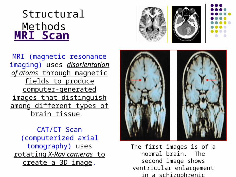
MRI Scan
MRI (magnetic resonance imaging) uses
disorientation of atoms through magnetic fields to
produce computer-generated images that
distinguish among different types of brain tissue.
CAT/CT Scan (computerized axial
tomography) uses rotating X-Ray cameras to create a
3D image.
The first images is of a normal brain. The second image shows
ventricular enlargement in a schizophrenic patient.
Structural Methods

Combination Method (structure & function)
An fMRI (functional MRI) is a comparison of blood flow before and during the performance of
mental functions to map the parts of the brain that control those
functions.
It sort of combines elements of the MRI (structure) and PET
(function – oxygen consumption).
The fMRI image shows brain regions that are active when
a participants lies.
fMRI

MA
KE I
T
STO
P!

The BrainAreas & Parts of the Brain

The Brainstem
The Brainstem is the oldest part of the brain, beginning where the spinal cord swells and enters the skull. It is responsible for automatic survival
functions.
brainstem
http://www.npr.org/templates/story/story.php?storyId=129027124&sc=fb&cc=fp

Parts of the Brain Stem:
The Medulla is the base of the brainstem that
controls heartbeat and breathing.
Pons deals with facial expressions. Also plays a role in sleep & dreaming.
Reticular Formation (RAS) is a nerve network
in the brainstem that plays an important role in
controlling arousal.
Pons

http://www.miketheheadlesschicken.org/story.php

Parts of the Brain Stem
The Thalamus is the brain’s sensory
switchboard, located on top of the brainstem. It directs messages to the
sensory areas in the cortex and transmits
replies to the cerebellum and
medulla.
It receives information for all of the senses EXCEPT for smell.

The Cerebellum is often called the “little
brain” and is attached to the rear of the brainstem.
It helps coordinate voluntary movements
and balance.
It also plays a part in memory, emotion
modulation & regulation, timing
and sensory discrimination.
Cerebellum
Brainstem

The Limbic System is a doughnut-shaped system of neural structures at
the border of the brainstem and cerebrum, associated with emotions such as fear, aggression and drives for food and
sex.
It includes the hippocampus, amygdala, and hypothalamus.
The Limbic System

The Hippocampus processes declarative
memories.
Hippocampus
Hippocampus

Amygdala
The Amygdala consists of two almond-shaped neural clusters
linked to emotions and
aggression

Hypothalamus
The Hypothalamus lies below (hypo) the thalamus.
It directs several maintenance activities like
eating, drinking, body temperature, and control of
sexual arousal.
It also helps control the endocrine system by giving directions to the pituitary
gland. Pituitary

Olds and Milner (1954) discovered that Rats cross an electrified grid for self-
stimulation when electrodes are placed in the reward (hypothalamus) center.
When the limbic system is manipulated, a rat will
navigate fields or climb up a tree (bottom picture).
It is possible that some addictive behavior may be
related to a genetic disorder (reward deficiency
syndrome).
The Limbic System contains many Reward/Pleasure Centers

Cerebral CortexThe intricate fabric of interconnected neural cells
that covers the cerebral hemispheres. It is the body’s ultimate control and information
processing center.

Structure of the Cerebral Cortex
Each brain hemisphere is divided into four
lobes that are separated by prominent
fissures.
These lobes are the: a. frontal lobe –
judgement/reasoning b. parietal lobe –
senses
c. occipital lobe – vision d. temporal lobe –
hearing
A.B.
C.D.

Functions of the Cerebral Cortex
The Motor Cortex is the area at the rear of the frontal lobes that controls voluntary movements.
The Sensory Cortex is the area at the front of the parietal lobes that receives information from
skin surface and sense organs.

Homunculus (“little man”)

Functions of the Cerebral Cortex
The visual cortex is located in the
occipital lobe of the brain.
The functional MRI scan shows the visual cortex is active as the
subject looks at faces.

Functions of the Cerebral Cortex
The auditory cortex is located in the temporal
lobe of the brain.
http://www.ted.com/talks/lang/eng/oliver_sacks_what_hallucination_reveals_about_our_minds.html

The association areas integrate sensory information and stored memories. More
intelligent animals have increased “uncommitted” or association areas of the
cortex.
Association Areas

The Curious Story of Phineas Gage (1848)
Frontal lobe damage showed effects on personality and social functioning

Banjo Dan’s Phineas Gage

The BrainThe Divided Brain

Lateralization (Specialization)
Our brain is divided into two hemispheres. The Left Hemisphere
Processes logical tasks/analytical (reading, writing, speaking, mathematics, and comprehension skills) Controls the right side of our body In the 1960s, it was termed as the dominant brain.
The Right Hemisphere Processes non-verbal tasks/perceptual (spatial relationships, musical/artistic ability and mental
imagery) Controls the left side of our body May also be related to some negative emotions
The Corpus Callosum is a wide band of axon fibers that connects the two hemispheres and allow them to communicate.

Language
Aphasia is an impairment of language, usually caused by left hemisphere damage
either to Broca’s area (impaired speaking) or to
Wernicke’s area (impaired understanding).

Specialization & Integration
Brain activity when hearing, seeing, and speaking words.

Corpus Callosotomy – Splitting the Brain
A procedure in which the two hemispheres of the brain are isolated by cutting the connecting fibers (mainly those of
the corpus callosum) between them. Usually done to prevent uncontrollable seizures in patients with severe
epilepsy.Corpus Callosum
Mark Gazzaniga

Split Brain Patients
With the corpus callosum
severed, objects (apple) presented in the right visual
field can be named.
Objects (pencil) in the left visual
field cannot.
Temporal & nasal portions

Divided Consciousness
SO
- which creates an artificial visual field
Can I have two volunteers (one needs to
have laced shoes)?

The brain is sculpted by our genes but also by our experiences.
Plasticity refers to the brain’s ability to modify or reorganize itself after some type of injury or illness.
Usually the brain areas that are related to the damaged/missing part develop the ability to function as a part of the new system. For example, in blind people the visual cortex may register and process touch and/or hearing also (heightening those senses)
Our brains demonstrate more plasticity when we are children.
The Brain’s Plasticity

2
1
3
4
5
6 Pop Quiz
7

http://www.liveleak.com/view?i=829_1360099797


