Embed Size (px)
Citation preview

Review
s�K
EYNOTEREVIEW
REVIEWS Drug Discovery Today � Volume 12, Numbers 17/18 � September 2007
Novel in vitro models based on undifferentiated or selectively differentiatedhuman stem cells will be instrumental for increasing the R&D
productivity in the pharmaceutical industries.
The application of human embryonic stemcell technologies to drug discoveryPeter Sartipy, Petter Bjorquist, Raimund Strehl andJohan Hyllner
Cellartis AB, Arvid Wallgrens Backe 20, 413 46 Goteborg, Sweden
PETER SARTIPY is a
Senior Scientist and Project
Manager at Cellartis AB. He
received his M.Sc. in
Chemical Engineering in 1994
,
The isolation of human embryonic stem cells about a decade ago
marked the birth of a new era in biomedical research. These pluripotent
stem cells possess unique properties that make them exceptionally
useful in a range of applications. Discussions about human stem cells are
most often focused around the area of regenerative medicine and indeed,
the possibility to apply these cells in cell replacement therapies is
highly attractive. More imminent, however, is the employment of
stem cell technologies for drug discovery and development. Novel
improved in vitro models based on physiologically relevant human
cells will result in better precision and more cost-effective assays
ultimately leading to lower attrition rates and safe new drugs.
from Chalmers University of
Technology (Goteborg,
Sweden). He then went on to
earn his Ph.D. in 2000 from
the Faculty of Medicine at Goteborg University.
After working as a post-doc at the Department of
Cell Biology at The Scripps Research Institute (La Jolla
CA, USA) he returned to Goteborg and joined
Cellartis AB in 2002. His current research is
mainly directed at exploring human embryonic
stem cell differentiation towards cardiomyocytes and
development of novel drug discovery applications
based on these cells.
PETTER BJORQUIST
RAIMUND STREHL
JOHAN HYLLNER
IntroductionDrug discovery and development are dynamic processes and the challenges are ever changing.
There are many external forces that affect the pharmaceutical industry today and the complexity of
designing a winning R&D strategy include several elements such as focus on diseases with the
highest potential return on investment, increase of R&D productivity, achievement of unques-
tionable benefit versus risk, and the drive to be ‘‘best-in-class’’. During recent years, the pharma-
ceutical industry has been struggling with declining R&D productivity. Some of the factors causing
the lower productivity are long R&D cycles and approval times, drug attrition and large clinical trial
sizes. From a regulatory perspective, it appears difficult to speed up the approval times and to
decrease the clinical trial sizes. However, the long R&Dcycles and high attrition rates are factors that
could be addressed by the pharmaceutical companies using novel or improved technologies for
drug discovery. The drug development costs for 2005 have been calculated to be at least in the order
of US$40 Billion. In 2004, 36 new drugs were approved by the US FDA while only 20 new drugs were
approved during 2005. The success rate of self-originating new chemical entities has historically
been between 10% and 30%, depending on therapeutic class [1]. One major issue today is the high
failure rate inphase III clinical trials.More than 40%of the trials areunsuccessful, whichcontributes
substantially to the high costs for drug development [2].
In order to increase R&D productivity, one possibility is that the pharmaceutical industry
evaluates and implements novel technologies that could positively affect the efficiency of drug
Corresponding author: Sartipy, P. ([email protected])
688 www.drugdiscoverytoday.com 1359-6446/06/$ - see front matter � 2007 Elsevier Ltd. All rights reserved. doi:10.1016/j.drudis.2007.07.005

Drug Discovery Today � Volume 12, Numbers 17/18 � September 2007 REVIEWS
FIGURE 1
Number of published original papers describing experimental work using hES
cells. The figure is redrawn and updated from [14]. Notably, scientific reviews,
commentaries, and papers on ethical or legal aspects of hES cell research areexcluded from the figure.
Reviews�KEYNOTEREVIEW
discovery. For instance, the impact of high-throughput technol-
ogies, such as high-throughput screening, high-throughput
organic synthesis, high-throughput crystallography, and high-
throughput metabolism, needs to be maximized. Other opportu-
nities for increasing R&D productivity are to use technologies to
expedite clinical proof-of-concept such as biomarker discovery
(e.g. proteomics and microarrays), imaging for rapid identification
of efficacy, and pharmacogenetics. Furthermore, in order to
achieve unquestionable benefit versus risk and to reduce late stage
attrition it is imperative to assess potential toxicity or other
adverse effects of a compound as early as possible in the drug
development project [3]. In many cases, the toxicity is not dis-
covered before clinical trials are conducted. Animal models have
an important role for detecting adverse effects of compounds, but
these are costly and, besides the issue of clinical relevance of these
models, there are ethical and political concerns associated with the
use of experimental animals. Many of the needs in drug develop-
ment could make use of human stem cell technologies and the
discovery process could be made more efficient and ultimately
reduce the need for in vivo experimentation.
The common denominator in most in vitro drug discovery
applications is the biological component (i.e. the cell) whose
functionalities and responses are being assayed. There is a sub-
stantial need for physiological cell models and, in particular, for
efficacy and safety studies. The properties of stem cells that make
them so attractive to use for in vitro testing are that they have the
capacity of self-renewal and differentiation into virtually any cell
type. Stem cells can be genetically modified using reporter gene
construct to improve the throughput of the assays and they can
provide specific disease models [4]. In addition, they are important
for understanding differentiation pathways and for identifying
factors needed to manipulate cell lineages, as well as for identify-
ing and validating disease targets using, for example, siRNA-library
or compound-library screens. In the adult organism, stem cells
mediate tissue homeostasis and repair. There are several tissue
sources of adult stem cells including brain, bone marrow, and skin
[5]. It is currently being speculated if every tissue has its own stem
cell population. When a stem cell is removed from its niche (i.e.
site of self-renewal), it usually starts to differentiate along a certain
pathway. The niche provides various stimuli such as extracellular
matrix, growth factors, and cell–cell interactions [6]. One area of
extensive exploration is the identification of low molecular weight
compounds that control stem cell fate [7]. Potentially, such com-
pounds could be able to activate resident tissue stem cells and thus
could lead to the development of drugs for treatment of degen-
erative diseases provided they are selective and sufficiently potent.
There are a number of different types of human stem cells and
these cell populations present varying degrees of developmental
potency, but they also share similarities related to molecular
mechanisms involved in maintenance of the stem cell state [8].
Multipotent adult stem cells can be derived from specific organs or
bone marrow [9], whereas populations of multipotent fetal stem
cells can be obtained from fetal tissues or umbilical cord blood
[10]. In the following sections, we will discuss the current and
future prospects of human embryonic stem (hES) cells, and illus-
trate some of the possibilities these cells provide for the develop-
ment of novel and improved tools for drug discovery. The use of
mouse ES cells in several areas of the drug discovery process was
recently reviewed and supports the efforts undertaken with the
human analogues [11].
Human pluripotent stem cellsPluripotent hES cells are isolated from early stage human embryos
(i.e. blastocysts) and have the potential for unlimited self-renewal.
Human ES cell lines were originally derived about a decade ago
from supernumerary fertilized eggs created for the purpose of
assisted reproduction [12]. Before this scientific breakthrough,
researchers had been employing mouse ES cells for various pur-
poses. Most dramatic is the impact of genetically engineered mice
in which gain-of-function and loss-of-function of specific genes
can be studied in an in vivo setting, and transgenic mice are now
used routinely for pharmaceutical target validation [13]. In recent
years, these animal models have been further developed and have
become more sophisticated, providing researchers with tools for
switching on and off the expression of certain genes in specific
tissues at selected time points. This has become especially valu-
able, since genetic engineering can lead to unpredictable devel-
opmental complications or lethal consequences. Furthermore,
studies using mouse ES cells have resulted in a considerable wealth
of data regarding the molecular mechanisms dictating lineage
specification. However, there are noteworthy differences between
humans and rodents and this has left a gap in our understanding of
human embryonic development and tissue specification. The
expanding interest in hES cells and the use of these cells in research
is illustrated well by the number of scientific original papers
published annually since the initial derivation of hES cells in
1998 (Figure 1) [14]. Notably, in 2003 the number of papers started
to increase substantially, probably reflecting the fact that a few
years were required to successfully implement hES cell research in
different laboratories, and the hES cell research field has been
growing rapidly over recent years.
Ethical and legal issuesThe procurement of hES cell lines has been surrounded by ethical
and legal considerations which, in most parts of the world, have
led to the establishment of guidelines and regulations concerning
www.drugdiscoverytoday.com 689

REVIEWS Drug Discovery Today � Volume 12, Numbers 17/18 � September 2007
FIGURE 2
Schematic of the establishment and propagation of hES cells. The inner cell mass cells are isolated from the human blastocysts at day 5–7 post-fertilization. The
pluripotent cell population is cultured on top of a growth inhibited feeder layer that provides the necessary signals that sustain undifferentiated proliferation of
the stem cells. The cultures are passaged regularly using either mechanical or enzymatic dissociation, and the cells are transferred to fresh culture dishes.
FIGURE 3
Micrograph showing undifferentiated hES cells (cell line SA002, Cellartis AB)
on a mouse embryonic fibroblast feeder layer. Within the colony, the tightly
packed, small hES cells with a high nucleus-to-cytoplasma ratio can be
observed. The colony has a sharply delineated border towards thesurrounding elongated feeder cells.
Review
s�K
EYNOTEREVIEW
hES cell research. For instance, in the US, federal funding is only
available for hES cell research using cell lines established before
9 August 2001, but in the private sector there are no such limita-
tions. In addition, individual states have allocated specific state
funding for hES cell research, whereas others have introduced local
restrictions. In Europe, the situation is complex and certain coun-
tries have a very permissive legislation concerning hES cell
research (e.g. UK, Sweden, and Belgium), whereas others have a
complete prohibition (e.g. Ireland and Austria). Based on the
diversity in regulations between member states, the European
Union currently only funds hES cell research programs performed
on existing cell lines. The complicated situation in the US and
Europe is also mirrored in the rest of the world. In order to promote
uniform practices worldwide, there are several organizations that
have published guidelines for the conduct of hES cell research
which specify rigorous ethical standards for scientists. These orga-
nizations include The International Society for Stem Cell Research
(http://www.isscr.org/), The National Academy of Sciences (http://
www.nasonline.org/), the National Institutes of Health (http://
www.nih.gov/), and the UK Stem Cell Bank (http://www.
ukstemcellbank.org.uk/).
Isolation, characterization, and propagation of hES cellsThe experimental approach for the derivation of hES cell lines was
initially adapted from the previously developed methods for
mouse and primate ES cells, as well as the early attempts to culture
inner cell mass cells from human blastocyts [12,15–18]. The pro-
cedure involves proteolytic digestion of the zona pellucida of an
expanded blastocyst followed by immunosurgery to lyse the tro-
phectoderm by an antibody/complement reaction. The isolated
inner cell mass cells are subsequently placed on a layer of growth-
inhibited mouse embryonic fibroblasts feeder cells in tissue culture
dishes. After 1–2 weeks, the initial outgrowth from the inner cell
mass is dissected manually and transferred to new culture dishes as
schematically illustrated in Figure 2. Successful propagation of the
inner cell mass is associated with the appearance of cells with
undifferentiated hES cell morphology (Figure 3) [19]. Till date, hES
cell lines have been derived in a number of independent labora-
tories worldwide using this traditional derivation method [20–22],
but alternative approaches such as whole embryo culture or partial
embryo culture have also been applied [23,24]. In total, more than
400 hES lines have been reported by various investigators, though
690 www.drugdiscoverytoday.com
the level of characterization of these lines varies substantially [14].
Under appropriate culture conditions, hES cell lines can be main-
tained in culture indefinitely and exhibit a stable developmental
potential to differentiate into all the cells of the human body. In
contrast to mouse ES cells, hES cells can also give rise to trophec-
toderm-like cells in vitro [25].
Undifferentiated hES cells are characterized by their expression
of a number of molecular markers, largely consisting of markers
previously used to distinguish human embryonic carcinoma cells,
mouse ES cells, and hematopoietic stem cells [26]. The markers
include the cell surface molecules SSEA-3, SSEA-4, TRA-1-60, and
TRA-1-81. In addition, expression of POU5F1/Oct3/4 and Nanog is
tightly associated with the undifferentiated state of the cells and
these transcription factors are commonly used as identifiers of hES
cells [27,28]. Additional markers linked to pluripotent human
stem cells have also been suggested, although no functional
connection between pluripotency and the expression level of

Drug Discovery Today � Volume 12, Numbers 17/18 � September 2007 REVIEWS
Reviews�KEYNOTEREVIEW
the individual markers has been reported [29]. The availability of
appropriate molecular markers of undifferentiated hES cells is
instrumental for the rapid detection of these cells in various
experimental settings. In particular, translational research of
hES cell-based therapeutic applications relies on the ability to
detect, with high sensitivity, the presence of any undifferentiated
hES cells in a population of differentiated progenies.
Pluripotent hES cells have the capacity for extensive, or possibly
even indefinite, self-renewal. Expansion of hES cells up to several
hundred population doublings has been reported by several inves-
tigators and we have similar experiences from our own laboratories
and cell lines [30]. However, the most important feature of hES cells
is their ability to differentiate into virtually any cell type present in
theadult (reviewedin: [31]).Thispluripotencycan beassayed in vitro
by removing the hES cells from the culture conditions that are
designed to inhibit their differentiation and subsequently maintain
the cells under differentiation promoting culture conditions. In the
resulting heterogeneous cell populations, differentiated progenies
representing all three embryonic germ layers (endoderm, ectoderm,
and mesoderm) can be identified using standard laboratory tech-
niques. Currently, the gold standard to demonstrate the pluripo-
tency of hES cells is by xeno-grafting undifferentiated hES cells to
TABLE 1
Summary of specialized cell types derived from hES cells
Cell type Referencea
Trophoblast [25]
Endothelial cell [94]
Cardiomyocyte [79]
Smooth muscle cell [95]
Hepatocyte [62]
Insulin producing endocrine cell [96]
Keratinocyte [97]
Oligodendrocyte [98]
Neuron and astrocyte [99]
Glia [100]
Germ cells [101]
Adipocyte [102]
Chondrocyte [103]
Osteoblast [104]
Natural killer cell [105]
T cells [106]
Dendritic cell [107]
Megakaryocyte [108]
Erythrocyte [109]
Macrophage [110]
Melanocyte [111]
Retinal neurons [112]
Motor neurons [113]
Type II pneumocytes [114]
Prostate tissue [115]
Lung alveolar epithelial type II cells [116]
a The reference list includes examples of key studies related to each cell type and should
not be considered a comprehensive list of all related published studies.
immuno-deficient mice where the human cells give rise to terato-
mas [12,19,21]. The teratomas contain various types of tissues
representing all three embryonic germ layers. Striated muscle, car-
tilage, bone, gut epithelium, and neural rosettes are commonly
observed in the teratomas [19]. These tissues show a sufficiently
high degree of differentiation to allow histological evaluation and
can thusprovide experimental proofofpluripotency in vivo. Inorder
specifically to derive functionally differentiated cells from hES cells,
investigators are working intensively to improve protocols beyond
the spontaneous differentiation of hES cells obtained in vitro or in
vivo. This topic will be discussed in more detail below and Table 1
summarizes various cell types that have been successfully derived
from hES cells till date.
One of the most important issues at the very basis of hES cell
research today is the proper maintenance and expansion of the
undifferentiated cells. Pluripotent hES cell lines are traditionally
propagated in co-culture with a mitotically inactivated feeder
layer. This feeder layer provides certain currently unknown fac-
tors, which support undifferentiated growth of hES cells [32].
There are substantial differences between the culture systems
for mouse – and hES cells. In particular, unlike in feeder-free
mouse ES cell cultivation, leukemia inhibitory factor (LIF) does
not prevent hES cells from differentiating [12,21]. The most widely
used method to propagate hES cells has been manual microdissec-
tion. In this delicate process, manually drawn or commercially
available micropipettes are used for dissection of individual hES
cell colonies into small clumps. These clusters of hES cells are
subsequently transferred to fresh culture dishes. The main advan-
tage of the mechanical transfer method is the possibility to per-
form a positive selection at every passage by isolating
undifferentiated hES cells from differentiated cells. This method,
however, is labor-intensive and time-consuming, making it very
difficult to process many cells simultaneously. This has incited the
development of alternative methods for hES cell expansion. The
use of enzymes for cell dissociation during passage is obviously
considerably faster and simpler than microdissection, and differ-
ent enzymes such as collagenase IV [33], trypsin [34], dispase [35],
and TrypleTM Select [36] have been employed for the expansion of
hES cells. Interestingly, in a recent study, enzymatic passaging was
combined with a synthetic ROCK-inhibitor resulting in increased
hES cell survival during propagation of the cells [37]. One dis-
advantage with the use of enzymes for hES cell passaging is the
increased risk of introducing genomic aberrations during propaga-
tion in vitro [38]. It is still not clear, however, in which way culture
conditions and the occurrence of chromosomal abnormalities
relate to each other. In order to expand hES cells in the absence
of feeder cells, the commonly used feeder-free culture systems take
advantage of the fact that soluble factors, which are necessary for
maintenance of undifferentiated hES cells, are secreted into the
culture medium by the feeder cells. Thus, the hES cells can be
grown in the absence of feeder cells on a suitable growth substrate
such as MatrigelTM using feeder cell conditioned medium [33].
Other reports indicate that culture additives which activate the
canonical Wnt pathway [39], a combination of growth factors
such as LIF, transforming growth factor-b1, and basic fibroblast
growth factor (bFGF) [40], a combination of noggin and bFGF [41]
or high levels of bFGF alone [42], may be sufficient to sustain
undifferentiated hES cells in the absence of supporting feeders.
www.drugdiscoverytoday.com 691

REVIEWS Drug Discovery Today � Volume 12, Numbers 17/18 � September 2007
Review
s�K
EYNOTEREVIEW
Ultimately, the development of chemically defined media is pre-
ferable [43,44]. Despite the large body of reports on various culture
methods for hES cells, no universal protocol has been adapted
though attempts along this line are currently being undertaken,
for example in a large international consortium [45].
Much progress has been made over recent years concerning
establishment, expansion, and characterization of hES cells. There-
fore, it is now realistic to believe that the scientific community can
soon generate hES cell lines in a standardized manner for various
applications, including future clinical use. Major challenges remain
concerning the scale-up of hES cell production, but these issues are
being addressed seriously by the launch of several focusedprograms.
We consider the use of bioreactor technology, as well as the auto-
mation of processes, as promising paths toward hES cell production
to an industrial level [46,47]. In parallel, it is equally important to
develop adequate robust and efficient characterization methods to
verify the quality of the cells that are being manufactured. These
issues are currently being addressed as well [48].
Human pluripotent stem cells: applications for drugdiscoveryDiscovering one new drug and bringing it to the market typically
takes 10–15 years and costs around US$900 million [3]. The com-
plex process of drug discovery and development is designed to
make certain that only safe and effective new medicines are
brought to the public. Unmet medical need is the constant driver
for the development of new therapies, and the evolution of the
drug discovery process is driven by the need to add medical value,
but, at the same time, limit the costs for the pharmaceutical and
biotechnological companies. The industry is now taking a more
disease-based approach, in which the understanding and treat-
ment of the underlying human pathology is emphasized instead of
simply focusing only on symptomatic relief. This approach
requires model systems based on humans rather than animals.
For obvious reasons, the use of animal-derived cells or tissues to
develop selective drugs has its pitfalls, since activity in animals or
animal models does not always translate into efficacy in humans.
Technical advancement in, for example, non-invasive imaging,
genomics, and clinical genetics, together with the sequence of the
human genome will certainly be instrumental for employing
the biological systems approach in drug discovery. In addition,
the development of human cell-based models will also be impor-
tant for evaluating novel targets and compounds in a close to
physiological environment. Presumably, attrition, because of lack
of efficacy, when new mechanisms are applied to humans will be
diminished if better validation could be performed in human
models and subjects as early as possible. Besides failure from lack
of efficacy, other major reasons for attrition are toxicity, poor
biopharmaceutical properties, and market reasons [3]. Identifica-
tion of the toxicity potential of novel drug candidates at an early
stage of development is essential in order to determine any unac-
ceptable safety profile. The provision of predictive high-through-
put cell-based in vitro toxicity screens is likely to be highly
beneficial in order to address this issue. Technical advances in
instrumentation have contributed substantially to improve the
throughput of these assays. However, these models are still
restricted by the lack of relevant and validated cell types. The
optimal cell types would obviously be human that display the
692 www.drugdiscoverytoday.com
appropriate organ phenotype. Specialized cell types can be derived
by differentiation of pluripotent human stem cells. One advantage
of human stem cell technologies is that the use of cells derived
from one single hES cell line would minimize the donor and
preparation variability. On the other hand, pharmacogenetic
information can be obtained by applying cell lines representing
different genotypes. In addition, disease-specific cell lines could be
isolated from patients carrying specific genetic traits or by genetic
manipulation of normal cell lines. Such models could be critical
for understanding the pathogenic progression of the disease as
well as testing drug efficacy. Here we highlight some hES cell-based
approaches for drug discovery and toxicity testing. To exemplify
the possibilities we use hepatocytes and cardiomyocytes, since
these two cell types are central in the drug development process
and have implications in specific disease areas in addition to wide
general applicability. Recent results also indicate the usefulness of
neural derivatives of hES cells for toxicity testing [49]. It should be
pointed out that the possibilities are far from limited to these cell
types and in principle any application requiring normal human
cells can be envisioned. Furthermore, undifferentiated hES cells
also provide novel opportunities for embryotoxicity testing [50].
Hepatocytes – derivation and characteristicsStem cell differentiation into hepatocytes is of great interest, since
access to large numbers of these cells would enable their use in
place of whole organ transplantation as a potential treatment for
severe liver diseases [51]. Of specific interest in this review is,
however, the idea that a convenient source of hepatocytes could
also substantially facilitate the development of new drug discovery
strategies and provide possibilities to perform in vitro metabolism
studies and toxicity assessment. Notably, the complexity and
function of the liver is not mirrored by any cell type available
today. Although primary human liver cells are available, they
rapidly lose functional properties when cultured in vitro, and
therefore the usefulness of these cells relies on repeated sourcing,
which is a major limitation [52]. Available hepatic cell lines con-
tain very low levels of metabolizing enzymes and they have a
distribution of other important proteins that is substantially dif-
ferent from the native hepatocyte [53].
Similar to many other aspects of human development, the
molecular program that initiates and sustains human liver devel-
opment remains elusive, though many molecular details have
been discovered in rodents [54]. Human hepatocytes could poten-
tially be derived from either adult or EC cells. A natural source of
mature hepatic cells is the intrahepatic stem cells [55], but also
extrahepatic stem cells can differentiate to the hepatic lineage
[56]. Hepatocyte-like cells have been derived from mouse ES cells
using a variety of different culture conditions resulting in hetero-
geneous cultures containing several other cell types besides the
hepatic lineage [57,58]. In a recent study, the efficiency of mouse
ES cell differentiation and maturation was improved using a
combination of growth factors (activin A, BMP-4, and bFGF)
and a high proportion of cells positive for a-fetoprotein and
albumin was generated [59].
Interestingly, cells with hepatocyte-like morphology and func-
tion have been derived from hES cells and reported in a few studies.
The initial studies reported spontaneous differentiation of hES
cells without specific efforts to enrich for hepatocyte-like cells [60].

Drug Discovery Today � Volume 12, Numbers 17/18 � September 2007 REVIEWS
Reviews�KEYNOTEREVIEW
These observations were later followed by more directed differ-
entiation strategies and modified culture conditions that sup-
ported hepatic differentiation of hES cells [61–65]. The cells
obtained displayed appropriate morphology and expressed some
hepatocyte-associated markers, for example albumin, a-1-anti-
trypsin, and cytokeratin 8 and 18. In addition, hepatic transcrip-
tion factors, such as Fox A2, HNF-1, and GATA-4 were expressed by
the hepatocyte-like cells. Functional analysis of the cells indicated
glycogen accumulation, inducible cytochrome P450 activity, pro-
duction of urea and albumin, and uptake of indocyanine green.
The first study to report on actual drug-metabolizing effects in hES
cell-derived hepatic cells was recently published and the authors
showed that lidocaine was significantly metabolized by the cells
[66]. We have obtained similar results in our laboratories and the
capacity of hES cells to differentiate into early stage hepatoblast-
like cells, as well as more differentiated hepatocyte-like cells, was
recently reported [67]. Besides the expression of several mature
liver markers, these cells express functional glutathione transferase
activity at levels comparable to human hepatocytes. For the future
industrial use of stem cell-derived hepatocytes, the presence of
specific biotransforming enzymes in the cells are of utmost impor-
tance. From a developmental biology stand point, it is currently
unclear whether the hES cell-derived hepatocyte-like cells
described above are derived from a population of cells differentiat-
ing from definitive endoderm, which is considered the origin of
the liver in mammalian development. Interestingly, cells with de-
toxifying capacity and a phenotype resembling hepatocytes in
certain aspects are also part of the extraembryonic endoderm.
Because of the limited availability of specific markers capable of
discriminating between extra- and definitive endoderm, the
results from previous studies have been somewhat inconclusive
in this respect. However, recent studies have begun to shed some
light on this issue and the derivation of definitive endoderm from
hES cells [68], and later the derivation of hepatocyte-like cells from
definitive endoderm, was reported [69]. For hepatocyte differen-
tiation, hES cells were induced by activin A, and further treated
with FGF-4 and BMP-2. The resulting cells showed expression of
hepatic genes and the presence of protein markers, in addition to
exhibiting functions similar to adult liver cells. However, no
metabolism, biotransformation, or transport of pharmaceutical
compounds was reported. In our own laboratories, we have taken a
slightly different approach for exploiting activin A induction of
definitive endoderm and subsequent derivation of hepatocyte-like
cells (G. Brolen, N. Heins, Manuscript in preparation). Besides
general hepatocyte-specific properties, these cells also metabolize
several commonly used pharmaceuticals. It will be interesting to
ascertain the further differentiation of the definitive endoderm
cells toward the hepatic lineage and specifically determine if these
cells are functional enough to be useful for broad drug discovery
and toxicology applications. Critical functions to be further inves-
tigated are metabolic competence, biotransformation capacity,
and transportation of exogenous compounds.
Hepatocytes – applicationsAs indicated above, hepatocytes represent key cells in the drug
discovery process and they are used for investigations of novel
targets in, for example, metabolic and dyslipidemic diseases. In
addition, these cells have broad uses in studies of liver metabolism
and pharmacokinetic properties of novel compounds, as well as in
hepatoxicity assessment. Notably, unexpected human metabo-
lism and pharmacokinetic problems are major causes for removal
of potential new drugs from pharmaceutical projects, and some
researchers place adverse drug reactions between the fourth and
sixth leading cause of death in hospitalized patients in the USA
[70]. Furthermore, liver toxicity and alterations of liver function
are the most frequently occurring reasons for toxicology among
drug molecules. Hence, there is a great need for novel improved
models for human hepatocytes.
The pharmaceutical industry has made major investments to
screen for metabolic properties early in the drug discovery process
[71]. The questions addressed at the early metabolism testing
stages are related to metabolic degradation of the compound,
mechanisms of the metabolism, and induction or inhibition of
any drug metabolizing enzymes. Hepatocytes are currently used as
the gold standard in drug metabolism studies, but the tools avail-
able today still lack accurate predictive power [72]. Among the
important human drug metabolizing enzymes, the CYPs and the
enzymes involved in drug conjugation, such as UDP glucuruno-
syltransferases and flavin monooxygenases are recognized [73]. In
addition, transporter proteins may be important for the clearance
and elimination of a drug when it passes the liver and in particular
the role of transporters in the hepato-biliary disposition has been
highlighted [74]. Numerous transporters are available on the
sinusoidal side of the hepatocyte to mediate uptake of drugs from
the blood as well as flux them back into the blood stream. Hepatic
transporters may also play an important role in the excretion of
drugs and their metabolites from the hepatocyte into bile.
It is very difficult to foresee hepatotoxic actions of new com-
pounds in humans and, unfortunately, the toxicity is often
observed only in the late phases of the drug discovery process.
The underlying reason for this problem is mainly related to
pronounced species differences and is based on the extensive
use of animal models. Much effort has been spent in order to
establish predictive human hepatic cell populations that could be
assayed in vitro. The models available today are based on human
cancer cell lines or human primary cells isolated from tissue
biopsies, but these have significant drawbacks. For example, a
commonly used human hepatoma cell line, HepG2, is limited
by its poor phenotypic and functional match to in vivo hepato-
cytes. In addition, HepG2 cells have low basal and inducible drug
metabolizing capacity. The best option today, with respect to
functional differentiation, is human primary isolated hepatocytes,
but issues related to the acquisition and variability in this material
result in practical constraints that limit its usefulness. Further-
more, the purity of the primary isolations is also a point of concern
and non-parenchymal cells and hepatocytes with limited viability
are, to a varying degree, contaminating the final cell preparations.
Importantly, a proof-of-concept test was recently reported which
demonstrated the potential of mouse ES cells to differentiate into
functional hepatic cell thus providing access to cell material for
assessing hepatotoxicity [75]. These results lend support to the
development of analog models based on human cells. Among the
challenges when trying to use hES cell derived hepatic cells for
toxicity testing is to apply the cells in formats for drug discovery
use. We have in our laboratories been able to reseed the hepato-
cyte-like cells into 96-well plates and to maintain the morphology
www.drugdiscoverytoday.com 693

REVIEWS Drug Discovery Today � Volume 12, Numbers 17/18 � September 2007
FIGURE 4
Micrograph showing hepatocyte-like cells derived from hES cell line SA002(Cellartis AB). The hES cells were differentiated for 4 weeks on mouse
embryonic fibroblasts as previously described [67] and subsequently
transferred to a 96-well plate, coated with collagen I, for further culture. Thecells are large, rhombic, multi-nucleated, and highly granulated.
Review
s�K
EYNOTEREVIEW
of the cells for several weeks (Figure 4). The 96-well format is
expected to enable a useful throughput of novel assays. Clearly,
hepatocytes derived from hES cells may combine a high degree of
specific differentiation with an excellent availability for in vitro
testing of potential new drug candidates.
Finally, the human liver is an organ consisting of many cell
types besides the hepatocyte. For example Kuppffer cells, stellate
cells, and cholangiocytes are adding important pieces to the
complex architecture of the liver. Therefore, to be able to fully
understand, and thereby predict, positive and negative effects of
new pharmaceutical compounds in vitro, more complex models
must be developed. This further underscores the potential for hES
cells as a source for human hepatotoxicity models, since basically
any cell type can be generated from the pluripotent stem cells.
Although speculative, there is a great hope that hES cell research
will pave the first way to mimic simple liver tissue, thereby
dramatically improve the chances to accurately predict human
toxicity in vitro.
Cardiomyocytes – derivation and characteristicsBecause of the lack of donor material as well as the somewhat
problematic procedure of cell isolation, human primary cardio-
myocytes are not currently available for preclinical drug discovery.
Thus, much hope is placed on the use of pluripotent human stem
cells to derive functional cardiomyocytes for in vitro applications
in drug development.
During embryonic development, the formation of the heart,
and the initiation of its functions are among the earliest events,
but knowledge about the molecular program that governs cardi-
ogenesis in humans is still in its infancy. Although a complete
understanding of these events remains to be determined, some
crucial factors, including members of the GATA family of tran-
694 www.drugdiscoverytoday.com
scription factors, Nkx2.5, Mef2C, Tbx5/20, and Hand1/2, have
been identified together with important signaling pathways invol-
ving Wnts, BMPs, and FGFs [76]. The initial observations that
mouse ES cells readily differentiate into cells with cardiomyocyte-
like properties was reported about two decades ago [77]. Although
the mouse system has proven very useful in certain aspects, it is
clear that there are substantial disparities in cardiogenesis between
mouse and human that are most probably attributed to the
fundamental species differences. Following the first isolations of
hES cells, several studies reported the establishment and charac-
terization of spontaneously contracting cells derived from hES
cells (for a review see [78]).
The most common method for obtaining cardiomyocyte-like
cells from hES cultures is by inducing cell differentiation through
embryoid body formation [79]. This technique is however labor-
ious and the yield is relatively low, making it necessary to develop
improved protocols for directed differentiation of hES cells to
cardiomyocytes. Several studies have been performed toward this
goal and some progress has been made [80,81]. However, one of
the major obstacles, today, for the utilization of hES cell-derived
cardiomyocytes is the insufficient number of cells achieved by the
currently described differentiation protocols.
Supporting the initiatives to increase the yield and cell number
are the mounting data on the characteristics of the cardiomyocyte-
like cells that can be derived from hES cells. During recent years, a
number of papers have described, in various ways, the basic
characteristics of hES cell-derived cardiomyocytes. In these
reports, cell analysis has been based on the expression of molecular
markers for cardiomyocytes, structural architecture, and function-
ality. The morphology and ultrastructure of hES cell-derived car-
diomyocytes share similarities with adult cardiomyocytes
although the myofibrillar and sarcomeric organization indicate
an immature phenotype in the stem cell-derived population
[79,81–83]. This is, however, not unexpected since in vitro differ-
entiation differs substantially from the in vivo situation in many
aspects. Interestingly, there are indications that suggest the possi-
bility to modulate the maturation of hES cell-derived cardiomyo-
cytes in vitro [82]. On a molecular level, several markers expressed by
cardiomyocytes are also expressed by hES cell-derived cardiomyo-
cytes, including transcription factors, structural proteins, hor-
mones, ion-channels, and tight junction proteins [79–81,83–86].
Taken together, the molecular and structural properties of
the hES cell-derived cardiomyocytes suggest that these cells
share similarities with their adult counterparts. More importantly,
however, are the functional characteristics of the cells, and different
pharmacological and electrophysiological approaches have been
used to examine these properties. One major advantage of cardio-
myocytes derived from hES cells is that they can be maintained in
culture for extended time periods without losing their spontaneous
contractile capacity. This allows for repeated non-invasive exam-
ination of the same cell preparations. Several studies have demon-
strated that hES cell-derived cardiomyocytes respond to a/b-
adrenergic and muscarinic stimuli suggesting that the cells express
specific surface membrane receptors coupled to a signaling pathway
that activate ion channels, membrane transporters, and myofila-
ment proteins [79,83,84,87]. In addition, action potentials indica-
tive of nodal-like, atrial-like and ventricular-like origin have been
identified in hES cell-derived cardiomyocytes using intracellular

Drug Discovery Today � Volume 12, Numbers 17/18 � September 2007 REVIEWS
FIGURE 5
Micrograph showing cardiomyocyte-like cells derived from hES cell line
SA002 (Cellartis AB) in a MEA. The hES cells were differentiated and
spontaneously contracting clusters of cells were isolated and transferred tothe MEA. The asterisks indicate contracting clusters.
Reviews�KEYNOTEREVIEW
electrophysiological measurements [84–86]. Taken together, these
results indicate that in vitro developed stem cell-derived cardiomyo-
cytes have a basal functionality that makes them attractive for
further evaluation in terms of applicability in drug discovery.
Cardiomyocytes – applicationsThe use of cardiomyocytes derived from hES cells in drug devel-
opment can generally be divided into two segments. The first
segment represents cardiac drug discovery where the heart is
the diseased organ and pharmaceutical modulation of cardiomyo-
cyte function is a potential intervention. The other segment
represents cardiac safety assessment of novel compounds in devel-
opment. Importantly, according to recommendations from the
regulatory agencies all compounds in development should be
tested for cardiac safety. In either of these segments, the key cell
type is the cardiomyocyte but unfortunately the pharmaceutical
industries currently lack human material for preclinical drug dis-
covery. As described above, hES cells have the capacity to differ-
entiate into spontaneously contracting cells with cardiomyocyte-
like properties and, as such, they represent a potential unlimited
source for human cardiomyocytes that can be utilized for in vitro
testing.
In the area of cardiac drug discovery, there are ample oppor-
tunities to employ stem cell-derived cardiomyocytes. For target
identification, validation, and evaluation studies, the access
to representative human cells would certainly improve the
precision of almost any assay. The possibilities to evaluate a
new target in a close-to-physiologic environment are clearly
advantageous to the use of animal models or transfected abnor-
mal cell lines. Although the efficiency and costs associated with
the creation of genetically modified mice have been improved
during recent years [88], the issues related to species differences
and extensive usage of experimental animals for research still
remain problematic. The possibilities to introduce or delete genes
in the stem cell-derived cardiomyocytes, either already at the
undifferentiated stage of the hES cells or conditionally in the
cardiomyocyte-like cells, open up novel avenues for the devel-
opment of in vitro cell-based assays for initial target studies. Such
assays could also prove very useful for lead optimization. Heart
diseases with monogenetic causes (e.g. congenital long QT syn-
drome) can be modeled using human pluripotent stem cells and
genetic engineering. In the case of more complex polygenetic
disorders, new hES cell lines could potentially be created using
patient specific embryos or somatic cell nuclear transfer technol-
ogy, and the isolated stem cells subsequently differentiated to
cardiomyocytes. The phenotype of those cells could provide new
information about disease mechanisms, and screens using siRNA
and compound libraries could potentially reveal new targets. In
addition, there are several relevant parameters for different car-
diac disease processes that could be successfully assayed using
hES cell-derived cardiomyocytes. These cellular responses
include, but are not limited to, contractile function, cardiac
arrhythmia, response to oxidative stress, resistance to apoptosis,
and protection from ischemia.
All major pharmaceutical companies have directed attention
specifically toward testing drug safety and assessing a substance
potential for delaying the repolarization of the cardiac ventricular
action potential. One major reason for this is that the primary
safety issue in development of new drugs is QT interval prolonga-
tion, which can lead to ventricular arrhythmia (Torsade de
Points). Unanticipated QT interval prolongation is the primary
cause of drug withdrawal from the market over the last ten years.
Notably, QT prolonging drugs belong to diverse therapeutic
classes including both cardiovascular and non-cardiovascular
drugs [89]. Hence, there is a substantial and general need for
strategies aimed at identifying the risk of drug-induced QT pro-
longation during early preclinical and clinical phases of drug
development. One of the present problems is the lack of avail-
ability of preclinical models representative for human physiology
in which a large library of substances can be screened rapidly.
Various in vitro models are currently being utilized and examples
include: cell lines heterologously expressing human cardiac ion
channels, cardiac cell cultures, isolated tissue preparations, and
perfused animal hearts. Because of the limited availability of
human myocytes, researchers are currently restricted to using
animal-derived cells or tissues. Many of these experimental mod-
els have limited predictivity for human in vivo response and their
phenotypic and functional match to human cardiomyocytes is
poor. For assessment of drug-induced QT prolongation, the opti-
mal sub-type is the ventricular cardiomyocyte. Electrophysiolo-
gical properties of these cells can be investigated in single cell
preparations using standard patch-clamp techniques [85]. Aggre-
gates of hES cell-derived cardiomyocytes can be applied in other
platforms such as Micro Electrode Arrays (MEA) in which rhythm,
route and origin of excitation, repolarization, and conduction can
be analyzed (Figure 5) [90]. Evidence supporting this approach of
drug safety testing has started to appear in the scientific literature,
and, recently, the effect of D-sotalol on delayed repolarization was
demonstrated in hES cell-derived cardiomyocytes using a MEA
system [91]. Furthermore, incubation of hES cell-derived cardio-
myocyteswith the hERG-specific channelblocker E-4031 resulted in
www.drugdiscoverytoday.com 695
REVIEWS Drug Discovery Today � Volume 12, Numbers 17/18 � September 2007
696 www.drugdiscoverytoday.com
Review
s�K
EYNOTEREVIEW

Drug Discovery Today � Volume 12, Numbers 17/18 � September 2007 REVIEWS
Reviews�KEYNOTEREVIEW
action potential prolongation, as well as early afterdepolarization
[86]. Automated platforms for high-throughput electrophysiologi-
cal recordings in combination with relevant hES cell-derived cardi-
omyocytes and novel in silico modeling approaches will provide
cost-effective methods for investigating potential proarrhythmic
risk of novel compounds [92].
The application of hES cell-derived cardiomyocytes for drug
discovery and safety assessment holds great promise. However,
initial observations need to be carefully validated and cells with
appropriate phenotypes need to be established using scalable
culture methods. These and other challenges are being addressed
by further research in this exciting new field, which has the
potential to contribute with substantial improvements of existing
technologies.
Concluding remarksResearch on hES cells holds great promise for the understanding
and treatment of human disease. In this regard, there are huge
expectations on the future application of these cells for therapeu-
tic interventions by permitting the creation of transplantable cells
to be used in regenerative medicine. In addition, hES cells provide
unprecedented opportunities for basic research of human devel-
opment and studies of the molecular programs that control early
lineage commitment and cell differentiation. In the context of this
review, we have discussed some exciting novel opportunities for
hES cells and their progenies in drug discovery. The major reason
for the interest in hES cell-based systems is that they offer an
alternative for obtaining a large number of different specialized
cell types which otherwise are difficult or impossible to acquire
from other sources. Although progress has been made regarding
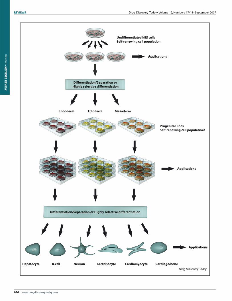
FIGURE 6
Schematic of the generation of functional cell populations from hES cells. Undiffe
quantities of cells, which subsequently are differentiated into germ layer commit
separation step might be required to generate pure populations of progenitor linexpanded before subsequent directed differentiation takes place. The final steps g
cell types are shown in the figure. As indicated, the cells can be applied, at the v
directed differentiation of hES cells, there is still a need to improve
the homogeneity and yield of the target cells by using enrichment
and selection techniques. Furthermore, the generation of defined
populations of specialized cells from hES cells usually takes several
weeks, making it advantageous to isolate and maintain intermedi-
ate precursor cells that can be cryopreserved and still have the
capacity to proliferate and differentiate upon thawing (Figure 6).
It is anticipated that there will be a widespread use of hES cell-
derived hepatocytes and cardiomyocytes in predictive toxicology
since two of the leading causes of preclinical failure of new
compounds are hepatoxicity and cardiotoxicity [93]. Thus, novel
improved models to assess adverse effects of new drugs early in the
development phase are needed. However, it is important to
appreciate that hES cell research represents an emerging area of
investigation, and there are still many fundamental issues related
to hES cell culture and differentiation that needs to be addressed.
Nevertheless, the research community is now at the very early
stages of putting into practice some of the new opportunities that
hES cells provide. With the right resources, together with sound
guidelines and regulations of stem cell research, the field of hES
cells has a real potential to revolutionize many aspects of human
biomedicine and the understanding of normal and abnormal
human development. Specifically, the development of many cell
culture tools in the pharmaceutical industry will most probably
emerge over the coming years. It is likely that pharmaceutical
companies that successfully integrate stem cell technologies will
have a competitive advantage.
AcknowledgementWe thank Dr. Daniella Steel for editorial assistance.
References
1 Dimasi, J.A. (2001) Risks in new drug development: approval success rates for
investigational drugs. Clin. Pharmacol. Ther. 69, 297–307
2 Pearson, H. (2006) The bitterest pill. Nature 444, 532–533
3 Kola, I. and Landis, J. (2004) Can the pharmaceutical industry reduce attrition
rates? Nat. Rev. Drug Discov. 3, 711–715
4 Friedrich Ben-Nun, I. and Benvenisty, N. (2006) Human embryonic stem cells as a
cellular model for human disorders. Mol. Cell Endocrinol. 252, 154–159
5 Vats, A. et al. (2005) Stem cells. Lancet 366, 592–602
6 Scadden, D.T. (2006) The stem-cell niche as an entity of action. Nature 441, 1075–
1079
7 Ding, S. and Schultz, P.G. (2004) A role for chemistry in stem cell biology. Nat.
Biotechnol. 22, 833–840
8 Cai, J. et al. (2004) In search of ‘‘stemness’’. Exp. Hematol. 32, 585–598
9 Jiang, Y. et al. (2002) Pluripotency of mesenchymal stem cells derived from adult
marrow. Nature 418, 41–49
10 Broxmeyer, H.E. et al. (2003) High-efficiency recovery of functional hematopoietic
progenitor and stem cells from human cord blood cryopreserved for 15 years. Proc.
Natl. Acad. Sci. U. S. A. 100, 645–650
11 McNeish, J. (2004) Embryonic stem cells in drug discovery. Nat. Rev. Drug Discov. 3,
70–80
12 Thomson, J.A. et al. (1998) Embryonic stem cell lines derived from human
blastocysts. Science 282, 1145–1147
13 Zambrowicz, B.P. and Sands, A.T. (2003) Knockouts model the 100 best-selling
drugs – will they model the next 100? Nat. Rev. Drug Discov. 2, 38–51
14 Guhr, A. et al. (2006) Current state of human embryonic stem cell research:
an overview of cell lines and their use in experimental work. Stem Cells 24,
2187–2191
15 Martin, G.R. (1981) Isolation of a pluripotent cell line from early mouse embryos
cultured in medium conditioned by teratocarcinoma stem cells. Proc. Natl. Acad.
Sci. U. S. A. 78, 7634–7638
16 Evans, M.J. and Kaufman, M.H. (1981) Establishment in culture of pluripotential
cells from mouse embryos. Nature 292, 154–156
17 Bongso, A. et al. (1994) Isolation and culture of inner cell mass cells from human
blastocysts. Hum. Reprod. 9, 2110–2117
18 Thomson, J.A. et al. (1995) Isolation of a primate embryonic stem cell line. Proc.
Natl. Acad. Sci. U. S. A. 92, 7844–7848
19 Heins, N. et al. (2004) Derivation, characterization, and differentiation of human
embryonic stem cells. Stem Cells 22, 367–376
20 Amit, M. and Itskovitz-Eldor, J. (2002) Derivation and spontaneous differentiation
of human embryonic stem cells. J. Anat. 200 (Pt 3), 225–232
21 Reubinoff, B.E. et al. (2000) Embryonic stem cell lines from human blastocysts:
somatic differentiation in vitro. Nat. Biotechnol. 18, 399–404
22 Stojkovic, M. et al. (2004) Derivation of human embryonic stem cells from day-8
blastocysts recovered after three-step in vitro culture. Stem Cells 22, 790–797
rentiated hES cells can be propagated extensively in vitro, generating large
ted progenitor cells. Depending on the efficiency of the differentiation a
es. The progenitor cells also have the capacity for self-renewal and can beenerate terminally differentiated specialized cells, and a few examples of key
arious stages, for in vitro use in drug discovery.
www.drugdiscoverytoday.com 697

REVIEWS Drug Discovery Today � Volume 12, Numbers 17/18 � September 2007
Review
s�K
EYNOTEREVIEW
23 Baharvand, H. et al. (2004) Establishment and in vitro differentiation of a new
embryonic stem cell line from human blastocyst. Differentiation 72, 224–229
24 Kim, H.S. et al. (2005) Methods for derivation of human embryonic stem cells. Stem
Cells 23, 1228–1233
25 Xu, R.H. et al. (2002) BMP4 initiates human embryonic stem cell differentiation to
trophoblast. Nat. Biotechnol. 20, 1261–1264
26 Draper, J.S. et al. (2004) Culture and characterization of human embryonic stem
cells. Stem. Cells Dev. 13, 325–336
27 Pesce, M. and Scholer, H.R. (2001) Oct-4: gatekeeper in the beginnings of
mammalian development. Stem Cells 19, 271–278
28 Chambers, I. et al. (2003) Functional expression cloning of Nanog, a pluripotency
sustaining factor in embryonic stem cells. Cell 113, 643–655
29 Bhattacharya, B. et al. (2005) Comparison of the gene expression profile of
undifferentiated human embryonic stem cell lines and differentiating embryoid
bodies. BMC Dev. Biol. 5, 22
30 Heins, N. et al. (2006) Clonal derivation and characterization of human embryonic
stem cell lines. J. Biotechnol. 122, 511–520
31 Pera, M.F. and Trounson, A.O. (2004) Human embryonic stem cells: prospects for
development. Development 131, 5515–5525
32 Lim, J.W. and Bodnar, A. (2002) Proteome analysis of conditioned medium from
mouse embryonic fibroblast feeder layers which support the growth of human
embryonic stem cells. Proteomics 2, 1187–1203
33 Xu, C. et al. (2001) Feeder-free growth of undifferentiated human embryonic stem
cells. Nat. Biotechnol. 19, 971–974
34 Cowan, C.A. et al. (2004) Derivation of embryonic stem-cell lines from human
blastocysts. N. Engl. J. Med. 350, 1353–1356
35 Richards, M. et al. (2002) Human feeders support prolonged undifferentiated
growth of human inner cell masses and embryonic stem cells. Nat. Biotechnol. 20,
933–936
36 Ellerstrom, C. et al. (2006) Derivation of a xeno-free human embryonic stem cell
line. Stem. Cells 24, 2170–2176
37 Watanabe, K. et al. (2007) A ROCK inhibitor permits survival of dissociated human
embryonic stem cells. Nat. Biotechnol. 25, 681–686
38 Mitalipova, M.M. et al. (2005) Preserving the genetic integrity of human
embryonic stem cells. Nat. Biotechnol. 23, 19–20
39 Sato, N. et al. (2004) Maintenance of pluripotency in human and mouse
embryonic stem cells through activation of Wnt signaling by a pharmacological
GSK-3-specific inhibitor. Nat. Med. 10, 55–63
40 Amit, M. et al. (2003) Human feeder layers for human embryonic stem cells. Biol.
Reprod. 68, 2150–2156
41 Wang, G. et al. (2005) Noggin and bFGF cooperate to maintain the pluripotency of
human embryonic stem cells in the absence of feeder layers. Biochem. Biophys. Res.
Commun. 330, 934–942
42 Levenstein, M.E. et al. (2006) Basic fibroblast growth factor support of human
embryonic stem cell self-renewal. Stem Cells 24, 568–574
43 Yao, S. et al. (2006) Long-term self-renewal and directed differentiation of human
embryonic stem cells in chemically defined conditions. Proc. Natl. Acad. Sci. U. S. A.
103, 6907–6912
44 Ludwig, T.E. et al. (2006) Feeder-independent culture of human embryonic stem
cells. Nat. Methods 3, 637–646
45 Andrews, P.W. et al. (2005) The International Stem Cell Initiative: toward
benchmarks for human embryonic stem cell research. Nat. Biotechnol. 23, 795–797
46 Thomson, H. (2007) Bioprocessing of embryonic stem cells for drug discovery.
Trends Biotechnol. 25, 224–230
47 Terstegge, S. et al. (2007) Automated maintenance of embryonic stem cell cultures.
Biotechnol. Bioeng. 96, 195–201
48 Loring, J.F. and Rao, M.S. (2006) Establishing standards for the characterization of
human embryonic stem cell lines. Stem Cells 24, 145–150
49 Zeng, X. et al. (2006) An in vitro model of human dopaminergic neurons derived
from embryonic stem cells: MPP+ toxicity and GDNF neuroprotection.
Neuropsychopharmacology 31, 2708–2715
50 Pellizzer, C. et al. (2005) Developmental toxicity testing from animal towards
embryonic stem cells. Altex 22, 47–57
51 Nussler, A. et al. (2006) Present status and perspectives of cell-based therapies for
liver diseases. J. Hepatol. 45, 144–159
52 Rodriguez-Antona, C. et al. (2002) Cytochrome P450 expression in human
hepatocytes and hepatoma cell lines: molecular mechanisms that determine lower
expression in cultured cells. Xenobiotica 32, 505–520
53 Wilkening, S. et al. (2003) Comparison of primary human hepatocytes and
hepatoma cell line Hepg2 with regard to their biotransformation properties. Drug
Metab Dispos. 31, 1035–1042
54 Zaret, K.S. (2002) Regulatory phases of early liver development: paradigms of
organogenesis. Nat. Rev. Genet. 3, 499–512
698 www.drugdiscoverytoday.com
55 Suzuki, A. et al. (2002) Clonal identification and characterization of self-renewing
pluripotent stem cells in the developing liver. J. Cell Biol. 156, 173–184
56 Schwartz, R.E. et al. (2002) Multipotent adult progenitor cells from bone
marrow differentiate into functional hepatocyte-like cells. J. Clin. Invest 109,
1291–1302
57 Hamazaki, T. et al. (2001) Hepatic maturation in differentiating embryonic stem
cells in vitro. FEBS Lett. 497, 15–19
58 Soto-Gutierrez, A. et al. (2006) Reversal of mouse hepatic failure using an
implanted liver-assist device containing ES cell-derived hepatocytes. Nat.
Biotechnol. 24, 1412–1419
59 Gouon-Evans, V. et al. (2006) BMP-4 is required for hepatic specification of mouse
embryonic stem cell-derived definitive endoderm. Nat. Biotechnol. 24, 1402–1411
60 Schuldiner, M. et al. (2000) Effects of eight growth factors on the differentiation of
cells derived from human embryonic stem cells. Proc. Natl. Acad. Sci. U. S. A. 97,
11307–11312
61 Rambhatla, L. et al. (2003) Generation of hepatocyte-like cells from human
embryonic stem cells. Cell Transpl. 12, 1–11
62 Lavon, N. et al. (2004) Differentiation and isolation of hepatic-like cells from
human embryonic stem cells. Differentiation 72, 230–238
63 Schwartz, R.E. et al. (2005) Defined conditions for development of functional
hepatic cells from human embryonic stem cells. Stem Cells Dev. 14, 643–655
64 Baharvand, H. et al. (2006) Differentiation of human embryonic stem cells
into hepatocytes in 2D and 3D culture systems in vitro. Int. J. Dev. Biol. 50,
645–652
65 Hay, D.C. et al. (2007) Direct differentiation of human embryonic stem cells to
hepatocyte-like cells exhibiting functional activities. Cloning Stem Cells 9,
51–62
66 Soto-Gutierrez, A. et al. (2006) Differentiation of human embryonic stem cells to
hepatocytes using deleted variant of HGF and poly-amino-urethane-coated
nonwoven polytetrafluoroethylene fabric. Cell Transpl. 15, 335–341
67 Soderdahl, T. et al. (2007) Glutathione transferases in hepatocyte-like cells derived
from human embryonic stem cells. Toxicol. In Vitro 21, 929–937
68 D’Amour, K.A. et al. (2005) Efficient differentiation of human embryonic stem
cells to definitive endoderm. Nat. Biotechnol. 23, 1534–1541
69 Cai, J. et al. (2007) Directed differentiation of human embryonic stem cells into
functional hepatic cells. Hepatology 45, 1229–1239
70 Lazarou, J. et al. (1998) Incidence of adverse drug reactions in hospitalized patients:
a meta-analysis of prospective studies. JAMA 279, 1200–1205
71 Masimirembwa, C.M. et al. (2001) In vitro high throughput screening of
compounds for favorable metabolic properties in drug discovery. Comb. Chem.
High Throughput Screen 4, 245–263
72 Cross, D.M. and Bayliss, M.K. (2000) A commentary on the use of hepatocytes in
drug metabolism studies during drug discovery and development. Drug Metab Rev.
32, 219–240
73 Rendic, S. and Di Carlo, F.J. (1997) Human cytochrome P450 enzymes: a status
report summarizing their reactions, substrates, inducers, and inhibitors. Drug
Metab. Rev. 29, 413–580
74 Chandra, P. and Brouwer, K.L. (2004) The complexities of hepatic drug transport:
current knowledge and emerging concepts. Pharm. Res. 21, 719–735
75 Kulkarni, J.S. and Khanna, A. (2006) Functional hepatocyte-like cells derived from
mouse embryonic stem cells: a novel in vitro hepatotoxicity model for drug
screening. Toxicol. In Vitro 20, 1014–1022
76 Olson, E.N. (2004) A decade of discoveries in cardiac biology. Nat. Med. 10, 467–474
77 Doetschman, T.C. et al. (1985) The in vitro development of blastocyst-derived
embryonic stem cell lines: formation of visceral yolk sac, blood islands and
myocardium. J. Embryol. Exp. Morphol. 87, 27–45
78 Goh, G. et al. (2005) Molecular and phenotypic analyses of human embryonic
stem cell-derived cardiomyocytes: opportunities and challenges for clinical
translation. Thromb Haemost. 94, 728–737
79 Kehat, I. et al. (2001) Human embryonic stem cells can differentiate into myocytes
with structural and functional properties of cardiomyocytes. J. Clin. Invest 108,
407–414
80 Passier, R. et al. (2005) Increased cardiomyocyte differentiation from human
embryonic stem cells in serum-free cultures. Stem Cells 23, 772–780
81 Yoon, B.S. et al. (2006) Enhanced differentiation of human embryonic stem cells
into cardiomyocytes by combining hanging drop culture and 5-azacytidine
treatment. Differentiation 74, 149–159
82 Snir, M. et al. (2003) Assessment of the ultrastructural and proliferative properties
of human embryonic stem cell-derived cardiomyocytes. Am. J. Physiol. Heart Circ.
Physiol. 285, H2355–H2363
83 Norstrom, A. et al. (2006) Molecular and pharmacological properties of human
embryonic stem cell-derived cardiomyocytes. Exp. Biol. Med. (Maywood) 231, 1753–
1762

Drug Discovery Today � Volume 12, Numbers 17/18 � September 2007 REVIEWS
Reviews�KEYNOTEREVIEW
84 Xu, C. et al. (2002) Characterization and enrichment of cardiomyocytes derived
from human embryonic stem cells. Circ. Res. 91, 501–508
85 Mummery, C. et al. (2003) Differentiation of human embryonic stem cells to
cardiomyocytes: role of coculture with visceral endoderm-like cells. Circulation
107, 2733–2740
86 He, J.Q. et al. (2003) Human embryonic stem cells develop into multiple
types of cardiac myocytes: action potential characterization. Circ. Res. 93,
32–39
87 Xue, T. et al. (2005) Functional integration of electrically active cardiac derivatives
from genetically engineered human embryonic stem cells with quiescent recipient
ventricular cardiomyocytes: insights into the development of cell-based
pacemakers. Circulation 111, 11–20
88 Poueymirou, W.T. et al. (2007) F0 generation mice fully derived from gene-
targeted embryonic stem cells allowing immediate phenotypic analyses. Nat.
Biotechnol. 25, 91–99
89 Shah, R.R. (2002) The significance of QT interval in drug development. Br. J. Clin.
Pharmacol. 54, 188–202
90 Meyer, T. et al. (2004) Micro-electrode arrays in cardiac safety pharmacology: a
novel tool to study QT interval prolongation. Drug Saf. 27, 763–772
91 Reppel, M. et al. (2005) The electrocardiogram of human embryonic stem cell-
derived cardiomyocytes. J. Electrocardiol. 38 (4 Suppl), 166–170
92 Muzikant, A.L. and Penland, R.C. (2002) Models for profiling the potential QT
prolongation risk of drugs. Curr. Opin. Drug Discov. Dev. 5, 127–135
93 Schuster, D. et al. (2005) Why drugs fail–a study on side effects in new chemical
entities. Curr. Pharm. Des. 11, 3545–3559
94 Wang, Z.Z. et al. (2007) Endothelial cells derived from human embryonic stem
cells form durable blood vessels in vivo. Nat. Biotechnol. 25, 317–318
95 Huang, H. et al. (2006) Differentiation of human embryonic stem cells into smooth
muscle cells in adherent monolayer culture. Biochem. Biophys. Res. Commun. 351,
321–327
96 D’Amour, K.A. et al. (2006) Production of pancreatic hormone-expressing
endocrine cells from human embryonic stem cells. Nat. Biotechnol. 24, 1392–1401
97 Green, H. et al. (2003) Marker succession during the development of keratinocytes
from cultured human embryonic stem cells. Proc. Natl. Acad. Sci. U. S. A. 100,
15625–15630
98 Nistor, G.I. et al. (2005) Human embryonic stem cells differentiate into
oligodendrocytes in high purity and myelinate after spinal cord transplantation.
Glia 49, 385–396
99 Zhang, S.C. et al. (2001) In vitro differentiation of transplantable neural precursors
from human embryonic stem cells. Nat. Biotechnol. 19, 1129–1133
100 Nat, R. et al. (2007) Neurogenic neuroepithelial and radial glial cells generated
from six human embryonic stem cell lines in serum-free suspension and adherent
cultures. Glia 55, 385–399
101 Kee, K. et al. (2006) Bone morphogenetic proteins induce germ cell differentiation
from human embryonic stem cells. Stem Cells Dev. 15, 831–837
102 Xiong, C. et al. (2005) Derivation of adipocytes from human embryonic stem cells.
Stem Cells Dev. 14, 671–675
103 Vats, A. et al. (2006) Chondrogenic differentiation of human embryonic stem cells:
the effect of the micro-environment. Tissue Eng. 12, 1687–1697
104 Bielby, R.C. et al. (2004) In vitro differentiation and in vivo mineralization of
osteogenic cells derived from human embryonic stem cells. Tissue Eng. 10, 1518–
1525
105 Woll, P.S. et al. (2005) Human embryonic stem cell-derived NK cells acquire
functional receptors and cytolytic activity. J. Immunol. 175, 5095–5103
106 Galic, Z. et al. (2006) T lineage differentiation from human embryonic stem cells.
Proc. Natl. Acad. Sci. U. S. A. 103, 11742–11747
107 Slukvin, I.I. et al. (2006) Directed differentiation of human embryonic stem cells
into functional dendritic cells through the myeloid pathway. J. Immunol. 176,
2924–2932
108 Gaur, M. et al. (2006) Megakaryocytes derived from human embryonic stem cells: a
genetically tractable system to study megakaryocytopoiesis and integrin function.
J. Thromb. Haemost. 4, 436–442
109 Chang, K.H. et al. (2006) Definitive-like erythroid cells derived from human
embryonic stem cells coexpress high levels of embryonic and fetal globins with
little or no adult globin. Blood 108, 1515–1523
110 Anderson, J.S. et al. (2006) Derivation of normal macrophages from human
embryonic stem (hES) cells for applications in HIV gene therapy. Retrovirology 3, 24
111 Fang, D. et al. (2006) Defining the conditions for the generation of melanocytes
from human embryonic stem cells. Stem Cells 24, 1668–1677
112 Lamba, D.A. et al. (2006) Efficient generation of retinal progenitor cells from
human embryonic stem cells. Proc. Natl. Acad. Sci. U. S. A. 103, 12769–12774
113 Lim, U.M. et al. (2006) Derivation of motor neurons from three clonal human
embryonic stem cell lines. Curr. Neurovasc. Res. 3, 281–288
114 Samadikuchaksaraei, A. et al. (2006) Derivation of distal airway epithelium from
human embryonic stem cells. Tissue Eng. 12, 867–875
115 Taylor, R.A. et al. (2006) Formation of human prostate tissue from embryonic stem
cells. Nat. Methods 3, 179–181
116 Wang, D. et al. (2007) A pure population of lung alveolar epithelial type II cells
derived from human embryonic stem cells. Proc. Natl. Acad. Sci. U. S. A. 104, 4449–
4454
www.drugdiscoverytoday.com 699


![STEM CELLS EMBRYONIC STEM CELLS/INDUCED PLURIPOTENT STEM CELLS Stem Cells.pdf · germ cell production [2]. Human embryonic stem cells (hESCs) offer the means to further understand](https://img.dokumen.tips/doc/110x75/6014b11f8ab8967916363675/stem-cells-embryonic-stem-cellsinduced-pluripotent-stem-cells-stem-cellspdf.jpg)