Embed Size (px)
Citation preview
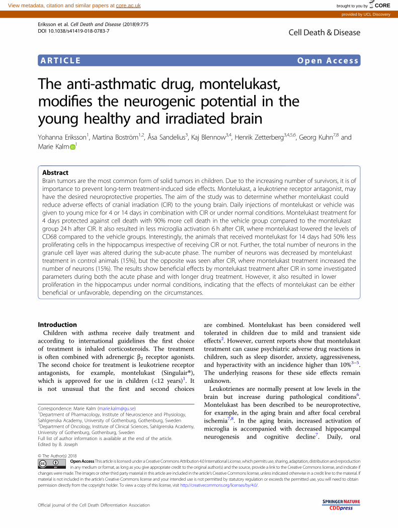
Eriksson et al. Cell Death and Disease (2018) 9:775
DOI 10.1038/s41419-018-0783-7 Cell Death & Disease
ART ICLE Open Ac ce s s
The anti-asthmatic drug, montelukast,modifies the neurogenic potential in theyoung healthy and irradiated brainYohanna Eriksson1, Martina Boström1,2, Åsa Sandelius3, Kaj Blennow3,4, Henrik Zetterberg3,4,5,6, Georg Kuhn7,8 andMarie Kalm 1
AbstractBrain tumors are the most common form of solid tumors in children. Due to the increasing number of survivors, it is ofimportance to prevent long-term treatment-induced side effects. Montelukast, a leukotriene receptor antagonist, mayhave the desired neuroprotective properties. The aim of the study was to determine whether montelukast couldreduce adverse effects of cranial irradiation (CIR) to the young brain. Daily injections of montelukast or vehicle wasgiven to young mice for 4 or 14 days in combination with CIR or under normal conditions. Montelukast treatment for4 days protected against cell death with 90% more cell death in the vehicle group compared to the montelukastgroup 24 h after CIR. It also resulted in less microglia activation 6 h after CIR, where montelukast lowered the levels ofCD68 compared to the vehicle groups. Interestingly, the animals that received montelukast for 14 days had 50% lessproliferating cells in the hippocampus irrespective of receiving CIR or not. Further, the total number of neurons in thegranule cell layer was altered during the sub-acute phase. The number of neurons was decreased by montelukasttreatment in control animals (15%), but the opposite was seen after CIR, where montelukast treatment increased thenumber of neurons (15%). The results show beneficial effects by montelukast treatment after CIR in some investigatedparameters during both the acute phase and with longer drug treatment. However, it also resulted in lowerproliferation in the hippocampus under normal conditions, indicating that the effects of montelukast can be eitherbeneficial or unfavorable, depending on the circumstances.
IntroductionChildren with asthma receive daily treatment and
according to international guidelines the first choiceof treatment is inhaled corticosteroids. The treatmentis often combined with adrenergic β2 receptor agonists.The second choice for treatment is leukotriene receptorantagonists, for example, montelukast (Singulair®),which is approved for use in children (<12 years)1. Itis not unusual that the first and second choices
are combined. Montelukast has been considered welltolerated in children due to mild and transient sideeffects2. However, current reports show that montelukasttreatment can cause psychiatric adverse drug reactions inchildren, such as sleep disorder, anxiety, aggressiveness,and hyperactivity with an incidence higher than 10%3–5.The underlying reasons for these side effects remainunknown.Leukotrienes are normally present at low levels in the
brain but increase during pathological conditions6.Montelukast has been described to be neuroprotective,for example, in the aging brain and after focal cerebralischemia7,8. In the aging brain, increased activation ofmicroglia is accompanied with decreased hippocampalneurogenesis and cognitive decline7. Daily, oral
© The Author(s) 2018OpenAccessThis article is licensedunder aCreativeCommonsAttribution 4.0 International License,whichpermits use, sharing, adaptation, distribution and reproductionin any medium or format, as long as you give appropriate credit to the original author(s) and the source, provide a link to the Creative Commons license, and indicate if
changesweremade. The images or other third partymaterial in this article are included in the article’s Creative Commons license, unless indicated otherwise in a credit line to thematerial. Ifmaterial is not included in the article’s Creative Commons license and your intended use is not permitted by statutory regulation or exceeds the permitted use, you will need to obtainpermission directly from the copyright holder. To view a copy of this license, visit http://creativecommons.org/licenses/by/4.0/.
Correspondence: Marie Kalm ([email protected])1Department of Pharmacology, Institute of Neuroscience and Physiology,Sahlgrenska Academy, University of Gothenburg, Gothenburg, Sweden2Department of Oncology, Institute of Clinical Sciences, Sahlgrenska Academy,University of Gothenburg, Gothenburg, SwedenFull list of author information is available at the end of the article.Edited by B. Joseph
Official journal of the Cell Death Differentiation Association
1234
5678
90():,;
1234
5678
90():,;
1234567890():,;
1234
5678
90():,;
brought to you by COREView metadata, citation and similar papers at core.ac.uk
provided by UCL Discovery
administration of montelukast to rats for 6 weeksimproved these negative changes in the aging brain7,effects that could also be beneficial in radiotherapy-induced injury in children.Brain tumors are the most common form of solid
tumors in children9, with surgery, chemotherapy, andradiotherapy as the main treatment strategies10. All ofthese treatments cause late side effects, with radiotherapyhaving the highest severity when graded11. Side effectsrange from sleep disturbance to cognitive impairment12.The mechanisms of chronic radiation-induced damageinvolve, for example, long-term toxicity to neural celltypes, including stem and progenitor cells, loss of oligo-dendrocytes, and inflammatory responses13. Cranialradiotherapy also changes the chemical milieu and affectssupporting cells such as microglia14,15. The subgranularzone (SGZ) in the hippocampus, an area in the brain thatharbor stem cells, is very sensitive to radiotherapy in boththe young and adult brain, and loss of these cells maycontribute to cognitive deficits16–18. Finding means toameliorate radiation-induced injury is of great interest forthe increasing number of long-term childhood cancersurvivors.Targeting the irradiation-induced inflammatory response
is of interest to prevent negative effects on cognitionand neurogenesis. Inhibiting microglia with MW-151, aselective inhibitor of proinflammatory microglial cytokineproduction, restored hippocampal-dependent learning,improved synaptic function, and partially protected neuro-genesis after cranial irradiation (CIR) to the adult ratbrain19. Further, it has been shown that indomethacin,a common nonsteroidal anti-inflammatory drug, hasthe potential to partly increase neurogenesis after CIRin adults20. Blocking chemokine (C–C motif) receptor 2(Ccr2) has also prevented neuronal dysfunction andhippocampal-dependent memory dysfunction inducedby irradiation toward the adult mouse brain21. We havepreviously shown that the juvenile and adult brain havedifferent radiation-induced inflammatory responses22,which is of importance if using an anti-inflammatoryapproach to protect the brain from CIR-induced injury. Inthe developing brain, it has been shown that blocking thecomplement cascade can improve reversal learning afterCIR, but without effects on neurogenesis23. As mentionedearlier, montelukast is a leukotriene receptor antagonist.Leukotrienes are lipid mediators of inflammation andare metabolized from arachidonic acid through the5-lipoxygenase (5-LOX) pathway. It has been shownthat minocycline, a tetracycline antibiotic that blocksthe activation of 5-LOX, had positive effects on cognitiveimpairment and decreased apoptosis in newborn neurons(DCX+) following a single dose of 20Gy irradiation to thebrain of 1 month old rats24. Inhibiting the 5-LOX pathwayto target the inflammatory response could therefore be an
interesting strategy to investigate when trying to protect thenormal tissue during radiotherapy. The purpose of thisstudy was to investigate the effect of montelukast in com-bination with CIR to the young brain.
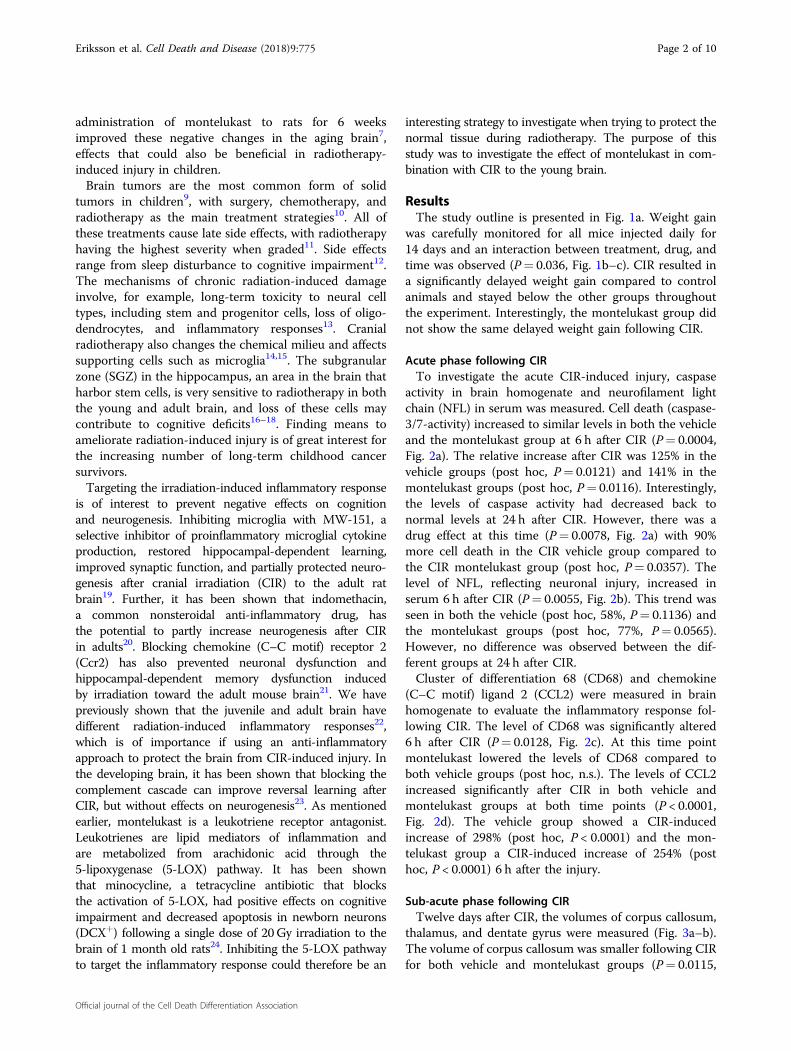
ResultsThe study outline is presented in Fig. 1a. Weight gain
was carefully monitored for all mice injected daily for14 days and an interaction between treatment, drug, andtime was observed (P= 0.036, Fig. 1b–c). CIR resulted ina significantly delayed weight gain compared to controlanimals and stayed below the other groups throughoutthe experiment. Interestingly, the montelukast group didnot show the same delayed weight gain following CIR.
Acute phase following CIRTo investigate the acute CIR-induced injury, caspase
activity in brain homogenate and neurofilament lightchain (NFL) in serum was measured. Cell death (caspase-3/7-activity) increased to similar levels in both the vehicleand the montelukast group at 6 h after CIR (P= 0.0004,Fig. 2a). The relative increase after CIR was 125% in thevehicle groups (post hoc, P= 0.0121) and 141% in themontelukast groups (post hoc, P= 0.0116). Interestingly,the levels of caspase activity had decreased back tonormal levels at 24 h after CIR. However, there was adrug effect at this time (P= 0.0078, Fig. 2a) with 90%more cell death in the CIR vehicle group compared tothe CIR montelukast group (post hoc, P= 0.0357). Thelevel of NFL, reflecting neuronal injury, increased inserum 6 h after CIR (P= 0.0055, Fig. 2b). This trend wasseen in both the vehicle (post hoc, 58%, P= 0.1136) andthe montelukast groups (post hoc, 77%, P= 0.0565).However, no difference was observed between the dif-ferent groups at 24 h after CIR.Cluster of differentiation 68 (CD68) and chemokine
(C–C motif) ligand 2 (CCL2) were measured in brainhomogenate to evaluate the inflammatory response fol-lowing CIR. The level of CD68 was significantly altered6 h after CIR (P= 0.0128, Fig. 2c). At this time pointmontelukast lowered the levels of CD68 compared toboth vehicle groups (post hoc, n.s.). The levels of CCL2increased significantly after CIR in both vehicle andmontelukast groups at both time points (P < 0.0001,Fig. 2d). The vehicle group showed a CIR-inducedincrease of 298% (post hoc, P < 0.0001) and the mon-telukast group a CIR-induced increase of 254% (posthoc, P < 0.0001) 6 h after the injury.
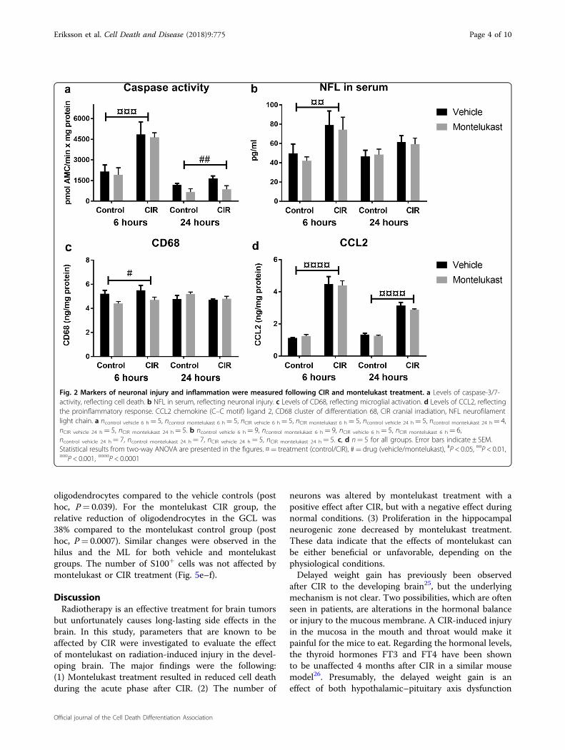
Sub-acute phase following CIRTwelve days after CIR, the volumes of corpus callosum,
thalamus, and dentate gyrus were measured (Fig. 3a–b).The volume of corpus callosum was smaller following CIRfor both vehicle and montelukast groups (P= 0.0115,
Eriksson et al. Cell Death and Disease (2018) 9:775 Page 2 of 10
Official journal of the Cell Death Differentiation Association
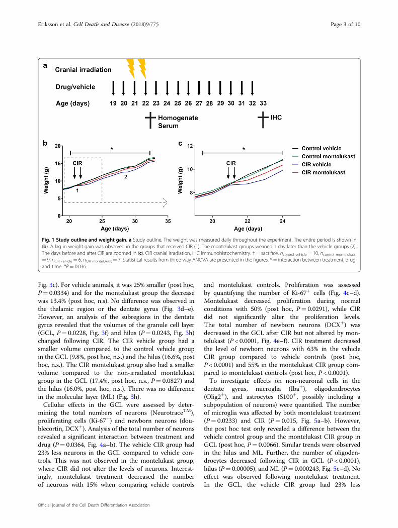
Fig. 3c). For vehicle animals, it was 25% smaller (post hoc,P= 0.0334) and for the montelukast group the decreasewas 13.4% (post hoc, n.s). No difference was observed inthe thalamic region or the dentate gyrus (Fig. 3d–e).However, an analysis of the subregions in the dentategyrus revealed that the volumes of the granule cell layer(GCL, P= 0.0228, Fig. 3f) and hilus (P= 0.0243, Fig. 3h)changed following CIR. The CIR vehicle group had asmaller volume compared to the control vehicle groupin the GCL (9.8%, post hoc, n.s.) and the hilus (16.6%, posthoc, n.s.). The CIR montelukast group also had a smallervolume compared to the non-irradiated montelukastgroup in the GCL (17.4%, post hoc, n.s., P= 0.0827) andthe hilus (16.0%, post hoc, n.s.). There was no differencein the molecular layer (ML) (Fig. 3h).Cellular effects in the GCL were assessed by deter-
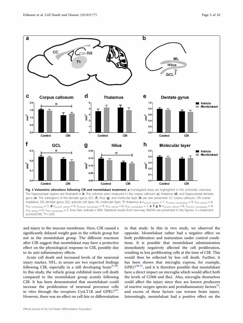
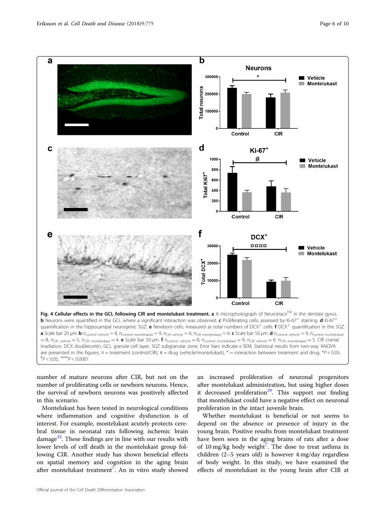
mining the total numbers of neurons (NeurotraceTM),proliferating cells (Ki-67+) and newborn neurons (dou-blecortin, DCX+). Analysis of the total number of neuronsrevealed a significant interaction between treatment anddrug (P= 0.0364, Fig. 4a–b). The vehicle CIR group had23% less neurons in the GCL compared to vehicle con-trols. This was not observed in the montelukast group,where CIR did not alter the levels of neurons. Interest-ingly, montelukast treatment decreased the numberof neurons with 15% when comparing vehicle controls
and montelukast controls. Proliferation was assessedby quantifying the number of Ki-67+ cells (Fig. 4c–d).Montelukast decreased proliferation during normalconditions with 50% (post hoc, P= 0.0291), while CIRdid not significantly alter the proliferation levels.The total number of newborn neurons (DCX+) wasdecreased in the GCL after CIR but not altered by mon-telukast (P < 0.0001, Fig. 4e–f). CIR treatment decreasedthe level of newborn neurons with 63% in the vehicleCIR group compared to vehicle controls (post hoc,P < 0.0001) and 55% in the montelukast CIR group com-pared to montelukast controls (post hoc, P < 0.0001).To investigate effects on non-neuronal cells in the
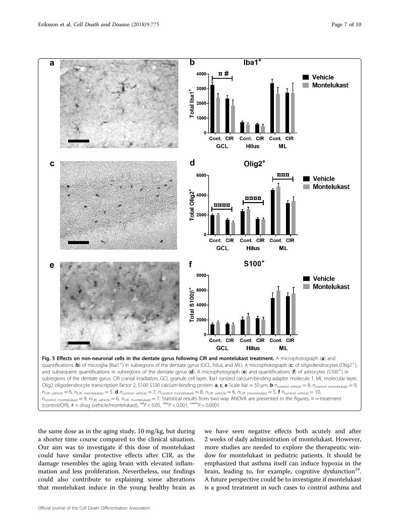
dentate gyrus, microglia (Iba+), oligodendrocytes(Olig2+), and astrocytes (S100+, possibly including asubpopulation of neurons) were quantified. The numberof microglia was affected by both montelukast treatment(P= 0.0233) and CIR (P= 0.015, Fig. 5a–b). However,the post hoc test only revealed a difference between thevehicle control group and the montelukast CIR group inGCL (post hoc, P= 0.0066). Similar trends were observedin the hilus and ML. Further, the number of oligoden-drocytes decreased following CIR in GCL (P < 0.0001),hilus (P= 0.00005), and ML (P= 0.000243, Fig. 5c–d). Noeffect was observed following montelukast treatment.In the GCL, the vehicle CIR group had 23% less
Fig. 1 Study outline and weight gain. a Study outline. The weight was measured daily throughout the experiment. The entire period is shown in(b). A lag in weight gain was observed in the groups that received CIR (1). The montelukast groups weaned 1 day later than the vehicle groups (2).The days before and after CIR are zoomed in (c). CIR cranial irradiation, IHC immunohistochemistry. †= sacrifice. ncontrol vehicle= 10, ncontrol montelukast
= 9, nCIR vehicle= 6, nCIR montelukast= 7. Statistical results from three-way ANOVA are presented in the figures, *= interaction between treatment, drug,and time. *P= 0.036
Eriksson et al. Cell Death and Disease (2018) 9:775 Page 3 of 10
Official journal of the Cell Death Differentiation Association
oligodendrocytes compared to the vehicle controls (posthoc, P= 0.039). For the montelukast CIR group, therelative reduction of oligodendrocytes in the GCL was38% compared to the montelukast control group (posthoc, P= 0.0007). Similar changes were observed in thehilus and the ML for both vehicle and montelukastgroups. The number of S100+ cells was not affected bymontelukast or CIR treatment (Fig. 5e–f).
DiscussionRadiotherapy is an effective treatment for brain tumors
but unfortunately causes long-lasting side effects in thebrain. In this study, parameters that are known to beaffected by CIR were investigated to evaluate the effectof montelukast on radiation-induced injury in the devel-oping brain. The major findings were the following:(1) Montelukast treatment resulted in reduced cell deathduring the acute phase after CIR. (2) The number of
neurons was altered by montelukast treatment with apositive effect after CIR, but with a negative effect duringnormal conditions. (3) Proliferation in the hippocampalneurogenic zone decreased by montelukast treatment.These data indicate that the effects of montelukast canbe either beneficial or unfavorable, depending on thephysiological conditions.Delayed weight gain has previously been observed
after CIR to the developing brain25, but the underlyingmechanism is not clear. Two possibilities, which are oftenseen in patients, are alterations in the hormonal balanceor injury to the mucous membrane. A CIR-induced injuryin the mucosa in the mouth and throat would make itpainful for the mice to eat. Regarding the hormonal levels,the thyroid hormones FT3 and FT4 have been shownto be unaffected 4 months after CIR in a similar mousemodel26. Presumably, the delayed weight gain is aneffect of both hypothalamic–pituitary axis dysfunction
Fig. 2 Markers of neuronal injury and inflammation were measured following CIR and montelukast treatment. a Levels of caspase-3/7-activity, reflecting cell death. b NFL in serum, reflecting neuronal injury. c Levels of CD68, reflecting microglial activation. d Levels of CCL2, reflectingthe proinflammatory response. CCL2 chemokine (C–C motif) ligand 2, CD68 cluster of differentiation 68, CIR cranial irradiation, NFL neurofilamentlight chain. a ncontrol vehicle 6 h= 5, ncontrol montelukast 6 h= 5, nCIR vehicle 6 h= 5, nCIR montelukast 6 h= 5, ncontrol vehicle 24 h= 5, ncontrol montelukast 24 h= 4,nCIR vehicle 24 h= 5, nCIR montelukast 24 h= 5. b ncontrol vehicle 6 h= 9, ncontrol montelukast 6 h= 9, nCIR vehicle 6 h= 5, nCIR montelukast 6 h= 6,ncontrol vehicle 24 h= 7, ncontrol montelukast 24 h= 7, nCIR vehicle 24 h= 5, nCIR montelukast 24 h= 5. c, d n= 5 for all groups. Error bars indicate ± SEM.Statistical results from two-way ANOVA are presented in the figures. ¤= treatment (control/CIR), #= drug (vehicle/montelukast), #P < 0.05, ¤¤P < 0.01,¤¤¤P < 0.001, ¤¤¤¤P < 0.0001
Eriksson et al. Cell Death and Disease (2018) 9:775 Page 4 of 10
Official journal of the Cell Death Differentiation Association
and injury in the mucous membrane. Here, CIR caused asignificantly delayed weight gain in the vehicle group butnot in the montelukast group. The different reactionsafter CIR suggest that montelukast may have a protectiveeffect on the physiological response to CIR, possibly dueto its anti-inflammatory effects.Acute cell death and increased levels of the neuronal
injury marker, NFL, in serum are two expected findingsfollowing CIR, especially in a still developing brain27,28.In this study, the vehicle group exhibited more cell deathcompared to the montelukast group acutely followingCIR. It has been demonstrated that montelukast couldincrease the proliferation of neuronal precursor cellsin vitro through the receptors CysLT1R and GPR1729.However, there was no effect on cell fate or differentiation
in that study. In this in vivo study, we observed theopposite. Montelukast rather had a negative effect onboth proliferation and maturation under control condi-tions. It is possible that montelukast administrationimmediately negatively affected the cell proliferation,resulting in less proliferating cells at the time of CIR. Thiswould then be reflected by less cell death. Further, ithas been shown that microglia express, for example,GPR1730,31, and it is therefore possible that montelukasthave a direct impact on microglia which would affect boththe levels of CD68 and Iba1. Also, microglia themselvescould affect the injury since they are known producersof reactive oxygen species and proinflammatory factors32,and excess of these factors can worsen brain injury.Interestingly, montelukast had a positive effect on the
Fig. 3 Volumetric alterations following CIR and montelukast treatment. a Investigated areas are highlighted in the schematic overview.The hippocampal regions are illustrated in b. The volumes were measured in the corpus callosum (c), thalamus (d), and hippocampal dentategyrus (e). The subregions of the dentate gyrus; GCL (f), hilus (g), and molecular layer (h) are also presented. CC corpus callosum, CIR cranialirradiation, DG dentate gyrus, GCL granule cell layer, ML molecular layer, Th thalamus. c ncontrol vehicle= 7, ncontrol montelukast= 9, nCIR vehicle= 6,nCIR montelukast= 5. d ncontrol vehicle= 9, ncontrol montelukast= 9, nCIR vehicle= 6, nCIR montelukast= 7, e, f, g, h ncontrol vehicle= 8, ncontrol montelukast= 9,nCIR vehicle= 6, nCIR montelukast= 5. Error bars indicate ± SEM. Statistical results from two-way ANOVA are presented in the figures, ¤= treatment(control/CIR), ¤P < 0.05
Eriksson et al. Cell Death and Disease (2018) 9:775 Page 5 of 10
Official journal of the Cell Death Differentiation Association
number of mature neurons after CIR, but not on thenumber of proliferating cells or newborn neurons. Hence,the survival of newborn neurons was positively affectedin this scenario.Montelukast has been tested in neurological conditions
where inflammation and cognitive dysfunction is ofinterest. For example, montelukast acutely protects cere-bral tissue in neonatal rats following ischemic braindamage33. These findings are in line with our results withlower levels of cell death in the montelukast group fol-lowing CIR. Another study has shown beneficial effectson spatial memory and cognition in the aging brainafter montelukast treatment7. An in vitro study showed
an increased proliferation of neuronal progenitorsafter montelukast administration, but using higher dosesit decreased proliferation29. This support our findingthat montelukast could have a negative effect on neuronalproliferation in the intact juvenile brain.Whether montelukast is beneficial or not seems to
depend on the absence or presence of injury in theyoung brain. Positive results from montelukast treatmenthave been seen in the aging brains of rats after a doseof 10 mg/kg body weight7. The dose to treat asthma inchildren (2–5 years old) is however 4 mg/day regardlessof body weight. In this study, we have examined theeffects of montelukast in the young brain after CIR at
Fig. 4 Cellular effects in the GCL following CIR and montelukast treatment. a A microphotograph of NeurotraceTM in the dentate gyrus.b Neurons were quantified in the GCL where a significant interaction was observed. c Proliferating cells, assessed by Ki-67+ staining. d Ki-67+
quantification in the hippocampal neurogenic SGZ. e Newborn cells, measured as total numbers of DCX+ cells. f DCX+ quantification in the SGZ.a Scale bar 20 µm. b ncontrol vehicle= 8, ncontrol montelukast= 9, nCIR vehicle= 6, nCIR montelukast= 6. c Scale bar 50 µm. d ncontrol vehicle= 9, ncontrol montelukast
= 8, nCIR vehicle= 5, nCIR montelukast= 4. e Scale bar 50 µm. f ncontrol vehicle= 8, ncontrol montelukast= 9, nCIR vehicle= 6, nCIR montelukast= 5. CIR cranialirradiation, DCX doublecortin, GCL granule cell layer, SGZ subgranular zone. Error bars indicate ± SEM. Statistical results from two-way ANOVAare presented in the figures, ¤= treatment (control/CIR), #= drug (vehicle/montelukast), *= interaction between treatment and drug. *P < 0.05,#P < 0.05, ¤¤¤¤P < 0.0001
Eriksson et al. Cell Death and Disease (2018) 9:775 Page 6 of 10
Official journal of the Cell Death Differentiation Association
the same dose as in the aging study, 10 mg/kg, but duringa shorter time course compared to the clinical situation.Our aim was to investigate if this dose of montelukastcould have similar protective effects after CIR, as thedamage resembles the aging brain with elevated inflam-mation and less proliferation. Nevertheless, our findingscould also contribute to explaining some alterationsthat montelukast induce in the young healthy brain as
we have seen negative effects both acutely and after2 weeks of daily administration of montelukast. However,more studies are needed to explore the therapeutic win-dow for montelukast in pediatric patients. It should beemphasized that asthma itself can induce hypoxia in thebrain, leading to, for example, cognitive dysfunction34.A future perspective could be to investigate if montelukastis a good treatment in such cases to control asthma and
Fig. 5 Effects on non-neuronal cells in the dentate gyrus following CIR and montelukast treatment. A microphotograph (a) andquantifications (b) of microglia (Iba1+) in subregions of the dentate gyrus (GCL, hilus, and ML). A microphotograph (c) of oligodendrocytes (Olig2+),and subsequent quantifications in subregions of the dentate gyrus (d). A microphotograph (e) and quantifications (f) of astrocytes (S100+) insubregions of the dentate gyrus. CIR cranial irradiation, GCL granule cell layer, Iba1 ionized calcium-binding adapter molecule 1, ML molecular layer,Olig2 oligodendrocyte transcription factor 2, S100 S100 calcium-binding protein. a, c, e Scale bar= 50 µm. b ncontrol vehicle= 8, ncontrol montelukast= 9,nCIR vehicle= 6, nCIR montelukast= 5. d ncontrol vehicle= 7, ncontrol montelukast= 8, nCIR vehicle= 6, nCIR montelukast= 5. f ncontrol vehicle= 10,ncontrol montelukast= 9, nCIR vehicle= 6, nCIR montelukast= 7. Statistical results from two-way ANOVA are presented in the figures, ¤= treatment(control/CIR), #= drug (vehicle/montelukast), ¤#P < 0.05, ¤¤¤P < 0.001, ¤¤¤¤P < 0.0001
Eriksson et al. Cell Death and Disease (2018) 9:775 Page 7 of 10
Official journal of the Cell Death Differentiation Association
at the same time minimize injuries from the hypoxia forthis group of children.In summary, montelukast has negative effects on the
maturation of the GCL during normal conditions,whereas during a pathological condition, such as follow-ing CIR, the effects can be protective. These findings,with the affected proliferation during normal conditions,in combination with the new profile for psychiatricadverse drug reactions, suggests that prescribing mon-telukast to young children should be a well thoughtthrough decision. However, more studies are needed toinvestigate if the negative effects are occurring also atlower dose spans and if the effect is chronic if ending thetreatment with montelukast.
Material and methodsAnimalsC57BL/6J mice with six female pups per mother were
ordered from Charles River Laboratories, Germany.Animals were housed according to normal procedures atthe Experimental Biomedicine animal facility (Universityof Gothenburg, Gothenburg, Sweden). The mice werekept on a 12-h light cycle with food and water providedad libitum. The room temperature was 19–21 °C with40–70% relative humidity. Animal experiments wereapproved by the Gothenburg committee of the SwedishAnimal Welfare Agency (2015–72).
Montelukast treatmentMontelukast sodium powder (Sigma, USA) was dis-
solved in 99.5% ethanol, diluted 1:10 with 0.9% saline(NaCl), and administered by intraperitoneal injectionsat a dose of 10 mg/kg. Animals (n= 6–10) received dailyinjections of montelukast or vehicle (0.9% NaCl with 10%ethanol) starting 2 days before CIR, resulting in fourdoses for the acute study and 14 doses for the sub-acutestudy (Fig. 1a).
Cranial irradiation procedureA linear accelerator True Beam STX (600 MU/min, 5.6
Gy/min; Radiation Oncology Systems, USA) with 6 MVnominal photon energy was used for CIR as describedearlier28. Briefly, all animals were anesthetized on post-natal day 21 with a mixture of oxygen and isoflurane(Attane Vet, VM Pharma AB, Sweden) to immobilize theanimals during the procedure. The whole brain wasirradiated with a clinically relevant dose of 2 × 4 Gy with12-h interval, using a radiation field of 2 × 2 cm, a sourceto skin distance of 99.5 cm, and a dose variation of ±5%.After CIR, animals were returned to their dams. Controlanimals were anesthetized but did not receive CIR.Typically, pediatric patients with brain tumors are treatedwith radiotherapy, once a day, 5 days per week, for severalweeks. The treatment protocol varies depending on tumor
type and other relevant factors, but often it is 50–59.4 Gyin 28–33 fractions of 1.8 Gy. This study was designed toinvestigate the radiation-induced injury in the normaltissue (8 Gy given in two fractions), hence a much lowerdose than the tumor bed receive.
Acute tissue preparationBrains were quickly removed after sacrifice, put in
liquid nitrogen and stored at −80 °C. Brains werehomogenized by sonication in phosphate-buffered salinecontaining Triton X-100 (Merck KGaA, Germany),ethylenediaminetetraacetic acid (EDTA, Sigma-Aldrich,USA), and protease inhibitor cocktail (cOmplete, EDTA-free, Roche, Switzerland). Samples were then centrifugedand supernatant stored at −80 °C. Protein concentrationwas measured using the Pierce BCA protein Assay Kit(Thermo Scientific, USA) according to the protocol pro-vided by the manufacturer.
Sub-acute tissue preparationAnimals were anesthetized with sodium pentobarbital
(Pentothal, Electra-box Pharma, Sweden) and transcar-dially perfused with 0.1M phosphate buffer (pH 7.5) torinse the vascular system, followed by 6% formaldehyde(pH 7.4; Histofix; Histolab Products AB, Sweden). Brainswere gently removed, immersion-fixed in Histofix for24 h and stored in a sucrose solution (30% sucrose in0.1M phosphate buffer, pH 7.5). The right hemispherewas cut sagittally into 25 µm sections in a series of12, using a sliding microtome (SM2010R, Leica Micro-systems, Germany), and stored at 4 °C in a cryoprotectantsolution (25% ethylene glycol and 25% glycerol).
Blood collectionMice were anesthetized with a mixture of oxygen and
isoflurane, blood was drawn from the heart with a 1 mLsyringe (Omnifix®, BRAUN, Germany) and centrifugedfor 5 min. Serum was collected and stored at −80 °C.
Fluorometric assay of caspase-3-like activityCaspase-dependent cell death was measured using a
caspase activity assay. An aliquot of 20 µl tissue homo-genate was added to a microplate and mixed with 80 µlextraction buffer (n= 5, duplicate samples) and analyzedas described earlier35. Cleavage of Ac-DEVD-AMC(for caspase-3/7-activity, Peptide Institute, Japan, cat.no.3171-v) was measured and expressed as pmol AMCreleased per mg protein and minute.
Enzyme-linked immunosorbent assayEnzyme-linked immunosorbent assays (ELISA) were
used to investigate chemokine (C–C motif) ligand 2(CCL2, MJE00, R&D Systems, USA) and cluster of dif-ferentiation 68 (mouse CD68, EKM1518, Nordic BioSite,
Eriksson et al. Cell Death and Disease (2018) 9:775 Page 8 of 10
Official journal of the Cell Death Differentiation Association
Sweden) expression. Analyses were performed accordingto instructions of the manufacturers and the amount ofinvestigated proteins measured using a SpectraMax i3x(Molecular Devices, USA).
Serum NFLSerum sample neurofilament light chain (NFL) con-
centration was determined using an in-house NFL assayon the single molecule array (Simoa) platform, which hasbeen described in detail previously36. Briefly, para-magnetic carboxylated beads (Quanterix Corp, USA) werecoated with a mouse anti-NFL antibody (UD1, Uman-Diagnostics, Sweden) and incubated with sample and abiotinylated mouse anti-NFL antibody (UD2, Uman-Diagnostics, Sweden) in a Simoa HD-1 instrument(Quanterix).
ImmunohistochemistrySections were treated as follows: rinsed (always in Tris-
buffered saline [TBS], 50 mM Tris-HCl in 150mM NaCl),incubated in 0.6% H2O2 in TBS for 30min, rinsed andincubated for 30min in a TBS block solution with 3%donkey serum (Jackson ImmunoResearch LaboratoriesInc, USA), and 0.1% Triton X-100 (Merck KGaA, Ger-many). Sections were incubated overnight at 4 °C in blocksolution using the following primary antibodies: oligo-dendrocyte transcription factor 2 (1:500, rabbit anti-Olig2,ab109186, Abcam, UK), marker of proliferation Ki-67(1:1000, rabbit anti-Ki-67, ab15580, Abcam, UK), S100calcium-binding protein (1:2000, mouse anti-S100, MA5-12969, ThermoFisher Scientific, USA), rabbit anti-ionizedcalcium-binding adapter molecule 1 (1:1000, rabbit anti-Iba1, 019-19741, WAKO Pure Chemical Industries ltd,Japan), and doublecortin (1:500, polyclonal goat anti-DCX, Santa Cruz Biotechnology, USA). Sections wererinsed, incubated for 1 h with block solution and bioti-nylated secondary antibodies (1:1000, Jackson Immu-noResearch Laboratories Inc, USA), rinsed and incubatedwith avidin-biotin-peroxidase (10 µL/mL TBS of A and B,Vectastain Elite ABC Kit, Vector Laboratories, USA) for1 h. Sections were rinsed and developed in 3,3′-diamino-benzidine (DAB, Saveen Werner AB, Sweden) diluted inTBS with H2O2 and NiCl2, until sufficient color wasnoted. After rinsing in tap water, sections were mountedusing 0.1M phosphate buffer, pH 7.5, and dried over-night, then cover slipped with X-Tra-Kitt (Medite GmbH,Germany).
NeuroTrace Fluorescent Nissl stainSections were rinsed in TBS and then washed in TBS
with 0.1% Triton X-100 for 10min. After further washing,sections were incubated with NeuroTraceTM 500/525Green Fluorescent Nissl stain for 20 min (N21480,ThermoFisher Scientific, USA). Following several rinsing
steps in TBS, the sections were mounted and cover slip-ped with ProLong® Gold Antifade Reagent (Thermo-Fisher Scientific, USA).
Stereological proceduresCells were counted in every 12th section using
systematic-random sampling (Stereoinvestigator, Micro-BrightField, USA) and a Leica DM6000 B microscope(Leica Microsystems, Germany). Counting startedon sections containing a clearly divided dorsal andventral hippocampus (only the dorsal granule cell layer[GCL] was measured). Total volumes were calculatedaccording to the Cavalieri principle (V= SA × P × T,where V is the total volume, SA is the sum of area mea-surements, P is the inverse of the sampling fraction, and Tis the section thickness). The total number of cells wasobtained by dividing the number of counted cells with thesampling fraction. Volumes for corpus callosum andthalamus were also measured in sections eligible forhippocampal quantifications.NeuroTrace was used to quantify neurons in GCL (×40
objective). A 275 × 75-µm grid was randomly placed overthe traced area and counting frames (25 × 25 µm) wereplaced within the grid. The total number of GCL neuronsper animal was calculated by dividing the number ofcounted cells with the sampling fractions, i.e., fraction ofsampling area/total traced area × series fraction (1/12) ×optical dissector height/physical section thickness.
StatisticsFor statistical analyses, two-way ANOVA was used
(drug and treatment as main effects), followed by a posthoc test (Sidak, corrected for multiple testing usingGraphPad Prism 7.02). Weight was analyzed using three-way ANOVA (Stata). Statistical significance was con-sidered if P < 0.05.
AcknowledgementsWe are grateful for the skillful technical assistance of Rita Grandér and thankour former and current students, Jolie Danial, Alba Rodríguez-Rodríguez, andZainab Jasim. This study was supported by grants from the Swedish andEuropean Research Councils, the Knut and Alice Wallenberg Foundation, theSwedish Brain Foundation, Swedish State Support for Clinical Research, theWolfson Foundation, Alzheimerfonden, the Swedish Society of Medicine, theSwedish Childhood Cancer Foundation (Barncancerfonden), the FrimurareBarnhus Foundations of Gothenburg, the Lions Cancer Foundation West, theWilhelm and Martina Lundgren Foundation, the Edith Jacobssons DonationFund, the Torsten Söderberg Foundation, and Jubileumskliniken’s anti-cancerresearch fund.
Author details1Department of Pharmacology, Institute of Neuroscience and Physiology,Sahlgrenska Academy, University of Gothenburg, Gothenburg, Sweden.2Department of Oncology, Institute of Clinical Sciences, Sahlgrenska Academy,University of Gothenburg, Gothenburg, Sweden. 3Department of Psychiatryand Neurochemistry, Institute of Neuroscience and Physiology, SahlgrenskaAcademy, University of Gothenburg, Mölndal, Sweden. 4ClinicalNeurochemistry Laboratory, Sahlgrenska University Hospital, Gothenburg,Sweden. 5Department of Molecular Neuroscience, UCL Institute of Neurology,
Eriksson et al. Cell Death and Disease (2018) 9:775 Page 9 of 10
Official journal of the Cell Death Differentiation Association
Queen Square, London, UK. 6UK Dementia Research Institute, UCL, London, UK.7Center for Brain Repair and Rehabilitation, Institute of Neuroscience andPhysiology, Sahlgrenska Academy, University of Gothenburg, Gothenburg,Sweden. 8Center for Stroke Research Berlin, Charité – Universitätsmedizin,Berlin, Germany
Conflict of interestAuthors K.B. and H.Z. are co-founders of Brain Biomarker Solutions inGothenburg AB, a GU Ventures-based platform company at the Universityof Gothenburg. H.Z. has served at advisory boards of Eli Lilly and RocheDiagnostics and has received travel support from Teva. The remaining authorsdeclare that they have no conflict of interest.
Publisher's noteSpringer Nature remains neutral with regard to jurisdictional claims inpublished maps and institutional affiliations.
Received: 16 March 2018 Revised: 8 June 2018 Accepted: 11 June 2018
References1. Global Initiative for Asthma. Global Strategy for Asthma Management and
Prevention www.ginasthma.org (2016).2. Bisgaard, H. et al. Safety and tolerability of montelukast in placebo-controlled
pediatric studies and their open-label extensions. Pediatr. Pulmonol. 44,568–579 (2009).
3. Benard, B. et al. Neuropsychiatric adverse drug reactions in childreninitiated on montelukast in real-life practice. Eur. Respir. J. 50, 700148 (2017).
4. Bygdell, M., Brunlof, G., Wallerstedt, S. M. & Kindblom, J. M. Psychiatric adversedrug reactions reported during a 10-year period in the Swedish pediatricpopulation. Pharmacoepidemiol. Drug Saf. 21, 79–86 (2012).
5. Wallerstedt, S. M., Brunlof, G., Sundstrom, A. & Eriksson, A. L. Montelukast andpsychiatric disorders in children. Pharmacoepidemiol. Drug Saf. 18, 858–864(2009).
6. Phillis, J. W., Horrocks, L. A. & Farooqui, A. A. Cyclooxygenases, lipoxygenases,and epoxygenases in CNS: their role and involvement in neurological dis-orders. Brain Res. Rev. 52, 201–243 (2006).
7. Marschallinger, J. et al. Structural and functional rejuvenation of theaged brain by an approved anti-asthmatic drug. Nat. Commun. 6, 8466(2015).
8. Zhao, R., Shi, W. Z., Zhang, Y. M., Fang, S. H. & Wei, E. Q. Montelukast, a cysteinylleukotriene receptor-1 antagonist, attenuates chronic brain injury afterfocal cerebral ischaemia in mice and rats. J. Pharm. Pharmacol. 63, 550–557(2011).
9. Segal, D. & Karajannis, M. A. Pediatric brain tumors: an update. Curr. Probl.Pediatr. Adolesc. Health Care 46, 242–250 (2016).
10. Zapotocky, M., Ramaswamy, V., Lassaletta, A. & Bouffet, E. Adolescents andyoung adults with brain tumors in the context of molecular advances inneuro-oncology. Pediatr. Blood Cancer 65, (2018) https://doi.org/10.1002/pbc.26861.
11. Han, J. W. et al. Comprehensive clinical follow-up of late effects in childhoodcancer survivors shows the need for early and well-timed intervention.Ann. Oncol. 20, 1170–1177 (2009).
12. Meyers, C. A. & Brown, P. D. Role and relevance of neurocognitive assessmentin clinical trials of patients with CNS tumors. J. Clin. Oncol. 24, 1305–1309(2006).
13. Belka, C., Budach, W., Kortmann, R. D. & Bamberg, M. Radiation inducedCNS toxicity–molecular and cellular mechanisms. Br. J. Cancer 85, 1233–1239(2001).
14. Kalm, M. et al. Transient inflammation in neurogenic regions after irradiationof the developing brain. Radiat. Res. 171, 66–76 (2009).
15. Kalm, M., Lannering, B., Bjork-Eriksson, T. & Blomgren, K. Irradiation-inducedloss of microglia in the young brain. J. Neuroimmunol. 206, 70–75 (2009).
16. Fike, J. R., Rosi, S. & Limoli, C. L. Neural precursor cells and central nervoussystem radiation sensitivity. Semin. Radiat. Oncol. 19, 122–132 (2009).
17. Kalm, M., Karlsson, N., Nilsson, M. K. & Blomgren, K. Loss of hippocampalneurogenesis, increased novelty-induced activity, decreased home cageactivity, and impaired reversal learning one year after irradiation of the youngmouse brain. Exp. Neurol. 247, 402–409 (2013).
18. Makale, M. T., McDonald, C. R., Hattangadi-Gluth, J. A. & Kesari, S. Mechanismsof radiotherapy-associated cognitive disability in patients with brain tumours.Nat. Rev. Neurol. 13, 52–64 (2017).
19. Jenrow, K. A. et al. Selective inhibition of microglia-mediated neuroin-flammation mitigates radiation-induced cognitive impairment. Radiat. Res.179, 549–556 (2013).
20. Monje, M. L., Toda, H. & Palmer, T. D. Inflammatory blockade restores adulthippocampal neurogenesis. Science 302, 1760–1765 (2003).
21. Belarbi, K., Jopson, T., Arellano, C., Fike, J. R. & Rosi, S. CCR2 deficiency preventsneuronal dysfunction and cognitive impairments induced by cranial irradia-tion. Cancer Res. 73, 1201–1210 (2013).
22. Blomstrand, M., Kalm, M., Grander, R., Bjork-Eriksson, T. & Blomgren, K. Differentreactions to irradiation in the juvenile and adult hippocampus. Int. J. Radiat.Biol. 90, 807–815 (2014).
23. Kalm, M. et al. C3 deficiency ameliorates the negative effects of irradiation ofthe young brain on hippocampal development and learning. Oncotarget 7,19382–19394 (2016).
24. Zhang, L. et al. Minocycline ameliorates cognitive impairment induced bywhole-brain irradiation: an animal study. Radiat. Oncol. 9, 281 (2014).
25. Barlind, A. et al. The growth hormone secretagogue hexarelin increasescell proliferation in neurogenic regions of the mouse hippocampus.Growth Horm. IGF Res. 20, 49–54 (2010).
26. Roughton, K., Bostrom, M., Kalm, M. & Blomgren, K. Irradiation to the youngmouse brain impaired white matter growth more in females than in males.Cell Death Dis. 4, e897 (2013).
27. Fukuda, A. et al. Age-dependent sensitivity of the developing brain to irra-diation is correlated with the number and vulnerability of progenitor cells.J. Neurochem. 92, 569–584 (2005).
28. Kalm, M., et al. Serum concentrations of the axonal injury marker neurofila-ment light protein are not influenced by blood-brain barrier permeability.Brain Res. 1668,12-19 (2017).
29. Huber, C. et al. Inhibition of leukotriene receptors boosts neural progenitorproliferation. Cell. Physiol. Biochem. 28, 793–804 (2011).
30. Ballerini, P. et al. P2Y1 and cysteinyl leukotriene receptors mediate purineand cysteinyl leukotriene co-release in primary cultures of rat microglia.Int. J. Immunopathol. Pharmacol. 18, 255–268 (2005).
31. Lecca, D. et al. The recently identified P2Y-like receptor GPR17 is a sensor ofbrain damage and a new target for brain repair. PLoS ONE 3, e3579 (2008).
32. Bordt, E. A. & Polster, B. M. NADPH oxidase- and mitochondria-derivedreactive oxygen species in proinflammatory microglial activation: a bipartisanaffair? Free Radic. Biol. Med. 76, 34–46 (2014).
33. Liu, J. L., Zhao, X. H., Zhang, D. L., Zhang, J. B. & Liu, Z. H. Effect of montelukaston the expression of interleukin-18, telomerase reverse transcriptase, and Bcl-2in the brain tissue of neonatal rats with hypoxic-ischemic brain damage.Genet. Mol. Res. 14, 8901–8908 (2015).
34. Guo, R. B. et al. Chronic asthma results in cognitive dysfunction in immaturemice. Exp. Neurol. 247, 209–217 (2013).
35. Wang, X. et al. Caspase-3 activation after neonatal rat cerebral hypoxia-ischemia. Biol. Neonate 79, 172–179 (2001).
36. Kuhle, J. et al. Comparison of three analytical platforms for quantificationof the neurofilament light chain in blood samples: ELISA, electro-chemiluminescence immunoassay and Simoa. Clin. Chem. Lab. Med. 54,1655–1661 (2016).
Eriksson et al. Cell Death and Disease (2018) 9:775 Page 10 of 10
Official journal of the Cell Death Differentiation Association