Embed Size (px)
Citation preview
THE ANATOMY AND PHYSICS OFAVIAN STRUCTURAL COLOURS
in 1999. Proceedings of the 22nd International Ornithological Congress. Adams,N. J. & Slotow, R. H. (eds.) Durban, South Africa: Bird Life South Africa.
Richard O. PrumNatural History Museum and Department of Ecology & Evolutionary Biology,University of Kansas, Lawrence, Kansas 66045-2454 USAEmail: [email protected]
ABSTRACTThe anatomical and biophysical bases of avian structural colours are reviewed. The physicsof light scattering and structural colour production by biological tissues is reviewed in anontechnical way. Various physical models that have been cited in ornithology are bestviewed as variations on incoherent and coherent scattering. Structural colours known fromthe feather barbules, feather barbs, avian dermis, and eye are described. For each, theevidence concerning the physical basis of the colours of these structures is reviewed. Recentadvances indicate that all known avian structural colours are produced by constructiveinterference among coherently scattered light waves. No known structures produce coloursby incoherent scattering alone: i.e.. Rayleigh scattering or Mie scattering. An understandingof the anatomical and physical basis of structural colouration is essential to studies of thedevelopment, function, and evolution of structural colours in avian species and populations.
INTRODUCTIONThe colouration of the avian integument and eyes can be produced by chemical
pigments, structures that interact physically with light waves, or by a combination of both ofthese (Lucas and Stettenheim 1972; Dyck 1976, 1978; Fox 1976; Durrer 1986). Pigmentsare molecules that differentially absorb and emit wavelengths of visible light. The specificsof the wavelengths absorbed and reemitted are determined by the molecular structure of thepigment molecule. Pigmentary colours are common in the plumage, egg shells, dermis, andeyes of birds (Lucas and Stettenheim 1972; Dyck 1976, 1978; Fox 1976; Durrer 1986).
Structural colours differ from pigmentary colours in that they are produced by thephysical interaction of light with biological structures. Structural colours are importantcomponents of colouration of the plumage, skin, and irides of many bird species and arebroadly distributed throughout Aves.
The classes, distribution, and production mechanisms of structural colours of birdfeathers have been extensively researched over the last century (see Dyck 1971b; Fox 1976for historical reviews). Despite this body of advanced research, there are still a number ofbasic questions about the physics of structural colours of feathers that remain to beaddressed. Furthermore, relatively few papers have investigated the anatomy and physics ofstructural colours found in the avian skin (Prum et al. 1994) and iris (Oehme 1969; Ferrisand Bagnara 1972; Oliphant 1981, 1987a, 1987b; Oliphant et al. 1992) . Recentdevelopments in this field indicate an important new direction for studies of aviancolouration.
Since structural colours comprise an important part of the phenotype of many birds,a thorough understanding of the physics of structural colour production is essential toanalyzing the function, development, and evolution of this component of the phenotype in
R. O. Prum - Avian Structural Colours– 2
bird species and populations. Advances in analyses of the physics and anatomy of avianstructural colours should provide corroborated biophysical basis for models of the functionof these colours in the lives of birds.
Furthermore, ornithologists have recently become interested in the consequences ofultraviolet (UV) vision for avian sensory biology, social signaling, and mate choice (Bennettand Cuthill 1994; Finger and Burkhardt 1994; Andersson 1996). Although somepigmentary colours may produce UV-stimulating reflectance peaks, most of these pigmentsalso have reflectance peaks in the longer wavelength areas of the visible spectrum (Fingerand Burkhardt 1994). (Purely UV-coloured pigments are rare in animals for the samechemical reasons that blue pigments are; see Fox 1976). So, in order to focus their studieson the importance of UV stimuli to avian vision, many researchers have turned to researchon UV structural colours that are predominantly expressed in the UV range of avian vision.This renewed interest in the biology of these structural colours underscores the importanceof having an accurate anatomical and physical understanding of these phenomena.
In this paper, I review the anatomy of avian structurally coloured tissues and theevidence concerning the physical mechanisms by which these tissues produce colours. First,I will briefly discuss the distinction between pigmentary and structural colours. Then, Ireview and categorise the various physical models of light scattering physics and theirapplications for structural colour production in birds. Lastly, I present the available dataconcerning the anatomy and physics of the structural colours of avian feathers, skin, andiris.
STRUCTURAL VS. PIGMENTARY COLOURSPigmentary and structural colours both contribute to the colouration of birds. Many of themost striking colours are created by a combination of pigmentary and structural colours.For example, most non-iridescent green plumages are created by a combination of astructural blue hue and a superficial yellow carotenoid pigment in the feather barbs (e.g.Dyck 1978). But what about the brilliantly iridescent hummingbird feather barbs that arecomposed of matrix of melanin and keratin; are these colours pigmentary or structural?
The complex relationship between structural and pigmentary colours can createsome confusion about the distinction between the two. To reiterate the definitions above,pigmentary colours are those created solely as a result of molecular absorbance andemission light. The hue of a pigmentary colour is determined by the molecular structure ofthe pigment and the density of its distribution in the tissue: i.e. the more densely distributedthe pigment is the more saturated the hue. In contrast, structural colours result from thephysical interactions of light scattered at the interfaces of biological materials of differentrefractive indices. Although light scattering may produce colours by various physicalmechanisms (discussed below), all mechanisms depend on the nanoscale physicalproperties of the structures and their refractive indices.
The composition structurally coloured tissues varies extensively, but typicallyincludes a combination of higher and lower refractive index substances: e. g. keratin and air,melanin and keratin, collagen and mucopolysaccaride, purines and cytoplasm. Theconfusion arises in that some the components of these structurally coloured tissues arethemselves pigments, such as melanin or purines. However, the colours produced by thesetissues are structural nevertheless because the hues produced are a consequence of the size,spatial distribution, and refractive indices of the pigment granules, not merely theirmolecular properties. Although the molecular structure of a pigment is important indetermining its refractive index, the colours produced by nanometer scale physicalstructures that are composed partially of pigments are structural colours and are notdetermined solely by the molecular properties of the pigments. In this regard, the distinctionbetween structural and pigmentary colouration mechanisms is clear. Structural colours arenot necessarily "non-pigmentary" colours; they are hues produced by large-scale physicalmechanisms that can some time involve pigments.
R. O. Prum - Avian Structural Colours– 3
So, the iridescent hue of a hummingbird feather is a structural colour even though itis created by a matrix of melanin and keratin in the feather barbule (see below). In complexsystems where a combination of structural and pigmentary colours interact to create theobserved hue (such as most noniridescent green birds), it is best to analyze and discuss thestructural and pigmentary components of the colour separately.
LIGHT SCATTERING PHYSICS AND STRUCTURAL COLOURA long list of physical mechanisms have been cited as responsible for the
production of various avian structural colours, including Rayleigh scattering, Tyndallscattering, Mie scattering, constructive interference, thin-film reflexion, and Bragg reflexion.An exact, electromagnetic model of the physical mechanism of structural colour productionhas never been attempted for any biological structure. All the hypothesized physicalmechanisms of structural colour production are theoretical models of how light wavesinteract with optically heterogeneous tissues (i.e. tissues that vary in refractive index) toproduce the reflectance spectra humans and birds observe as colours.
The most physically sensible way to view the diversity of proposed mechanisms isas various models of light scattering (van de Hulst 1981; Bohren and Huffman 1983).Whenever a light wave travels from one medium to another medium with a differentrefractive index, some of the total energy of the light wave is absorbed as heat and theremainder is scattered as light. Some of the scattered light waves may travel forward andfurther propagate the wave, while others scatter in all directions. The absorbance andscattering behaviour of light waves in a optically heterogeneous tissue is determined by thesize, shape, and spatial distribution of the scattering objects and their refractive indices.
Incoherent vs. Coherent scatteringStructural colours of avian tissues are produced by light scattering from the
interfaces between structures of different refractive indices. The observed colour of a tissueis the result of scattering by numerous scattering structures in the tissue which compriseplanes or arrays in the tissues. Light scattering models differ in whether these scatterers arehypothesized to be incoherent or coherent (van de Hulst 1981; Bohren and Huffman1983). Incoherent scattering models assume that the phase relationships among thescattered waves are random so that they can be ignored in describing the behaviour of lightscattered by the array. Consequently, an incoherent array of scatterers is characterized byspatial independence of the scatterers. Spatially independent scatterers are separated bydistances that are larger than the wavelength of light.
In contrast, coherent scattering models account for the phase relationships of wavesscattered by the array. A coherent array is characterized by scatterers that are closer togetherthan the wavelength so that the waves scattered from nearby scatterers are nonrandom inphase. The interactions of the waves scattered by adjacent scatterers produces coherentscattering, or selective reinforcement of a subset of visible wavelengths.
The various physical models hypothesized to explain colour production by variousavian tissues include both incoherent and coherent scattering models. The distinctionbetween them can be complex. For example, an array may be appropriately sized to be anincoherent array at visible wavelengths but coherent for far infrared radiation (e.g. snow;Bohren 1987). However, understanding the basic differences between these models canprovide a better understanding of the testable predictions and explanatory limits of themodels.
Incoherent Scattering ModelsIncoherent scattering can result in structural colour production because individual
scatterers can differentially scatterer and absorb visible wavelengths of light. The mostcommonly cited incoherent scattering models are Rayleigh and Tyndall scattering. Rayleighscattering is an approximate model of scattering by objects that are smaller than wavelengthof visible light, including small spheres or molecules (van de Hulst 1981; Bohren and
R. O. Prum - Avian Structural Colours– 4
Huffman 1983). The term Rayleigh scattering has been used confusingly by physicists torefer to a number of different light scattering phenomena, but is now generally agreed torefer to light scattering by small particles (Young 1982). The Rayleigh scattering modelpredicts that the scattering efficiency of a particle of a given size will be inversely related tothe fourth power of the wavelength. As a consequence of Rayleigh's Inverse Fourth PowerLaw, particles of a given size will scatterer smaller wavelengths of light much moreefficiently than longer wavelengths. The result should be a distinct blue, violet, or UVcolour, depending on the size of the scatterer. Rayleigh scattering can only produce coloursfrom the smaller end of the visible spectrum since larger particles would scatter allwavelengths of light equivalently producing a white appearance.
Tyndall scattering is often synonymized with Rayleigh scattering, but actuallyTyndall scattering refers to scattering by particles smaller than visible light wavelengths butlarger than molecules; Rayleigh scattering explicitly includes molecular scattering (Young1982). In one sense, Tyndall scattering may be accurately applied to organismal structuralcolours since light scattering by biological tissues is hypothesized to be produced byparticles that are much larger than molecules. However, Tyndall scattering does not includethe Inverse Fourth Power Law (Young 1982), and use of the term in biology shouldprobably be eliminated in favour of the more predictive Rayleigh scattering model.
Mie scattering is an exact, exhaustive electromagnetic model of incoherent scatteringof light by spherical particles of a given size and refractive index (van de Hulst 1981;Bohren and Huffman 1983). Rayleigh scattering is essentially a simplified approximationof Mie scattering for particles that are smaller than the wavelengths of visible light (van deHulst 1981; Bohren and Huffman 1983). Mie theory is complex and cumbersome, butcomputer programs are available at various websites to calculate scattering distributions forparticles of given sizes and refractive indices.
Coherent Scattering ModelsCoherent scattering models can produce differential propagation of visible light
waves as a result of the interactions among the waves scattered by different scatterers(Benedek 1971; Dyck 1971a; Land 1972). The light observed after scattering by a tissue isthe summation of the waves scattered by a larger number of elements in the tissue. Lightwaves scattered by different scatterers travel different distances before observation, and sohave the potential to shift in phase before summation. The difference in the distance traveledby waves scattered by adjacent scatterers is called the path length addition. In general, thepath length addition depends on the distance between the scatterers and the angle ofobservation.
Scattered waves that are out-of-phase will destructively interfere with one anotherand cancel out. For a given path length addition, most wavelengths will be substantially out-of-phase and will therefore cancel out. In contrast, scattered waves that are near to the pathlength addition in wavelength (or an integer multiple of the path length addition) will be in-phase at summation. This limited subset of wavelengths will constructively reinforce oneanother, and be coherently scattered by the array.
The phase relationships among scattered waves are determined by the size andspatial distribution of the scatterers. Periodic spatial relationships among scatterers willproduce predictable path length differences among scattered waves, and reinforcement of alimited set of wavelengths. Genuinely random spatial relationships among scatterers will notresult in reinforcement of a specific subset of wavelengths and will result in incoherentscattering (see above).
In biological tissues, coherent scattering is produced by the predictable distributionof substances of different refractive indices at spatial scales near the size of the wavelengthsof visible light. So, biological scatterers are structures of a different refractive index that areimmersed in another medium with a different refractive index. The best known coherentscattering model is scattering by thin-film reflectors (Land 1972; Macleod 1986). A thin-
R. O. Prum - Avian Structural Colours– 5
film system is characterized by single layers or repeated pairs of layers of materials ofdifferent refractive indices. An 'ideal' thin-film system is characterized by having layers ofboth media of same optical thickness: n (the refractive index) x d (the actual thickness).In an ideal thin-film system (Land 1972), the wavelength of maximum reflection (λmax) isgiven by λmax= 4nada = 4nbdb , where na and nb are the refractive indices of the twomedia, and da and db are the actual thicknesses of the layers of the two media. Thus, onesimple prediction of the thin-film optics is that ideal thin-film systems should have layersthat are approximately λmax/4 in thickness. Such systems are frequently referred to as λ/4or "quarter wave" reflectors. A 'non-ideal' thin film is characterized by layers of differentoptical thicknesses (nada ≠nbdb) (Land 1972). Similarly, calculating λmax for a nonidealsystem is given by λmax= 2(nada +nbdb).
Land (1972) demonstrates that, in addition to the primary reflectance peak centeredon λmax, thin-film systems can give rise to second and third order reflection peaks. Landpresents the optical theory establishing the height and breadth of the first, second, and thirdorder reflectance peaks based on the refractive indices, thicknesses, and number of layers inthe system. Theoretically in ideal systems, the second order and third order reflectancepeaks are given by λmax/3 and λmax/5. Non-ideal thin film systems should yield lowerbut narrower primary reflectance peaks around λmax (perhaps creating structural colourswith purer hues). But the second and third order reflectance peaks of nonideal systems aregiven by λmax/2 and λmax/3, which means that they should be closer in wavelength toλmax, and would be more likely to be within the visible spectrum.
Coherent scattering by thin-films is common in arthropod cuticles and elsewhere inanimals (Fox 1976; Neville 1993), and even plants (Lee 1991, 1997). But, except for thestructurally coloured, iridescent feather barbules of hummingbirds (Trochilidae) andquetzals (Pharomachros, Trogonidae; see below), few of the colour producing tissues ofbirds can be accurately modeled as thin-films reflectors (Land 1972). Instead of beingorganized as parallel planes, many avian colour producing structures are composed ofsquare, hexagonal, or quasirandom arrays of bars, tubes, fibers, or bubbles. For the mostregular or squarely-oriented of these structures, thin-film optics may provide anapproximation of the colour production mechanism, but for most structures more detailedphysical models are required.
Bragg reflexion was first developed as a model of X-ray scattering by atoms inelemental crystals(Bragg and Bragg 1915). As modified for biological systems, Bragg'sLaw provides an expression for the relationship between angle of incident light, the size,spacing, and refractive index of a lattice-like array of scatterers and the maximumwavelength of reflexion, λmax (Durrer 1962; Durrer and Villiger 1966, 1970). Accordingthe Bragg's Law, the wavelength of maximum reflectance, λmax is given by: λmax = n 2dsin α ; where n is the refractive index of the medium between the reflectors, d is the spacelattice dimension (the minimum distance between parallel planes of reflection), and α is theangle of incident light.
The application of Bragg's Law is limited to anatomical systems that are highlyregular or nearly crystal-like in spatial organization. However, there are also less ordered,quasirandom arrays of scatterers in other structurally coloured tissues. In these instances, amore detailed theory which accounts for the variation in size and spacing of the scatterers isrequired. An appropriate theory has been developed for analysis of the optical transparencyof the cornea, and this theory has only recently been applied to structural colour production.The cornea is composed of a quasirandom array of parallel collagen fibers (Maurice1984).The cornea is optically transparent because the collagen fibers that compose it arevery small and close together, resulting in destructive interference among all scatteredwavelengths of visible light (Benedek 1971; Maurice 1984; Vaezy and Clark 1991, 1993).Previous research on structural colouration by collagen arrays in the avian skin (Prum et al.
R. O. Prum - Avian Structural Colours– 6
1994) led me to consider applying Benedek's theory of corneal transparency to structuralcolour production by the spongy medullary keratin matrix of feather barbs– an opticallysimilar phenomenon on a different spatial scale.
According to Benedek's theory, in a quasirandom array of scatterers (i.e. a less thanperfect lattice), significant reinforcement is predicted only for those light waves in themedium that are twice the size of the largest components of the Fourier transform of thespatial variation in refractive index (Benedek 1971). The discrete Fourier transform is abasic mathematical tool used to describe how a signal or image is composed of differentperiodic components (Briggs and Henson 1995). Discrete data are transformed into a sumof component sine waves of different amplitudes and frequencies (Briggs and Henson1995). The relative squared amplitudes of these component waves, called the Fourier powerspectrum, express the contributions of each frequency of variation to the original data, andindicate which frequencies carry the most energy (Briggs and Henson 1995).
Benedek's theory predicts that the Fourier power spectrum of spatial variation inrefractive index of a tissue will predict which wavelengths will be constructively reflected bythat tissue. This is because the distribution of the Fourier coefficients of the spatial variationin refractive index is directly related to the distribution of the path length additionsexperienced by light waves scattered by the tissue.
Both one-dimensional and two-dimensional (2-D) Fourier Analysis have been usedto analyze spatial variation in optical density in optically transparent, opaque, and whitetissues of the mammalian eye (Gisselberg et al. 1991; Vaezy and Clark 1991, 1993).Currently, my colleagues and I are applying 2D Fourier analysis to a number of aviantissues that are characterized by imperfect lattice structure. This technique should also proveinteresting for the analysis of complex multilayer systems composed of multiple scatteringmedia (e.g. keratin, melanin, and air).
Role of a Basal Pigment Layers in the Production of Structural ColoursAll structurally coloured tissues are some how connected to the rest of the organism. If thenumber of scatterers is extremely large (e.g. many white contour feathers in a bodyplumage), then the colour producing structure can be essentially infinitely optically thick(Bohren 1987), and no light can scatter from the tissue below. However, in most cases thenumber of scattering layers or arrays is limited. Typically, the tissue below the colourproducing tissue cannot also be structured for the purpose of colour production, and couldproduce incoherent scattering of visible wavelengths (i.e. white light scattering up toward theobserver). Such incoherent white scattering would greatly reduce the efficiency of any wave-length specific structurally coloured tissue.
To prevent this, most structurally coloured tissues that have a limited number ofscatterers are anatomically underlain by a layer of pigment granules which can absorbincident light that has penetrated the entire colour producing tissue and prevent incoherentscattering from the tissue below (Fox 1976). Most avian structural colour systems areunderlain by a layer of melanin granules, but in some cases the absorbing pigment is acarotenoid (e.g. feather barbs of Cotinga).
STRUCTURAL COLOURS OF FEATHERSThere are three classes of colour producing structures in feathers. The first is
unspecialized, unpigmented feather keratin, which produces white by incoherent scatteringof all visible light waves, perhaps by randomly distributed air vacuoles in the feather.
The second class includes structurally coloured feather barbules, which produceiridescent structural colours by scattering from arrays of melanin granules and/or airvacuoles suspended in the barbule keratin. Here, iridescence refers only to colours thatchange hue with the angle of observation.
The third class of colour-producing feather structures includes the specialized,spongy, medullary layer of feather barbs. Structural barb colours include generallynoniridescent blue, violet, green, and UV hues, which are broadly distributed among bird

R. O. Prum - Avian Structural Colours– 7
species and must have arisen numerous times independently within the phylogenetic historyof birds. In combination with yellow carotenoid pigments in the feather barb cortex,structural barb colours are an essential component of almost all green bird plumages.
White of Unpigmented FeathersThe white appearance of unpigmented feathers is generally recognized as a structural"colour" (Fox 1976). White is probably the result of incoherent scattering fromunpigmented feather keratin, perhaps by the surfaces of the feather structures and airvacuoles within unpigmented feather keratin. All parts of the feather can be white, thoughthe barbules are often so thin that they can appear transparent. White feathers appear in abroad diversity of avian families and have clearly evolved many times independently.
In a detailed analysis of the brilliantly white Winter plumage of the Rock PtarmiganLagopus mutus, Dyck (1979) demonstrated that white Lagopus feathers are whiter thanunpigmented feathers of other species. Single Winter plumage Lagopus feathers reflectalmost 50% of visible light of all wavelengths, compared to 15-18% reflectance of visiblewavelengths for single feathers of white Domestic Hen Gallus gallus. Using transmissionelectron microscopy, Dyck demonstrated that this white colour is produced by large,randomly organized air vacuoles in the barbules of the Lagopus feathers (FIG. 1), and thatthese vacuoles are absent from white feathers of other species. Dyck hypothesized thatmany feathers overlapping in the plumage of the bird would eventually approach the highlyefficient 80% reflectance of fallen snow. Furthermore, the reflectance spectra of whiteLagopus feathers are slightly bluish (scattering smaller wavelengths more efficiently) evenfurther approaching the genuinely "bluish" reflectance spectrum of snow. This white colouris probably produced by incoherent scattering by the irregularly shaped, randomlydistributed air vacuoles in the Lagopus feather barbules. Whether the slightly bluish hue ofthe feathers is due to differential scattering (Rayleigh scattering) or differential absorptionof longer visible wavelengths is unknown.
Dyck's (1979) fascinating analysis of white Lagopus feathers indicates convincinglythat structural colours can function as cryptic plumages in certain environments, and thatthis colour likely evolved by natural selection as an adaptation against detection by visualpredators.
Structural Colours of Feather BarbulesStructural colours of avian feather barbules are produced by scattering from arrays ofgranules of melanin, or melanin and air, that are distributed in the keratin of the featherbarbules. Although some aspects of the nature of these arrays were determinedexperimentally by researchers early in this century (Häcker and Meyer 1902; Mason 1922),the description and analysis of these structures advanced tremendously with the invention oftransmission electron microscopy (Greenewalt et al. 1960; Schmidt and Ruska 1961;Durrer 1962; Schmidt and Ruska 1962; Durrer and Villiger 1966; Rutschke 1966; Durrerand Villiger 1970; Dorst et al. 1974; Dyck 1987). These colour-producing structures arefound in many avian orders and families, and have evolved numerous times independentlywithin groups of birds that vary widely in size, behaviour, and ecology (Dyck 1976; Durrer1986).
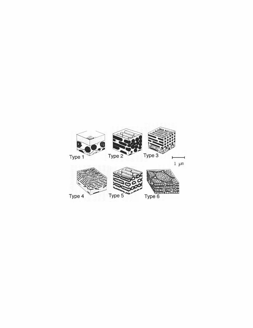
Given the diversity of avian taxa in which structural barbule colours have evolved, itis not surprising that there is a tremendous diversity in the composition, size, and shape ofthese structures. The diversity of light scattering arrays in avian barbules has been describedand categorized by Durrer (1986). They can be roughly categorized in the following classes(FIG. 2):
Type 1: a thin layer of keratin above a layer of large spherical melanocytes and airvacuoles
Type 2: single or multiple layers of adjacent rod-like melanin granules (1-2µmlong, 0.2 µm diameter),
R. O. Prum - Avian Structural Colours– 8
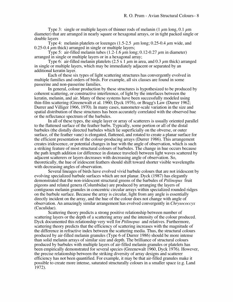
Type 3: single or multiple layers of thinner rods of melanin (1 µm long, 0.1 µmdiameter) that are arranged in nearly square or hexagonal arrays, or in tight packed single ordouble layers
Type 4: melanin platelets or lozenges (1.5-2.5 µm long; 0.25-0.4 µm wide, and0.25-0.4 µm thick) arranged in single or multiple layers;
Type 5: air-filled melanin tubes (1.2-1.6 µm long; 0.12-0.27 µm in diameter)arranged in single or multiple layers or in a hexagonal array;
Type 6: air-filled melanin platelets (2.5 x 1 µm in area, and 0.3 µm thick) arrangedin single or multiple layers, which may be immediately adjacent or separated by anadditional keratin layer.
Each of these six types of light scattering structures has convergently evolved inmultiple families and orders of birds. For example, all six classes are found in somepasserine and non-passerine families.
In general, colour production by these structures is hypothesized to be produced bycoherent scattering, or constructive interference, of light by the interfaces between thekeratin, melanin, and air. Many of these systems have been successfully modeled usingthin-film scattering (Greenewalt et al. 1960; Dyck 1976), or Bragg's Law (Durrer 1962;Durrer and Villiger 1966, 1970). In many cases, nanometer-scale variation in the size andspatial distribution of these structures has been accurately correlated with the observed hueor the reflectance spectrum of the barbules.
In all of these types, the single layer or array of scatterers is usually oriented parallelto the flattened surface of the feather barbs. Typically, some portion or all of the distalbarbules (the distally directed barbules which lie superficially on the obverse, or outersurface, of the feather vane) is elongated, flattened, and rotated to create a planar surface forthe efficient presentation of the colour-producing arrays (Durrer 1986). This arrangementcreates iridescence, or potential changes in hue with the angle of observation, which is sucha striking feature of most structural colours of barbules. The change in hue occurs becausethe path length addition (or difference in distance traveled) between light waves scattered byadjacent scatterers or layers decreases with decreasing angle of observation. So,theoretically, the hue of iridescent feathers should shift toward shorter visible wavelengthswith decreasing angles of observation.
Several lineages of birds have evolved vivid barbule colours that are not iridescent byevolving specialized barbule surfaces which are not planar. Dyck (1987) has elegantlydemonstrated that the non-iridescent structural greens of the barbules of Ptilinopus fruitpigeons and related genera (Columbidae) are produced by arranging the layers ofcontiguous melanin granules in concentric circular arrays within specialized rounded ridgeson the barbule surface. Because the array is circular, light from any angle is essentiallydirectly incident on the array, and the hue of the colour does not change with angle ofobservation. An amazingly similar arrangement has evolved convergently in Chrysococcyx(Cuculidae).
Scattering theory predicts a strong positive relationship between number ofscattering layers or the depth of a scattering array and the intensity of the colour produced.Dyck documented this relationship very well for Ptilinopus and relatives. Furthermore,scattering theory predicts that the efficiency of scattering increases with the magnitude ofthe difference in refractive index between the scattering media. Thus, the structural coloursproduced by air-filled melanin granules (Type 6 of Durrer 1986) should be more intensethan solid melanin arrays of similar size and depth. The brilliance of structural coloursproduced by barbules with multiple layers of air-filled melanin granules or platelets hasbeen empirically demonstrated for several species (Greenewalt 1960, Dyck 1976). However,the precise relationship between the striking diversity of array designs and scattererefficiency has not been quantified. For example, it may be that air-filled granules make itpossible to create more intense, saturated structurally colours in a smaller space (e.g. Land1972).

R. O. Prum - Avian Structural Colours– 9
Scattering theory also predicts the existence of second order and third orderreflectance peaks. These peaks are due to reinforcement among wavelengths that are close toone half, one third, twice, or three times the path length addition. If the primary reflectancepeak is at either extreme of the visual spectrum, then it is possible for the secondary peak tobe at the other end of the visual spectrum producing complex, combined hues (e.g. purple).Dyck (1976) reports the presence of visible first and second order reflectance peaks iniridescent feathers of Pica which has a air-filled melanin granules in what appear to bequasi-ordered layers. The first order peak is near 600 nm and the second order peak is inthe near UV. Recent development of fiber optic concave gradient diode spectrophotometershas allowed for convenient measurement of avian reflectance spectra into the UV (e.g.Andersson 1996). This advance will enable researchers to measure more broadly in theelectromagnetic spectrum and identify more cases second and third order reflectance peaksthat are visible to birds.
Thin-film scattering theory predicts that second and third order peaks will be closerin wavelength to λmax in non-ideal systems, where the optical thickness of the layers ofmedia are not equal, than in ideal systems (Land 1972). Although the application of thin-film theory to arrays of rods is not exact, it is interesting that the barbule arrays of Picaappear to be less ordered than in other some other taxa (e.g. Trogon) which do not exhibitvisible second order peaks. Perhaps application of a more exact scattering theory to themultimedia arrays will yield a more precise understanding of array order and the presenceof visible second order reflectance peaks. This is particularly important to analyzes of signalevolution, since second order peaks could greatly influence the perceived hue of a structuralcolour.
Structural Colours of Feather BarbsStructural colours of avian feather barbs are created by light scattering from specialized"boxy" cells or "spongy" cells in the medullary layer of the barbs (Auber 1957, 1971/72;Dyck 1971b, 1971a, 1978; Lucas and Stettenheim 1972; Fox 1976). The spongy medullarylayer is composed of a matrix of keratin rods and air vacuoles of varying shapes, sizes, andcomplexity. To prevent incoherent backscattering from underlying feather structures or thelarge nuclear vacuole at the center of most spongy medullary cells, most species have a layerof eumelanin granules that surrounds the large air-filled nuclear vacuoles or in the layer ofcells below the medullary layer. A few species use phaeomelanin ("brown" melanin) orcarotenoids to prevent backscattering (Auber 1957; Dyck 1971a). The spongy, medullarykeratin is absent in noncolour-producing feather barbs (Dyck 1978).
Auber (1957) described and classified the variation in shape, size, position, andpigmentary composition of the spongy medullary tissue of many families of birds withstructurally coloured barbs. In general, the medullary cells can form a ring around thesurface of the barb, a central core, a basal layer, or a even a divided pair of obverse andreverse cell bundles. Auber (1971/72) also described the development of the medullarycells using histology and light microscopy. He documented that during the development ofthe feather barbs, the medullary keranitocytes first expand in size to create the box-like,polygonal, keratin cell boundaries. Then, the medullary cells themselves gradually shrink involume as they create the spongy medullary keratin matrix on the inner surface of theoriginal polygonal cell boundaries. Eventually, the cells cease to produce keratin, detachesfrom the inner walls of the nuclear vacuole (creating the central vacuole), and then die.
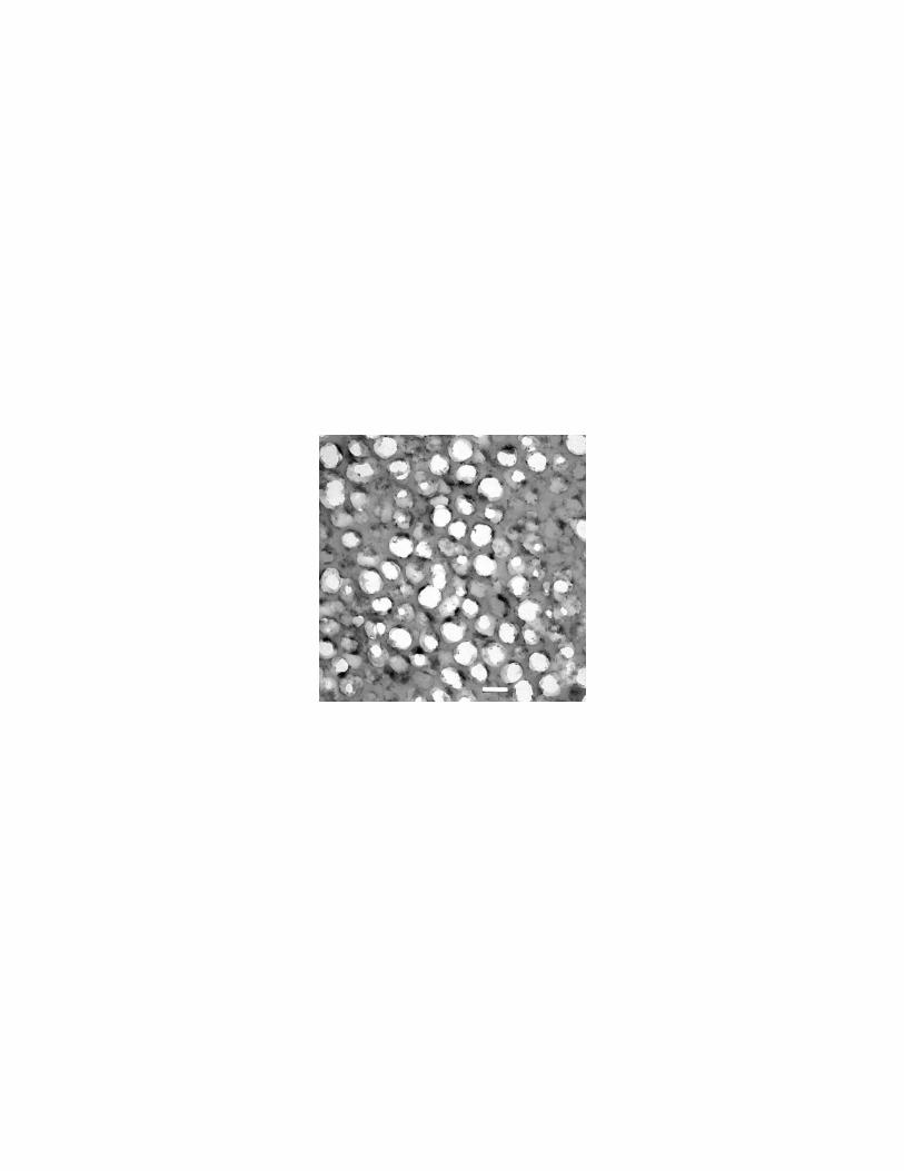
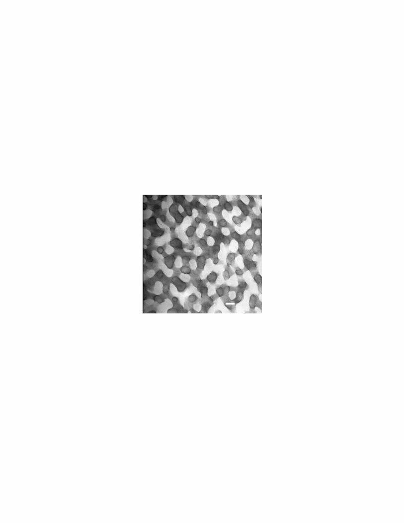
The colour-producing medullary keratin matrix varies in structure among taxa. Onegeneral form is characterized by a series of nearly circular, equivalently sized air-filled tubesseparated by keratin bridges (FIG. 3). Avian taxa known to demonstrate this type ofstructure include Cotinga (Cotingidae), Megalaima (Ramphastidae), and Poephila(Estrildidae) (Dyck 1976; R. O. Prum pers. obs.). The other structural type is characterizedby air-filled channels of more variable diameter separated by essentially equivalently sized
R. O. Prum - Avian Structural Colours– 10
keratin bars (FIG. 4). This type is known from many psittacids (Psittacidae), Coracias(Coraciidae), Chloropsis (Irenidae), and others (Dyck 1976; R. O. Prum pers. obs.).
Despite a more than a century of research (reviewed in Dyck 1971a; Fox 1976), thephysical mechanism of colour production by feather barbs is still debated (Dyck 1971a,1971b, 1976; Fox 1976; Finger et al. 1992; Finger 1995; Andersson 1996). Raleighscattering, or Tyndall scattering, has been the predominant explanation of structural coloursof feather barbs for nearly a century (see reviews in Dyck 1971a, and Fox 1976). Thesupport for this hypothesis first came from reflectance spectra of blue feather barbs byHäcker and Meyer (1902) which appeared to show adherence to Rayleigh's predictedinverse fourth power relationship for a limited set of visible wavelengths (480-650 nm).Subsequently, Dyck (1971a, 1976) demonstrated that many structurally coloured featherbarbs have distinct peaks in the visible spectrum. These distinct peaks falsified a majorprediction of Rayleigh scattering theory which is that reflectance should continue to increasein value into the UV, following the inverse fourth power law. Häcker and Meyer (1902)probably did not measure a broad enough section of the visible spectrum to detect a peak inthe lower portion of the human-visible spectrum, and erroneously interpreted their data assupport for Rayleigh scattering. Despite the compelling evidence against Raleigh scatteringthat has been available for more than 25 years, Rayleigh scattering is currently cited as theexclusive or primary explanation of feather barb structural colours in most currentornithology texts and references (e.g. Brooke and Birkhead 1991; Campbell and Lack1985; Gill 1995).
Finger (1995) has recently hypothesized that Mie scattering theory, an exactelectromagnetic theory of incoherent scattering by a particle, is an accurately explanation ofstructural colour production by feather barbs. Finger argues that medullary keratin-airmatrix is appropriately sized to scatter the smaller wavelengths of visible light and thatcortical filtering, or absorption of light by the keratin cortex, is responsible for creating thedistinct peaks in the visible spectrum.
Dyck proposed a coherent scattering, constructive interference, "hollow cylinder"hypothesis as an alternative to Raleigh scattering. The hollow cylinder model hypothesizedthat the sizes of the keratin bars and air vacuoles in the spongy medullary keratin createopportunities for constructive interference and vivid structural colour production. Althoughthis hypothesis was apparently accepted by Auber (1970/1971) and Durrer (1986), it hasreceived limited general acceptance in ornithology and zoology, perhaps because of the lackof evidence demonstrating that the spongy medullary keratin was sufficiently ordered toproduce coherent phase relationships among scattered waves.
Advances in the understanding of the scattering physics of the optically transparentcornea have demonstrated that tissues need not have a precise, lattice-like structure toproduce predictable phase relationships among scattered light waves (Benedek 1971, Vaezyand Clark 1991, 1993; Gisselberg et al. 1991). Recently, Prum, Dyck and colleagues haveapplied 2-D discrete Fourier analysis of spatial variation in refractive index to investigate thenanostructural periodicity of the spongy medullary keratin matrix. Our results demonstratethat the spongy medullary keratin is highly nanostructured and is appropriately scaled toproduce the observed hues by coherent scattering, or constructive interference, alone (Prumet al. 1998; Prum et al. 1999).
Furthermore, these Fourier analyses demonstrate that the spongy medullary keratinmatrices are spatially ordered at scales that are smaller than visible light. This result is directevidence against the independence of scatterers that is assumed by the incoherent scatteringmodels such as Rayleigh and Mie scattering. Incoherent scattering of visible light requiresthat scatterers be separated by distances that are greater than the wavelengths of visible light.The scatterers in the medullary keratin matrices of all bird species known are generallymuch closer together than 350 - 800 nm, the general limits of the avian-visible spectrum.Given direct evidence that the scatterers in the spongy medullary tissues of structurallycoloured barbs are coherent and not spatially independent, incoherent scattering models thatdo not take into account the phase relationships among the scattered light waves, such as
R. O. Prum - Avian Structural Colours– 11
Rayleigh and Mie scattering, cannot be considered to be complete or accurate descriptionsof the light scattering behaviour of these tissues.
These Fourier analysis results strongly support Dyck's original "hollow cylinder"hypothesis (Dyck 1971a, b) that feather barb structural colours are produced by coherentscattering, or constructive interference. Furthermore, these results document convincinglythat the conditions for incoherent scattering required by Rayleigh and Mie theory are notmet by any feathers, and that these mechanistic hypotheses provide incomplete explanationsof the phenomenon.
This finding indicates that structural colours of the feather barbs and barbules areproduced by the same physical mechanism, but differ in the spatial position, organization,and physical composition of the scatterers. The result is vivid structural colour productionbut without obvious, macroscopic iridescence. The lack of iridescence arises because themedullary keratin is appropriately structured on a nanoscale to produce constructiveinterference, but is structurally random at higher spatial scales. The observed colour is thesummation of many scatterers that are randomly oriented to the surface, but a change inangle of observation does not change the average path length addition to waves scattered bythe array.
STRUCTURAL COLOURS OF THE AVIAN DERMISGiven the intricate structural beauty, variation, and frequently striking colours of feathers, itis perhaps understandable that ornithologists have almost completely neglected toinvestigate the biology and physics of the colours of the avian skin. However, it is clear thatpigmentary and structural colours of the avian skin can play an important, even critical rolein social communication of many species of birds. Obviously, an understanding of thebiology and biophysics of dermal structural colours is essential to understanding theevolution of this component of avian phenotype and social signaling.
There have been several cursory investigations of structural blue colours in bird skin. Allprevious authors have hypothesized that these colours are produced by Rayleigh scattering frommelanin granules distributed in the dermis, but the anatomical structures responsible for thesecolours were never described (Rawles 1960; Lucas and Stettenheim 1972; Fox 1976; Durrer 1986).Prum et al. (1994) were the first to use both light and transmission electron microscopy toinvestigate the anatomy and biophysics of apparent structural colour production in the vividly greenand blue caruncles of male Philepitta castanea (Eurylaimidae).
Prum et al. (1994) found that the colour of the caruncle of Philepitta was produced byextensive, nearly perfectly hexagonal arrays of parallel collagen fibers in the dermis (FIG. 5). Thehexagonal arrays are organized into large macrofibrils that are as wide as dozens of the smallesthexagonal fiber units. These macrofibrils are chaotically or randomly organized within cone-shapedpapillae on the surface of the skin that are covered with an epidermal keratinous cone. The dermalcollagen macrofibrils are underlain by a thick layer of melanocytes which prevent incoherent backscattering from the optically disorganized tissue below.
Prum et al. (1994) demonstrated that the colour of the blue preserved caruncles of adultmale Philepitta castanea conformed well to the wavelengths predicted by the application of theBragg equation to the dimensions of collagen arrays in the caruncle papillae. The colour producedby the caruncles of Philepitta castanea is the summation of constructive reflections from thousandsof collagen arrays arranged in macrofibrils within each papilla. This paper was based on formalin-fixed caruncle tissues that had changed substantially in size and colour. In subsequent research onglutaraldehyde preserved caruncle tissues that retained the original colour almost exactly, I havedocumented that the difference between the green and blue portions of the caruncle can beaccurately described as a result of nanoscale differences in the size and spacing of the collagenfibers in the arrays (Prum et al. In Prep.).
This mechanism of structural colour production had never been described before in anyorganisms. However, similar collagen-based dermal colours have been implicated in primates. Thisstudy was also the first detailed microscopic study of any structurally-coloured, avian dermaltissue. These exciting results demonstrate the possibilities for future structural colour research in
R. O. Prum - Avian Structural Colours– 12
birds. A general survey of structural colouration in birds is required to further document thedistribution of this and other colour-production mechanisms in the avian dermis.
There are additional structural colours of other dermal derivatives that have been verypoorly studied. The ramphotheca and scutellated hindlimbs of many bird species are obviouslystructurally coloured (i.e. blue or green colours that are unlikely to be purely pigmentary). Further,many of these tissues that are colourful in life turn black in museum specimens, indicating that theyare underlain with melanin and may have a structural basis. Among the few papers specificallyaddressing the nature of a structural colour of a dermal derivative is a paper on the bright light bluebill of the Ruddy Duck Oxyura jamaicensis (Anatidae; Hays and Habermann 1969). Hays andHabermann (1969) demonstrate that the colour is produced by a spongy layer beneath theramphotheca that is underlain with melanin. They did not examine the tissues with transmissionelectron microscopy, though it is a distinct possibility that this spongy layer could be composed ofcollagen arrays, as in Philepitta (Prum et al. 1994). Currently, I know of no other studies ofstructural colours of non-feather dermal derivatives of birds.
STRUCTURAL COLOURS OF THE AVIAN EYETraditionally, the colour of the avian iris has been hypothesized to be produced by melaninand carotenoid pigments. Although there are obviously many iris colours that could not becaused merely these pigments, little research has been done on the nature of avian iriscolours.
Oehme (1969) produced an extensive treatment of the pigments of avian iris.Although he did not separate the pigments chromotographically, Oehme identified manybird species with carotenoid, pteridine, and purine pigments in their irides. The carotenoidswere present within intracellular lipid droplets. The pteridine or purine pigments were foundin crystalline form in some species, but they were not chemically identified.
The detection of crystalline pigments in the avian iris by Oehme (1969) implied thepresence of iridiphores or leucophores: special pigment cells, commonly distributed inpoikilothermic vertebrates, that produce structural colours by constructive interference (i.e.coherent scattering ) from arrays of purine or pteridine pigments. Purines are only presentin pigment cells in crystalline form, but colour producing pteridines can be found in bothcrystalline and non-crystalline form.
The first detailed anatomical analysis of iridphores in the avian iris was by Ferrisand Bagnara (1972). They documented that the irides of Inca Dove Columbina inca and theCommon Ground-Dove Columbina passerina (Columbidae) include two structurallydistinct types of iridiphores. One type of iridiphore included melanin granules andabundant, spherical guanine crystals, and the second type included many thinner, rod-shaped guanine crystals. The two types of purine-containing pigment cells were distributedin different parts of the iris.
Subsequently, Oliphant (1981, 1987a, 1987b; Oliphant et al. 1992) has identifiednumerous species of birds from many families that have crystalline purines and pteridinesin the iris. Oliphant (1981) identified pteridine-containing leucophores in the iris of theGreat Horned Owl Bubo virginianus (Strigidae). Subsequently, in a sample of 28 speciesfrom 11 families, Oliphant identified crystalline purines in the irides of 20 species in 11families and crystalline pteridines in 3 species. In all species with purine crystals, Oliphantidentified the presence of non-crystalline pteridines. He hypothesized that these pteridinepigments were superficial to the purine crystal-containing iridiphores and were modifyingtheir structural colour. Similar structural-pigmentary colour combinations are found widelyin poikilothermic vertebrates.
Obviously, there is much more to be learned about pigmentary and structuralcolours of avian iris. However, based even on the small sample of current data, it is clear thatthe colouration of the iris is based on entirely different pigmentary and cellular mechanismsthan the avian integument. To date, no purine- or pteridine-containing pigment cells havebeen identified in the dermis of birds (Oliphant et al. 1992). Currently, nothing is knownabout the ontogeny or evolution of these structures in the avian iris.

R. O. Prum - Avian Structural Colours– 13
The other structural colouration of the avian eye is the well-known phenomenon ofeye-shine: the reflection of incident light off of the retina of the eye. Although eye-shine iswell-known from a wide variety of nocturnal and even diurnal species, its mechanistic basisremains a mystery (Walls 1963). In mammals, eye-shine is created by the tapetum lucidum :a specially structured layer of the choroid membrane within the retina (Land 1972). In birds,the tapetum lucidum is apparently absent although a choroid is present (Walls 1963).Apparently, no one has microscopically examined the eye of a nocturnal bird to try toidentify the anatomical source of this well known phenomenon. One can predict that itshould be made of a nearly lattice-like array of reflecting crystal or fibers, given theiridescent quality of the eye-shine of some species.
DISCUSSIONStructural colours are present in all parts of the avian integument and eyes, and theydoubtless play an important role in the social communication and cryptic colouration ofbirds. Our understanding of the anatomical basis of structural colours has expandedtremendously in the last 50 years with the application of electron microscopy. However,there are still woefully few published studies of structural colours of the avian dermis,dermal derivatives, and eyes. The fact that the anatomical basis of the structural colours ofthe toucans bill and the eye-shine of nightjars have not be determined anatomically is anunfortunate indictment of ornithological curiosity!
Our understanding of the physics of structural colour has been consistentlyadvancing as well. The hypothesis that the iridescent structural colours of feather barbulesare caused by constructive interference among coherently scattered light waves has beenaccepted for over a century (Fox 1976). However, recent advances in anatomy and physicsimply that the noniridescent colours of feather barbs and dermis are produced by the samemechanism.
Early in this century, ornithologists assumed that iridescent colours must be causedby constructive interference, whereas non-iridescent colours must be caused by incoherentscattering by small particles, such as Rayleigh scattering (e.g. Mason 1922). Now, severaldifferent coherently scattering structures have been described in birds that produce non-iridescent colours by constructive interference: blue barbs of Cotinga (Prum et al. 1998),and the dermal collagen arrays in Philepitta (Prum et al. 1994). The constructiveinterference is the result of structural organization at a nanoscale, but the lack of iridescenceis a consequence of the lack of higher scale spatial organization. It appears likely thatconstructive interference of coherently scattered light waves is the sole physical explanationof all avian structural colours. It appears that evolution has created tissues with theappropriate nanostructured periodicity in refractive index to produce visible colours byconstructive interference. However, there are no clear examples in birds where a tissue hasevolved to include appropriately sized and distributed incoherent scatterers. That is, there areno avian tissues for which Rayleigh or Mie scattering are accurate descriptions of thecolours created. Any description of the physics of structural colour produced by knownavian tissues must include an analysis of the phase relationships among the scatteredwaves.
However, there is the distinct possibility that an exact description of the lightscattering behaviour by a tissue will include both coherent scattering by particles that arebest described by Rayleigh or Mie scattering models. For example, Mie scattering couldgive an accurate description of the differential scattering by an individual medullary airvacuole in a feather barb, but a coherent scattering model may be required to describe thesubsequent interactions among the scattered waves from different scatterers. A complete,exact description of scattering and absorption of light by any of these tissues would be anenormous but very interesting undertaking.
Several basic, and important questions remain in the physics of avian structuralcolours. First, why are feather barb structural colours limited to the lower end of the visiblespectrum? Second, what is the anatomical basis of structural colours of the dermis and

R. O. Prum - Avian Structural Colours– 14
dermal derivatives in other birds? Lastly, why have no structurally coloured pigment cells(i.e. iridiphores or leucophores) been found in the avian dermis when they are close toubiquitous in the irides of birds with brightly coloured eyes? These questions shouldreceive top priority in subsequent investigations.
A review of the implications of structural colours for the evolution of signal functionin birds is too large to accomplish in this paper. However, there is an expanding literature onthe evolution of carotenoid pigmentation as an honest indicator of quality (e.g. Hill 1991,1992, 1993, 1994; Andersson 1994). Some of this literature reflects an ignorance ofstructural colouration in birds. For example, Price et al. (1993) hypothesized a model ofsexual selection in which the male trait varied with the amount of exogenous carotenoidpigments deposited in the feathers; high quality males with high concentrations ofcarotenoids were hypothesized to be bright green, whereas low quality males with lowconcentrations of carotenoids were hypothesized to be drab brown. Unfortunately, theseauthors did not realize that the absence of carotenoids in a bright green plumage wouldproduce bright blue. Obviously, further discussions on the quality information of aviancolouration should be based on an accurate understanding of the differences betweenexogenous pigments, endogenous pigments, and structural colours.
Most important among the questions regarding the evolution of structural coloursfor social signaling is the question of condition dependence. Do structural colours varywithin populations in relation to the quality or condition of the individuals? This questionneeds to be addressed experimentally with each class of structural colours. However, Iwould predict that the fine levels of developmental control required to create colourproducing structures of the correct size are likely to be under tight genetic control. I wouldexpect that experimental studies will not find a high degree of quality correlated variation inavian structural colours. This result would have a fascinating impact on sexual selectiontheory. The Peacock's tail (Pavo cristatus, Phasiandiae) is a famous example a secondarysexual character. The question of whether the brilliant colours of the tail evolved becausethey have quality information or because they are merely preferred by females remainsentirely unanswered.
LITERATURE CITED
Andersson, M. 1994. Sexual Selection. Princeton University Press: Princeton, NJ.Andersson, S. 1996. Bright ultraviolet colouration in the Asian whistling-thrushes
(Myiophonus spp..). Proceedings of the Royal Society of London. Series B.263:843-848.
Auber, L. 1957. The structures producing "non-iridescent" blue color in bird-feathers.Proc. Zool. Soc. London 129:455-486.
Auber, L. 1971/72. Formation of 'polyhedral' cell cavities in cloudy media of bird feathers.Proceedings of the Royal Society, Edinborough 74:27-41.
Benedek, G. B. 1971. Theory of transparency of the eye. Appl. Optics 10:459-473.Bennett, A. T. D., and I. C. Cuthill. 1994. Ultraviolet Vision in birds: what is its function?
Vision Research 34:.Bohren, C. F. 1987. Multiple scattering of light and some of its observable consequences.
American Journal of Physics 55:524-533.Bohren, C. F., and D. R. Huffman. 1983. Absorption and Scattering of Light by Small
Particles. John Wiley and Sons.: New York.Bragg, W. H., and W. L. Bragg. 1915. X-rays and Crystal Structure. G. Bell: London.Briggs, W. L., and V. E. Henson. 1995. The DFT. Society for Industrial and Applied
Mathematics: Philadelphia, Penn.Brooke, M., and T. Birkhead. 1991. The Cambridge Encyclopedia of Ornithology.
Cambridge University Press: Cambridge.Campbell, B., and E. Lack. 1985. A Dictionary of Birds. T. A. & D. Poyser: London.

R. O. Prum - Avian Structural Colours– 15
Dorst, J., G. Gastaldi, R. Hagege, and J. Jacquemart. 1974. Différents aspects des barbulesde quelques Paradisaeidés sur coupes en microscopie électronique. ComptesRendus de l'Academie des Sciences, Paris 278:285-290.
Durrer, H. 1962. Schillerfarben beim Pfau (Pavo cristatus L.). Verhandl. Naturf. GesBasel 73:204-224.
Durrer, H. 1986. The Skin of Birds: Colouration. 239-247. 239-247 in Biology of theIntegument: 2 Vertebrates. (J. Bereiter-Hahn, A. G. Matoltsky, and K. S. Richards,Ed.), Springer-Verlag: Berlin.
Durrer, H., and W. Villiger. 1966. Schillerfarben der Trogoniden. Journal fürOrnithologie 107:1-26.
Durrer, H., and W. Villiger. 1970. Shilleradien des Goldkuckucks. Zeitschrift fürZellforschung 109:407-413.
Dyck, J. 1971a. Structure and colour-production of the blue barbs of Agapornisroseicollis and Cotinga maynana. Z. Zellforsch. 115:17-29.
Dyck, J. 1971b. Structure and spetral reflectance of green and blue feathers of theLovebird (Agapornis roseicollis). Biol. Skrift. 18:1-67.
Dyck, J. 1976. Structural colours. Proc. Internat. Ornithol. Congr 16:426-437.Dyck, J. 1978. Olive green feathers: Reflection of light from the rami and their structure.
Anser, Supplement 3:57-75.Dyck, J. 1979. Winter plumage of the Rock Ptarmigan: structure of the air-filled barbules
and function of the white colour. Dansk. Orn. Foren. Tidsskr. 73:41-58.Dyck, J. 1987. Structure and light reflection of green feathers of fruit doves (Ptilinopus
spp.) and an Imperial Pigeon (Ducula concinna). Biol. Skrift. (Copenhagen)30:2-43.
Ferris, W., and J. T. Bagnara. 1972. Reflecting pigment cells in the dove iris. 181-192.181-192 in Pigmentation: Its Genesis and Biological Control. (V. Riley, Ed.),Appleton-Century-Crofts: New York.
Finger, E. 1995. Visible and UV Coloration in Birds: Mie Scattering as the Basis of ColorProduction in Many Bird Feathers. Naturwissenschaften 82:570-573.
Finger, E., and D. Burkhardt. 1994. Biological aspects of bird coloration and avian visionincluding the ultraviolet range. Vision Research 34:1509-1514.
Finger, E., D. Burkhardt, and J. Dyck. 1992. Avian Plumage Colors: Origin of UVReflection in a Black Parrot. Naturwissenschaften 79:187-188.
Fox, D. L. 1976. Animal Biochromes and Structural Colors. Univ. California Press:Berkeley,CA.
Gill, F. B. 1995. Ornithology. Second Edition ed., W. H. Freeman and Co.: New York.Gisselberg, M., J. I. Clark, S. Vaezy, and T. Osgood. 1991. A quantitative evaluation of
Fourier components in transparent and opaque calf cornea. American Journal ofAnatomy 191:408-418.
Greenewalt, C. H., W. Brandt, and D. Friel. 1960. The iridescent colors of humminbirdfeathers. Journal of the Optical Society of America 50:1005-1013.
Häcker, V., and G. Meyer. 1902. Die blaue Farbe der Voelfedern. Zool. Jb., Abt. Syst.Geog. Biol. Tiere 15:267-294.
Hays, H., and H. Habermann. 1969. Note on bill color of the Ruddy Duck, Oxyurajamaicensis rubida. Auk 86:765-766.
Hill, G. 1991. Plumage coloration is a sexually selected indicator of male quality. Nature350:337-339.
Hill, G. E. 1992. Proximate basis of variation in carotenoid pigmentation in male HouseFinches. Auk 109:1-15.
Hill, G. E. 1993. Geographic variation in the carotenoid plumage pigmentation of malehouse finches (Carpodacus mexicanus). Biol. J. Linn. Soc 49:63-86.
Hill, G. E. 1994. Geographic variation in male ornamentation and female mate preferencein the House Finch: a comparative test of models of sexual selection. BehavioralEcology 5:64-73.
R. O. Prum - Avian Structural Colours– 16
Land, M. F. 1972. The physics and biology of animal reflectors. Progress in Biophysicsand Molecular Biology 24:77-106.
Lee, D. W. 1991. Ultrastructural basis and function of iridescent blue colour of fruits inElaeocarpus. Nature 349:260-262.
Lee, D. W. 1997. Iridescent Blue Plants. American Scientist 85:56-63.Lucas, A. M., and P. R. Stettenheim. 1972. Avian Anatomy- Integument. U.S. Dept.
Agricult. Handbook: Washington, DC.Macleod, H. A. 1986. Thin-film Optical Filters. 2nd ed., Adam Hilger, Ltd.: Bristol.
Mason, C. W. 1922. Structural Colors of Feathers. I. Journal of Physical Chemistry27:201-251.
Maurice, D. M. 1984. The cornea and sclera. 1-158. 1-158 in The Eye. (H. Davson, Ed.),Academic Press: New York.
Neville, A. C. 1993. Biology of Fibrous Composites. Cambridge University Press:Cambridge.
Oehme, H. 1969. Vergleichende Untersuchungen über die Farbung der Vogeliris.Biologische Zentralblatt 88:3-35.
Oliphant, L. W. 1981. Crystalline Pteridines in the stromal pigment cells of the iris of theGreat Horned Owl. Cell and Tissue Research 217:387-395.
Oliphant, L. W. 1987a. Observations on the pigmentation of the pigeon iris. Pigment CellResearch 1:202-208.
Oliphant, L. W. 1987b. Pteridines and purines as major pigments of the avian iris.Pigment Cell Research 1:129-131.
Oliphant, L. W., J. Hudon, and J. T. Bagnara. 1992. Pigment cell refugia in homeotherms–the unique evolutionary position of the iris. Pigment Cell Research 5:367-371.
Price, T., D. Schluter, and N. E. Heckman. 1993. Sexual selection when the female directlybenefits. Biological Journal of the Linnean Society 49:187-211.
Prum, R. O., R. L. Morrison, and G. R. Ten Eyck. 1994. Structural color production byconstructive reflection from ordered collagen arrays in a bird (Philepitta castanea:Eurylaimidae). Journal of Morphology 222: 61-72.
Prum, R. O., R. H. Torres, S. Williamson, and J. Dyck. 1998. Constructive Interference ofLight by Blue Feather Barbs. Nature 396: 28-29.
Prum, R. O., R. H. Torres, S. Williamson, and J. Dyck. 1999. Two-dimensional Fourieranalysis of the spongy medullary keratin of structurally coloured feather barbs.Proceedings of the Royal Society, London, Series B. 266: 13-22.
Rawles, M. E. 1960. The integumentary system. 189-240. 189-240 in Biology andComparative Physiology of Birds. (A. J. Marshall, Ed.), 1. Academic Press: NewYork.
Rutschke, E. 1966. Die submikroskopische struktur schillernder federn von entenvögeln.Zeitschrift für Zellforschung 73:432-443.
Schmidt, W. J., and H. Ruska. 1961. Elektronenmikroskopische Untersuchung derPigmentgranula in den Shillerenden Federstahlen der Taube Columba trocaz H.Zeitschrift für Zellforschung 55:379-388.
Schmidt, W. J., and H. Ruska. 1962. Über das schillernde federmelanin bei Heliangelusund Lophophorus. Zeitschrift für Zellforschung 57:1-36.
Vaezy, S., and J. I. Clark. 1991. A quantitative analysis of transparency in the humansclera and cornea using Fourier methods. Journal of Microsopy 163:85-94.
Vaezy, S., and J. I. Clark. 1993. Quantitative analysis of the microstructure of the humancornea and sclera using 2-D Fourier methods. Journal of Microscopy 175:93-99.
van de Hulst, H. C. 1981. Light Scattering by Small Particles. Dover: New York.Walls, G. L. 1963. The Vertebrate Eye. Hafner Pub. Co.: New York.Young, A. T. 1982. Rayleigh Scattering. Physics Today 35:42-48.
R. O. Prum - Avian Structural Colours– 17
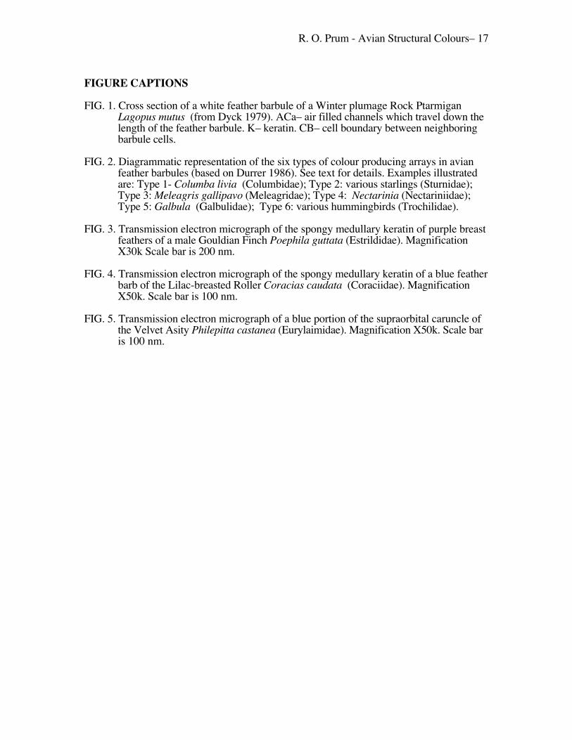
FIGURE CAPTIONS
FIG. 1. Cross section of a white feather barbule of a Winter plumage Rock PtarmiganLagopus mutus (from Dyck 1979). ACa– air filled channels which travel down thelength of the feather barbule. K– keratin. CB– cell boundary between neighboringbarbule cells.
FIG. 2. Diagrammatic representation of the six types of colour producing arrays in avianfeather barbules (based on Durrer 1986). See text for details. Examples illustratedare: Type 1- Columba livia (Columbidae); Type 2: various starlings (Sturnidae);Type 3: Meleagris gallipavo (Meleagridae); Type 4: Nectarinia (Nectariniidae);Type 5: Galbula �(Galbulidae); Type 6: various hummingbirds (Trochilidae).
FIG. 3. Transmission electron micrograph of the spongy medullary keratin of purple breastfeathers of a male Gouldian Finch Poephila guttata (Estrildidae). MagnificationX30k Scale bar is 200 nm.
FIG. 4. Transmission electron micrograph of the spongy medullary keratin of a blue featherbarb of the Lilac-breasted Roller Coracias caudata (Coraciidae). MagnificationX50k. Scale bar is 100 nm.
FIG. 5. Transmission electron micrograph of a blue portion of the supraorbital caruncle ofthe Velvet Asity Philepitta castanea�(Eurylaimidae). Magnification X50k. Scale baris 100 nm.