Embed Size (px)
Citation preview

Clinical StudyThe Accordion Maneuver: A Noninvasive Strategy for Absent orDelayed Callus Formation in Cases of Limb Lengthening
Asim M. Makhdom,1,2 Adrian Sever Cartaleanu,1 Juan Sebastian Rendon,1
Isabelle Villemure,3 and Reggie C. Hamdy1
1Division of Orthopaedic Surgery, Shriners Hospital for Children, Montreal Children Hospital, McGill University, 1529 Cedar Avenue,Montreal, QC, Canada H3G 1A62Department of Orthopaedic Surgery, King Abdulaziz University, Jeddah 21589, Saudi Arabia3Canada Research Chair in Mechanobiology of the Pediatric Musculoskeletal System, Department of Mechanical Engineering,Polytechnique Montreal, Montreal, QC, Canada
Correspondence should be addressed to Asim M. Makhdom; [email protected]
Received 19 June 2015; Accepted 27 September 2015
Academic Editor: Robert F. Ostrum
Copyright © 2015 Asim M. Makhdom et al. This is an open access article distributed under the Creative Commons AttributionLicense, which permits unrestricted use, distribution, and reproduction in any medium, provided the original work is properlycited.
The distraction osteogenesis (DO) technique has been used worldwide to treat many orthopaedic conditions. Although successful,absent or delayed callus formation in the distraction gap can lead to significant morbidities. An alternate cycle of distraction-compression (accordion maneuver) is one approach to accelerate bone regeneration. The primary aim of our study is to reportour experience with the accordion maneuver during DO and to provide a detailed description of this technique, as performed inour center. The secondary aim is to present a review of the literature regarding the use of accordion maneuver. We reviewed thedatabase of all patients undergoing limb lengthening from the year of 1997 to 2012. Four patients (6.15%) out of 65 showed poorbone regenerate in their tibiae and therefore accordion maneuver was applied for a mean of 6.75 weeks. Of these, three patientshave had successful outcome with this technique. The literature showed that this technique is successful approach to trigger bonehealing. However, details of how andwhen to apply this combination of distraction-compression forces were lacking. In conclusion,the accordion technique is safe noninvasive approach to promote bone formation, thus avoiding more invasive surgical proceduresin cases of poor callus formation in limb lengthening.
1. Introduction
Distraction osteogenesis (DO) is a surgical technique usedworldwide to treat a broad variety of musculoskeletal andcraniofacial conditions, including correction of angulardeformities or management of bone defects secondary toinfection, trauma, or tumor via limb lengthening or segmen-tal bone transport [1, 2]. This technique was popularized byIlizarov in the early 1950s who demonstrated that when con-trolled gradual distraction is applied to the two ends of a bonefollowing a low energy osteotomy, new bone will form in thedistracted gap [2]. Its principle is based on the intrinsic capac-ity of the bone to regenerate under a controlled mechanicalenvironment and is considered the best type of in vivo bone
tissue engineering technique [3]. Both the rate and rhythm ofdistraction are vital to the quality of the regenerate bone.
Although DO is associated with satisfactory outcomesin most cases, absent or delayed callus formation in thedistraction gap may occur. This could lead to significantmorbidities, as the fixator needs to be kept in place for anextended period of time until the bone is completely con-solidated. Consequently, unfavorable psychological impact,increased pin tract infections, persistent pain, and increasedrisk of osteopenia might be encountered [4–6]. In somecases, subsequent surgical interventions might be required[1, 4, 6]. Numerous techniques have been described in themanagement of poor regenerate in cases of DO, includingsystemic administration of pharmaceutical agents such as
Hindawi Publishing CorporationAdvances in OrthopedicsVolume 2015, Article ID 912790, 8 pageshttp://dx.doi.org/10.1155/2015/912790

2 Advances in Orthopedics
bisphosphonates, local exogenous administration of growthfactors (GFs) such as BMPs, bone marrow cells (BMC), andthe use of externally applied low-intensity pulsed ultrasound(LIPU) and pulsed electromagnetic fields (PEMF) [3, 7–10].
There are several modalities where the use of compressiveforces in the context of DO could be used in order to acceler-ate bone formation in the distracted gap, and these includeearly and increasing weight bearing on the operated limb,dynamization of the fixator, overdistraction, and then short-ening and alternating cycles of distraction and compression[11–13]. This last technique—the accordion maneuver—hasoriginally been described by Ilizarov in order accelerate boneregeneration in DO [2]. However, despite several reports inthe English literature on the successful use of this techniquein the management of poor regenerate, they are mostly anec-dotal without a detailed description of thismaneuver [14–23].
The aim of this study is to report our experience with theaccordion maneuver in a small series of cases with absent ordelayed bone formation during DO and to provide a detaileddescription of this technique, as performed in our center. Wealso present a review of the literature regarding the use ofalternating cycles of distraction and compression in cases ofDO, nonunions, and fractures in both human and animalstudies.
2. Patients and Methods
After approval from our local institutional review board, weretrospectively reviewed all patients who underwent straightlower limb lengthening at our institution between 1997 and2012. The medical records of 65 patients (forty-one malesand twenty-four females, M : F = 1.7 : 1) who underwent 72interventions (35 on right side, 37 on left side), in which72 bone segments were lengthened (44 femora and 28tibiae), were reviewed. Of these 4 patients underwent theaccordion technique. The demographic data, clinical courseand imaging information, diagnosis, surgery, lengtheningdetails, and complications were all collected from themedicalrecord system. In all patients, a low energy osteotomy wasperformed by creating multiple small drill holes at the siteof osteotomy followed by completion of the osteotomy withan osteotome. Immediate weight bearing as tolerated wasinitiated in all patients with intense physiotherapy.
The specific indication for using the accordion maneuverwas an absent or delayed callus formation in the distrac-tion gap, judged radiographically. The accordion maneuverconsisted in alternating distraction with compression asfollows: distraction (0.25mm) in the morning and thencompression (0.25mm) in the afternoon, followed by distrac-tion (0.25mm) in the evening, resulting in an overall dailylengthening of 0.25mm.
3. Results
The decision to apply the accordion maneuver was takenduring initial distraction when imaging has shown absent orsignificantly delayed callus formation in the distraction gap.This was the case of tibiae in four patients (6.15%) of the 65investigated. Their mean age was 16.5 years (range, 10 to 20
years). After lengthening initiation, their X-rays showed anabsent or very timid bone regenerate in the distraction gap(Figure 1(a)). In these four cases, the accordion maneuverwas applied at a mean of 4.5 weeks after surgery (range,3–7 weeks), which corresponds to a mean of 3.62 weeks(range, 2–6 weeks) after initiation of the distraction phase.The accordion maneuver was carried out on a daily basis,alternating distraction with compression three times per day,for an average of 6.75 weeks, as previously described. Thetotal distraction period (the routine distraction period + theaccordion maneuver period) was of an average of 12.5 weeks(range, 11–14 weeks) to obtain a mean lengthening of 3.92 cm(range 3–5 cm). The residual limb length discrepancy was onaverage 1.12 cm (range, 0.7–2 cm). A mean healing index of75.38 days/cm was noted. Details on clinical and accordionmaneuver details are provided in Table 1.
Favorable progression of the bone regenerate was notedafter an average of 5.3 weeks (range, 4–6 weeks) afterstarting the accordion maneuver in three out of four patients(Figure 1(b)). These patients continued to have full boneconsolidation in the distraction gap. However, in one patient(case number 3), infection has complicated the course andthere was absent bone formation after using the accordionmaneuver (Figure 2(a)). Antibiotic treatment, additionalbone grafting, and administration of bone morphogeneticprotein-7 (OP-1) ultimately resulted in bone union for thispatient (Figure 2(b)).
4. Discussion
Several host related, local, and iatrogenic causes can lead topoor bone regenerate during DO [8]. These include systemicillness, infection, immunosuppression, poor tissue envelope,exposure to radiation, instability of the external fixator,suboptimal osteotomy technique, and rapid distraction rate[8]. We were unable to identify any of these risk factors in 3out of the 4 patients with poor regenerate and the applicationof the accordion maneuver in these 3 patients resulted insuccessful bone regeneration in the distracted gap, while, inthe fourth patient (case number 3), the accordion maneuverfailed to stimulate the regenerative process. We believe thisis most likely due to the presence of underlying infection.This emphasizes the importance of identifying all risk factorsthat may lead to a poor regenerate in DO before the use ofthe accordion technique. However, a firm conclusion can bemade when a larger sample size is studied.
A review of the English literature revealed several clinicalstudies in humans reporting the use of the accordion tech-nique in cases with poor regenerate bone formation in DO,the majority of them with positive outcome. However, thedescription of the alternate distraction-compression regimenin these studies is anecdotal and lacks details as of when,how, and for how long this technique is applied (Table 2) [15–17, 19, 24–28].
The accordion maneuver has also been used clinicallyto stimulate bone formation in the context of fracturehealing. Similar to its reported use in DO, most of thesestudies also reported positive outcome, however still withpoor description of the technique (Table 3) [14, 18, 29–31].

Advances in Orthopedics 3
Table 1: Clinical data for patients who underwent the accordion maneuver.
Data Case number 1 Case number 2 Case number 3 Case number 4Age and gender 18 y.o., female 10 y.o., male 20 y.o., male 18 y.o., maleDiagnosis Blount disease Fibular hemimelia Tibial hemimelia HME1
Indication forsurgery, LLD LLD, L < R 6 cm LLD, L < R 5.2 cm LLD, R < L 5 cm LLD, L < R 4 cm
Surgical procedure Tibial lengthening withcircular Ilizarov
Tibial lengthening withcircular Ilizarov
Tibial lengthening withcircular Ilizarov
Tibial lengthening withcircular Ilizarov
Comorbidities None None None NoneStart time ofaccordionmaneuver
5 weeks after surgery 4 weeks after surgery 3 weeks after surgery 7 weeks after surgery
Lengtheningprocess description
Latency period: 4 daysDistraction: 0.25mm × 4times (1mm)/day for 4.5weeks and then theaccordion maneuver2 wasinitiated and lasted for 7weeksFinal distraction: 0.25mm× 2 times (0.5mm)/day for2 weeks
Latency period: 6 daysInitial distraction: 0.25mm× 2 times (0.5mm)/day for2 weeks and thenthe accordion maneuverwas initiated and lasted for4 weeksFinal distraction: 0.25mm× 4 times (1mm)/day for 2weeks and then 0.25mm ×2 times (0.5mm)/day for 4weeks
Latency period: 8 daysInitial distraction: 0.25mmdistraction × 3 times(0.75mm)/day for 2 weeks,and thenthe accordion maneuverwas initiated and lasted for9 weeks, stopping thelengthening thereafter
Latency period: 6 daysInitial distraction: 0.25mmdistraction × 2 times(0.5mm)/day for 6 weeks,and thenthe accordion maneuverwas initiated and lasted for7 weeks, stopping thelengthening thereafter
Total lengtheningduration 14 weeks 12 weeks 11 weeks 13 weeks
Lengtheningachieved 5 cm 4.4 cm 3 cm 3.3 cm
Residual LLD3 1 cm 0.8 cm 2 cm 0.7 cmLengthening index 19.6 days/cm 19.09 days/cm 25.66 days/cm 27.57 days/cmHealing index 52.8 days/cm 54.1 days/cm 122.5 days/cm 72.12 days/cm
Outcome andcomplications
Bone regenerate observedat 6 weeks within theaccordion. Had transientperoneal nerve palsy
Bone regenerate observedat 4 weeks within theaccordion. Had 5 degrees ofknee flexion contracture. 3weeks after frame removal(9-month post-op), theregenerate was fracturedand was nailed, ultimatelyhealed
No bone regenerate formedafter the accordionmaneuver. Had infection,was successfully treatedwith antibiotics, and wasfollowed up 7 months laterby bone grafting and OP-14.Residual bowing of thetibia. 40 degrees fixedequinus R ankle
Good bone regenerateobserved at 6 weeks withinthe accordion course withno complications
Comments Underwent concomitantcorrection of valgus
After 4 weeks of accordion,continued with 1mmdistraction/day for 2 weeks,had fibular prematurefusion, and underwentreosteotomy and thencontinued distraction foranother 4 weeks
Infection treated withantibiotics. Absent boneformation after using theaccordion maneuver
Had correction of valgus.After finishing theaccordion, starteddistraction 1mm/day, for 2days had pain, and stopped
1HME: Hereditary Multiple Exostoses.2Accordion maneuver: 0.25mm distraction in AM, followed by 0.25mm compression early PM, and then distraction of 0.25mm late PM (0.25mm oflengthening/day).3LLD: limb length discrepancy.4OP-1: osteogenic protein-1.

4 Advances in Orthopedics
Table 2: Previously published clinical studies reporting the accordion technique during delayed or absent callus formation of distractionosteogenesis (DO).
Authors Number ofpatients Indication Successful
outcome Technique for accordion maneuver
Iacobellis et al.2010 [26] 3 Poor regenerate during
bone transport100%(3/3)
Compression followed by distraction ofthe transport segment (no details)
Hatzokos et al.2011 [24] 8 Delayed consolidation 75%
(6/8) Accordion technique (no details).
Kawoosa et al.2003 [25] 1 Delayed consolidation 100%
(1/1)Alternate compression and distraction ofthe regenerate (no details)
El-Mowafi et al.2005 [27] 𝑁 = ? Delayed consolidation ? Compression and distraction of a moving
segment (no details)El-Sayed et al.2010 [17] 25 Absence of callus formation 76%
(19/25)Distraction-compression technique (nodetails)
Tsuchiya et al.1997 [28] 𝑁 = ? Poor regenerate during
bone transport ? Compression and distraction of a movingsegment (no details)
Vidyadhara andRao 2007 [15] 𝑁 = ? Poor regenerate callus
during bone transport ? Compression and distraction of a movingsegment (no details).
Simpson andKenwright 2000[16]
2 Poor callus formation 0%(0/2)
Changes in the dynamics of distraction(no details)
Krishnan et al.2006 [19] 2 Poor regenerate during
bone transport 100% (2/2)Reported as distraction, discontinued,reversed, and restarted at a reduced rate(0.25mm/12 h, instead of 0.25mm/6 h)
(a)
(b)
Figure 1: (a) Anteroposterior (on the right) and lateral radiographic (on the left) views of left tibia (case 4) showing very discrete boneregenerate after 6 weeks of distraction at a rate of 0.25mm × 2 times/day. (b) Anteroposterior (on the right) and lateral radiographic (on theleft) views of left tibia (case 4) after 7 weeks of the accordion technique showing significantly improved osteoformation.

Advances in Orthopedics 5
Table 3: Previous reports on the use of distraction and compression in treatment of long bone fractures, delayed unions, and nonunions.
Authors Number ofpatients Indication Successful
outcome Technique for distraction-compression
Kulkarni 2004 [29] N/A Hypertrophic nonunion N/ADistraction 0.5mm/day for 20 days,then stopping for the next 20 days, andfinally compression
Inan et al. 2005 [30] 11 Femoral pseudarthrosis 100% (11/11) Cyclic compression and distraction atthe nonunion site
Madhusudhan et al. 2008 [14] 2 Tibial nonunion 100% (2/2) Compression and distraction (nodetails)
Laursen et al. 2000 [18] 2 Tibial nonunions 50% (1/2)Alternating distraction (1 week) withcompression (1 week), until callusvisible on X-ray
Chand et al. 2010 [31] 2 Nonunion of long bone fractures 100% (2/2) Compression and distractiontechnique (no details)
(a)
(b)
Figure 2: (a) Anteroposterior (on the right) and lateral radiographic (on the left) views of left tibia (case 3) showing insufficient response after4 weeks of accordion maneuver. (b) Anteroposterior (on the right) and lateral radiographic view (on the left) of left tibia (case 3) showingcomplete healing after bone grafting and BMP-7 administration.
Interestingly, only experimental studies performed in animalshave provided details of this technique. Mofid et al. showedthat daily sequential compression and distraction for 3 weeksduring the consolidation phase at rate of 1mm/day increasedsignificantly the bone formation when compared to thecontrol group in mandibular DO in rabbit model [21]. Claeset al. investigated the effect of temporary distraction andcompression on bone regeneration in fracture healing [11].The authors noted higher bone formation in the treatmentgroup when compared with the control group. On the otherhand, Greenwald et al. used a rat mandibular DO modeland reported that there were no differences histologically
and radiographically between groups of rats with distraction-compression protocol versus a control group with standardDO technique [23]. We could not explain why these negativeresults were obtained, except that the regimen used by theseauthors was not an accordion technique with alternatingcycles of distraction and compression, but rather 5 days of dis-traction followed by 2 days of compression. However, takentogether, both clinical and experimental studies demon-strated the positive role of accordion maneuver in accelera-tion of bone regeneration in the context of both fracture heal-ing and DO. However, the rate and rhythm of the accordiontechnique varied between experimental studies and were not
6 Advances in Orthopedics
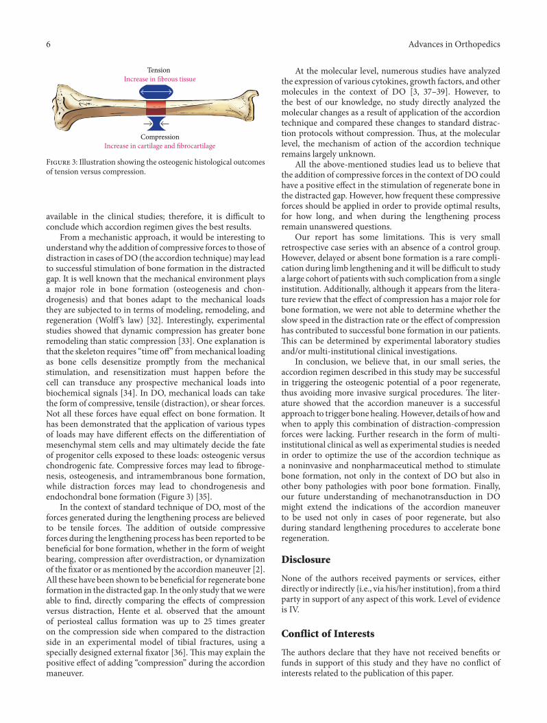
CompressionIncrease in cartilage and fibrocartilage
TensionIncrease in fibrous tissue
Figure 3: Illustration showing the osteogenic histological outcomesof tension versus compression.
available in the clinical studies; therefore, it is difficult toconclude which accordion regimen gives the best results.
From a mechanistic approach, it would be interesting tounderstandwhy the addition of compressive forces to those ofdistraction in cases ofDO (the accordion technique)may leadto successful stimulation of bone formation in the distractedgap. It is well known that the mechanical environment playsa major role in bone formation (osteogenesis and chon-drogenesis) and that bones adapt to the mechanical loadsthey are subjected to in terms of modeling, remodeling, andregeneration (Wolff ’s law) [32]. Interestingly, experimentalstudies showed that dynamic compression has greater boneremodeling than static compression [33]. One explanation isthat the skeleton requires “time off” frommechanical loadingas bone cells desensitize promptly from the mechanicalstimulation, and resensitization must happen before thecell can transduce any prospective mechanical loads intobiochemical signals [34]. In DO, mechanical loads can takethe form of compressive, tensile (distraction), or shear forces.Not all these forces have equal effect on bone formation. Ithas been demonstrated that the application of various typesof loads may have different effects on the differentiation ofmesenchymal stem cells and may ultimately decide the fateof progenitor cells exposed to these loads: osteogenic versuschondrogenic fate. Compressive forces may lead to fibroge-nesis, osteogenesis, and intramembranous bone formation,while distraction forces may lead to chondrogenesis andendochondral bone formation (Figure 3) [35].
In the context of standard technique of DO, most of theforces generated during the lengthening process are believedto be tensile forces. The addition of outside compressiveforces during the lengthening process has been reported to bebeneficial for bone formation, whether in the form of weightbearing, compression after overdistraction, or dynamizationof the fixator or as mentioned by the accordionmaneuver [2].All these have been shown to be beneficial for regenerate boneformation in the distracted gap. In the only study that wewereable to find, directly comparing the effects of compressionversus distraction, Hente et al. observed that the amountof periosteal callus formation was up to 25 times greateron the compression side when compared to the distractionside in an experimental model of tibial fractures, using aspecially designed external fixator [36]. This may explain thepositive effect of adding “compression” during the accordionmaneuver.
At the molecular level, numerous studies have analyzedthe expression of various cytokines, growth factors, and othermolecules in the context of DO [3, 37–39]. However, tothe best of our knowledge, no study directly analyzed themolecular changes as a result of application of the accordiontechnique and compared these changes to standard distrac-tion protocols without compression. Thus, at the molecularlevel, the mechanism of action of the accordion techniqueremains largely unknown.
All the above-mentioned studies lead us to believe thatthe addition of compressive forces in the context of DO couldhave a positive effect in the stimulation of regenerate bone inthe distracted gap. However, how frequent these compressiveforces should be applied in order to provide optimal results,for how long, and when during the lengthening processremain unanswered questions.
Our report has some limitations. This is very smallretrospective case series with an absence of a control group.However, delayed or absent bone formation is a rare compli-cation during limb lengthening and it will be difficult to studya large cohort of patientswith such complication froma singleinstitution. Additionally, although it appears from the litera-ture review that the effect of compression has a major role forbone formation, we were not able to determine whether theslow speed in the distraction rate or the effect of compressionhas contributed to successful bone formation in our patients.This can be determined by experimental laboratory studiesand/or multi-institutional clinical investigations.
In conclusion, we believe that, in our small series, theaccordion regimen described in this study may be successfulin triggering the osteogenic potential of a poor regenerate,thus avoiding more invasive surgical procedures. The liter-ature showed that the accordion maneuver is a successfulapproach to trigger bone healing.However, details of how andwhen to apply this combination of distraction-compressionforces were lacking. Further research in the form of multi-institutional clinical as well as experimental studies is neededin order to optimize the use of the accordion technique asa noninvasive and nonpharmaceutical method to stimulatebone formation, not only in the context of DO but also inother bony pathologies with poor bone formation. Finally,our future understanding of mechanotransduction in DOmight extend the indications of the accordion maneuverto be used not only in cases of poor regenerate, but alsoduring standard lengthening procedures to accelerate boneregeneration.
Disclosure
None of the authors received payments or services, eitherdirectly or indirectly {i.e., via his/her institution}, from a thirdparty in support of any aspect of this work. Level of evidenceis IV.
Conflict of Interests
The authors declare that they have not received benefits orfunds in support of this study and they have no conflict ofinterests related to the publication of this paper.

Advances in Orthopedics 7
References
[1] J. G. Birch and M. L. Samchukov, “Use of the Ilizarov methodto correct lower limb deformities in children and adolescents,”The Journal of the American Academy of Orthopaedic Surgeons,vol. 12, no. 3, pp. 144–154, 2004.
[2] G. A. Ilizarov, “Clinical application of the tension-stresseffect for limb lengthening,” Clinical Orthopaedics and RelatedResearch, no. 250, pp. 8–26, 1990.
[3] A. M. Makhdom and R. C. Hamdy, “The role of growthfactors on acceleration of bone regeneration during distractionOsteogenesis,” Tissue Engineering—Part B: Reviews, vol. 19, no.5, pp. 442–453, 2013.
[4] R. J. Velazquez, D. F. Bell, P. F. Armstrong, P. Babyn, and R.Tibshirani, “Complications of use of the Ilizarov technique inthe correction of limb deformities in children,” The Journal ofBone and Joint Surgery—American Volume, vol. 75, no. 8, pp.1148–1156, 1993.
[5] E. Garcia-Cimbrelo, B. Olsen, M. Ruiz-Yague, N. Fernandez-Baillo, and L. Munuera- Martinez, “Ilizarov technique: resultsand difficulties,”Clinical Orthopaedics and Related Research, no.283, pp. 116–123, 1992.
[6] J. C. Eldridge and D. F. Bell, “Problems with substantial limblengthening,” Orthopedic Clinics of North America, vol. 22, no.4, pp. 625–631, 1991.
[7] J. Aronson, “Experimental and clinical experience with distrac-tion osteogenesis,” Cleft Palate-Craniofacial Journal, vol. 31, no.6, pp. 473–482, 1994.
[8] S. Sabharwal, “Enhancement of bone formation during dis-traction osteogenesis: pediatric applications,” Journal of theAmerican Academy of Orthopaedic Surgeons, vol. 19, no. 2, pp.101–111, 2011.
[9] D. Gebauer and J. Correll, “Pulsed low-intensity ultrasound: anew salvage procedure for delayed unions and nonunions afterleg lengthening in children,” Journal of Pediatric Orthopaedics,vol. 25, no. 6, pp. 750–754, 2005.
[10] K. S. Eyres, M. Saleh, and J. A. Kanis, “Effect of pulsedelectromagnetic fields on bone formation and bone loss duringlimb lengthening,” Bone, vol. 18, no. 6, pp. 505–509, 1996.
[11] L. Claes, P. Augat, S. Schorlemmer, C. Konrads, A. Ignatius,and C. Ehrnthaller, “Temporary distraction and compressionof a diaphyseal osteotomy accelerates bone healing,” Journal ofOrthopaedic Research, vol. 26, no. 6, pp. 772–777, 2008.
[12] R. Mora, Nonunion of the Long Bones: Diagnosis and Treatmentwith Compression-Distraction Techniques, Springer, Milan, Itlay,2006.
[13] R. C. Hamdy, J. S. Rendon, and M. Tabrizian, “Distractionosteogenesis and its challenges in bone regeneration,” in BoneRegeneration, H. Tal, Ed., chapter 8, pp. 177–204, InTech, Rijeka,Croatia, 2012.
[14] T. R. Madhusudhan, B. Ramesh, K. Manjunath, H. M. Shah, D.C. Sundaresh, and N. Krishnappa, “Outcomes of Ilizarov ringfixation in recalcitrant infected tibial non-unions—a prospec-tive study,” Journal of Trauma Management & Outcomes, vol. 2,no. 1, article 6, 2008.
[15] S. Vidyadhara and S. K. Rao, “A novel approach to juxta-articular aggressive and recurrent giant cell tumours: resectionarthrodesis using bone transport over an intramedullary nail,”International Orthopaedics, vol. 31, no. 2, pp. 179–184, 2007.
[16] A. H. R. W. Simpson and J. Kenwright, “Fracture after distrac-tion osteogenesis,” Journal of Bone and Joint Surgery B, vol. 82,no. 5, pp. 659–665, 2000.
[17] M. M. El-Sayed, J. Correll, and K. Pohlig, “Limb sparing recon-structive surgery and Ilizarov lengthening in fibular hemimeliaof Achterman-Kalamchi type II patients,” Journal of PediatricOrthopaedics B, vol. 19, no. 1, pp. 55–60, 2010.
[18] M. B. Laursen, P. Lass, andK. S. Christensen, “Ilizarov treatmentof tibial nonunions results in 16 cases,” Acta OrthopaedicaBelgica, vol. 66, no. 3, pp. 279–285, 2000.
[19] A. Krishnan, C. Pamecha, and J. J. Patwa, “Modified Ilizarovtechnique for infected nonunion of the femur: the principle ofdistraction-compression osteogenesis,” Journal of OrthopaedicSurgery, vol. 14, no. 3, pp. 265–272, 2006.
[20] S. Mori, M. Akagi, A. Kikuyama, Y. Yasuda, and C. Haman-ishi, “Axial shortening during distraction osteogenesis leads toenhanced bone formation in a rabbit model through the HIF-1alpha/vascular endothelial growth factor system,” Journal ofOrthopaedic Research, vol. 24, no. 4, pp. 653–663, 2006.
[21] M. M. Mofid, N. Inoue, A. Atabey et al., “Callus stimulationin distraction osteogenesis,” Plastic and Reconstructive Surgery,vol. 109, no. 5, pp. 1621–1629, 2002.
[22] E. G. Loboa, T. D. Fang, D. W. Parker et al., “Mechanobiologyof mandibular distraction osteogenesis: finite element analyseswith a rat model,” Journal of Orthopaedic Research, vol. 23, no.3, pp. 663–670, 2005.
[23] J. A. Greenwald, J. S. Luchs, B. J. Mehrara et al., “‘Pumping theregenerate’: an evaluation of oscillating distraction osteogenesisin the rodent mandible,”Annals of Plastic Surgery, vol. 44, no. 5,pp. 516–521, 2000.
[24] I. Hatzokos, S. I. Stavridis, E. Iosifidou, D. Karataglis, and A.Christodoulou, “Autologous bone marrow grafting combinedwith demineralized bone matrix improves consolidation ofdocking site after distraction osteogenesis,”The Journal of Bone& Joint Surgery—American Volume, vol. 93, no. 7, pp. 671–678,2011.
[25] A. A. Kawoosa, S. Majid, M. R. Mir, and G. R. Mir, “Resultsof tibial lengthening by Ilizarov technique,” Indian Journal ofOrthopaedics, vol. 37, no. 7, p. 7, 2003.
[26] C. Iacobellis, A. Berizzi, and R. Aldegheri, “Bone transportusing the Ilizarovmethod: a review of complications in 100 con-secutive cases,” Strategies in Trauma and Limb Reconstruction,vol. 5, no. 1, pp. 17–22, 2010.
[27] H. El-Mowafi, B. Elalfi, and K. Wasfi, “Functional outcomefollowing treatment of segmental skeletal defects of the forearmbones by Ilizarov application,” Acta Orthopaedica Belgica, vol.71, no. 2, pp. 157–162, 2005.
[28] H. Tsuchiya, K. Tomita, K. Minematsu, Y. Mori, N. Asada,and S. Kitano, “Limb salvage using distraction osteogenesis. Aclassification of the technique,” Journal of Bone and Joint SurgeryB, vol. 79, no. 3, pp. 403–411, 1997.
[29] G. S. Kulkarni, “Principles and practice of deformity correc-tion,” Indian Journal of Orthopaedics, vol. 38, no. 3, pp. 191–198,2004.
[30] M. Inan, S. Karaoglu, F. Cilli, C. Y. Turk, and A. Harma,“Treatment of femoral nonunions by using cyclic compressionand distraction,” Clinical Orthopaedics and Related Research,vol. 436, pp. 222–228, 2005.
[31] P. Chand, R. L. Shrestha, B. R. Kc, B. C. Shah, A. Joshi, and B.N. Thapa, “Managing difficult fractures due to Ballistic traumawith Ilizarov ring fixation,” Medical Journal of Shree BirendraHospital, vol. 9, no. 1, pp. 1–8, 2010.
[32] C. Huang and R. Ogawa, “Mechanotransduction in bone repairand regeneration,”The FASEB Journal, vol. 24, no. 10, pp. 3625–3632, 2010.

8 Advances in Orthopedics
[33] L. K. Saxon, A. G. Robling, I. Alam, and C. H. Turner, “Mechan-osensitivity of the rat skeleton decreases after a long period ofloading, but is improved with time off,” Bone, vol. 36, no. 3, pp.454–464, 2005.
[34] A. G. Robling, F. M. Hinant, D. B. Burr, and C. H.Turner, “Improved bone structure and strength after long-termmechanical loading is greatest if loading is separated into shortbouts,” Journal of Bone and Mineral Research, vol. 17, no. 8, pp.1545–1554, 2002.
[35] L. R. Amir, V. Everts, and A. L. J. J. Bronckers, “Bone regenera-tion during distraction osteogenesis,”Odontology, vol. 97, no. 2,pp. 63–75, 2009.
[36] R. Hente, B. Fuchtmeier, U. Schlegel, A. Ernstberger, and S. M.Perren, “The influence of cyclic compression and distractionon the healing of experimental tibial fractures,” Journal ofOrthopaedic Research, vol. 22, no. 4, pp. 709–715, 2004.
[37] L. Tong, S. R. Buchman, M. A. Ignelzi Jr., S. Rhee, and S. A.Goldstein, “Focal adhesion kinase expression during mandibu-lar distraction osteogenesis: evidence for mechanotransduc-tion,” Plastic and Reconstructive Surgery, vol. 111, no. 1, pp. 211–224, 2003.
[38] S. T. Rhee and S. R. Buchman, “Colocalization of c-Src(pp60src) and bone morphogenetic protein 2/4 expressionduring mandibular distraction osteogenesis: in vivo evidenceof their role within an integrin-mediatedmechanotransductionpathway,” Annals of Plastic Surgery, vol. 55, no. 2, pp. 207–215,2005.
[39] D. Lewinson, A. Rachmiel, S. Rihani-Bisharat et al., “Stimula-tion of Fos- and Jun-related genes during distraction osteogen-esis,” Journal of Histochemistry and Cytochemistry, vol. 51, no. 9,pp. 1161–1168, 2003.