Embed Size (px)
Citation preview

Tensile Properties and Local Stiffness of Cells
K. Hayashi
Osaka University, Department of Mechanical Science and Bioengineering, [email protected]
Cytoskeletal structure is closely related to cell function, and it is reflected inthe mechanical properties of cells. Therefore, it is very important to determinethe properties for the study of the mechanisms of tissue and organ physiol-ogy and diseases, and also for better understanding of cell biomechanics andphysiology. For these reasons, we have studied tensile properties and local stiff-ness of cells (fibroblasts, vascular endothelial cells, and smooth muscle cells)using a specially designed tensile test system and atomic force microscopes,respectively. These studies showed that (i) there are large differences in tensileproperties and local stiffness among cells, (ii) the distribution of stiffness in acell is not uniform, and (iii) the local stiffness of cells is affected by diseaseslike atherosclerosis.
1 Introduction
It is now well recognized that cells change their shape, structure, and mechan-ical properties in response to mechanical stress (Frangos (1993)). Cytoskeletalstructure is closely related to cell function, and is reflected in the mechanicalproperties of cells. Therefore, the determination of the properties should con-tribute much to the study of the mechanisms of tissue and organ physiology,diseases, and other events that occur in the body. Moreover, it is basicallyvery important to know the mechanical properties of cells to understand cellphysiology and cell mechanics.
Various methods and techniques have been applied to the determinationof the mechanical properties of cells, which are roughly divided into two cat-egories: (i) measurement of the properties of a single, whole cell, and (ii)measurement of the local properties in a cell. The mechanical properties ofa whole cell have been studied mainly on blood cells and muscle cells us-ing the methods of micropipette aspiration of a whole cell (Hochmuth et al.(1993)), compression and stretch of a whole cell with a pair of micro-plates

138 K. Hayashi
(Thoumine and Ott (1997)), and stretch of a whole cell with a pair of mi-cropipettes (Glerum et al. (1990), Palmer et al. (1996), Miyazaki et al. (2000),Matsumoto et al. (2000)). Local mechanical properties have been studied ona variety of cells using the techniques of, for example, cell poking (Zaha-lak et al. (1990)), micropipette aspiration of local cell surface layer (Sato etal. (1987)), twisting of embedded (Valberg and Feldman (1987)) or surface-attached (Wang and Ingber (1994)) magnetic particles with external magneticfield, bending of an extended portion of an adherent cell with micro needles(Albrecht-Buehler (1987)), scanning acoustic microscopy (Bereiter-Hahn etal. (1995)), and atomic force microscopy (Hoh and Schoenenberger (1994),Miyazaki and Hayashi (1999)).
We have been using a tensile test method and atomic force microscopy(AFM) for studying tensile properties and local stiffness of cells, respectively(Hayashi (2003)). This article first deals with our newly developed tensile testsystem for cells, and shows several results obtained from fibroblasts, vascularendothelial cells, and smooth muscle cells using this test system. Then, ourrecent AFM studies on the local stiffness of cultured smooth muscle cells,and also of endothelial cells on intact normal and atherosclerotic arteries arebriefly described.
2 Tensile Properties of Cells
Simple tensile tests are useful for determining the basic mechanical propertiesof cells. However, only a few studies have been done on the tensile properties ofcells. For example, Thoumine and Ott (1997) and Palmer et al. (1996) studiedthe viscoelastic and contractile properties of fibroblasts and cardiac myocytes,respectively, and also described their tensile properties. However, they did notdetermine the tensile force-elongation relations or the strength of these cells.Glerum et al. (1990) determined the tensile properties of smooth muscle cellsobtained from the pig urinary bladder and human uterus. They knotted theends of a single cell around the tips of a pair of micropipettes, and appliedforce to the cell by moving one of the micropipettes. Their gripping technique(knotting), however, can be used only for relatively long and stiff cells. Morerecently, we have developed a novel tensile test system applicable to a varietyof cells (Miyazaki et al. (2000)), and used it to determine the tensile propertiesof fibroblasts, smooth muscle cells, and vascular endothelial cells. AlthoughMatsumoto et al. (2000) also designed a tensile tester similar to our system,they applied it only to aortic smooth muscle cells obtained from the rat andbovine.
2.1 Tensile Test System
The test system is composed of a thermostatic test chamber, an inverted mi-croscope attached with a CCD camera, two micromanipulators, a direct drive

Tensile Properties and Local Stiffness of Cells 139
Fig. 1. Major part of the tensile test system for cells (Miyazaki et al. (2002)).
linear actuator, a cantilever-type load cell, and a video dimension analyzer(VDA) (Miyazaki et al. (2000)). The test chamber has a window of glasscover slip at the bottom, and is mounted on the microscope (Fig. 1). Themicroscope is equipped with a confocal laser scanner to observe the internalstructure of cells. A cell is attached to the fine tips of the two micropipettesusing cell adhesives. One of the micropipettes is connected to a micromanip-ulator, which is moved by the linear actuator to apply force to the cell. Theother micropipette is attached to the cantilever-type load cell; load applied tothe cell is determined from the deflection of the cantilever measured by meansof a laser displacement meter. The elongation of the cell is obtained from thedistance between the tips of the two micropipettes, which is measured fromtheir CCD images using the VDA.
The maximum stroke is 10mm, and the rate of displacement can bechanged from 1µm to 10mm per second. The data demonstrated in this arti-cle were obtained at the rate of 6µm/sec. The accuracy of force measurementis +0.05µN, and the resolution of displacement measurement is 0.24µm.
2.2 Tensile Properties
Tensile tests were performed on fibroblasts (FBs), vascular endothelial cells(ECs), and vascular smooth muscle cells (VSMCs).
FBs were isolated from the rabbit patellar tendon by an enzymatic di-gestion method, and sub-cultured in Dulbecco’s Modified Eagle Medium(DMEM) supplemented with 10% fetal calf serum (FCS) till the passagenumber 9 (Miyazaki et al. (2000)). They were spherical under non-loaded con-dition; their non-loaded diameter measured immediately before tensile testswere 20.6 ± 3.7µm (mean ± standard deviation for 6 cells from 6 animals).Shape of the cells was irregular during tensile testing, which indicates thattheir internal structure is inhomogeneous. The averaged load-elongation rela-tion of those 6 cells was almost linear until fracture (Fig. 2). The maximumstrength of fibroblasts was approximately 1µN, and the elongation to fracturewas 85µm.

140 K. Hayashi
Fig. 2. Load-elongation relationship of fibroblasts (Miyazaki et al. (2000)).
Table 1. Initial dimensions and tensile test results of vascular endothelial cells(ECs): (mean±SD, n = 5; Nabeshima (2002)).
Vascular ECs were obtained from the common carotid artery and the ex-ternal jugular vein in rats by means of a mechanical method, and then storedin Hanks’ balanced salt solution (HBSS). They had elongated shape beforetensile tests, having the length of approximately 20µm and width of 3µm(Table 1); see Nabeshima (2002). Force was applied in their longitudinal di-rection during tensile tests; the tests were completed within 6 hours after thecollection of cells. There were almost no differences in size and tensile prop-erties between arterial and venous ECs. In comparison to FBs, vascular ECshad much lower strength and elongation at fracture, but higher stiffness insmall elongation range.
VSMCs have two phenotypes: synthetic and contractile phenotypes. Wedetermined the tensile properties of these two phenotypes of smooth mus-cle cells obtained from the rabbit thoracic aorta (Miyazaki et al. (2002)).

Tensile Properties and Local Stiffness of Cells 141
Synthetic phenotype of cells were isolated with an explant (scratching) method,and then sub-cultured in DMEM supplemented with 10% FCS up to twicefor 2 weeks. On the other hand, contractile phenotype of cells were isolatedby enzymatic digestion (collagenase), and cultured in the above-mentionedsolution only for 3 days. To confirm phenotypes, we observed smooth mus-cle myosin heavy chain (SM-MHC), a contractile phenotype marker, using aconfocal laser scanning microscope. Abundant SM-MHCs were observed inthe cells that were isolated with the enzymatic digestion method. On theother hand, almost no myosin heavy chains were observed in the cells ob-tained from explants. Moreover, we studied the response of these cells tonorepinephrine (10−5 M), and observed that the cells isolated by enzymaticdigestion contracted and formed remarkable membranous evagination on cellsurfaces. However, explanted cells did not exhibit these phenomena. Theseresults indicated that the cells isolated with the enzymatic digestion methodwere of contractile phenotype, and the cells obtained from the explant methodwere of synthetic phenotype.
Non-loaded VSMCs of both phenotypes were spherical. Their diameterswere approximately 30µm; there was almost no difference between the twophenotypes (Fig. 3). Almost the same load-elongation curves were observed atsmaller elongation than 15µm. At larger elongation, however, the slope of thecurve became higher in contractile than in synthetic phenotypes of cells, andthe difference in the stiffness between 10 and 25µm elongation was statistically
Fig. 3. Load-elongation relationships of vascular smooth muscle cells of contractileand synthetic phenotypes (Miyazaki et al. (2002)).

142 K. Hayashi
significant. These cells did not break even at the force of 2µN, which indicatesthat VSMCs are much stronger than FBs and vascular ECs. The stiffness ofthese VSMCs were much higher than that observed by Matsumoto et al. (2000)from bovine VSMCs. This difference may be attributable to the difference instrain rate between the two experiments, as they stated in their report.
To see the reason for such higher stiffness in contractile phenotype ofcells than in synthetic phenotype of cells, we treated both cells with FITC-phalloidin for the fluorescence observation of F-actin with confocal laser mi-croscopy. Actin bundles were much thicker in contractile cells than in syntheticones, which suggests that there are differences in cytoskeletal structure andcontractile apparatus between the two phenotypes of cells. These differencesmay be one of the reasons for the difference in tensile properties.
Then, we studied the effect of contraction on the tensile properties ofVSMCs (Kajino (2003), Kajino et al. (2003)). The contraction induced by theadministration of norepinephrine (10−5 M) to HBSS significantly decreasedthe diameter (15.1 ± 1.5mm) and increased the stiffness (Fig. 4), which isessentially similar to the phenomenon observed in contracted arterial wall. Theactive, contracted cells did not break even at the tensile force of 6.5µN. Tensilecurves obtained under a physiologically normal condition (in HBSS) werealmost the same as those under a passive, relaxed condition (administrationof 10−4 M papaverine to HBSS) (Kajino (2003); data not shown).
Table 2 shows a summary of the tensile properties of the three cells. Therewere large differences in strength among these cells. As stated above, FBs werebroken at the force of approximately 1µN with large elongation to failure.However, VSMCs did not break even at 2µN, and they had much larger stiff-ness compared with FBs. Although FBs and synthetic phenotype of VSMCs
Fig. 4. Effect of activation on tensile characteristics of vascular smooth muscle cells(Kajino et al. (2003)).

Tensile Properties and Local Stiffness of Cells 143
Table 2. Summary of tensile test results of fibroblasts (FBs), vascular endothelialcells (ECs), and vascular smooth muscle cells (VSMCs): (mean±SD).
have the same function of synthesizing extracellular matrix components, therewas a great difference in tensile properties. VSMCs had the highest strength,and vascular ECs were weakest. Stiffness was significantly larger in VSMCsthan in the other two cell types. The stiffness of VSMCs is larger in con-tractile phenotype than in synthetic phenotype, and was greatly increased bycontraction.
Such large differences in tensile properties among cells might be attribut-able to differences in cytosekeletal structures like the distribution and densityof stress fibers and contractile apparatus. For example, actin bundles seemedto be thicker and denser in VSMCs than in FBs (Miyazaki et al. (2002)).Moreover, the network of stress fibers in vascular ECs seemed to be less or-ganized compared with the other two cell types. However, the details are notknown yet, and are issues for future studies.
3 Local Stiffness in Cells
As stated above, there are several methods and techniques for the measure-ment of local properties in a cell. Of those methods, atomic force microscopy(AFM) can easily be used for the study of the local stiffness of cells.
AFM has been developed for micro- to nano-scale topography, and is veryuseful for the high resolution imaging of such biological specimens as cells, pro-teins, and DNA with no specific treatment of samples (Lal and John (1994)).
Measurements of local mechanical properties of cells are also possible withAFM nano-indentation techniques (Weisenhorn et al. (1993)). This techniquehas been applied to several kinds of cultured cells; for example, MDCK cells(Hoh and Schoenenberger (1994)), myocytes (Shroff et al. (1995)), carcinomacells (Goldmann and Ezzell (1996)), 3T6 cells (Ricci et al. (1997)). We haveapplied this technique to cultured vascular smooth muscle cells (VSMCs) ofsynthetic and contractile phenotypes. Moreover, it is also powerful for the

144 K. Hayashi
Fig. 5. Atomic force microscopic measurement of local force-indentation relationshipin a cell.
measurement of the local stiffness of intact cells. To our knowledge, however,there has been no report on the AFM measurement of the mechanical prop-erties of living cells in situ. Thus, we have determined the local stiffness ofintact endothelial cells (ECs) in fresh vascular segments obtained from aorticbifurcations in healthy and atherosclerotic rabbits.
3.1 AFM Stiffness Measurement
If we indent a very tiny pyramidal tip of an AFM indenter (V-shaped siliconnitride cantilever) into a cell, we can obtain force-indentation data and thenlocal stiffness (Fig. 5). Force is automatically calculated from the cantileverdeflection measured with a laser beam and a photodiode, while indentationis obtained from the difference between the displacement of the AFM samplestage and the movement of the cantilever tip caused by cantilever deflection.
Force (F)-indentation (δ) relations are expressed by Miyazaki and Hayashi(1999) as
F = a (exp(bδ)− 1) , (1)
where a and b are constants. The slope of the force-indentation curve is writtenas:
dF/dδ = bF + ab = bF + c. (2)
These equations are empirical, and provide a measure of the structural stiffnessof cells, but not the material stiffness. The parameter a is an index associatedwith the shape of force-indentation curves; this parameter is related to thespatial change of inhomogeneous structure inside a cell. The parameter c (=ab) is the initial modulus, and can be used to represent the stiffness of a cell.The parameter b corresponds to the rate of modulus change induced by stress,which is related to structural inhomogeneity.
3.2 Local Stiffness of Vascular Smooth Muscle Cells
In this section, the stiffness of VSMCs of two phenotypes, synthetic and con-tractile phenotypes, are dealt with (Ohara (2000)). Cells were obtained with

Tensile Properties and Local Stiffness of Cells 145
the same methods as those applied to the cells used for tensile tests (seeabove). First, the surface topograph of each cell was obtained by the scanningof an AFM indenter tip (Fig. 6), from which the highest point of the cell wasdetermined. Usually this point is just over the cell nucleus. At this point, aforce-indentation curve was obtained.
An example of the results is shown in Fig. 7. The parameter c representingcell stiffness was significantly higher in the cells of contractile phenotype than
Fig. 6. Atomic force microscopic image of a vascular smooth muscle cell (left) andits profile along a line A-B (right) (Ohara (2000)).
Fig. 7. Local stiffness of vascular smooth muscle cells of synthetic and contractilephenotypes under normal (non-activated) condition (Ohara (2000)).

146 K. Hayashi
Fig. 8. Effect of activation on stiffness of vascular smooth muscle cells (HBSS =Hanks’ balanced salt solution; NE = norepinephrine) (Ohara (2000)).
in those of synthetic phenotype. The higher stiffness in contractile phenotypeof VSMCs than in synthetic ones is essentially similar to the results obtainedfrom tensile tests of single whole cells that were explained above (see Fig. 3).However, the AFM stiffness of these cells calculated from parameters b andc (approximately 5N/m at 2.5µN force) was one order in magnitude largerthan the stiffness determined from the tensile tests (0.1 to 0.2N/m at 0.5 to3µN force; see Table 2). This difference is attributable to the fact that AFMstiffness is primarily affected by stress fibers concentrated in cell surface layersas well as by stiff cell nuclei, while tensile stiffness reflects the whole structureof a cell including stress fibers, nucleus, and intracellular liquid.
The activation of contractile VSMCs by the administration of norepinephrineto the concentration of 10−5 M significantly increased AFM local stiffness(Fig. 8). This phenomenon was also similar to that observed in the tensiletests of single whole cells (Fig. 4).
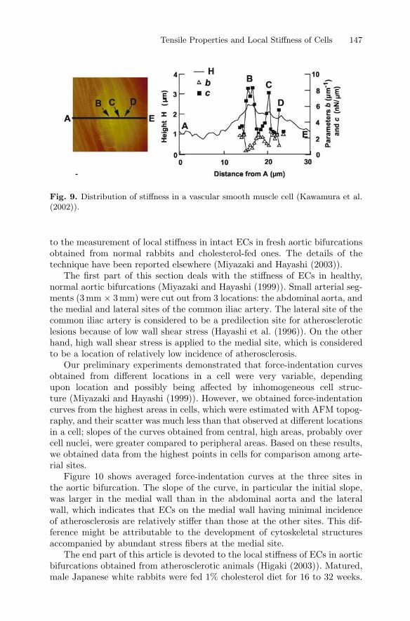
Figure 9 shows an example of stiffness distribution in a VSMC. The photoon the left-hand side is a topograph of the cell (Kawamura et al. (2002)).The variation of cell height along the line A–E is shown in the figure on theright-hand side. If we compare the trace of cell height with the AFM image,the height of the cell increases at the locations B, C and D (right) where thereseems to be actin bundles or stress fibers (left). It is interesting to see fromthe right figure that stiffness is high at these locations, which implies thatlocal stiffness measured by AFM sensitively reflects underlying cytoskeletalstructures.
3.3 Local Stiffness in Endothelial Cells in Normal andAtherosclerotic Wall
As far as the author knows, there has been no report on the AFMmeasurementof local stiffness of living cells in situ. Therefore, we applied the AFM technique
Tensile Properties and Local Stiffness of Cells 147
Fig. 9. Distribution of stiffness in a vascular smooth muscle cell (Kawamura et al.(2002)).
to the measurement of local stiffness in intact ECs in fresh aortic bifurcationsobtained from normal rabbits and cholesterol-fed ones. The details of thetechnique have been reported elsewhere (Miyazaki and Hayashi (2003)).
The first part of this section deals with the stiffness of ECs in healthy,normal aortic bifurcations (Miyazaki and Hayashi (1999)). Small arterial seg-ments (3mm × 3mm) were cut out from 3 locations: the abdominal aorta, andthe medial and lateral sites of the common iliac artery. The lateral site of thecommon iliac artery is considered to be a predilection site for atheroscleroticlesions because of low wall shear stress (Hayashi et al. (1996)). On the otherhand, high wall shear stress is applied to the medial site, which is consideredto be a location of relatively low incidence of atherosclerosis.
Our preliminary experiments demonstrated that force-indentation curvesobtained from different locations in a cell were very variable, dependingupon location and possibly being affected by inhomogeneous cell struc-ture (Miyazaki and Hayashi (1999)). However, we obtained force-indentationcurves from the highest areas in cells, which were estimated with AFM topog-raphy, and their scatter was much less than that observed at different locationsin a cell; slopes of the curves obtained from central, high areas, probably overcell nuclei, were greater compared to peripheral areas. Based on these results,we obtained data from the highest points in cells for comparison among arte-rial sites.
Figure 10 shows averaged force-indentation curves at the three sites inthe aortic bifurcation. The slope of the curve, in particular the initial slope,was larger in the medial wall than in the abdominal aorta and the lateralwall, which indicates that ECs on the medial wall having minimal incidenceof atherosclerosis are relatively stiffer than those at the other sites. This dif-ference might be attributable to the development of cytoskeletal structuresaccompanied by abundant stress fibers at the medial site.
The end part of this article is devoted to the local stiffness of ECs in aorticbifurcations obtained from atherosclerotic animals (Higaki (2003)). Matured,male Japanese white rabbits were fed 1% cholesterol diet for 16 to 32 weeks.

148 K. Hayashi
Fig. 10. Force-indentation curves of endothelial cells at three different sites in theaortic bifurcation of normal rabbits (Miyazaki and Hayashi (1999)).
Age-matched, regular chow-fed rabbits were used to obtain control data. Themicrographic observation of luminal surfaces stained with Sudan IV indicatedthat atherosclerotic plaques were more or less developed at all the three sitesin the aortic bifurcation. Moreover, AFM surface topography demonstratedthat ECs in the abdominal aorta and at the medial site of the iliac artery wereelongated in the axial direction of each vessel. However, cells at the lateralsite were more round or oval than those at the other two sites. This differencein cell morphology may be attributable to differences in the magnitude anddirection of wall shear stress. There were almost no differences in cell morphol-ogy between atherosclerotic and normal arteries and also between locationson atherosclerotic plaques and off plaques.
Force-indentation curves of ECs in the normal, control vessels (Fig. 11,left) were almost the same as those shown in Fig. 10, which implies highreproducibility of the experiments. As already mentioned above, the curvesobtained from the medial site of the iliac artery were significantly shiftedtowards the left compared with those obtained from the abdominal aorta andat the lateral site of the iliac artery. Even in cholesterol-fed animals, the resultsobtained from locations apart from atherosclerotic plaques were very similarto control data (Fig. 11, center). However, the locations on plaques at themedial site gave very different results, that is, force-indentation curves fromthese locations were very similar to those from the other two sites (Fig. 11,right). These results indicate that only the stiffness of endothelial cells onplaques at the medial site is affected by atherosclerosis.
Again, eq. (2) was applied to these force-indentation curves for the cal-culation of the values of parameters c and b, which represent initial stiffnessand stiffening rate, respectively. Then, those values were plotted against thearea fraction of sudanophilic lesions that represents the degree of atheroscle-rosis (Hayashi et al. (1994)). Parameter b at all the three sites and parameter

Tensile Properties and Local Stiffness of Cells 149
Fig. 11. Force-indentation curves of endothelial cells at three different sites of theaortic bifurcation in atherosclerotic rabbits (Higaki (2003)).
c in the aorta and at the lateral site of the iliac artery were not affected byatherosclerosis (data not shown). However, only parameter c at the medial sitedecreased with increase in the area fraction of lesions. That is, the stiffnessof ECs at this site significantly decreases with the progression of atheroscle-rosis, which might be due to lipid uptake and/or some disorganization of thecytoskeletal structure, such as stress fibers. At this moment, however, mech-anisms for this phenomenon are not known.
4 Conclusions
From these results, we can say:(i) there are large differences in tensile properties and local stiffness among
cells, depending upon cell kind, location, phenotype, passive or active state,and so on. Fibroblasts have the tensile strength of approximately 1µN, whilevascular smooth muscle cells do not break even at 2µN. Vascular endothelialcells are much weaker than these two cells. The stiffness of vascular smoothmuscle cells, which is significantly larger than those of the other two cells, islarger in contractile phenotype than in synthetic phenotype, and is greatlyincreased by contraction.
(ii) The distribution of stiffness in a cell is not uniform, possibly due toinhomogeneous cytoskeletal structure. The stiffness of cells is higher in thecentral areas over cell nuclei than in peripheral areas, and greater at thelocations where there exist actin bundles or stress fibers underneath.

150 K. Hayashi
(iii) The stiffness of vascular endothelial cells at the medial site of the iliacartery significantly decreases with the progression of atherosclerosis, probablydue to lipid uptake and/or some disorganization of cytoskeletal structure, suchas stress fibers.
Acknowledgements. The author appreciates Hiroshi Miyazaki, YoshitakaHasegawa, Yuji Ohara, Michitaka Higaki, Shohei Kajino, Akihide Kawamura,and Yuki Nabeshima for their great contribution to the experiments. Thiswork was financially supported in part by Grant-in-Aids for Scientific Research(A)(2) (nos. 09358020, 12308047, 15200036) and (B)(2) (no.13558111) fromthe Ministry of Education, Culture, Sports, Science and Technology, Japan.
References
Albrecht-Buehler, G. (1987). Role of cortical tension in fibroblast shape andmovement. Cell Motil. Cytoskel. 7:54–67.
Bereiter-Hahn, J., Karl, I., Luers, H., and Voth, M. (1995). Mechanical basis ofcell shape: Investigations with the scanning acoustic microscope. Biochem.Cell Biol. 73:337–348.
Frangos, J. A., ed. (1993). Physical Forces and the Mammalian Cell. NewYork: Elsevier Science & Technology Books.
Glerum, J. J., Van Mastrigt, R., and Van Koeveringe, A. J. (1990). Me-chanical properties of mammalian single smooth muscle cells. III. Passiveproperties of pig detrusor and human a terme uterus cells. J. Muscle Res.Cell Motil. 11:453–462.
Goldmann, W. H., and Ezzell, R. M. (1996). Viscoelasticity in wild-type andvinculin-deficient (5.51) mouse F9 embryonic carcinoma cells examined byatomic force microscopy and rheology. Exp. Cell Res. 226:234–237.
Hayashi, K., Ide, K., and Matsumoto, T. (1994). Aortic walls in atheroscleroticrabbits – Mechanical study. J. Biomech. Eng. 116:284–293.
Hayashi, K., Yanai, Y., and Naiki, T. (1996). A 3d-lda study of the relationbetween wall shear stress and intimal thickness in a human aortic bifurca-tion. J. Biomech. Eng. 118:273–279.
Hayashi, K. (2003). Mechanical properties of soft tissues and arterial walls.In Holzapfel, G. A., and Ogden, R. W., eds., Biomechanics of Soft Tissuein Cardiovascular Systems, 15–64. Wien: Springer-Verlag. CISM Coursesand Lectures No. 441, International Centre for Mechanical Sciences.
Higaki, M. (2003). Atomic force microscopic measurement of local stiffnessof vascular endothelial cells in atherosclerotic rabbits. MS dissertation, De-partment of Systems and Human Science, Graduate School of EngineeringScience, Osaka University. Japanese text with English abstract, figures, andtables.

Tensile Properties and Local Stiffness of Cells 151
Hochmuth, R. M., Ting-Beall, H. P., Beaty, B. B., Needham, D., and SonTay, R. T. (1993). Viscosity of passive human neutrophils undergoingsmall deformations. Biophys. J. 64:1596–1601.
Hoh, J. H., and Schoenenberger, C. A. (1994). Surface morphology andmechanical properties of MDCK monolayers by atomic force microscopy.J. Cell Sci. 107:1105–1114.
Kajino, S., Miyazaki, H., and Hayashi, K. (2003). Tensile properties of con-tracted or relaxed vascular smooth muscle cells. In Proc. 79th AnnualConf. JSME Kansai Branch, I–37–I–38. JSME. Japanese text with Englishabstract and figures.
Kajino, S. (2003). Tensile properties of contracted or relaxed vascular smoothmuscle cells. MS dissertation, Department of Systems and Human Science,Graduate School of Engineering Science, Osaka University. Japanese textwith English abstract, figures, and tables.
Kawamura, A., Miyazaki, H., and Hayashi, K. (2002). Effects of the phenotypemodulation on the local stiffness of vascular smooth muscle cells. In Proc.Mech. Eng. Cong., 2002, Japan, volume 1, 103–104. JSME. Japanese textwith English abstract, figures, and tables.
Lal, R., and John, S. A. (1994). Biological applications of atomic forcemicroscopy. Am. J. Physiol. 266:C1–C21.
Matsumoto, T., Sato, J., Yamamoto, M., and Sato, M. (2000). Smooth musclecells freshly isolated from rat thoracic aortas are much stifer than culturedbovine cells: Possible effect of phenotype. JSME Intern. J., Ser. C 43:867–874.
Miyazaki, H., and Hayashi, H. (1999). Atomic force microscopic measurementof the mechanical properties of intact endothelial cells in fresh arteries. Med.Biol. Eng. Comput. 37:530–536.
Miyazaki, H., and Hayashi, K. (2003). Measurement of mechanical propertiesof intact endothelial cells in fresh arteries. In Braga, P. C., and Ricci,D., eds., Atomic Force Microscopy: Biomedical Methods and Applications.Totowa, New Jersey: Human Press. 307–313.
Miyazaki, H., Hasegawa, Y., and Hayashi, K. (2000). A newly designed tensiletester for cells and its application to fibroblast. J. Biomech. 33:97–104.
Miyazaki, H., Hasegawa, Y., and Hayashi, K. (2002). Tensile properties ofcontractile and synthetic vascular smooth muscle cells. JSME Intern. J.,Ser. C 45:870–879.
Nabeshima, Y. (2002). Tensile properties of vascular endothelial cells. BAthesis, Department of Systems, Graduate School of Engineering Science,Osaka University. Japanese text with English figures and tables.
Ohara, Y. (2000). Atomic force microscopic measurement of mechanical prop-erties of vascular smooth muscle cells. MS dissertation, Department of Me-chanical Science, Graduate School of Engineering Science, Osaka University.Japanese text with English figures and tables.

152 K. Hayashi
Palmer, R. E., Brady, A. J., and Roos, K. P. (1996). Mechanical measurementsfrom isolated cardiac myocytes using a pipette attachment system. Am. J.Physiol. 270:C697–C704.
Ricci, D., Tedesco, M., and Grattarola, M. (1997). Mechanical and morpho-logical properties of living 3T6 cells probed via scanning force microscopy.Microsc. Res. Tech. 36:165–171.
Sato, M., Levesque, M. J., and Nerem, R. M. (1987). An application of themicropipette technique to the measurement of the mechanical properties ofcultured bovine aortic endothelial cells. J. Biomech. Eng. 109:27–34.
Shroff, S. G., Saner, D. R., and Lal, R. (1995). Dynamic micromechanicalproperties of cultured rat atrial myocytes measured by atomic force mi-croscopy. Am. J. Physiol. 269:C286–C292.
Thoumine, O., and Ott, A. (1997). Time scale dependent viscoelastic andcontractile regimes in fibroblasts probed by microplate manipulation. J.Cell Sci. 110:2109–2116.
Valberg, P. A., and Feldman, H. A. (1987). Magnetic particle motions withinliving cells. measurement of cytoplasmic viscosity and motile activity. Bio-phys. J. 52:551–561.
Wang, N., and Ingber, D. E. (1994). Control of cytoskeletal mechanics byextracellular matrix, cell shape, and mechanical tension. Biophys. J. 66:2181–2189.
Weisenhorn, A. L., Khorsandi, M., Kasas, S., Gotzos, V., and Butt, H. J.(1993). Deformation and height anomaly of soft surfaces studied with anAFM. Nanotechnology 4:106–113.
Zahalak, G. I., McConnaughey, W. B., and Elson, E. L. (1990). Determina-tion of cellular mechanical properties by cell poking, with an application toleukocytes. J. Biomech. Eng. 112:283–294.


![Parametric stiffness analysis of the Orthoglide · manipulator is intended to become a Parallel Kinematic Machine (PKM), stiffness becomes a very important issue in its design [4,5,6]](https://img.dokumen.tips/doc/110x75/5f6b1611676852030075e2c4/parametric-stiiness-analysis-of-the-orthoglide-manipulator-is-intended-to-become.jpg)