Embed Size (px)
Citation preview

Temporal Region
The temporal region includes the temporal and infratemporal fossae, superior and inferior to the zygomatic arch, respectively
Temporal fossaIn which the temporal muscle (L. temporalis) is locatedIs bounded :
Posteriorly and superiorly by the temporal linesAnteriorly by the frontal and zygomatic bonesLaterally by the zygomatic archInferiorly by the infratemporal crest Floor by frontal, parietal, temporal, and greater wing of the sphenoid (bone of pterion)Roof by the temporal fascia


Temporal fossaTemporal fascia covers the temporal muscle, attaching superiorly to the superior temporal line. Inferiorly, the fascia splits into two layers, which attach to the lateral and medial surfaces of the zygomatic arch. The temporal fascia also tethers the zygomatic arch superiorly. When the powerful masseter muscle, which is attached to the inferior border of the arch, contracts and exerts a strong downward pull on the arch, the temporal fascia provides resistance.


Temporal fossa
Contents: 1. Temporalis ms arises from the bony floor and
the overlying temporal fascia2. Superficial temporal nerve and vessels3. Deep temporal nerve and vessels4. auriculotemporal nerve

Infratemporal fossa
is an irregularly shaped space deep and inferior to the zygomatic arch, deep to the ramus of the mandible and posterior to the maxilla.It communicates with the temporal fossa through the interval between (deep to) the zygomatic arch and (superficial to) the cranial bones.

Infratemporal fossa- BoundariesLateral = ramus of mandible Medial = lateral pterygoid plate Anterior = posterior aspect of maxilla Posterior = tympanic plate and the mastoid & styloid process of temporal bone. Superior = infratemporal surface of the greater wing of sphenoid bone. Inferior = medial pterygoid muscle attaches to the mandible near the angle


Infratemporal fossa, contents.
Inferior part of the temporal muscle Lateral and Medial Pterygoid muscle Maxillary artery (2nd part) and its branchesPterygoid venous plexus Mandibular nerve and its branches :
inferior alveolar lingual buccal
Otic Ganglionchorda tympani nerve , from facial nerve


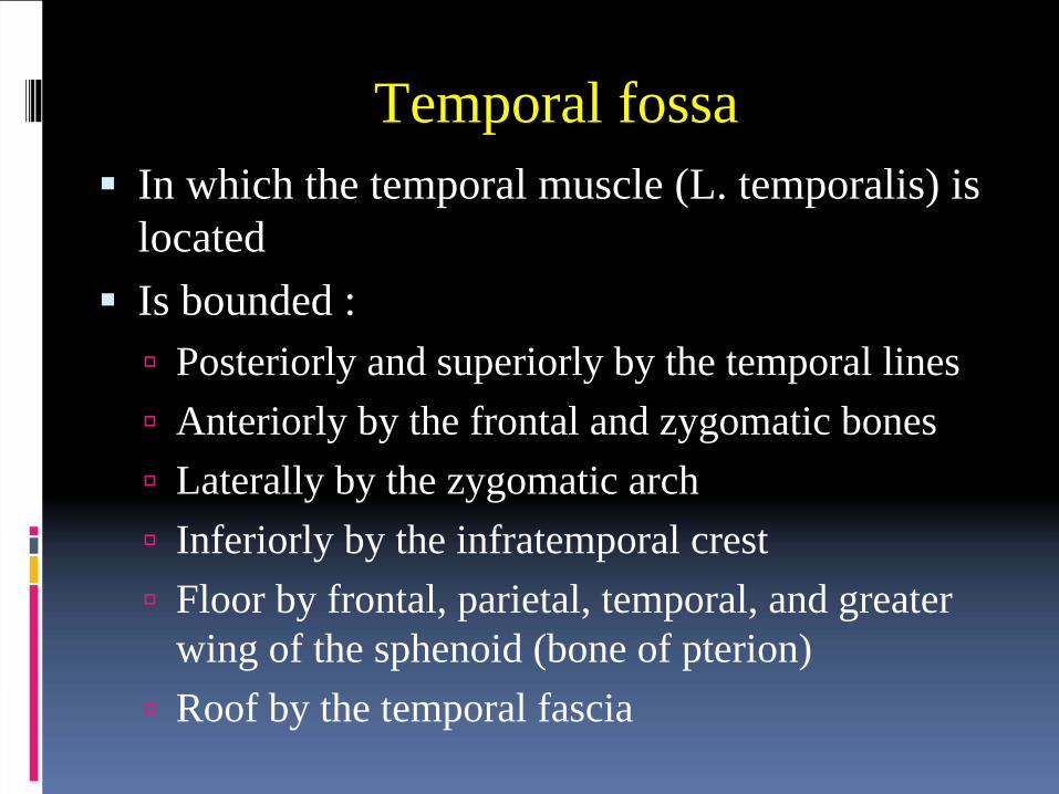
Maxillary artery
The maxillary artery is the larger of the two terminal branches of the external carotid artery It arises posterior to the neck of the mandible and is divided into three parts based on its relation to the lateral pterygoid muscle.

Course of 1st partBranches DistributionProximal (posterior) to lateral pterygoid muscle; runs horizontally, deep (medial) to neck of condylar process of mandible and lateral to stylomandibular ligament
Deep auricular artery
Supplies external acoustic meatus, external tympanic membrane, and temporomandibular joint
Anterior tympanic artery
Supplies internal aspect of tympanic membrane
Middle meningeal artery
Enters cranial cavity via foramen spinosum to supply periosteum, bone, red bone marrow, dura mater of lateral wall and calvaria of neurocranium, trigeminal ganglion, facial nerve and geniculate ganglion, tympanic cavity, and tensor tympani muscle
Accessory meningeal artery
Enters cranial cavity via foramen ovale; its distribution is mainly extracranial to muscles of infratemporal fossa, sphenoid bone, mandibular nerve, and otic ganglion
Inferior alveolar artery
Descends to enter mandibular canal of mandible via mandibular foramen; supplies mandible, mandibular teeth, chin, mylohyoid

Course fo 2nd part Branches DistributionAdjacent (superficial or deep) to lateral pterygoid muscle; ascends obliquely anterosuperiorly, medial to temporal muscle
Masseteric artery Traverses mandibular notch, supplying temporomandibular joint and masseter
Deep temporal arteries
Anterior and posterior arteries ascend between temporal muscle and bone of temporal fossa, supplying mainly muscle
Pterygoid branches Irregular in number and origin; supply pterygoid muscle
Buccal artery Runs anteroinferiorly with buccal nerve to supply buccal fat-pad, buccinator, and buccal oral mucosa
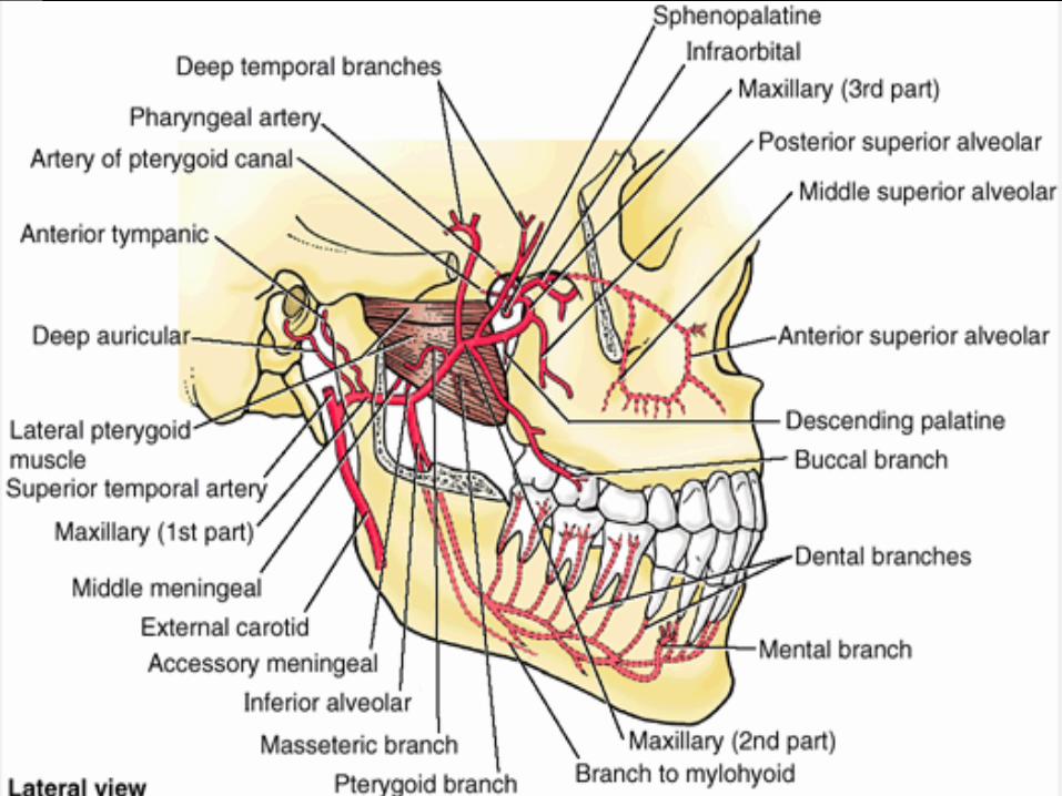
Infratemporal fossa, contentsThe pterygoid venous plexus:
is located partly between the temporal and the pterygoid muscles. It is the venous equivalent of most of the maxillary artery ,that is, most of the veins that accompany the branches of the maxillary artery drain into this plexus. Anastomoses anteriorly with the facial vein via the deep facial vein and superiorly with the cavernous sinus via emissary veins. The extensive nature and volume of this plexus is difficult to appreciate in the cadaver

Infratemporal fossa, contents
The mandibular nerve descends through the foramen ovale into the infratemporal fossa and divides into sensory and motor branches.The branches of CN V3 are the auriculotemporal, inferior alveolar, lingual, and buccal nerves.Branches of the CN V3 also supply the four muscles of mastication but not the buccinator, which is supplied by the facial nerve.

Infratemporal fossa, contentsThe otic ganglion (parasympathetic):
Is located in the infratemporal fossa, just inferior to the foramen ovale, medial to CN V3 and posterior to the medial pterygoid muscle.Presynaptic parasympathetic fibers, derived mainly from the glossopharyngeal nerve, synapse in the otic ganglion. Postsynaptic parasympathetic fibers, which are secretory to the parotid gland, pass from the otic ganglion to this gland through the auriculotemporal nerve.

Infratemporal fossa, contentsThe auriculotemporal nerve:
Encircles the middle meningeal arteryDivides into numerous branches, the largest of which passes posteriorly, medial to the neck of the mandible, and supplies sensory fibers to the auricle and temporal region. Also sends articular fibers to the TMJ and parasympathetic secretomotor fibers to the parotid gland.
The inferior alveolar nerve:Enters the mandibular foramen and passes through the mandibular canal, forming the inferior dental plexus, which sends branches to all mandibular teeth on its side. Another branch of the plexus, the mental nerve, passes through the mental foramen and supplies the skin and mucous membrane of the lower lip, the skin of the chin, and the vestibular gingiva of the mandibular incisor teeth.

Infratemporal fossa, contents
The lingual nerve:lies anterior to the inferior alveolar nerve.sensory to the anterior two thirds of the tongue, the floor of the mouth, and the lingual gingivae. Enters the mouth between the medial pterygoid muscle and the ramus of the mandible and passes anteriorly under cover of the oral mucosa, just inferior to the 3rd molar tooth.
The chorda tympani nerve:branch of CN VII carrying taste fibers from the anterior two thirds of the tongue, joins the lingual nerve in the infratemporal fossa .Also carries secretomotor fibers for the submandibular and sublingual salivary glands.

Infratemporal fossa, clinicalMandibular Nerve Block
An anesthetic agent is injected near the mandibular nerve where it enters the infratemporal fossa. In the extraoral approach, the needle passes through the mandibular notch of the ramus of the mandible into the infratemporal fossa. The injection usually anesthetizes the auriculotemporal, inferior alveolar, lingual, and buccal branches of CN V3.
Inferior Alveolar Nerve BlockAnesthetizes the inferior alveolar nerve, a branch of CN V3. The site of the anesthetic injection is around the mandibular foramen, the opening into the mandibular canal on the medial aspect of the ramus of the mandible. This canal gives passage to the inferior alveolar nerve, artery, and vein. When this nerve block is successful, all mandibular teeth are anesthetized to the median plane. The skin and mucous membrane of the lower lip, the labial alveolar mucosa and gingivae, and the skin of the chin are also anesthetized because they are supplied by the mental nerve,a branch of the inferior alveolar nerve.


Pterygopalatine fossa A small pyramidal space inferior to the apex of the Orbit. Lies between the pterygoid process of the sphenoid posteriorly and the posterior aspect of the maxilla anteriorly. Is a major distributing center for branches of the maxillary nerve and the pterygopalatine (third) part of the maxillary artery. Boundaries:
Medial wall = vertical plate of palatine bone Roof (incomplete)= greater wing of the sphenoid bone Floor = pyramidal process of the palatine bone Superior larger end opens into the inferior orbital fissureInferior end is closed except the palatine foramina


Pterygopalatine fossa It is located between, and has communications with, the infratemporal fossa, nasal cavity, orbit, middle cranial fossa, pharyngeal vault, maxillary sinus, and oral cavity (palate)It communicates with the following:
Laterally thru the pterygomaxillary fissure= infratemporal fossa Medially thru sphenopalatine foramen = nasal cavity Anterosuperiorly thru inferior orbital fissure = Orbit. Posterosuperiorly thru foramen rotundum and
pterygoid canal = Middle cranial fossa.

The fossa has been exposed through the floor of the orbit and maxillary sinus. The foramen rotundum, pterygoid canal, and pharyngeal canal are openings in the posterior wall of the pterygopalatine fossa.

Pterygopalatine fossa , contentsContents:
1. Terminal 3rd or the pterygopalatine part of the maxillary artery and branches. +accompaning veins
2. Maxillary nerve ( enters thru the foramen rotundum) and branches:
Zygomatic NervesZygomaticofacial + zygomaticotemporal nerves
Pterygopalatine nerves3. Nerve of the pterygoid canal. 4. Pterygopalatine ganglion.5. Surrounding fatty matrix

Third part Branches DistributionDistal (anteromedial) to lateral pterygoid muscle; passes between heads of lateral pterygoid and through pterygomaxillary fissure into pterygopalatine fossa
Posterior superior alveolar artery
Descends on maxilla's infratemporal surface with branches traversing alveolar canals to supply maxillary molar and premolar teeth, adjacent gingiva, and mucous membrane of maxillary sinus
Infraorbital artery
Traverses inferior orbital fissure, infraorbital groove, canal, and foramen; supplies inferior oblique and rectus muscles, lacrimal sac, maxillary canines and incisors teeth, mucous membrane of the maxillary sinus, and skin of infraorbital region of face
Artery of pterygoid canal
Passes posteriorly through pterygoid canal; supplies mucosa of upper pharynx, pharyngotympanic tube, and tympanic cavity

Third part Branches Distribution
Pharyngeal branchPasses through palatovaginal canal to supply mucosa of nasal roof, nasopharynx, sphenoidal air sinus, and pharyngotympanic tube
Descending palatine artery
Descends through palatine canal, dividing into greater and lesser palatine arteries to mucosa and glands of hard and soft palate
Sphenopalatine artery
Terminal branch of maxillary artery, traverses sphenopalatine foramen to supply walls and septum of nasal cavity; frontal, ethmoidal, sphenoid, and maxillary sinuses; and anteriormost palate as posterior lateral nasal + posterior septal branches


The maxillary nerve:enters the fossa through the foramen rotundum and runs anterolaterally
in the posterior part of the fossa .Within it, it gives off the zygomatic nerve, which divides into
zygomaticofacial and zygomaticotemporal nerves . These nerves emerge from the zygomatic bone through cranial foramina of the same name and supply general sensation to the lateral region of the cheek and temple.
The zygomaticotemporal nerve also gives rise to a communicatingbranch, which conveys parasympathetic secretomotor fibers to thelacrimal gland by way of the heretofore purely sensory lacrimal nerve from CN V1 .
Also gives off the two pterygopalatine nerves that suspend the parasympathetic pterygopalatine ganglion in the superior part of the pterygopalatine fossa . These nerves convey general sensory fibers of the maxillary nerve, which pass through the pterygopalatine ganglionwithout synapsing and supply the nose, palate, tonsil, and gingivae. The maxillary nerve leaves the pterygopalatine fossa through the inferior orbital fissure, after which it is known as the infraorbital nerve.


The parasympathetic fibers to the pterygopalatine ganglion come from the facial nerve by way of its first branch, the greater petrosal nerve . This nerve joins the deep petrosal nerve as it passes through the foramen lacerum to form the nerve of the pterygoid canal, which passes anteriorly through this canal to the pterygopalatine fossa. The parasympathetic fibers of the greater petrosal nerve synapse in the pterygopalatine ganglion.The deep petrosal nerve is a sympathetic nerve arising from the internal carotid plexus as the artery exits the carotid canal. It conveys postsynaptic fibers from nerve cell bodies in the superior cervical sympathetic ganglion to the pterygopalatine ganglion by joining the nerve to the pterygoid canal. The fibers do not synapse in the ganglion but pass directly through it into the branches (of CN V2) arising from it. The postsynaptic parasympathetic and the sympathetic fibers pass to the lacrimal gland, the palatine glands, and the mucosal glands of the nasal cavity and superior pharynx.


External Cranial Fossae

Cranial Fossae