Embed Size (px)
Citation preview

Nucleic Acids Research, 1993, Vol. 21, No. 1 27-36
Telomere directed fragmentation of mammalianchromosomes
Michael A.Barnett, Veronica J.Buckle1, Edward P.Evans2, Andrew C.G.Porter, Derek Rout3,Austin G.Smith3 and William R.A.Brown*Biochemistry Department, Oxford University, South Parks Road, Oxford OX1 3QU, 1'nstitute ofMolecular Medicine, John Radcliffe Hospital, Headington, Oxford OX3 9DU, 2MRC Radiobiology Unit,Chilton, Didcot, Oxfordshire, OX1 1 ORD and 3AFRC Centre for Genome Research, University ofEdinburgh, King's Buildings, West Mains Road, Edinburgh EH9 3JQ, UK
Received October 29, 1992; Revised and Accepted November 27, 1992
ABSTRACT
Cloned human telomeric DNA can integrate intomammalian chromosomes and seed the formation ofnew telomeres. This process occurs efficiently in threeestablished human cell lines and in a mouse embryonicstem cell line. The newly seeded telomeres appear tobe healed by telomerase. The seeding of new telomeresby cloned telomeric DNA is either undetectable or veryinefficient in non-tumourigenic mouse or humansomatic cell lines. The cytogenetic consequences ofthe seeding of new telomeres include largechromosome truncations but most of the telomereseeding events occur close to the pre-existing ends ofnatural chromosomes.
INTRODUCTION
Mammalian artificial chromosomes (MACs) would offer new
opportunities for introducing large numbers of genes in a definedsequence environment into experimental animals, agriculturallivestock, human somatic cells in vivo or mammalian cells intissue culture (1). In the yeast Saccharomyces cerevisiaetransfonnation with telomeric DNA can be used to fragment bothnatural and artificial chromosomes (2) and has become the basisof a powerful technology for mapping sequences in natural andartificial yeast chromosomes (3, 4). Preliminary evidence suggeststhat cloned telomeric DNA can also fragment mammalianchromosomes (5). These observations indicate one route to theconstruction of a MAC. The first step in this involves fragmentinga natural mammalian chromosome with cloned telomeric DNAto produce a mini-chromosome. If such a mini-chromosome were
small enough then it might be shuttled into an experimentalenvironment where it could be analysed and manipulated more
easily than in a mammalian cell. The nucleus of S. cerevisiaewould be one such environment. If the mini-chromosome couldbe re-introduced into a mammalian cell and retain its integritythen it could become the basis of a MAC vector. Central to such
a project is the use of cloned telomeric DNA as a reagent forfragmenting mammalian chromosomes.The work described in this paper demonstrates that cloned
human telomeric DNA can efficiently fragment mammalianchromosomes in several mammalian cell types including mouseembryonic stem (ES) cells. We show that when the clonedtelomeric DNA seeds the formation of a new telomere it is healedby an enzymatic machinery with the characteristics of telomerase.We have characterized the products of fragmentation in a humancell line and in mouse ES cells by cytogenetic techniques. Theseinclude truncated centromere containing versions of naturalchromosomes. We have not detected acentric fragmentationproducts. These results demonstrate that cloned telomeric DNAcan be used as a reagent to manipulate the structure of mammalianchromosomes.
MATERIALS AND METHODSDNA manipulationsPlasmids were constructed by standard procedures from thefollowing fragments. We used the 2.2 kb AccI BamHI fragmentof pSV2neo (6) as the source of the G418 resistance gene in theplasmids pTZsvneoproTEL, pBSsvneoTEL' and pTZsvneo. Weused the 1.8 kb HindI BamHI fragment from pPGKneo(3(referred to as pDEneo in ref. 7) as the source of the G418resistance gene in the plasmid pBSPGKneoTEL. In theconstruction of this plasmid it was assumed that the early regionpolyadenylation sequence was in the same position with respectto the BamHI site as in pSV2neo. Subsequently we obtainedsequence information about a precursor of pPGKneo,B andrealised that the poladenylation sequence is inverted with respectto the BamHI site. Our construct therefore lacks a polyadenylationsequence. The telomeric DNA in pTZsvneoproTEL extendedfrom the PstI site at position 678, 2.4 kb to a Bal3l deletionendpoint within TelSau2.0 (8). The telomeric DNA inpBSsvneoTEL' (TEL' is referred to as TELHS in Itzahki et al.,
* To whom correspondence should be addressed
\.) 1993 Oxford University Press

28 Nucleic Acids Research, 1993, Vol. 21, No. 1
in press) and pBSPGKneoTEL extended from the TaqI site atpostion 1831 to the deletion end point in the same deleted variantof TelSau2.0.
Extraction of genomic DNA, restriction enzyme digestion,Bal3l digestion, gel electrophoresis and filter hybridization wereas described previously (8, 9)
PCR and DNA sequencingThe primers used to amplify the DNA healed onto the linearizedpBSsvneoTEL' were termed 5-amp and C-strand. Theirsequences were GACTGAGCTCAGGGGGAATTATCAAGC-TAT and TATAAGCTTCCCTAACCCTGACCCTAACCCrespectively. Both were tagged at the 5' end with a stretch ofDNA which was not complimentary to any known sequence inthe potential target, which included sites for either SacI (5-amp)or Hindm (C-strand) and which, in the case of C-strand, servedto enhance the amplification of the products of the initial roundof amplification through subsequent rounds. The PCR reactionswere carried out in a total volume of 0.1 mL of 50 mM KCl,10 mM Tris-HCl pH8.4, 2.0 mM MgC2, 0.1 mg/mL gelatin,10-7M 5-amp, 10-7M C-strand, 0.2 mM of each dNTP, 2.5units Taq polymerase (Cetus) and approximately 0.1 gg ofgenomic DNA. Annealing was at 66°C for 2 minutes, elongationat 720C for 2 minutes, and denaturation at 94°C for 2 minutesfor a total of 35 cycles. PCR products were partially purifiedby two cycles of geneclean (BiolOl), digested with HindM andSac, cloned into pBSKS+ and sequenced by standard doublestranded DNA sequencing tehniques using Sequenase II (USB).
Cell cultureThe human cell lines HeLa (10), EC27C4 (11), HT1080 (12)and the mouse cell line lOT1/2 (13) were all grown as adherentcells in Dulbecco's modified Eagles Medium supplemented with10% Foetal Calf Serum, 2 mM L-glutamine and antibiotics(DMEM). The primary human cell line was established as anoutgrowth from a portion of a foetal limb and was maintainedin supplemented Dulbecco's modified Eagles medium furthersupplemented with DMEM conditioned by the growth of rat ormouse thymocytes. Cells of this line displayed density dependentinhibition of proliferation and started to senesce after about 25passages. The mouse ES line, EFC-I, was maintained asdescribed in ref. 14. Cells were transformed with DNA byelectroporation using a Bio-Rad gene pulsar typically set to400V/250 ,uF and transformants were selected by the growth ofthe cells in medium containing G418 at 250-300 ,ug/mL. Thetransformation efficiencies and protocols varied beween differentcell lines and varied between experiments. The frequency oftelomere seeding however was consistent for one cell line fromexperiment to experiment. No systematic differences wereobserved between the transformation efficiencies observed withpTZsvneoproTEL and pSV2neo suggesting that the effect of anyadjacent telomere upon the expression of the svneo gene wassmall.
Cytogenetic analysisChromosomes were prepared from the HT1080 cells which hadbeen transfected with pTZsvneoproTEL by growing the cells for12-16 hours in medium supplemented with 100 ,ug/mL5-bromo-2-deoxyuridine and then for a further 5-12 hours inmedium further supplemented with 10-5M thymidine. Colcemidwas added to the culture ten minutes prior to harvesting to a final
trypsinization using a solution of 40 mM KCl, 0.5 mM EDTA,20 mM Hepes pH 7.4, fixed, spread and G banded by standardtechniques (15). In situ hybridization, detection and replicationbanding procedures were all performed as previously described(15). The probe used for the in situ hybridization was arecombinant of the svneo fragment of pSV2neo in pTZ18R,referred to as pTZsvneo, labelled with 3H by nick translation.After the hybridization, exposure and developing of thephotographic emulsion the chromosomes were banded withHoechst 33258 and Giemsa. In situ hybridization of thechromosomes isolated from the ES cells was performed afterchromosome isolation as described in ref. 16. After thehybridization, exposure and development of the photographicemulsion the chromosomes were banded by trypsin digestion andGiemsa staining.
RESULTSFunctional properties of cloned human telomeric DNA inHeLa cells
We chose to start our investigation of the functional propertiesof cloned telomeric DNA in the HeLa cell line for two reasons.Firstly, HeLa cells have been shown to contain telomerase activity(17). It seemed likely that this activity would be necessary toheal any newly seeded telomere. Secondly, HeLa cells areaneuploid and contain many rearranged chromosomes. Ittherefore seemed unlikely that any loss of chromosomal materialaccompanying a fragmentation event would be fatal to these cells.We constructed the plasmid pTZsvneoproTEL (Figure 1). Thisplasmid contains a 2.4 kb stretch of human telomeric DNA fromthe plasmid TelSau2.0 (8), a gene encoding resistance to theantibiotic G418 which is transcriptionally active in humanfibroblasts and plasmid vector sequences. The 2.4 kb stretch ofhuman telomeric DNA includes 1.4 kb of human proterminalDNA and 1.0 kb of the (TTAGGG)n array. The yeast telomericDNA in TelSau2.0 was deleted by Bal31 digestion prior toplasmid construction. pTZsvneoproTEL was linearized to revealthe human telomeric DNA in its natural orientation at one endof the molecule and introduced into HeLa cells by electroporation.Stably transfected cells were cloned. DNA extracted from theclones was analysed by restriction enzyme digestion, gelelechtWhoresis and filter hybridization. Digestion with an enzymecutting at a unique site witiin pTZsvneoproTEL and hybridizationwith a probe lying on the telomeric side of the site should giveinformation about the fate of the telomeric DNA in the integratedconstruct. If the telomeric DNA had seeded the formation of anew telomere then we should expect the filter hybridizationanalysis to detect a heterogeneous collection of fragments. If theconstruct had integrated into a chromosome witiout seeding the
svno p118 (rTAOQ3)npTZ svneo proTEL
8om"e10 PBSKS (TTAG0G)n
_46- ,)s (TTAGGG)n
-&* I-PGKno
_ m oo)n
pBS svneo TEL (Asp7l8)
pBS svneo TEL (Apal)
pBS PGKneo TEL
concentration of 10 Ag/mL. Cells were harvested without Figure 1. Structure of the constructs used in this work.

Nucleic Acids Research, 1993, Vol. 21, No. 1 29
formation of a new telomere then we should expect to detect adiscretely sized fragment. We analysed 40 stably transfectedclones in this way. Figure 2 illustrates the results of a typicalset of analysis of 20 clones. The 40 clones contained 44 stableintegration events of which 24 were associated withheterogeneously sized fragments (Table 1). These clonescontained either one or two copies of the construct. We mapped
s nsx s s s s s s nsnsnss s s s ns s ns snssns
f S _lfSfSS l fSSlSl
23.1
9.46.6
kb 4.4
't4 .4i:
2.32.0 -
0.56-a
Figure 2. Detection of Telomere seeding activity by gel electrophoresis andfilter hybridization. DNA extracted from clones of HeLa cells that had beenstably transfected with pTZsvneoproTEL was digested with HindIII, elec-trophoresed on a 0.8% agarose gel, filter transfered and hybridized to 32plabelled pTZ18R. The first track includes an end labelled HindIII digest of phageX DNA and is labelled X. Subsequent tracks are labelled s or ns to indicatewhether the pTZsvneoproTEL has seeded a new telomere.
restriction sites flanking the construct in four clones containinga single integration site and demonstrated that there was a singleconstruct at each and that the integration sites differed from oneanother. The enzyme used in the experiment illustrated in Figure2 was HindI; this cuts at a site 4.7 kb from the boundary withthe telomeric DNA in pTZsvneoproTEL. If the heterogeneouslysized fragments do correspond to newly seeded telomeres thenthe results of the experiment illustrated in Figure 2 suggest thatthey range between 1 kb and 20 kb in size. We wanted to confirmthat the construct in this sort of clone lay at the end of achromosomal DNA molecule and so we digested DNA extractedfrom three such clones with the exonucease Bal3 1. The digestswere sampled periodically and analysed by restriction enzymedigestion, gel elecrophoresis and filter hybridization as describedabove. In each of these three clones the cognate restrictionfragments were sensitive to the action ofthe Bal31 (Figure 3a-c).In order to check the specificity of our approach we also analyseda clone which contained a construct which had stably integratedinto a HeLa cell chromosome without appearing to seed theformation of a new telomere. As anticipated the cognatefragments in this clone were not detectably sensitive to the actionof Bal3l (Figure 3d). In the experiment illustrated in Figure 3the DNA was digested with XbaI after Bal3l digestion. XbaIdoes not cut within the integrated pTZneoproTEL but was usedin this experiment becase it allowed us to use a hybridizationprobe specific for the neo gene which produced lower non-specific background after filter hybridization than the vector probeused in Figure 2.We tried to confirm the chromosomal location of the integrated
construct by fluorescent in situ hybridization but were unableroutinely to detect the short stretch of heterologous DNA in thisconstruct using this technique.We were interested to know whether the ability to seed new
telomeres in this way required the precence of telomeric DNA
X 0 30 60 90 120 A 0 30 60 90 120 X 0 30 60 90 120 X 0 30 60 90 120 min Bal3l
23.1 - _
9.4 -
kb 6.6 - p*.*1 0
4.4 --
2.3-2.0-
0.56-
A B C D
Figure 3. Bal31 sensitivity of newly seeded telomeres. DNA extracted from each of four HeLa derived clones which had been stably transfected with pTZsvneoproTELwas incubated for the indicated time with Bal3 1. The reaction was terminated with EGTA and phenol. DNA was extracted and further digested with XbaI. Digestswere analysed by gel electrophoresis and filter hybridization with a 0.8 kb Pvull fragment from the the neomycin resistance gene in pSV2neo. One track fromeach panel includes an end labelled HindIll digest of phage X DNA. Panels A-C correspond to clones where the construct has seeded a new telomere, panelD corresponds to a clone where no detectable seeding has occurred. The clones used in this experiment do not correspond to any of those used in the experimentillustrated in Figure 2.
0 ;:::.:.-0
::t,I.:too - be

30 Nucleic Acids Research, 1993, Vol. 21, No. I
Table 1. Frequency of telomere seeding in mouse and human cell lines
Cell line Origin Construct Clones Events Seeded Not-Seeded Fraction
HeLa Human pTZsvneoproTEL 40 44 24 20 0.55HeLa Human pSV2neo 20 27 0 27 < 0.04HeLa Human pBSsvneoTEL'(Asp7l8It) 43 45 20 25 0.45HeLa Human pBSsvneoTEL'(ApaIt) 39 47 17 30 0.36EC27C4 Human pTZsvneoproTEL 39 54 34 20 0.63HT1080 Human pTZsvneoproTEL 22 27 13 14 0.551 0-FT Human pTZsvneoproTEL 40 54 0 54 < 0.02ES EFC Mouse pBSPGKneoTEL 29 30 11 19 0.3610T1/2 Mouse pTZsvneoproTEL 38 53 1 52 0.02
tindicates the enzyme used to linearize pBSsvneoTEL'.
in the construct and so we transfected HeLa cells with linearizedpSV2neo. We analysed 20 clones in the way described above.None of the 27 different integration events detectable in theseclones appeared to be associated with a new telomere (data notshown).
The fate of cloned telomeric DNA upon introduction intoHeLa cellsThe results described in the previous section suggested that thecloned telomeric DNA in the construct pTZsvneoproTEL hadintegrated into the HeLa cell chromosomes and seeded theformation of a new telomere. The results were also consistentwith the possibility that the construct had integrated into pre-existing telomeres. This seemed less likely when we comparedthe average lengths of the construct associated telomeres and ofthe endogenous HeLa cell telomeres. In order to make thiscomparison we measured the average lengths of the endogenoustelomeres by restriction enzyme digestion, pulsed field gelelectrophoresis and filter hybridization with the TelBam3.4 probe(8) (Figure 4). In this experiment we digested the DNA withBamHI prior to gel electrophoresis and filter hybridization.BamHI cuts 3.4 kb from the boundary between the telomeric andthe proterminal DNA in the TelBam3.4 cognate sequences andthus the results illustrated in Figure 4 demonstrate that the lengthsof the endogenous telomeres range between 15 kb and 50 kbwhich is consistent with earlier measurements by de Lange (18).The length of sequence added onto the telomeric construct in the24 clones in which the construct had appeared to seed theformation of a new telomere was within the range 0-20 kb(Figure 2) with an average of 4 kb. At the time of the analysisthe clones had been through approximately 24 doublings sincetransfection with pTZsvneoproTEL. The difference in lengthsbetween the endogenous telomeres and the construct-associatedtelomeres suggests that the construct had seeded the formationof new telomeres rather than integrated into pre-existingtelomeres.
In order to investigate the mechanism of healing of the newlyseeded telomeres we designed an experiment using a strategyestablished by Murray and colleagues (19) in an investigationof the mechanism of healing of cloned telomeric DNA inS.cerevisiae. These workers introduced a construct containingcloned telomeric DNA with a non-telomeric polylinker extensioninto yeast and observed that the construct was healed withtelomeric DNA but retained some or all of the polylinkersequences. They concluded that the construct was healed by amechanism which did not involve sequence conversion orrecombination and was likely to involve telomerase. We therefore
.Clones A
94 -
Figure 4. Lengths of endogenous telomeres in HeLa cells and in three transfectedclones. DNA from the HeLa cell line or from each of three stably transfectedclones was restricted with BamHI, size fractionated by pulsed field gel elec-trophoresis and analyzed by filter hybridization with the TelBam3.4 probe(Brown et al., 1991). Markers were a mixture of multimers of X DNA andHindlIl restricted lambda DNA.
assembled the plasmid pBSsvneoTEL' (Figures 1 and 5). Thisplasmid differs from pTZsvneoproTEL in two significantrespects: firstly, it contains only 176 bp of proterninal DNA andsecondly, the extreme terminus of the molecule has beenengineered to enable details of the processing of the end of theconstruct to be examined by PCR and sequencing (Figure 5a).When pBSsvneoTEL' is linearized by Apal the telomeric endof the molecule consists of 15 bp of non-telomeric DNA, 2 copiesof the TTAGGG repeat and then 28 bp and 4 unpaired 3' residuesof non-telomeric DNA (Figure 5a). The recessed 15 bp stretchof non-telomeric DNA acts as a primer binding site to enablethe DNA healed onto the construction to be specifically amplifiedby the PCR. The 32 residue stretch of non-telomeric DNA atthe very end of the molecule acts as an indicator of the specificityfor the healing reaction. We needed to establish that the presenceof only 176 bp of proterminal DNA did not impair the functionalproperties of the TTAGGG array before we used this constructto examine the mechanism of healing of the newly seededtelomeres. There is a unique Asp718I site between the terminal32 residue stretch of non-telomeric DNA and the adjacent TTA-GGG sequence and so in initial experiments we linearizedpBSsvneoTEL' with Asp718I, transfected the plasmid into HeLacells by electroporation, isolated clones and analyzed the extracted
Nucleic Acids Research, 1993, Vol. 21, No. 1 31
AAsp7181
T = __ jT=A = ;ACCGCGGCCGATCCASGTCG CCAATCCCA&TCCqCTTAATAGST GA TACCCAASCTl TOCOCCGCCGTAOCTACAGCTCCCCC
B
TTAGGGTTAGGGWGATATTCAAGCCT4T,AGGGTSATGGACCGCGGCCGASCGAGGGGG4TTAGGGTTAG?GTTAGGAATCCCAATC4CICCSAASAGSCG
GTTATQAGCS AGGGAATCCCARATTAC TCGA C
TTAGGGTSACGOGAATTATCAAGCT4TSAGGGTSAGGQACCGCOGGCGCATCGATGGAGGGQT?AGOGTTAG=GTAGGAASOCAATCOCSTAATAGTTC
TT AIG TT C __AACQSCq TTAATAGTTCGA1ATcAATcALcATcAccTCC
TTAGOGTTAGGG GGAATTATCAAGCT,TTAGGGTTAGGGACCGCGGGCGCAcG TCGATCSTAGGGTSAGGGSSAGGAASC ATC t CSAASGSTCGAIAACCWATCCtATGGCGCC
TZAGGGTTAA3G.GGAATTATCAAGCT4T!AGGGTTAGG4TJLcCGCGG4TTAGGGT7AGGGTTAGGGT!AGOG.
AATCCCAATCCqC TTAATAGTTCGA AATCCcTcCqATGGCGc4aATCAAcCCAATccCAATCC
0
x Clones
23.1 -Xg a1- ~ I * * mlW
9.4 - U'.kb 6.6-
Input
Clone
15
25-1
25-2
27
31
4.4 -,
2.0-
0.56-
37
39
a0
40
Figure 5. Specificity of telomere healing in HeLa cells. A. The sequence cor-
responds to the telomeric terminus of the ApaI linearized pBSsvneoTEL'. Theregion underlined is the cognate sequence of the specific primer 5-amp usedto amplify DNA healed onto the construct. The boxed sequences correspondto TTAGGG repeats. The Asp718I site used in the preliminary experiment isindicated. B. Sequences of DNA at the termini of healed ApaI cutpBSsvneoTEL'. Two sequences were detected in the PCR products if clone25 and these are indicated as 25-1 and 25-2.
x tHeLa [IC27C4 ILHT1080 L _x
145.5 -
97 -
48.5 -
23.1-
9.4-A6.6 -
4.4 -
Figure 6. Lengths of DNA healed onto the newly seeded telomeres in differentcell lines. DNA isolated from four clones derived by transfection of the in-dicated lines with either pTZsvneoproTEL (HeLa, EC27C4 and HT1080) or
pBSPGKneoTEL (ES) was digested with either XbaI (HeLa, EC27C4 and
HT1080) or HindIII (ES), size fractionated by pulsed field gel electrophoresisand analyzed by filter hybridization with the 0.8 kb PvuII fragment from the
neomycin resistance gene of pSV2neo. Markers were a mixture of multimers
of X DNA and HindIlI restricted lambda DNA.
DNA using the biochemical approach established above. In 20of the 43 stably transfected clones that we analysed the linearizedconstruct seeded the formation of a new telomere. We thereforeconcluded that, as anticipated, the proterminal sequences present
Figure 7. Telomere growth during cloning and proliferation of primary humanfibroblasts. Primary human fibroblasts of the 1 °FT line were transfected withlinearized pTZsvneoproTEL and stably transformed cells were cloned. DNAwas extracted from the clones, digested with BamHI and analyzed by gel elec-trophoresis and filter hybridization with the TelBam3.4 probe.
in pTZsvneoproTEL, but absent from pBSsvneoTEL', werewithout significance for this aspect of telomere function. In fiveof the clones in which the construct had seeded the formationof a new telomere we were able to generate a PCR product whichhybridized to 32P-(TTAGGG)4 when we amplified with primerscomplimentary to the G rich strand of the human telomeric repeatand to the 15 basepair recessed primer binding site. We clonedand sequenced one of the PCR products and demonstrated thatthe construct had been healed with (TTAGGG)n on or within the(TTAGGG)2 sequence (not shown). We interpreted the failureto generate a PCR product in other fifteen clones where theconstruct had seeded the formation of a new telomere as indicatingthat the construct had been healed behind the boundary of the(TTAGGG)2 repeat and thus had lost the sequencecomplimentary to the specific primer. In the next experiment welinearized the construction with ApaI, electroporated HeLa cellsand analysed 39 stably tranfected clones. In 17 of these theconstruct was associated with a newly seeded telomere suggestingthat the non-telomeric DNA extension did not significantly impairthe functional properties of the cloned telomeric DNA. Sevenof these clones yielded a PCR product. We cloned and sequencedtwo or three copies of the individual products. The results (FigureSb) demonstrate that the machinery responsible for healing theend of our construct does not require (TTAGGG)n to be presentat the very end of the molecule. This observation stronglysuggests that the construct has been healed by a mechanism thatdoes not involve recombination or gene conversion. It seemsprobable that our construct has been healed by telomerase. Theseresults also provide further evidence against the notion that inthese clones the construct has simply integrated into a pre-existingtelomere.The newly seeded telomeres were smaller than the endogenous
telomeres when we examined them first. We were therefore
TTAGWTTAGG;4-.,-TTATCAAGCTFTAGG.=.ITAGOr.TTAGGGTTAGOOTTAG=TAGM-AATCCCAATCcqccTTAATAG"CG&IAATCCCAATCCCRATCCCRATCCCILATCCCAATCCC

32 Nucleic Acids Research, 1993, Vol. 21, No. I
AA. pp..-.-inrm I OMe p p ppp0133DAAw,&m,m
2 3 4 5
7 8 9 o0 1 1 12
-* rM13 14 15 16 17 18
5P ,
19 20 21
l1q * 19 s
SCoDCCM22 X Y
22p*
2
6 7 8 9 10
13 14 15 16 17 18 19 20 21 22 x Y
5p . I lq -
1 2
6 7 8
13 14 15 16 17
p3M p&%=mMml
3 4 5
I 10Dm1129 10 1 12
cb &X pm I s
18 19 20 21 22 x Y
5pIT_1IA Ison
6
B
4 5
12eD.,mlK%D
11
C
crsr_X3dsi_r
m IaftFs -amf-vr
m
5p * I lq + iSo 13 q
Nucleic Acids Research, 1993, Vol. 21, No. 1 33
interested to know whether the lengths of these two classes oftelomere changed on prolonged culture and, if so, at what rate.We analysed the lengths of the newly seeded telomeres and ofthe endogenous telomeres at intervals corresponding to 60population doublings for a total of 300 population doublings. Theaverage lengths of the newly seeded telomeres progressivelyapproached those of the endogenous telomeres and became moreheterogeneous. Assuming a uniform growth rate for all the cellsin the culture, the maximum rate of telomere growth in the newlyseeded telomere was approximately 130 bp per cell doubling (datanot shown). We did not detect the appearance of any discretelysized fragments in the course of this analysis indicating that, oncethey are formed, the telomeres are stable.
In approximately half of the clones which had been stablytransfected with either of the telomeric constructs, the constructhad appeared to fail to seed the formation of a new telomere.We were also interested to know the fate of the cloned telomericDNA in these clones. In order to address this point we madeuse of a unique EcoNI site present in the proterminal DNA ofpTZsvneoproTEL. Restriction analysis demonstated that this sitewas undetectable in 13 out of 15 integration sites associated withthe failure to seed a new telomere (data not shown). The sitewas detectable in each of the 22 sites in the 22 clones in whichpTZsvneoproTEL had seeded the formation of a new telomere.These results suggest that the failure of the construct to seed anew telomere is associated with the loss of telomeric DNA fromthe construct. We analysed the structure of the integration sitein one such clone at intervals of 60 population doublings for atotal of 300 population doublings and, as anticipated, failed todetect any evidence of resolution of the construct into a telomere.Similarly, restriction site analysis of the DNA flanking theintegration sites in one clone in which the construct existed inboth telomeric and non-telomeric locations demonstated twoindependent sites of integration. These results thus suggest thata 1kb interstitial stretch of (TTAGGG)n is unstable and thatchromosome breakage and telomere seeding occurssimultaneously or very soon after integration of the construct intothe chromosome.
Telomere seeding in other mammalian cell typesThe results of the previous section demonstrate that clonedtelomeric DNA can seed the formation of new telomeresefficiently upon integration into HeLa cell chromosomes. Wewanted to examine the functionality of cloned telomeric DNAin a variety of other mammalian cell tpes for three reasons. Firstof all we wanted to determine the cytogenetic consequences oftelomere seeding and if possible to demonstrate directly thatcloned telomeric DNA could fragment mammalian chromosomes.We therefore needed to detect telomere directed chromosomefragmentation in a mammalian cell type with a easily definedset of chromosomes. Secondly, we want to be able to fragmentchromosomes in the mouse germ line. We therefore needed to
demonstrate telomere directed chromosome fragmentation inmouse embryonic stem (ES) cells. Thirdly, we were interestedto know how different cell types process cloned telomeric DNA.We started our analysis with a human embryonic fibroblast
line, 1°-FT, established in this laboratory. We chose to workwith a primary fibroblast line in the first experiments becausecytogenetic analysis of such lines is relatively straightforward.We transfected cells from the second passage of this line withlinearized pTZsvneoproTEL and analysed 40 stably transfectedclones by the molecular techniques established above. Fifty-fourdifferent integration events were detectable but in none of thesehad the construct seeded the formation of a new telomere. Wetherefore analysed the ability of cloned telomeric DNA to seedthe formation of new telomeres in two established human celllines; the teratocarcinoma EC27C4 and the fibrosarcoma HT1080both of which have been reported to contain a recognizable setof human chromosomes. We transfected cells of each line withthe linearized pTZsvneoproTEL and analyzed stably transfectedclones. The construct efficiently seeded the formation of newtelomeres in both lines. (Table 1). We next analyzed the abilityof the construct pBSPGKneoTEL (Figure 1) to seed the formationofnew telomeres in the ES line EFC. We chose to use a constructwhich included a G418 resistance gene driven by the promoterof the mouse phosphoglycerate kinase gene because the sv4O earlyregion is poorly expressed in ES cells. We analysed 29 stablytransfected ES clones and detected 30 independent integrationevents of which 11 were associated with the formation of a newtelomere. During the initial stages of this analysis we noted thatthe newly seeded telomeres were associated with a much longerstretch of telomeric DNA in the ES cells than in any of the othercell types (Figure 6). Two practical consequences of thisdifference are that reliable detection of telomere seeding in EScells requires the use of pulsed field gel electrophoresis and thatthe ES cell DNA is best extracted in agarose plugs in order toretain its integrity prior to analysis. The observation that theconstruct is often associated with a long stretch of telomeric DNAis also consistent with the possibility that in some of these celllines it has simply integrated into pre-existing telomeres. Howeverwe assume that ES cells are not qualitatively different from theother cells in which telomere seeding is observed and that theour construct has in fact seeded the formation of new telomeresin a majority of the ES cell clones where it has been healed witha large tract of DNA. Cytogenetical analyses described belowsupport this view. We were curious to know whether the abilityof the ES cells to heal the cloned telomeric DNA with a longstretch of (TTAGGG)2 was a consequence of their being ofmouse or of germ line origin. We therefore transfected mouselOT1/2 cells with linearized pTZsvneoproTEL. These are somaticcells but unlike many established mouse somatic cells share withES cells endogenous telomeres which range in length between25 and 75 kb (not shown). We analyzed 38 stably transfectedclones containing a total of 53 different integration events and
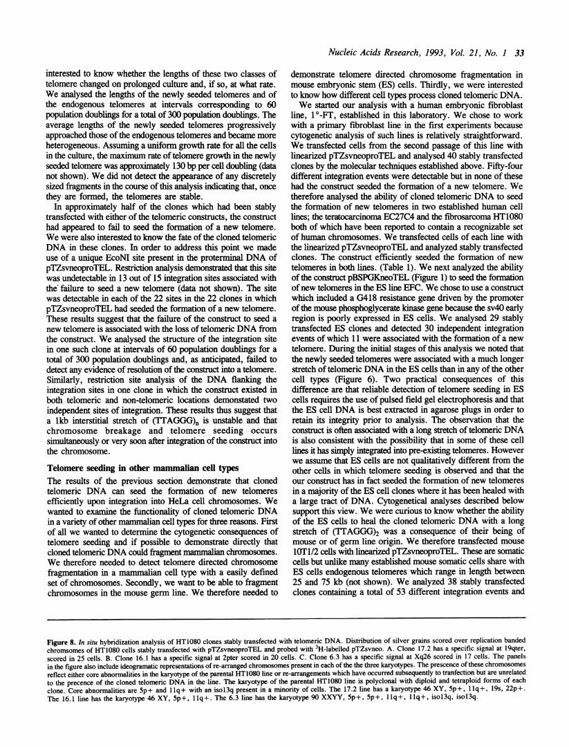
Figure 8. In situ hybridization analysis of HT1080 clones stably transfected with telomeric DNA. Distribution of silver grains scored over replication bandedchromsomes of HT1080 cells stably transfected with pTZsvneoproTEL and probed with 3H-labelled pTZsvneo. A. Clone 17.2 has a specific signal at l9qter,scored in 25 cells. B. Clone 16.1 has a specific signal at 2pter scored in 20 cells. C. Clone 6.3 has a specific signal at Xq26 scored in 17 cells. The panelsin the figure also include ideogramatic representations of re-arranged chromosomes present in each of the the three karyotypes. The prescence of these chromosomesreflect either core abnormalities in the karyotype of the parental HT1080 line or re-arrangements which have occurred subsequently to tranfection but are unrelatedto the precence of the cloned telomeric DNA in the line. The karyotype of the parental HT1080 line is polyclonal with diploid and tetraploid forms of eachclone. Core abnormalities are 5p+ and llq+ with an isol3q present in a minority of cells. The 17.2 line has a karyotype 46 XY, 5p+, llq+, 19s, 22p+.The 16.1 line has the karyotype 46 XY, 5p+, llq+. The 6.3 line has the karyotype 90 XXYY, 5p+, 5p+, llq+, llq+, isol3q, isol3q.
34 Nucleic Acids Research, 1993, Vol. 21, No. I
9F 4*
*9
4,
* 9
. I w 69
,~~~~"Itiq
~4 &
.: a *a
9 .1S,',,;*,i9~~~~~
* *
,
.iIw 44
A:
k6 ~~* 4*
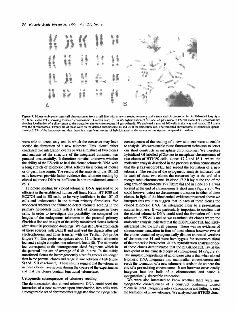
Figure 9. Mouse embryonic stem cell chromosomes from a cell line with a newly seeded telomere and a truncated chromosome 14. A. G-banded karyotypeof ES cell clone Tel 2 showing truncated chromosome 14 (arrowhead). B. In situ hybridization of 3H-labelled pTZsvneo to ES cell clone Tel 2 chromosomesshowing localization of a silver grain to the truncation site on chromosome 14 (arrowhead). We analyzed a total of 100 cells in this way and located 225 grainsover the chromosomes. Twenty six of these were on the deleted chromosome 14 and 25 at the truncation site. The truncated chromosome 14 comprises approx-imately 3.2% of the karyotype and thus there is a significant excess of hybridization to the truncation breakpoint compared to random.
were able to detect only one in which the construct may haveseeded the formation of a new telomere. This 'clone' eithercontained two integration events or was a mixture of two clonesand analysis of the structure of the integrated construct waspursued unsuccesfully. It therefore remains unknown whetherthe ability of the ES cells to heal the cloned telomeric DNA witha long stretch of telomeric DNA reflects their being of mouseor of germ line origin. The results of the analysis of the lOT1/2cells however provide futher evidence that telomere seeding bycloned telomeric DNA is inefficient in non-transformed somaticcells.Telomere seeding by cloned telomeric DNA appeared to be
efficient in the established human cell lines; HeLa, HT 1080 andEC27C4 and in ES cells, to be very inefficient in the lOTl/2cells and undetectable in the human primary fibroblasts. Wewondered whether the failure to detect telomere seeding in theprimary fibroblasts might reflect a lack of telomerase in thesecells. In order to investigate this possibility we compared thelengths of the endogenous telomeres in the parental primaryfibroblast line and in eight of the stably transfected clones isolatedafter about 20 population doublings. We digested DNA from eachof these sources with BamHI and analysed the digests after gelelectrophoresis and filter transfer with the TelBam 3.4 probe(Figure 7). This probe recognizes about 12 different telomericloci and a single complex non-telomeric locus (8). The telomericloci correspond to the heterogeneous sized fragments which inthe parental line are of average of 6 kb in size. In the stablytransfected clones the heterogeneously sized fragments are longerthan in the parental clones and range in size between 9.4 kb (clone5) and 15.0 kb (clone 2). These results suggest that the telomeresin these clones have grown during the course of the experimentsand that the clones contain functional telomerase.
Cytogenetic consequences of telomere seedingThe demonstration that cloned telomeric DNA could seed theformation of a new telomere upon introduction into cells witha recognizable set of chromosomes suggested that the cytogenetic
consequences of the seeding of a new telomere were amenableto analysis. We were unable to use fluorescent techniques to detectour short constructs in metaphase chromosomes. We thereforehybridized 3H-labelled pTZsvneo to metaphase chromosomes oftwo clones of HT1080 cells, clones 17.2 and 16.1, where themolecular analysis described in the previous section demonstratedthat the pTZsvneoproTEL had seeded the formation of a newtelomere. The results of the cytogenetic analysis indicated thatin each of these two clones the construct lay at the end of arecognizable chromosome. In clone 17.2 it lay at the end of thelong arm of chromosome 19 (Figure 8a) and in clone 16.1 it waslocated at the end of chromosome 2 short arm (Figure 8b). Wecould however detect no chromosome truncation in either of theseclones. In light of the biochemical evidence presented above weinterpret this result to suggest that in each of these clones thecloned telomeric DNA has integrated close to a pre-existingnatural telomere. It was particularly important to confirm thatthe cloned telomeric DNA could seed the formation of a newtelomere in ES cells and so we examined six clones where themolecular analysis indicated that the pBSPGKneoTEL had stablyintegrated into the ES cell genome. There was no evidence ofchromosome truncation in four of these clones however two ofthe clones contained cytogenetically distinct truncated versionsof chromosome 14 and were hemizygous for sequences distalof the truncation breakpoint. In situ hybridization analysis of oneof these clones demonstrated that the pPGKneoTEL lay at thebreakpoint of the truncated copy of chromosome 14 (Figure 9).The simplest interpretation of all of these data is that when clonedtelomeric DNA integrates into mammalian chromosomes andseeds the formation of a new telomere it tends to do so near theend of a pre-existing chromosome. It can however occasionallyintegrate into the bulk of a chromosome and cause acytogenetically detectable truncation.We were also interested to know whether there were any
cytogenetic consequences of a construct containing clonedtelomeric DNA integrating into a chromosome and failing to seedthe formation of a new telomere. We analysed one HT1080 clone,
a Ab
A
*
-0
*
Y
::.
B
41 *
i0 A
*6

Nucleic Acids Research, 1993, Vol. 21, No. 1 35
clone 6.3, where the molecular analysis demonstrated thatpTZsvneoproTEL had integrated but failed to seed the formationof a new telomere (Figure 8c). In clone 6.3 the construct hadintegrated into Xq26 without being associated with acytogenetically detectable rearrangement.
DISCUSSION
We initiated this project in order to determine whether we coulduse cloned telomeric DNA to fragment mammalianchromosomes. The combination of molecular and cytogeneticanalyses described here, together with the results of Itzahki andcolleagues (in press) showing truncation of human chromosome1 by targeted telomere directed breakage, demonstrate that wecan. This process occurs efficiently in the established cell linesHeLa, EC27C4 and HT1080 and in the mouse embryonic stemcell line EFC but is either inefficient or undetectable in mouse10Tl/2 cells or in human primary fibroblasts. The cytogeneticconsequences of the seeding of a new telomere include thegeneration of large chromosome truncations but most of theintegration events associated with the formation of newly seededtelomeres appear to occur close to the pre-existing ends of naturalchromosomes and to generate, consequently, truncations whichare too small to detect by cytogenetic techniques. Theseobservations raise two questions. Why does cloned telomericDNA not detectably seed the formation of new telomeres inhuman primary fibroblasts and why do most of the seeding eventsappear to occur close to the pre-existing ends of chromosomes?We analysed 54 sites where pTZsvneoproTEL had integrated
into the genome of a human primary fibroblast line but at noneof these had a new telomere been seeded. One possibleexplanation for a failure to detect telomere seeding might be anabsence of telomerase from the primary fibroblast line (20). Ifthis was the case then the cloned telomeric DNA could haveseeded the formation of new telomeres but the telomeres wouldhave progressively shorted during culture as a result ofexonuclease action and a failure to replicate the 5' end of thetelomeric DNA. If the newly seeded telomere had shortened untilit was too small to function then the remnants of the constructionwould either have been lost as a result of further sequencedegradation or would have become the junction of a fusion withanother chromosome. If the selectable marker gene in theconstruct had been destroyed then the clone, grown in thepresence of the antibiotic G418 would have died. If the telomerehad failed and the construction had fused with anotherchromosome then the construct would have appeared to havefailed to seed the formation of a new telomere. The observationthat the endogenous telomeres in the stably transfected cloneswere longer than those in the parental line however argues againsttelomerase deficiency being the basis of the correct explanationof our results. Chromosome structure is more stable in primaryfibroblasts than in transformed cells and it is possible that thefailure to detect telomere seeding by transfected telomeric DNAreflects active repair and recombination pathways which tend tointegrate sequences into the genomic DNA of these cells in sucha way as to prevent chromosome breakage. Our experimenthowever does not allow an unambiguous explanation of ourresults; it is, for example, possible that telomerase was not presentin the primary fibroblasts at the time of transfection and that anynewly seeded telomeres were lost prior to expression of anytelomerase activity. Alternatively we may have selected for thoseclones with long telomeres. Understanding chromosome
breakage, telomere seeding and the role of telomerase will requirereconstitution of the events in vitro from defined components.
It is striking that most of the seeding events that we detectedappear to occur close to the pre-existing ends of thechromosomes. It is unlikely that this will prevent cloned telomericDNA being used to systematically fragment mammalianchromosomes. In experiments described elsewhere we havetruncated human chromosome 1 at band lp35 by targetingtelomeric DNA to the 6-16 locus (Itzhaki et al. in press).Nevertheless more work will be needed to establish how reliablywe can use sequence targeting and telomere directed chromosomefragmentation to systematically fragment mammalianchromosomes. It seems particularly important that this work becarried out in ES cells where, suprisingly, two of the breaks thatwe detected were on chromosome 14. It might be thought thatthat our failure to detect a set of apparently random chromosometruncations was caused by the reduced viability of cellshemizygous for large chromosomal regions. This explanation isunsatisfactory for two reasons. Firstly cells with large truncationsof chromosomal material induced by therapeutic irradiation areviable in vivo for many years (21). Similarly mice hemizygousfor large chromosomal regions can be viable (Cattanach et al.,in press). Secondly the HT1080 karyotype appears variable andchanges in ploidy which might compensate for the geneticconsequences of any truncation should occur readily. Wespeculate that the non-random pattern of integration of thefunctioning telomeric DNA reflects an interaction between thecloned telomeric DNA in the construct and telomere bindingproteins located at the nuclear periphery prior to the integrationof the construct into the chromosome. Cytogenetic analysis alsofailed to detect large acentric fragments of chromosomal DNAin the clones containing newly seeded telomeres. This may reflectour analysis of relatively few clones by cytogenetic techniques,the fact that most of the breaks generate fragments too small todetect or the mitotic instability of such fragments.
Despite these limitations in our understanding of the molecularevents which occur when cloned telomeric DNA breaks achromosome and seeds the formation of a new telomere it seemsreasonable to conclude that we can use cloned telomeric DNAas a reagent for both structural and functional studies ofmammalian chromosomes in a variety of transformed cell linesand in the mouse germ line. Furthermore, the observation thatcloned telomeric DNA can break chromosomes in mammaliancells which readily integrate non-homologous DNA intochromosomes suggests that telomeric DNA might be used tomanipulate chromosome structure in a wide variety of plants andanimals.
ACKNOWLEDGEMENTSWe thank Chris Tyler-Smith and Karen Brown for theircomments upon a draft manuscript. This work was supportedby the Cancer Research Campaign and by the Medical ResearchCouncil as part of the UK Human Genome Mapping Programme.
REFERENCES1. Brown,W.R.A. (1992) Curr. Opin. Genet. Dev. 2, 479-486.2. Surosky,R.T. and Tye,B.-K. (1985) Proc. Natl. Acad. Sci. USA 82,
2106-2110.3. Vollrath,D., Davis,R. W., Connelly,C. and Hieter,P. (1988) Proc. Natl.
Acad. Sci. USA 85, 6027-6031.4. Pavan,W.J., Hieter,P. and Reeves,R.H. (1990) Proc. Natl. Acad. Sci. USA
87, 1300-1304.

36 Nucleic Acids Research, 1993, Vol. 21, No. I
5. Farr,C., Fantes,J., Goodfellow,P. and Cooke,H.J. (1991) Proc. Natl. Acad.Sci. US4 88, 7006-7010.
6. Mulligan,R.C. and Berg,P. (1980) Science 209, 1422-1428.7. Rathjen,P.D., Toth,S., Willis,A., Heath,J.K. and Smith,A.G. (1990) Cell
62, 1105-1114.8. Brown,W.R.A., MacKinnon,P.J., Villasante,A., Spurr,N., Buckle,V.J. and
Dobson,M.J. (1990) Cell 63, 119-132.9. Brown,W.R.A. (1988) EMBO J. 7, 2377-2385.
10. Scherer,W.F., Syverton,J.T. and Gey,G.O. (1953) J. Exp. Med. 97,695-709.
11. Pera,M.F., Cooper,S., Mills,J. and Parrington,J.M. (1989) Differentiation42, 10-23.
12. Rasheed,S., Nelson-Rees,W.A., Toth,E.M., Arnstein,P. and Gardner,M.B.(1974) Cancer 33, 1027-1033.
13. Reznikoff,C.A., Brankow,D.W. and Heidelburger,C. (1973) Cancer Res.33, 3231-3238.
14. Nichols,J., Evans,E.P. and Smith,A.G. (1990) Development 110,1341- 1348.
15. Buckle,V.J. and Craig,I.W. (1986) in Davies,K.E. (ed.), Human GeneticDiseases. IRL Press, Oxford.
16. Hogan,B.L.M., Costatini,F. and Lacy,E. (1980) Manipulating the MouseEmbryo;A Laboratory Manual. Cold Spring Harbor Laboratory Press, ColdSpring Harbor, NY.
17. Morin,G.B. (1991) Cell 59, 521-529.18. De Lange,T., Shiue,L., Myers,R.M., Cox,D.R., Naylor,S.L., Killery,A.M.
and Varmus,H.E. (1990) Mol. Cell. Biol. 10, 518-527.19. Murray,A.W., Claus,T.E. and Szostak,J.W. (1988) Mol. Cell. Biol. 8,
4642-4650.20. Counter,C.M., Avillon,A.A., LeFeuvre,C.E., Stewart,N.G., Grieder,C.W.,
Harley,C.B. and Baccheti,S. (1992) EMBO J. 11, 1921-1929.21. Adolph,S., Bartram,C.R. and Hameister,H. (1987) Cytogenet. Cell Genet.
44, 65-68.
![Intrarenal arteriosclerosis and telomere attrition ...€¦ · Telomere length is a well-established marker of biological age [4]. Although telomere length is partly heritable, there](https://img.dokumen.tips/doc/110x75/5f2629fb310cc83259516f06/intrarenal-arteriosclerosis-and-telomere-attrition-telomere-length-is-a-well-established.jpg)