Embed Size (px)
Citation preview

Tele-pathology Evolution and Usage
Jared N Schwartz MD PhD August 21, 2013 CLIAC Meeting
Atlanta, GA

Disclosures
All comments, opinions, and recommendations presented today are solely mine and not those of my employers or any other entity.

Disclosures
• Chief Medical Officer, Leica Biosystems • Consulting Professor Pathology, Stanford School of Medicine • Board of Directors, Personalized Medicine Coalition • Board of Directors, American Pathology Foundation • Advisory Board Cancer Commons • Past-President College American Pathologists • Past-Member of CLIAC

Tele-Medicine and Digitization Not New in Medical Devices
• Digital Otoscopes • Digital Stethoscopes • Digital Cameras on Microscopes • Digital Video on Microscopes • Endoscopy and many more • Are Images Good Enough to Make DX? Yes
Digital Microscopes (Whole Slide Imaging: WSI)
Hamamatsu Olympus Claro… Aperio Omnyx Phillips Ventana Leica Zeiss … and more

Different Tools - Same Image

“Whenever science makes a discovery, the devil grabs it while the angels are
debating the best way to use it.”
~Alan Valentine

… it’s not about the tools,
it is about the PEOPLE

Minimally Invasive Robotic Surgery eSurgery / In Vivo Imaging/ Sequencing on a Chip
The specimens we receive are getting smaller and smaller, yet expected to obtain so much more usable information

Does Viewing Pathology Images Place Patient in Harms Way
• Pathologists have 1:1 relationship with image be it glass or digital
• Not True for Chemistry/Hematology/Cytology Analyzers
• Glass Slide Always Available (Nearby, Courier Service) • Pathologists Can Always Go Back To Original Source
of Image

Why do Something Now?
Why paper isn’t the answer • This is a top Pathologist’s office
Glass slides are a challenge • I want to compare this to the last specimen? Where is the
patient’s past biopsy? • I need an expert, opinion, fast? • Cases ready to ship
Collaboration is essential • Where are the results?
Waiting times are too long Patients are worried

Digital Pathology Can Make a Difference

What is WSI DIGITAL PATHOLOGY ePathology?
• ePathology is a complete scan of a microscopic glass slide (eSlide) and the viewing of the eSlide on a computer monitor through a software system
• ePathology is the process by which a patient’s pathology results, including images, are available in their electronic medical record
• ePathology Means Much More to Pathology Research and Patient Care
• Engage Pathology more easily to support patient care • Ease of access limited specialty resources • Efficiently manage of turn-around time • Effectively communicate critical information across care settings with different
providers and patients • Expand Quality Assurance Tools • Enable use of Mobile Technologies for Sharing & Education • EXCITE and ENTICE Medical Students, Graduate Students, Histology Staff, and
Residents to an ever EVOLVING Specialty Career in Pathology Research and Clinical

All Pathology Images…
From procedure room – to pathologist – to patient’s EMR – to your mobile device!

eSlides Means All Pathology can be Integrated into the LIS and Ultimately the EHR and/or What Else?

Severe Shortage of Pathologists and Especially Subspecialists
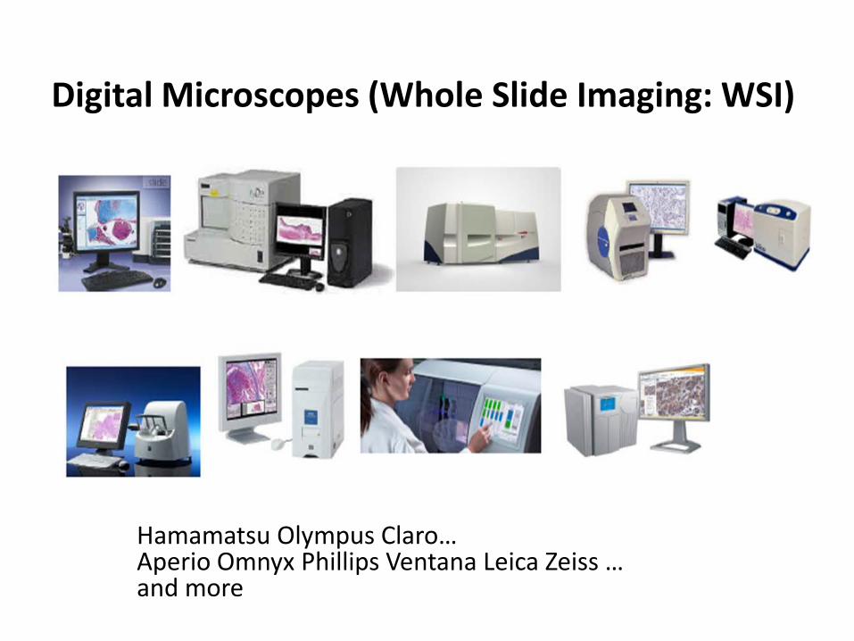
STRATEGIC GOAL 1: HEALTH CARE
Strategic Objective 1.1: Broaden health insurance and long-term
care coverage
Strategic Objective 1.2: Increase health care service availability and
accessibility
Strategic Objective 1.3: Improve health care quality, safety, cost,
and value
Strategic Objective 1.4: Recruit, develop, and retain a competent
health care workforce
HHS Digital Priorities
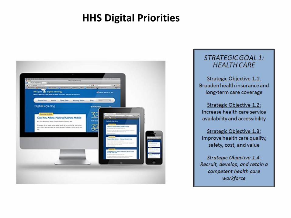
Pathologists Can and Are Providing Help to Hospitals/Communities with No Pathologists
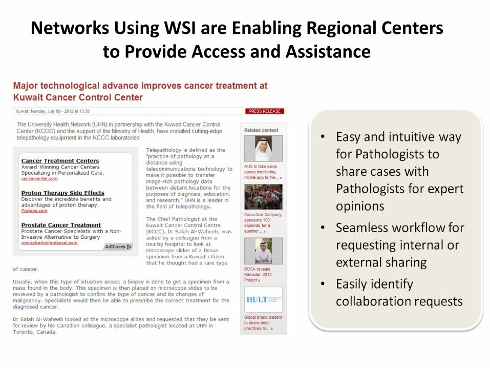
Networks Using WSI are Enabling Regional Centers to Provide Access and Assistance
• Easy and intuitive way for Pathologists to share cases with Pathologists for expert opinions
• Seamless workflow for requesting internal or external sharing
• Easily identify collaboration requests

Use of eSlides Enabling Us to Do More Without Traveling: A Sample
May 06, 2010
Live tele-consultation of complex cases, professional pathological diagnosis, regular packaged delivery---these are steps to guarantee the double check of diagnosis of every patient’s pathology by experts of both the Second Affiliated Hospital Zhejiang University and UCLA Medical Center. This is the perspective on joint pathology consultation by the Second Affiliated Hospital and UCLA stated by Prof. Rao Jianyu, tenured professor of pathology and epidemiology at UCLA Medical Center, Director of gynecologic pathology and cytopathology The medical diagnostic center at the Second Affiliated Hospital Zhejiang University is the first one UCLA Medical Center is involved in, and it is an exploratory pattern expected to benefit not only people of Zhejiang Province, but also of a wider area in near future. This is regarded as one step toward the goal of medical reform
It’s learned that UCLA Medical Center has a reservoir of more than 30 experts in pathology to provide remote medical consultation service. Most of these specialists in cell pathology, urinary pathology, etc., have rich clinical experiences with solid academic background and patients with complex cases or special requests can have their pathological images transmitted to UCLA for diagnosis.

CHINA-UCLA WSI Telepathology
TELEPATHOLOGY CLINICAL CARE AND MEDICAL EDUCATION SPANNING THE GLOBE
•Major Medical Centers Providing Increased Access and Education To China and Other Countries with Shortage of Pathologists
•The department also continues to develop a successful telepathology exchange with a prestigious health center in China
•The prestigious Second Affiliated Hospital Zhejiang University (SAHZU) currently sends an increasing number of challenging digitized slides/cases to UCLA Pathology for diagnostic purposes
•The goal is to collaborate even more closely over the next few years, and the result of this collaboration may be a new joint diagnostic center using the advanced technologies of telepathology, molecular pathology and genomics to create the most advanced cancer diagnostic center in China, under the leadership of both UCLA and SAHZU pathologists

Improving Access To Care in USA Using WSI?
• Under the leadership of Scott Binder, MD, senior vice chair for Clinical Services, the department continues to expand its pathology expertise via telepathology
• Binder is leading work, in conjunction with the University of California (UC) Office of the President, on a UC system-wide initiative to implement this new technology to a network of 8–10 remote hospitals throughout the state of California. These sites would have the ability to send high quality images of pathology slides to UCLA pathologists over a secure Internet connection, which would enable a group of pathologists without subspecialty expertise to consult with the department’s expert team of pathologists for advanced diagnostic purposes
• This technology also produces a windfall of educational opportunities for medical students, residents, and fellows in the David Geffen School of Medicine. It enhances clinical training by providing greater exposure to some of the world’s most complex pathological cases. It also benefits the UCLA scientific community by providing material for research

Major Medical Centers Enabling WSI to be Received by Their Subspecialist Experts

International Telepathology - Canada and Japan

USCAP Web Page

First Full Adoption Hospital
ADVANCE, February, 2010 First hospital in the world to
adopt digital pathology for 100% of their histology work (over 60,000 slides / yr.)

Medical Schools Not Waiting

Mobile Applications Use of Annotations

Whole slide imaging technology and computer accessibility have advanced to the point that virtual microscopy can be integrated into a pathology residents' educational activities. The digital teaching set we developed provided additional benefits of using the glass slides
Development and use of a genitourinary pathology digital teaching set for trainee education Li Li,1 Bryan J. Dangott,2 and Anil V. Parwani2 1Department of Pathology, Albany Medical Center, 43 New Scotland AveAlbany, NY, 12208, USA 2Center for Pathology Informatics, Department of Pathology, University of Pittsburgh Medical Center, Pittsburgh, PA 15232, USA Anil V. Parwani: [email protected] April 5, 2010.
A teaching set of over 295 glass slides has been used for resident training at the Division of Genitourinary Pathology, Department of Pathology, University of Pittsburgh Medical Center (UPMC)
ePathology - eMobile - eLearning
Education Using WSI is Now Critical Part Training Pathology Residents How to Diagnose
Prepare for Board Certification and in Board Testing

Education Using WSI is Now Critical Part Training Pathology Residents How to Diagnose
Prepare for Board Certification and in Board Testing
• Development and use of a genitourinary pathology digital teaching set for trainee education
• Li Li,1 Bryan J. Dangott,2 and Anil V. Parwani2 • 1Department of Pathology, Albany Medical Center, 43
New Scotland AveAlbany, NY, 12208, USA • 2Center for Pathology Informatics, Department of
Pathology, University of Pittsburgh Medical Center, Pittsburgh, PA 15232, USA
• Anil V. Parwani: [email protected] • April 5, 2010. A teaching set of over 295 glass slides has been used for resident training at the Division of Genitourinary Pathology, Department of Pathology, University of Pittsburgh Medical Center (UPMC) Whole slide imaging technology and computer accessibility have advanced to the point that virtual microscopy can be integrated into a pathology residents' educational activities. The digital teaching set we developed provided additional benefits of using the glass slides

Does Color Make A Difference in Routine
H&E Diagnosis • Colorblindness in pathologists same as general
population • Can pathologists with cataracts make accurate
diagnosis (color perception is impacted by cataracts) • Image quality not color is what matters • Wide variation in H&E recipes in different labs • Individual pathologists have personal preferences for
stain color • Different tissues/cold ischemia time/fixation many
other daily variables impact H&E slides

Pathologists are Trained for Slide Stain Variation
• Pathologists receive slides for consultation or patient referred to your institution and slides reviewed
• Some major cancer referral centers have half or more of all slides examined coming from outside histology labs with wide variation in staining color
• Pathologists are trained to reject images that are not of sufficient quality to make a diagnosis be they glass or digital

Observer Performance Using Virtual Pathology Slides: Impact of LCD Color Reproduction Accuracy Elizabeth A. Krupinski & Louis D. Silverstein & Syed F. Hashmi & Anna R. Graham & Ronald S. Weinstein & Hans Roehrig Published online: 1 May 2012 # Society for Imaging Informatics in Medicine 2012
In terms of the lack of a significant difference in diagnostic accuracy, it simply may be that color, although a very important aspect of the pathology images, is not the only diagnostic feature that pathologists use during the interpretation process, so completely accurate rendering may not be as important as one would think. There are many features that the pathologist processes visually when examining a typical specimen slide. The configuration of the cells and the cell structures are critical for example in determining whether a given specimen is benign or malignant, and although color may aid in visualizing these structures, it is the basic configuration and relationship between the structures that matter rather than color.

Heterogeneity in Cancer With Static Devices Person asking for Help is Selecting Area of Interest

WSI Enabler of Precision Medicine

FDA Class Determination • Device classification depends on the
intended use of the device and also upon indications for use. For example, a scalpel's intended use is to cut tissue. A subset of intended use arises when a more specialized indication is added in the device's labeling such as, "for making incisions in the cornea". Indications for use can be found in the device's labeling, but may also be conveyed orally during sale of the product. A discussion of the meaning of intended use is contained in Premarket Notification Review Program K86-3.
• In addition, classification is risk based, that is, the risk the device poses to the patient and/or the user is a major factor in the class it is assigned.
Class I includes devices with the lowest
risk and Class III includes those with the greatest risk.
Sec. 892.2030 Medical image digitizer. Identification. A medical image digitizer is a device intended to convert an analog medical image into a digital format. Examples include systems employing video frame grabbers, and scanners which use lasers or charge-coupled devices. Classification. Class II (special controls; voluntary standards—Digital Imaging and Communications in Medicine (DICOM) Standard, Joint Photographic Experts Group (JPEG) Standard

Class 2 Devices: Examples • Cardiovascular diagnostic devices
The arrhythmia detector and alarm device monitors an electrocardiogram
• Magnetic resonance diagnostic device A magnetic resonance diagnostic device is intended for general diagnostic
use to present images which reflect the spatial distribution and/or magnetic resonance spectra which reflect frequency and distribution of nuclei exhibiting nuclear magnetic resonance
• Gastroenterology-urology devices An endoscope and accessories is a device used to provide access,
illumination, and allow observation or manipulation of body cavities, hollow organs, and canals
• Clinical chemistry and clinical toxicology devices A blood gases (PCO2, PO2) and blood pH test system is a device intended
to measure certain gases in blood, serum, plasma or pH of blood, serum, and plasma
• Automated and semi-automated hematology devices An automated differential cell counter is a device used to identify one or
more of the formed elements of the blood.

In Conclusion Digital Pathology (WSI ) for primary diagnosis has been used around the world for
many years with no evidence of risk or harm to patients or user
• WSI for primary diagnosis should have a fast track for clearance, so all US patients can benefit from having access to same levels of pathology services as those available to patients anywhere else in the world
• The medical director should continue to use standard methods for validation and determine when and how to introduce WSI technology in the laboratory, as is the practice for other laboratory specialties under CLIA
• WSI should be treated as no more than a Class 2 device
My Recommendations

“Everyone here has the sense that right now is one of those moments
when we are influencing the future.”
– Steve Jobs