Embed Size (px)
Citation preview
1
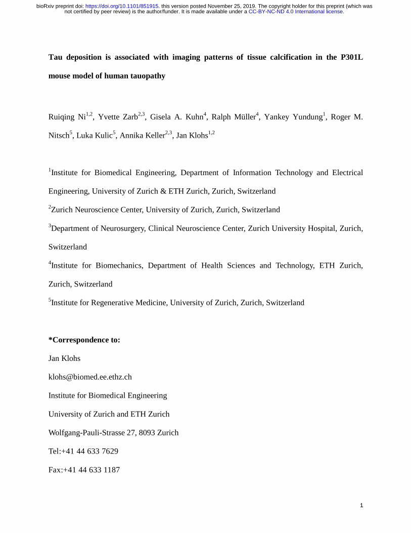
Tau deposition is associated with imaging patterns of tissue calcification in the P301L
mouse model of human tauopathy
Ruiqing Ni1,2, Yvette Zarb2,3, Gisela A. Kuhn4, Ralph Müller4, Yankey Yundung1, Roger M.
Nitsch5, Luka Kulic5, Annika Keller2,3, Jan Klohs1,2
1Institute for Biomedical Engineering, Department of Information Technology and Electrical
Engineering, University of Zurich & ETH Zurich, Zurich, Switzerland
2Zurich Neuroscience Center, University of Zurich, Zurich, Switzerland
3Department of Neurosurgery, Clinical Neuroscience Center, Zurich University Hospital, Zurich,
Switzerland
4Institute for Biomechanics, Department of Health Sciences and Technology, ETH Zurich,
Zurich, Switzerland
5Institute for Regenerative Medicine, University of Zurich, Zurich, Switzerland
*Correspondence to:
Jan Klohs
Institute for Biomedical Engineering
University of Zurich and ETH Zurich
Wolfgang-Pauli-Strasse 27, 8093 Zurich
Tel:+41 44 633 7629
Fax:+41 44 633 1187
.CC-BY-NC-ND 4.0 International licensenot certified by peer review) is the author/funder. It is made available under aThe copyright holder for this preprint (which wasthis version posted November 25, 2019. . https://doi.org/10.1101/851915doi: bioRxiv preprint

2
Abstract:
Brain calcification is associated with several neurodegenerative proteinopathies. Here, we report
a new phenotype of intracranial calcification in transgenic P301L mice overexpressing 4 repeat
tau. P301L mice (Thy1.2) of 3, 5, 9 and 18-25 months-of-age and age-matched non-transgenic
littermates were assessed using in vivo/ex vivo magnetic resonance imaging (MRI) with a
gradient recalled echo sequence and micro computed tomography (μCT). Susceptibility weighted
images computed from the gradient recalled echo data revealed regional hypointensities in the
hippocampus, cortex, caudate nucleus and thalamus of P301L mice, which in corresponding
phase images indicated diamagnetic lesions. Concomitantly, µCT detected hyperdense lesions.
Occurrence of diamagnetic susceptibility lesions in the hippocampus, increased with age.
Immunochemical staining of brain sections revealed bone protein-positive deposits. Furthermore,
intra-neuronal and vessel-associated protein-containing nodules co-localized with
phosphorylated-tau (AT8 and AT100) in the hippocampus. Protein-containing nodules were
detected also in the thalamus in the absence of phosphorylated-tau deposition. In contrast,
osteocalcin-containing nodules were vessel-associated, indicating ossified vessels, in the
thalamus in absence of phosphorylated-tau. In summary, MRI and µCT demonstrated imaging
pattern of intracranial calcification, concomitant with immunohistochemical evidence of
formation of protein deposits containing bone proteins along with phosphorylated-tau in the
P301L mouse model of human tauopathy. The P301L mouse model may thus serve as a future
model to study the pathogenesis of brain calcifications in tauopathies.
.CC-BY-NC-ND 4.0 International licensenot certified by peer review) is the author/funder. It is made available under aThe copyright holder for this preprint (which wasthis version posted November 25, 2019. . https://doi.org/10.1101/851915doi: bioRxiv preprint

3
Keywords: Alzheimer’s disease; calcification; computer tomography; frontal temporal lobe
dementia; magnetic resonance imaging; tauopathy; transgenic mouse model;
Introduction
Brain calcifications are detected in approximately 20% of aged individuals undergoing
diagnostic neuroimaging, particularly in the pineal gland and choroid plexus [9, 15, 20, 29].
Higher incidences of brain calcifications are found in patients with neurodegenerative diseases
such as Alzheimer’s disease (AD) [37, 38], cerebral amyloid angiopathy [4], frontotemporal
dementia (FTD) [36], Parkinson’s disease and Down syndrome [68]. Calcified deposits in the
human brain are also found distributed throughout the hippocampus and basal ganglia, and some
are associated with cerebral vessels and cerebral amyloid angiopathy [7, 9, 15, 20, 57]. In
patients with diffuse neurofibrillary tangles with calcification, pallidal calcification is the most
prominent feature and is accompanied by frontotemporal atrophy, neurofibrillary tangle (NFT)
deposition throughout the neocortex, without the occurrence of β-amyloid plaques [30].
Symmetrical calcification of the basal ganglia is a key diagnostic criterion in patients with
primary familial brain calcification [70]. The etiology of brain calcification is poorly understood,
despite high prevalence of calcification in neurodegenerative disorders.
The discovery of tau mutations has facilitated the generation of several mouse models of human
tauopathy e.g. P301S and P301L lines (transgenic for a human 4 repeat tau isoform) [6, 52-54],
which have become important tools to study the mechanisms of abnormal tau aggregation and
deposition in FTD (4 repeat tau) [61] and AD (3 and 4 repeat tau) [25, 33, 41]. In the P301L
.CC-BY-NC-ND 4.0 International licensenot certified by peer review) is the author/funder. It is made available under aThe copyright holder for this preprint (which wasthis version posted November 25, 2019. . https://doi.org/10.1101/851915doi: bioRxiv preprint

4
(Thy 1.2, pR5 line) [11, 18], P301L (CaMKIIa) [23, 43, 62] and P301L (tetO) [14] mouse
models, tau deposits begin forming before 3 months-of-age in neurons in the entorhinal cortex,
hippocampus and later in the cortex, and amygdala; with neuroinflammation and impaired
memory functions in hippocampus- and amygdala-dependent tasks manifesting at a later stage
[49, 50, 67]. Brain atrophy and white matter changes indicating neurodegeneration were reported
in P301L (CaMKIIa) mouse line around 9 months-of-age [16, 23, 43, 62], which was driven by
factors additional to human tau overexpression. However, unlike in human patients with
tauopathies, brain calcifications have not been reported in transgenic mouse models of human
disease.
Here, we report an early occurrence of intracranial calcifications in the P301L (Thy1.2) mouse
model of tauopathy. Evidence is provided by high-field magnetic resonance imaging (MRI),
micro computed tomography (μCT) which correspond to protein-containing nodules upon
immunohistochemical staining. Parenchymal and vascular calcifications increase with age and
are associated with intra-neuronal phosphorylated-tau deposition in the hippocampus. The P301L
mouse line may be a suitable animal model to study the role of brain calcification in human
tauopathy.
Materials and methods
Animals
Homozygous mice, transgenic for a human four repeat isoform with the P301L under Thy1.2
promoter (C57B6.Dg background) [19] and non-transgenic littermates were used (see groups in
Supplementary Table 1). Animals were housed in individually ventilated cages inside a
.CC-BY-NC-ND 4.0 International licensenot certified by peer review) is the author/funder. It is made available under aThe copyright holder for this preprint (which wasthis version posted November 25, 2019. . https://doi.org/10.1101/851915doi: bioRxiv preprint

5
temperature-controlled room, under a 12-hour dark/light cycle. Pelleted food (3437PXL15,
CARGILL) and water was provided ad-libitum. All experiments were performed in accordance
with the Swiss Federal Act on Animal Protection and approved by the Cantonal Veterinary Office
Zurich (permit number: ZH082/18).
Magnetic resonance imaging
MRI was performed as described previously [27, 44]. In vivo MRI was performed at 7T for
detecting calcifications and volumetry. A comparison between the two field strengths was
performed in order to determine if using a lower magnetic field at 7T (feasible in a clinical
setting), could detect the diamagnetic signal from calcification. In vivo MRI was completed on a
Bruker Biospec 70/40 (Bruker Biospin GmbH, Ettlingen, Germany) small animal MR system
equipped with an actively shielded gradient set of 760 mT/m and 80 μs rise time and operated by
a Paravision 6.0.1 software platform (Bruker Biospin GmbH, Ettlingen, Germany). Mice were
anesthetized with an initial dose of 4 % isoflurane in oxygen/air (200:800 ml/min) and
maintained at 1.5 % isoflurane in oxygen/air (100:400 ml/min). Body temperature was monitored
with a rectal temperature probe (MLT 415, AD Instruments, Spechbach, Germany) and kept at
36.5 ± 0.5 °C on a water-heated holder (Bruker BioSpin AG, Fällanden, Switzerland). For
susceptibility weighted imaging (SWI), a two-dimensional flow compensated gradient-recalled
echo (FLASH) sequence was applied with the following parameters: field-of-view = 20×20 mm;
image size = 256×256 mm, slice thicknesss = 0.8 mm, resulting in a resolution of 78×78 µm,
number of slices = 20. One echo with an echo time = 18 ms; repetition time = 698 ms; flip
angle= 30 °; and number of averages = 30 within an acquisition scan time of 1 h 29 min 2 s was
recorded. Global 1st-order shimming followed by fieldmap-based local shimming on the mouse
.CC-BY-NC-ND 4.0 International licensenot certified by peer review) is the author/funder. It is made available under aThe copyright holder for this preprint (which wasthis version posted November 25, 2019. . https://doi.org/10.1101/851915doi: bioRxiv preprint
6
brain was performed using the automated MAPshim routine, with an ellipsoid reference volume
covering the whole cerebrum.
To compare the image quality of data acquired in vivo at 7T, ex vivo MRI was done on a Bruker
Biospec 94/30 (Bruker Biospin GmbH, Ettlingen, Germany) small animal MR system. The
system was equipped with a cryogenic 2×2 radiofrequency surface coil probe (Bruker BioSpin
AG, Fällanden, Switzerland). After in vivo MRI, mice were perfused under
ketamine/xylazine/acepromazine maleate anesthesia (75/10/2 mg/kg body weight, i.p. bolus
injection) with 0.1 M PBS (pH 7.4) and decapitated. The mouse heads were post-fixed in 4 %
paraformaldehyde in 0.1 M PBS (pH 7.4) for 6 days and stored in 0.1 M PBS (pH 7.4) at 4 °C
afterwards. The heads were then placed in a 15 ml centrifuge tube filled with perfluoropolyether
(Fomblin Y, LVAC 16/6, average molecular weight 2700, Sigma-Aldrich, U.S.A.). Samples were
measured at room temperature. The brains with skull were scanned to detect calcification using
SWI sequence. A 3D gradient-recalled echo SWI sequence was recorded with the following
parameters: field-of-view = 15×12×15 mm; image size = 248×200×36 mm, resulting in a spatial
resolution of 60×60×417 µm. One echo with an echo time = 12 ms; repetition time = 250 ms;
flip angle= 15 °; number of averages = 4 within an acquisition scan time of 1 h 59 min 24 s was
recorded. To reduce field inhomogeneities, global 1st-order followed by fieldmap-based local
shimming on the mouse brain was performed. Phase maps and SW images were generated using
Paravision software 6.0.1 as described previously [27].
SW images were compared with their phase image counterparts to ensure that the signals were
due to a diamagnetic signal (i.e. presence of calcifications). The numbers of suspected calcified
.CC-BY-NC-ND 4.0 International licensenot certified by peer review) is the author/funder. It is made available under aThe copyright holder for this preprint (which wasthis version posted November 25, 2019. . https://doi.org/10.1101/851915doi: bioRxiv preprint

7
spots in a panel of an anatomical region (hippocampus, thalamus, caudate nucleus, midbrain and
cortex etc.) were quantified using the ex vivo datasets acquired at 9.4T. The Allen mouse brain
atlas was used for anatomical reference [24].
µCT
After the ex vivo MRI, the mouse heads were scanned in 0.1 M PBS (pH 7.4) in a microCT 40
(Scanco Medical AG, Brüttisellen, Switzerland) operated at 45 kVp, 177 µA intensity, an
integration of 200 ms and two-fold frame averaging. From 1000 projection images, 3D datasets
with isotropic voxels of 8µm were reconstructed and converted to DICOM format using the
scanner software. DICOM files were exported and analyzed using ITK SNAP [73].
Histochemistry & Immunohistochemistry
The 4 % paraformaldehyde fixed mouse brains were cut sagittally into two hemispheres. One
hemisphere was cut into 60 μm coronal sections using a vibratome (Leica VT1000S, Germany)
for fluorescence immunohistochemistry. The protocol used for fluorescence
immunohistochemistry was described previously [43, 46, 75]. Primary antibodies used for
immunofluorescence staining are listed in Supplementary table 2. All fluorescently-labelled
(Alexa 488, Cy3, DyLight 649) secondary antibodies (suitable for multiple labelling) were
hosted in donkey (anti-rabbit, anti-rat, anti-mouse and anti-goat, Jackson Immunoresearch, UK).
Brain sections were pre-treated with mouse-on-mouse kit (Vector Laboratories, USA) to quench
the endogenous IgG and subsequently incubated with primary and secondary antibodies.
Immunohistochemistry stainings were imaged using a confocal microscope (Leica SP5; 40 ×
numerical aperture: 1.25; 63 × numerical aperture: 1.4). Images were analyzed using the image-
.CC-BY-NC-ND 4.0 International licensenot certified by peer review) is the author/funder. It is made available under aThe copyright holder for this preprint (which wasthis version posted November 25, 2019. . https://doi.org/10.1101/851915doi: bioRxiv preprint

8
processing software Imaris 8.4.1 (Bitplane, USA) and Illustrator CS 6 (Adobe, USA).
For histochemistry and non-fluorescent immunostaining, the other brain hemisphere was
embedded in paraffin following routine procedures and cut into 2 μm thick sections. Sections
were stained using Hematoxylin & Eosin, Alcian blue, Periodic acid–Schiff or Prussian blue
using a standard protocol. The sections were deparaffinized and rehydrated before
immunostaining. For glial fibril acidic protein (GFAP) staining, sections were incubated with
rabbit anti-GFAP (DakoCytomation A/S, Denmark, #20334), followed by an incubation with
HRP-conjugated goat anti-rabbit (Jackson Immunoresearch, USA). For ionized calcium binding
adaptor molecule 1 (Iba1) staining, antigen retrieval was performed using hot citrate buffer (0.01
M; pH 6), followed by an incubation with rabbit anti-Iba1 (WAKO, Japan; 1:2500) and
subsequently a biotinylated secondary antibody (Vector laboratories, USA). Visualization ensued
after using the ABC complex solution (Vector laboratories, USA), 3,3′-Diaminobenzidine (DAB;
Sigma-Aldrich, Switzerland) and hydrogen peroxide (Sigma-Aldrich, Switzerland).
Counterstaining was performed using Hematoxylin. Stained paraffin sections were scanned using
a NanoZoomer HT (Hamamatsu Photonics, Japan) using a 40× objective. Images were analyzed
using Digital Image Hub software (SlidePath) and Adobe Illustrator CS6 (Adobe, USA).
Hematoxylin & Eosin stained whole sagittal mouse brain slices were imaged at 20×
magnification using Pannoramic 250 (3D HISTECH, Hungary). The images were analyzed using
CaseViewer (3D HISTECH, Hungary) and ImageJ (NIH, USA).
Statistics
Statistical analysis was performed using GraphPad Prism 7.0 (GraphPad Software, U.S.A).
.CC-BY-NC-ND 4.0 International licensenot certified by peer review) is the author/funder. It is made available under aThe copyright holder for this preprint (which wasthis version posted November 25, 2019. . https://doi.org/10.1101/851915doi: bioRxiv preprint
9
D’Agostino & Person normality test was used for assessing the normal distribution of the data.
One-way ANOVA with Turkey’s post-hoc analysis was used for group comparison. The
difference between groups was considered significant (*) at p value < 0.05. All error bars in the
figures are expressed as standard deviation.
Results
Phase imaging detects the presence of diamagnetic lesions in P301L mouse brain
Gradient recalled echo data of P301L mice and non-transgenic littermates were collected (Fig. 1-
3). We observed hypointensities in the SW images (Fig. 1-3). Corresponding phase images
showed positive phase shifts (hyperintensities) in corresponding locations, indicating the
diamagnetic nature of lesions (Fig. 1-3). The magnetic susceptibility [55] and appearance of the
lesions indicated the presence of calcified deposits. Blood degradation products such as
hemosiderin are paramagnetic and would induce opposite signal shifts. Hypointensities in the
SW images on reconstructed data from in vivo MRI at 7T (Fig. 1) correspond well to that from
ex vivo MRI at 9.4T (Fig. 3). However, boundaries of lesions were sharper in the 9.4T compared
to 7T images due to the difference in field strengths and scan parameters.
Hypointensities/positive phase shifts in P301L mice were detected in all age groups by using in
vivo and ex vivo SWI/phase image, prominently in the hippocampus, but also in the thalamus,
caudate nucleus, choroid plexus and midbrain (Fig. 1, 2, 3). In aged non-transgenic littermates
such lesions were only observed in the choroid plexus (e.g. positive phase shifts in the 4th
ventricle, which appeared as a cohesive structure) (Fig. 2a-c). In the other brain regions
hyperintenities/positive phase shifts on SW/phase images appeared as ovoid (Fig. 3b, c, f, g),
.CC-BY-NC-ND 4.0 International licensenot certified by peer review) is the author/funder. It is made available under aThe copyright holder for this preprint (which wasthis version posted November 25, 2019. . https://doi.org/10.1101/851915doi: bioRxiv preprint
10
elongated (Fig. 3e, i) lesions, and nests (Fig. 3d, h). D’Agostino & Person normality test results
in p = 0.0648, indicating passing the normality test. The number of SW/phase diamagnetic
deposits increased with age in the hippocampus, but not the other brain structures (18 months-old
vs 3 months-old, p <0.0001; vs 5 months-old, p <0.0001; and vs 9 months-old, p <0.0001, Fig.
3j). No statistically significant difference was observed between male and female mice within
age groups (Fig. 3k).
µCT detects hyperdense lesions in the brain of P301L mice
To further assess cerebral lesions in the brain of P301L mice, ex vivo μCT scans were performed
on the same brain samples. μCT images showed hyperdense lesions in 5, 9- and 18+ months-old
P301L mice. The higher X-ray density in these areas indicates the presence of material of higher
atomic number than soft tissue [56]. Hyperdense lesions were seen in regions that correspond to
the hippocampus, deep brain regions, cerebellum, and cortex when compared to SW and phase
MR images in the same mice (Fig. 4a-d). Hyperdense structures in the choroid plexus of the
third ventricle were observed in the μCT images of P301L mouse brains and also in aged
controls. However, fewer lesions were observed using μCT compared to MRI. Nevertheless, this
adds further evidence for the presence of intracranial calcification in P301L mice.
Vascular bone protein-containing nodules in the thalamus of P301L mice
As a next step, we used histochemistry and immunohistochemistry to investigate the nature of
the imaging lesions in different brain regions of P301L mice. To this end, we used
immunohistochemical staining with antibodies against bone proteins deposited in brain
calcifications [15, 75]. In the thalamus, osteocalcin- and osteopontin-positive nodules were
.CC-BY-NC-ND 4.0 International licensenot certified by peer review) is the author/funder. It is made available under aThe copyright holder for this preprint (which wasthis version posted November 25, 2019. . https://doi.org/10.1101/851915doi: bioRxiv preprint

11
associated with blood vessels, in the absence of phosphorylated-tau (AT8 and AT100) staining
(Fig. 5a-h). Interestingly, similar to previous reports [42, 74], we detect amyloid-precursor
protein (APP), a marker for damaged neurons [58], deposition in vascular nodules containing
bone proteins (Fig. 5i-l). These nodules were visualized using hematoxylin and eosin staining, a
standard histological stain, indicating the basophilic nature of these structures (Fig. 5m). In
addition, vascular calcification in the thalamus elicits a strong glial reactivity (Fig. 5n, o),
reminiscent to changes described in a mouse models of primary familial brain calcification [26,
42, 75].
We performed histology on sagittal brain sections of P301L mouse and non-transgenic littermate
brains to verify the presence of other neuropathology in the brain of P301L mice. Alcian blue,
Periodic acid–Schiff, and Hematoxylin & Eosin staining showed no pathological abnormalities
or infarction in the mouse brain P301L mice at 18 month-of-age (SFig. 1). Furthermore, Prussian
blue staining demonstrated that the imaging pattern in the P301L mouse brains were not cerebral
microbleeds (SFig. 1c, d).
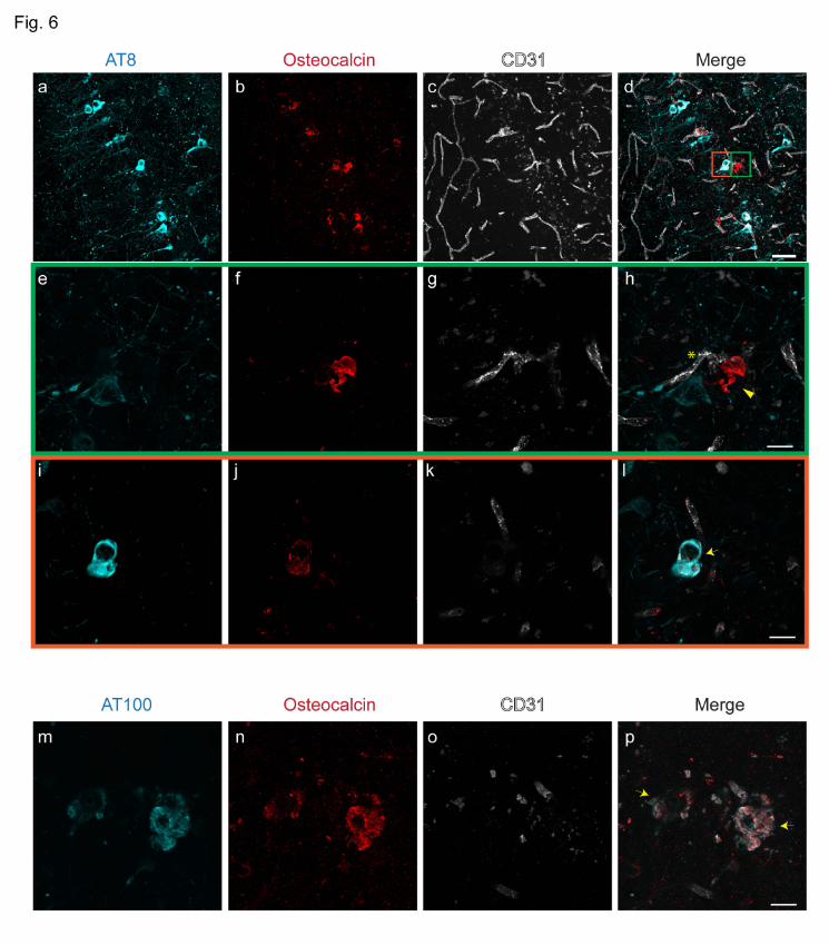
Vascular and intracellular bone protein-containing nodules s in the hippocampus
The hippocampus of P301L mice was found to be a prominent site of imaging pattern indicative
of tissue calcification (Fig. 3). Immunohistochemistry using antibodies against bone proteins
confirmed their presence in deposits in the hippocampus (Fig. 6a-p), which were vascular (Fig.
6e-h) and parenchymal (Fig. 6i-l). In contrast to the thalamus, we observed in the hippocampus
of P301L mice intracellular staining of osteocalcin which was co-localizing with phosphorylated-
tau detected using antibodies AT8 and AT100 (Fig. 6i-l).
.CC-BY-NC-ND 4.0 International licensenot certified by peer review) is the author/funder. It is made available under aThe copyright holder for this preprint (which wasthis version posted November 25, 2019. . https://doi.org/10.1101/851915doi: bioRxiv preprint

12
Discussion
In this study, we observed imaging pattern on high-resolution μCT and gradient recalled echo
MRI, indicative of intracranial calcification, in brains of P301L mice. Immunohistochemistry
revealed that these bone protein-containing lesions were either vessel-associated or intracellular
and co-localizing with phosphorylated-tau.
CT is considered the non-invasive gold standard for the identification of intracranial calcification
[3], with hyperdensity threshold >100 Hounsfield units [5]. In comparison, in conventional T1-,
T2-weighted spin echo MR images calcifications appear with various signal intensities, which
makes differentiation of calcified tissue from other sources of focal contrast (hemorrhagic lesions
or infarctions) difficult. On the other hand, SW and phase imaging, two gradient recalled echo
MRI techniques, allow distinguishing tissue calcification from cerebral microbleeds based on the
magnetic susceptibility of the lesions [55]. In our study, all hypointense inclusions on SW images
of P301L mouse brain were diamagnetic, indicating the presence of intracranial calcifications
(Fig. 1-3). Negativity for Prussian blue staining indicated the absence of cerebral microbleeds
and other iron deposition in the brain of P301L mice (SFig. 1c, d). Immunohistochemistry
revealed that lesions are positive for bone proteins, thus indicating that the characteristic
radiological pattern corresponds to markers of calcified tissue (Fig. 5, 6). Studies using clinical
brain imaging data suggest that the diagnostic accuracy of SWI outperformed conventional MRI
and CT in detecting brain calcifications [1, 32, 71, 72, 76], although the diagnostic accuracy of
different methods might depend on disease indication, MRI parameters and field strengths used.
.CC-BY-NC-ND 4.0 International licensenot certified by peer review) is the author/funder. It is made available under aThe copyright holder for this preprint (which wasthis version posted November 25, 2019. . https://doi.org/10.1101/851915doi: bioRxiv preprint

13
In non-transgenic littermates, imaging abnormalities were found confined to the choroid plexus.
While this has been reported to occur in aged individuals [20] it has so far not been reported as a
phenotype in aged mice. In P301L mice we observed high inter-individual variations in
hypointensities on SW images in all anatomical regions (Fig. 2g), similarly to previous studies in
patients with familial brain calcification and mouse models thereof [26, 75].
The etiology of most forms of brain calcifications are unknown, but they may occur under
several pathophysiological conditions, including inflammation, metabolic, infectious and genetic
syndromes and after exposure to toxins or radiotherapy [10, 12, 35]. Loss-of-function mutations
in genes involved in familial forms of diseases presenting with brain calcification have been
associated with disturbance in phosphate homeostasis and the dysfunction of the blood-brain
barrier [34]. This was supported by the finding of mutations in tight junction components in
other genetic diseases with intracranial calcifications [40, 45]. Moreover, studies using
hypomorphs of platelet derived growth factor subunit B suggested a connection between blood-
brain barrier impairment and brain calcification [26], however, later studies did not find a causal
evidence [65, 66]. Interestingly, blood-brain barrier breakdown has been reported in P301L
tauopathy mice, with erythrocyte and leukocyte infiltration occurring before accumulation of
hyperphosphorylated tau [4], thus it would be of interest for future studies to investigate if blood-
brain barrier impairment plays a role in the manifestation of brain calcification in this setting.
The genetic cause of basal ganglia calcification with dementia and bone cysts has been linked
with triggering receptor expressed on myeloid cells 2 (TREM2) mutation [28], and TYRO
protein tyrosine kinase binding protein (TYROBP, formerly DAP12) [48] on chromosome 19.
.CC-BY-NC-ND 4.0 International licensenot certified by peer review) is the author/funder. It is made available under aThe copyright holder for this preprint (which wasthis version posted November 25, 2019. . https://doi.org/10.1101/851915doi: bioRxiv preprint

14
Mutations in TREM2 presenting as a FTD-like syndrome without bone involvement were later
also reported [8, 17, 21, 22]. TREM2 deficiency exacerbates tau pathology through dysregulated
kinase signaling in the tauopathy mouse model [2], suggesting microglial activation may also
play a role in the tau pathology. We observed strong glial reactivity towards calcified nodules in
the thalamus of the P301L mice (Fig. 5n, o). Reactive astrocytes and microglia surrounded
calcified deposits and it will be interesting to study whether glia cells control the progression
of the pathology as in hereditary brain calcification [74]. Further studies will need to elucidate
if these calcifications in P301L are also TREM2 dependent and if there will be differences in
susceptibility between the thalamic and hippocampal ones.
Intracranial calcification may also be linked to tauopathy. Studies demonstrated hippocampal
calcification in the brain of patients with AD and FTD using CT and histopathology [51]. This is
in line with our study where hypointensities were most abundant in the hippocampus, increasing
in an age-related manner (Fig. 3j). Furthermore, we found co-localization of tau (AT8, AT100)
with osteocalcin in hippocampal neurons, an earliest brain region affected in AD and FTD (Fig.
6d, l). In P301L mice, tau accumulation is prominent in the hippocampus, amygdala, and cortical
regions, but sparse in the brain stem, thalamus, and caudate nucleus [18, 69]. The tau distribution
was not matched regionally with the hypointensity pattern seen on MR imaging at whole brain
level (Fig 1, 2, 3). The susceptibility to develop calcification may thus depend on a number of
other factors in addition to human tau overexpression, such as the biochemical, and genetic
properties. The individual and combined contribution of these factors to the formation of brain
calcification needs further research [39]. Given the non-invasive nature of the imaging read-outs,
longitudinal studies would be informative with respect to individual changes in calcium load and
.CC-BY-NC-ND 4.0 International licensenot certified by peer review) is the author/funder. It is made available under aThe copyright holder for this preprint (which wasthis version posted November 25, 2019. . https://doi.org/10.1101/851915doi: bioRxiv preprint

15
can be combined with other read-outs (e.g. perfusion measurement, elastography etc.).
We observed APP and osteopontin accumulation in the thalamic nodules in P301L mice. APP is a
marker for damaged neurons [58] and osteopontin is a bone protein upregulated in damaged
brain [59]. Both APP [58] and osteopontin [15, 75] are reported in the extracellular matrix of
calcified tissue. The presence of APP could be an indication of damaged neurons in the area [42].
Here, we observed that they accumulate in the thalamic calcifications in P301L mice similar to
other reports about brain calcifications, further supporting that these nodules are indeed calcified.
As the antibody for APP binds to amino acids 85-99 of the C99 fragment of APP, which might
bind to amyloid-beta deposits and APP C-terminal fragment C99 located in dystrophic neurites.
Once manifested, brain calcification can affect brain function by evoking expression of
neurotoxic astrocyte markers, interfering with neuronal circuitry and/or glucose metabolism [32,
60, 75]. However, the functional contribution of brain calcification to neuropsychiatric
symptoms in various brain disorders is still debated [13, 47]. Patients with primary familial brain
calcification often present with impaired movement (parkinsonism and dystonia), but also
cognitive impairment and psychiatric manifestations, including schizophrenia-like symptoms,
mood disorders, or obsessive–compulsive disorder [13]. Patients with diffuse neurofibrillary
tangles with calcification including early but progressive memory and verbal disturbances,
followed by psychiatric symptoms. While the P301L strain shows impaired memory functions in
hippocampus- and amygdala-dependent tasks [49, 50], future studies need to disentangle the
potential contributions of tauopathy-related brain calcification to functional deficits.
.CC-BY-NC-ND 4.0 International licensenot certified by peer review) is the author/funder. It is made available under aThe copyright holder for this preprint (which wasthis version posted November 25, 2019. . https://doi.org/10.1101/851915doi: bioRxiv preprint

16
Conclusions
We described a new brain phenotype of P301L mice, characterized by imaging patterns of
intracranial calcification. The study further suggests a potential link between tau deposition and
tissue calcification, where the underlying pathophysiology and functional consequences need to
be further investigated. The P301L mouse strain therefore may be a suitable model to study the
pathogenesis and pathophysiology of brain calcifications in FTD and AD, and other tauopathies.
Availability of data and material
The datasets generated and/or analyzed during the current study are available in the repository
(DOI: 10.5281/zenodo.3518986).
Declaration of conflict of interests
No competing interests declared.
Author Contributions
RN, AK, JK conceived and designed the study; RN, YZ, YY, GK performed the experiments; RN,
YZ, GK, LK, AK and JK interpreted the results; RN and JK wrote the manuscript; all coauthors
contributed constructively to the manuscript.
Acknowledgement
The authors acknowledge Prof. Daniel Razansky and Prof. Markus Rudin at the Institute for
Biomedical Engineering, ETH Zurich & University of Zurich for access to the infrastructure of
the animal imaging facility; Dr. Zsofia Kovacs, Dr. Mark-Aurel Augath at the Institute for
.CC-BY-NC-ND 4.0 International licensenot certified by peer review) is the author/funder. It is made available under aThe copyright holder for this preprint (which wasthis version posted November 25, 2019. . https://doi.org/10.1101/851915doi: bioRxiv preprint

17
Biomedical Engineering, ETH Zurich & University of Zurich, Daniel Schuppli at the Institute for
Regenerative Medicine, University of Zurich and Dr. Gabriella Bodizs, ScopeM, ETH Zurich for
technical support. Dr. Joanne Lim for language corrections.
Funding
JK received funding from the Swiss National Science Foundation (320030_179277), in the
framework of ERA-NET NEURON (32NE30_173678/1), the Synapsis Foundation and the
Vontobel foundation. RN received funding from the University of Zurich Forschungskredit (Nr.
FK-17-052), and Synapsis Foundation career development award (2017 CDA-03). AK received
funding from the Swiss National foundation (31003A_159514) and the Synapsis Foundation.
Figure legends
Figure 1. Susceptibility weighted/phase magnetic resonance imaging reveals characteristic
pattern in vivo in P301L mouse brain (a) Coronal sections from a mouse brain atlas [24] show
location of susceptibility weighted (SW) and phase images; (b) Representative in vivo SW and
(c) phase image at 7T showing hypointensities/positive phase shifts (red arrowheads) in the
thalamus of an 18 month-old P301L mouse and (d) choroid plexus of the third ventricle of a 5
month-old P301L mouse; (c, e) SW and corresponding phase image in the hippocampus of a 5
month-old P301L mouse.
Figure 2. Comparison of imaging findings in the choroid plexus of P301L and non-
transgenic mice (a) Representative ex vivo susceptibility weighted (SW) magnetic resonance
imaging at 9.4T and (b) corresponding phase image showing hyperintensities in the fourth
.CC-BY-NC-ND 4.0 International licensenot certified by peer review) is the author/funder. It is made available under aThe copyright holder for this preprint (which wasthis version posted November 25, 2019. . https://doi.org/10.1101/851915doi: bioRxiv preprint

18
ventricle choroid plexus of 5, 9 and 18 month-old P301L mouse (red arrowheads); (c)
Representative ex vivo SW images at 9.4T in the fourth ventricle choroid plexus of 9 month-old
and in 5 month-old non-transgenic littermates (NTL); (d) 3D mouse brain atlas from Allen
Institute with choroid plexus highlighted in green [24].
Figure 3. Magnetic resonance imaging reveals imaging pattern in ex vivo brain of P301L
mice (a) Coronal mouse brain atlas from Allen Institute (slices 46, 56, 76, 89) [24] showing
corresponding location on magnetic resonance images; (b-i): Representative ex vivo
susceptibility weighted (SW) and phase image at 9.4T showing (b, f) hypointensities/positive
phase shifts in the 5 month-old P301L mouse; (c, g) in the cortex of a 9 month-old P301L mouse;
(d, h) in the thalamus of a 18 month-old P301L mouse; (e, i) in the hippocampus of a 18 month-
old P301L mouse (red arrowheads) (j) Quantification of regional distribution of SW
hypointensities. 18+ month (n = 11) compared to 3 month (n = 4, p < 0.0001), 5 month (n = 11, p
< 0.0001), 9 month (n = 10, p < 0.0001); (k) Number of SWI hypointensities in male and female
P301L mice of all age groups; **p <0.01, *** p < 0.001 two-way ANOVA with Turkey’s post
hoc analysis.
Figure 4. Hyperdense lesion detected on micro computed tomography images of the brains
of P301L mice (a-d) Representative ex vivo micro computed tomography showing hyperdense
lesions in different brain regions (red arrowhead).
Figure 5. Characterization of vessel-associated nodules in the thalamic region of 18 month-
old P301L mice (a-l) Osteocalcin-positive (red) nodules (b, f, j) are associated with vessels (c, g,
.CC-BY-NC-ND 4.0 International licensenot certified by peer review) is the author/funder. It is made available under aThe copyright holder for this preprint (which wasthis version posted November 25, 2019. . https://doi.org/10.1101/851915doi: bioRxiv preprint

19
k, CD31; white) and do not stain with antibodies recognizing different phosphorylated residues
of tau, AT8 (a) and AT100 (e); Thalamic nodules are positive for amyloid-precursor protein (i,
APP, cyan) and osteopontin (j, red); (m) Hematoxylin and Eosin (HE) stain of thalamic nodules;
(n, o) Thalamic nodules elicit glial reactivity. Activated astrocytes (n) and microglia (o) surround
vascular nodules. Vessels are visualized using CD31 (c, g, k, white) and vessel adjacent to bone
protein containing nodule is marked using an asterix (d, h, l). Arrowheads (d, h, l) mark thalamic
nodules. Scale bars: 15 µm (a-l) and 50 µm (m-o).
Figure 6. Characterization of hippocampal inclusions in 18 month-old P301L mice (a-p)
Osteocalcin (b, f, j, red) staining in the hippocampus. (e-h, green inset) Extracellular osteocalcin-
positive (f, red) nodule is vessel-associated (g, CD31; white) but did not stain with the
phosphorylated-tau AT8 (e, cyan) antibody. (i-l, orange inset) Intracellular co-localization of
phosphorylated-tau AT8 (i, cyan) and osteocalcin (j, red) in a hippocampal neuron; (m-p) Co-
localization of osteocalcin (n, red) positivity with phosphorylated-tau AT100 (m, cyan) in the
hippocampus of P301L mice. Vessels are visualized using CD31 (c, g, k, o, white) and vessel
adjacent to nodule is marked using an asterix (h). Arrowheads (h) mark extracellular nodules and
arrows (l, p) mark intracellular co-localization of phosphorylated-tau stains and osteocalcin.
Scale bars: 50 µm (a-d) and 15 µm (e-p).
References
1 Adams LC, Boker SM, Bender YY, Diederichs G, Fallenberg EM, Wagner M et al (2017)
Diagnostic accuracy of susceptibility-weighted magnetic resonance imaging for the
evaluation of pineal gland calcification. PloS one 12: e0172764 Doi
.CC-BY-NC-ND 4.0 International licensenot certified by peer review) is the author/funder. It is made available under aThe copyright holder for this preprint (which wasthis version posted November 25, 2019. . https://doi.org/10.1101/851915doi: bioRxiv preprint
20
10.1371/journal.pone.0172764
2 Bemiller SM, McCray TJ, Allan K, Formica SV, Xu G, Wilson G et al (2017) TREM2
deficiency exacerbates tau pathology through dysregulated kinase signaling in a mouse
model of tauopathy. Mol Neurodegener 12: 74-74 Doi 10.1186/s13024-017-0216-6
3 Bergen D, Fagnou J, Sevick R (2007). In: Semmler MRaW (ed) MRT 3rd ed. edn.
Springer City, pp 310–347
4 Blair LJ, Frauen HD, Zhang B, Nordhues BA, Bijan S, Lin Y-C et al (2015) Tau depletion
prevents progressive blood-brain barrier damage in a mouse model of tauopathy. Acta
Neuropathol Commun 3: 8 Doi 10.1186/s40478-015-0186-2
5 Bockenheimer S (2002). In: Sartor K (ed) Diagnostic and Interventional Neuroradiology:
A Multimodal Approach. Thieme Medical, City, pp 160–169.
6 Brelstaff J, Tolkovsky AM, Ghetti B, Goedert M, Spillantini MG (2018) Living Neurons
with Tau Filaments Aberrantly Expose Phosphatidylserine and Are Phagocytosed by
Microglia. Cell Reports 24: 1939-1948.e1934 Doi 10.1016/j.celrep.2018.07.072
7 Bulk M, Moursel LG, Graaf LMvd, Veluw SJv, Greenberg SM, Duinen SGv et al (2018)
Cerebral Amyloid Angiopathy With Vascular Iron Accumulation and Calcification. Stroke
49: 2081-2087 Doi doi:10.1161/STROKEAHA.118.021872
8 Chouery E, Delague V, Bergougnoux A, Koussa S, Serre J-L, Mégarbané A (2008)
Mutations in TREM2 lead to pure early-onset dementia without bone cysts. Hum Mutat
29: E194-E204 Doi 10.1002/humu.20836
9 Deng H, Zheng W, Jankovic J (2015) Genetics and molecular biology of brain
calcification. Aging Res Rev 22: 20-38 Doi 10.1016/j.arr.2015.04.004
10 Flint J, Goldstein L (1992) Familial calcification of the basal ganglia: A case report and
.CC-BY-NC-ND 4.0 International licensenot certified by peer review) is the author/funder. It is made available under aThe copyright holder for this preprint (which wasthis version posted November 25, 2019. . https://doi.org/10.1101/851915doi: bioRxiv preprint
21
review of the literature. Pychol Med 22: 581-95 Doi 10.1017/s0033291700038046
11 Fontaine SN, Ingram A, Cloyd RA, Meier SE, Miller E, Lyons D et al (2017)
Identification of changes in neuronal function as a consequence of aging and tauopathic
neurodegeneration using a novel and sensitive magnetic resonance imaging approach.
Neurobiol Aging 56: 78-86 Doi 10.1016/j.neurobiolaging.2017.04.007
12 Forstl H, Burns A, Levy R, Cairns N (1994) Neuropathological correlates of psychotic
phenomena in confirmed Alzheimer's disease. Br J Psychiatry 165: 53-59 Doi
10.1192/bjp.165.1.53
13 Forstl H, Krumm B, Eden S, Kohlmeyer K (1992) Neurological disorders in 166 patients
with basal ganglia calcification: a statistical evaluation. J Neurol 239: 36-38 Doi
10.1007/bf00839209
14 Fu H, Rodriguez GA, Herman M, Emrani S, Nahmani E, Barrett G et al (2017) Tau
Pathology Induces Excitatory Neuron Loss, Grid Cell Dysfunction, and Spatial Memory
Deficits Reminiscent of Early Alzheimer's Disease. Neuron 93: 533-541.e5 Doi
10.1016/j.neuron.2016.12.023
15 Fujita D, Terada S, Ishizu H, Yokota O, Nakashima H, Ishihara T et al (2003)
Immunohistochemical examination on intracranial calcification in neurodegenerative
diseases. Acta Neuropathol 105: 259-264 Doi 10.1007/s00401-002-0640-7
16 Gamache J, Benzow K, Forster C, Kemper L, Hlynialuk C, Furrow E et al (2019) Factors
other than hTau overexpression that contribute to tauopathy-like phenotype in rTg4510
mice. Nat Commun 10: 2479 Doi 10.1038/s41467-019-10428-1
17 Giraldo M, Lopera F, Siniard AL, Corneveaux JJ, Schrauwen I, Carvajal J et al (2013)
Variants in triggering receptor expressed on myeloid cells 2 are associated with both
.CC-BY-NC-ND 4.0 International licensenot certified by peer review) is the author/funder. It is made available under aThe copyright holder for this preprint (which wasthis version posted November 25, 2019. . https://doi.org/10.1101/851915doi: bioRxiv preprint

22
behavioral variant frontotemporal lobar degeneration and Alzheimer's disease. Neurobiol
Aging 34: 2077.e11-8 Doi 10.1016/j.neurobiolaging.2013.02.016
18 Gotz J, Chen F, Barmettler R, Nitsch RM (2001) Tau filament formation in transgenic
mice expressing P301L tau. J Biol Chem 276: 529-534 Doi 10.1074/jbc.M006531200
19 Gotz J, Chen F, van Dorpe J, Nitsch RM (2001) Formation of neurofibrillary tangles in
P301l tau transgenic mice induced by Abeta 42 fibrils. Science 293: 1491-1495 Doi
10.1126/science.1062097
20 Grinberg LT, Thal DR (2010) Vascular pathology in the aged human brain. Acta
Neuropathol 119: 277-290 Doi 10.1007/s00401-010-0652-7
21 Guerreiro R, Bilgic B, Guven G, Brás J, Rohrer J, Lohmann E et al (2013) A novel
compound heterozygous mutation in TREM2 found in a Turkish frontotemporal
dementia-like family. Neurobiol Aging 34: 2890.e2891-2890.e2895 Doi
10.1016/j.neurobiolaging.2013.06.005
22 Guerreiro RJ, Lohmann E, Brás JM, Gibbs JR, Rohrer JD, Gurunlian N et al (2013)
Using Exome Sequencing to Reveal Mutations in TREM2 Presenting as a
Frontotemporal Dementia–like Syndrome Without Bone Involvement. JAMA Neurology
70: 78-84 Doi 10.1001/jamaneurol.2013.579
23 Ishikawa A, Tokunaga M, Maeda J, Minamihisamatsu T, Shimojo M, Takuwa H et al
(2018) In Vivo Visualization of Tau Accumulation, Microglial Activation, and Brain
Atrophy in a Mouse Model of Tauopathy rTg4510. J Alzheimers Dis 61: 1037-1052 Doi
10.3233/jad-170509
24 Jones AR, Overly CC, Sunkin SM (2009) The Allen Brain Atlas: 5 years and beyond. Nat
Rev Neurosci 10: 821-828 Doi 10.1038/nrn2722
.CC-BY-NC-ND 4.0 International licensenot certified by peer review) is the author/funder. It is made available under aThe copyright holder for this preprint (which wasthis version posted November 25, 2019. . https://doi.org/10.1101/851915doi: bioRxiv preprint

23
25 Josephs KA, Hodges JR, Snowden JS, Mackenzie IR, Neumann M, Mann DM et al
(2011) Neuropathological background of phenotypical variability in frontotemporal
dementia. Acta neuropathologica 122: 137-153 Doi 10.1007/s00401-011-0839-6
26 Keller A, Westenberger A, Sobrido MJ, Garcia-Murias M, Domingo A, Sears RL et al
(2013) Mutations in the gene encoding PDGF-B cause brain calcifications in humans and
mice. Nat Genet 45: 1077-1082 Doi 10.1038/ng.2723
27 Klohs J, Deistung A, Schweser F, Grandjean J, Dominietto M, Waschkies C et al (2011)
Detection of cerebral microbleeds with quantitative susceptibility mapping in the
ArcAbeta mouse model of cerebral amyloidosis. J Cereb Blood Flow Metab 31: 2282-
2292 Doi 10.1038/jcbfm.2011.118
28 Klunemann HH, Ridha BH, Magy L, Wherrett JR, Hemelsoet DM, Keen RW et al (2005)
The genetic causes of basal ganglia calcification, dementia, and bone cysts: DAP12 and
TREM2. Neurology 64: 1502-1507 Doi 10.1212/01.wnl.0000160304.00003.ca
29 Kockelkoren R, De Vis JB, Mali WP, Hendrikse J, de Jong PA, Rozemuller AM et al
(2016) Hippocampal Calcification on Computed Tomography in Relation to Cognitive
Decline in Memory Clinic Patients: A Case-Control Study. PloS one 11: e0167444 Doi
10.1371/journal.pone.0167444
30 Kosaka K (1994) Diffuse neurofibrillary tangles with calcification: a new presenile
dementia. J Neurol, Neurosurg & Psych 57: 594 Doi 10.1136/jnnp.57.5.594
31 Langlois NE, Grieve JH, Best PV (1995) Changes of diffuse neurofibrillary tangles with
calcification (DNTC) in a woman without evidence of dementia. J Neurol Neurosurg
Psychiatry 59: 103 Doi 10.1136/jnnp.59.1.103
32 Le Ber I, Marie RM, Chabot B, Lalevee C, Defer GL (2007) Neuropsychological and
.CC-BY-NC-ND 4.0 International licensenot certified by peer review) is the author/funder. It is made available under aThe copyright holder for this preprint (which wasthis version posted November 25, 2019. . https://doi.org/10.1101/851915doi: bioRxiv preprint

24
18FDG-PET studies in a family with idiopathic basal ganglia calcifications. J Neurol Sci
258: 115-122 Doi 10.1016/j.jns.2007.03.017
33 Lee VM, Goedert M, Trojanowski JQ (2001) Neurodegenerative tauopathies. Annu Rev
Neurosci 24: 1121-1159 Doi 10.1146/annurev.neuro.24.1.1121
34 Legati A, Giovannini D, Nicolas G, Lopez-Sanchez U, Quintans B, Oliveira JR et al
(2015) Mutations in XPR1 cause primary familial brain calcification associated with
altered phosphate export. Nat Genet 47: 579-581 Doi 10.1038/ng.3289
35 Lopez-Villegas D, Kulisevsky J, Deus J, Junque C, Pujol J, Guardia E et al (1996)
Neuropsychological alterations in patients with computed tomography-detected basal
ganglia calcification. Arch Neurol 53: 251-256 Doi
10.1001/archneur.1996.00550030061023
36 Lowe VJ, Curran G, Fang P, Liesinger AM, Josephs KA, Parisi JE et al (2016) An
autoradiographic evaluation of AV-1451 Tau PET in dementia. Acta Neuropathol
Commun 4: 58 Doi 10.1186/s40478-016-0315-6
37 Mahlberg R, Walther S, Kalus P, Bohner G, Haedel S, Reischies FM et al (2008) Pineal
calcification in Alzheimer's disease: an in vivo study using computed tomography.
Neurobiol Aging 29: 203-209 Doi 10.1016/j.neurobiolaging.2006.10.003
38 Mann DM (1988) Calcification of the basal ganglia in Down's syndrome and Alzheimer's
disease. Acta Neuropathol 76: 595-598 Doi 10.1007/bf00689598
39 Miklossy J, Mackenzie IR, Dorovini-Zis K, Calne DB, Wszolek ZK, Klegeris A et al
(2005) Severe vascular disturbance in a case of familial brain calcinosis. Acta
Neuropathol 109: 643-653 Doi 10.1007/s00401-005-1007-7
40 Mochida GH, Ganesh VS, Felie JM, Gleason D, Hill RS, Clapham KR et al (2010) A
.CC-BY-NC-ND 4.0 International licensenot certified by peer review) is the author/funder. It is made available under aThe copyright holder for this preprint (which wasthis version posted November 25, 2019. . https://doi.org/10.1101/851915doi: bioRxiv preprint

25
homozygous mutation in the tight-junction protein JAM3 causes hemorrhagic destruction
of the brain, subependymal calcification, and congenital cataracts. Am J Hum Genet 87:
882-889 Doi 10.1016/j.ajhg.2010.10.026
41 Murray ME, Graff-Radford NR, Ross OA, Petersen RC, Duara R, Dickson DW (2011)
Neuropathologically defined subtypes of Alzheimer's disease with distinct clinical
characteristics: a retrospective study. Lancet Neurol 10: 785-796 Doi 10.1016/S1474-
4422(11)70156-9
42 Nahar K, Lebouvier T, Andaloussi Mae M, Konzer A, Bergquist J, Zarb Y et al (2019)
Astrocyte-microglial association and matrix composition are common events in the
natural history of primary familial brain calcification. Brain Pathol: Doi
10.1111/bpa.12787
43 Ni R, Ji B, Ono M, Sahara N, Zhang MR, Aoki I, Nordberg A et al (2018) Comparative
in-vitro and in-vivo quantifications of pathological tau deposits and their association with
neurodegeneration in tauopathy mouse models. J Nucl Med 59: 960-966 Doi
10.2967/jnumed.117.201632
44 Ni R, Kindler DR, Waag R, Rouault M, Ravikumar P, Nitsch R et al (2019) fMRI Reveals
Mitigation of Cerebrovascular Dysfunction by Bradykinin Receptors 1 and 2 Inhibitor
Noscapine in a Mouse Model of Cerebral Amyloidosis. Front Aging Neurosci 11: 27-27
Doi 10.3389/fnagi.2019.00027
45 O'Driscoll MC, Daly SB, Urquhart JE, Black GC, Pilz DT, Brockmann K et al (2010)
Recessive mutations in the gene encoding the tight junction protein occludin cause band-
like calcification with simplified gyration and polymicrogyria. Am J Hum Genet 87: 354-
364 Doi 10.1016/j.ajhg.2010.07.012
.CC-BY-NC-ND 4.0 International licensenot certified by peer review) is the author/funder. It is made available under aThe copyright holder for this preprint (which wasthis version posted November 25, 2019. . https://doi.org/10.1101/851915doi: bioRxiv preprint

26
46 Ono M, Sahara N, Kumata K, Ji B, Ni R, Koga S et al (2017) Distinct binding of PET
ligands PBB3 and AV-1451 to tau fibril strains in neurodegenerative tauopathies. Brain
140: 764-780 Doi 10.1093/brain/aww339
47 Ostling S, Andreasson LA, Skoog I (2003) Basal ganglia calcification and psychotic
symptoms in the very old. Int J Geriatr Psychiatry 18: 983-987 Doi 10.1002/gps.997
48 Paloneva J, Kestilä M, Wu J, Salminen A, Böhling T, Ruotsalainen V et al (2000) Loss-
of-function mutations in TYROBP (DAP12) result in a presenile dementia with bone
cysts. Nat Genet 25: 357-361 Doi 10.1038/77153
49 Pennanen L, Welzl H, D'Adamo P, Nitsch RM, Gotz J (2004) Accelerated extinction of
conditioned taste aversion in P301L tau transgenic mice. Neurobiol Dis 15: 500-509 Doi
10.1016/j.nbd.2003.11.020
50 Pennanen L, Wolfer DP, Nitsch RM, Gotz J (2006) Impaired spatial reference memory
and increased exploratory behavior in P301L tau transgenic mice. Genes Brain Behav 5:
369-379 Doi 10.1111/j.1601-183X.2005.00165.x
51 Peters MEM, Kockelkoren R, de Brouwer EJM, Koek HL, Bleys RLAW, Mali WPTM et
al (2018) Histological validation of calcifications in the human hippocampus as seen on
computed tomography. PloS one 13: e0197073 Doi 10.1371/journal.pone.0197073
52 Ramsden M, Kotilinek L, Forster C, Paulson J, McGowan E, SantaCruz K et al (2005)
Age-dependent neurofibrillary tangle formation, neuron loss, and memory impairment in
a mouse model of human tauopathy (P301L). J Neurosci 25: 10637-10647 Doi
10.1523/jneurosci.3279-05.2005
53 Rösler TW, Tayaranian Marvian A, Brendel M, Nykänen N-P, Höllerhage M, Schwarz SC
et al (2019) Four-repeat tauopathies. Prog Neurobiol 180: 101644 Doi
.CC-BY-NC-ND 4.0 International licensenot certified by peer review) is the author/funder. It is made available under aThe copyright holder for this preprint (which wasthis version posted November 25, 2019. . https://doi.org/10.1101/851915doi: bioRxiv preprint

27
10.1016/j.pneurobio.2019.101644
54 SantaCruz K, Lewis J, Spires T, Paulson J, Kotilinek L, Ingelsson M et al (2005) Tau
Suppression in a Neurodegenerative Mouse Model Improves Memory Function. Science
309: 476-481 Doi 10.1126/science.1113694
55 Schenck JF (1996) The role of magnetic susceptibility in magnetic resonance imaging:
MRI magnetic compatibility of the first and second kinds. Med Phys 23: 815-850 Doi
10.1118/1.597854
56 Seibert JA, Boone JM (2005) X-ray imaging physics for nuclear medicine technologists.
Part 2: X-ray interactions and image formation. J Nucl Med Technol 33: 3-18
57 Sellal F, Wallon D, Martinez-Almoyna L, Marelli C, Dhar A, Oesterle H et al (2017) APP
Mutations in Cerebral Amyloid Angiopathy with or without Cortical Calcifications:
Report of Three Families and a Literature Review. J Alzheimers Dis 56: 37-46 Doi
10.3233/jad-160709
58 Shigematsu K, McGeer PL (1992) Accumulation of amyloid precursor protein in
damaged neuronal processes and microglia following intracerebral administration of
aluminum salts. Brain Res 593: 117-123 Doi 10.1016/0006-8993(92)91272-g
59 Shin T (2012) Osteopontin as a two-sided mediator in acute neuroinflammation in rat
models. Acta Histochem 114: 749-754 Doi 10.1016/j.acthis.2012.08.004
60 Sjögren M, Folkesson S, Blennow K, Tarkowski E (2004) Increased intrathecal
inflammatory activity in frontotemporal dementia: pathophysiological implications. J
Neurol, Neurosurg, and psychiatry 75: 1107-1111 Doi 10.1136/jnnp.2003.019422
61 Spillantini MG, Goedert M (2013) Tau pathology and neurodegeneration. Lancet Neurol
12: 609-622 Doi 10.1016/S1474-4422(13)70090-5
.CC-BY-NC-ND 4.0 International licensenot certified by peer review) is the author/funder. It is made available under aThe copyright holder for this preprint (which wasthis version posted November 25, 2019. . https://doi.org/10.1101/851915doi: bioRxiv preprint

28
62 Takeda S, Wegmann S, Cho H, DeVos SL, Commins C, Roe AD et al (2015) Neuronal
uptake and propagation of a rare phosphorylated high-molecular-weight tau derived from
Alzheimer’s disease brain. Nat Commun 6: 8490 Doi 10.1038/ncomms9490
63 Tsuchiya K, Nakayama H, Iritani S, Arai T, Niizato K, Haga C et al (2002) Distribution
of basal ganglia lesions in diffuse neurofibrillary tangles with calcification: a
clinicopathological study of five autopsy cases. Acta Neuropathol 103: 555-564 Doi
10.1007/s00401-001-0501-9
64 Ukai K, Kosaka K (2016) Diffuse neurofibrillary tangles with calcification (Kosaka-
Shibayama disease) in Japan. Psychiatry Clin Neurosci 70: 131-140 Doi
10.1111/pcn.12334
65 Vanlandewijck M, Lebouvier T, Andaloussi Mae M, Nahar K, Hornemann S, Kenkel D et
al (2015) Functional Characterization of Germline Mutations in PDGFB and PDGFRB in
Primary Familial Brain Calcification. PloS one 10: e0143407 Doi
10.1371/journal.pone.0143407
66 Wallingford MC, Chia JJ, Leaf EM, Borgeia S, Chavkin NW, Sawangmake C et al (2017)
SLC20A2 Deficiency in Mice Leads to Elevated Phosphate Levels in Cerbrospinal Fluid
and Glymphatic Pathway-Associated Arteriolar Calcification, and Recapitulates Human
Idiopathic Basal Ganglia Calcification. Brain Pathol 27: 64-76 Doi 10.1111/bpa.12362
67 Wang H, Li Y, Ryder JW, Hole JT, Ebert PJ, Airey DC et al (2018) Genome-wide
RNAseq study of the molecular mechanisms underlying microglia activation in response
to pathological tau perturbation in the rTg4510 tau transgenic animal model. Mol
Neurodegener 13: 65 Doi 10.1186/s13024-018-0296-y
68 Wegiel J, Kuchna I, Wisniewski T, de Leon MJ, Reisberg B, Pirttila T et al (2002)
.CC-BY-NC-ND 4.0 International licensenot certified by peer review) is the author/funder. It is made available under aThe copyright holder for this preprint (which wasthis version posted November 25, 2019. . https://doi.org/10.1101/851915doi: bioRxiv preprint

29
Vascular fibrosis and calcification in the hippocampus in aging, Alzheimer disease, and
Down syndrome. Acta Neuropathol 103: 333-343 Doi 10.1007/s00401-001-0471-y
69 Wegmann S, Bennett RE, Delorme L, Robbins AB, Hu M, McKenzie D et al (2019)
Experimental evidence for the age dependence of tau protein spread in the brain. Sci Adv
5: eaaw6404 Doi 10.1126/sciadv.aaw6404
70 Westenberger A, Balck A, Klein C (2019) Primary familial brain calcifications: genetic
and clinical update. Curr Opin Neurol 32: 571-578 Doi 10.1097/wco.0000000000000712
71 Wu Z, Mittal S, Kish K, Yu Y, Hu J, Haacke EM (2009) Identification of calcification
with MRI using susceptibility-weighted imaging: a case study. J Magn Reson Imaging
29: 177-182 Doi 10.1002/jmri.21617
72 Yamada N, Imakita S, Sakuma T, Takamiya M (1996) Intracranial calcification on
gradient-echo phase image: depiction of diamagnetic susceptibility. Radiology 198: 171-
178 Doi 10.1148/radiology.198.1.8539373
73 Yoo TS, Ackerman MJ, Lorensen WE, Schroeder W, Chalana V, Aylward S et al (2002)
Engineering and algorithm design for an image processing Api: a technical report on
ITK--the Insight Toolkit. Stud Health Technol Inform 85: 586-592
74 Zarb Y, Nassiri S, Utz SG, Schaffenrath J, Rushing EJ, Nilsson KPR et al (2019)
Microglia control small vessel calcification via TREM2. bioRxiv: 829341 Doi
10.1101/829341
75 Zarb Y, Weber-Stadlbauer U, Kirschenbaum D, Kindler DR, Richetto J, Keller D et al
(2019) Ossified blood vessels in primary familial brain calcification elicit a neurotoxic
astrocyte response. Brain 142: 885-902: Doi 10.1093/brain/awz032
76 Zulfiqar M, Dumrongpisutikul N, Intrapiromkul J, Yousem DM (2012) Detection of
.CC-BY-NC-ND 4.0 International licensenot certified by peer review) is the author/funder. It is made available under aThe copyright holder for this preprint (which wasthis version posted November 25, 2019. . https://doi.org/10.1101/851915doi: bioRxiv preprint

30
intratumoral calcification in oligodendrogliomas by susceptibility-weighted MR imaging.
AJNR Am J Neuroradiol 33: 858-864 Doi 10.3174/ajnr.A2862
.CC-BY-NC-ND 4.0 International licensenot certified by peer review) is the author/funder. It is made available under aThe copyright holder for this preprint (which wasthis version posted November 25, 2019. . https://doi.org/10.1101/851915doi: bioRxiv preprint

.CC-BY-NC-ND 4.0 International licensenot certified by peer review) is the author/funder. It is made available under aThe copyright holder for this preprint (which wasthis version posted November 25, 2019. . https://doi.org/10.1101/851915doi: bioRxiv preprint

.CC-BY-NC-ND 4.0 International licensenot certified by peer review) is the author/funder. It is made available under aThe copyright holder for this preprint (which wasthis version posted November 25, 2019. . https://doi.org/10.1101/851915doi: bioRxiv preprint

.CC-BY-NC-ND 4.0 International licensenot certified by peer review) is the author/funder. It is made available under aThe copyright holder for this preprint (which wasthis version posted November 25, 2019. . https://doi.org/10.1101/851915doi: bioRxiv preprint

.CC-BY-NC-ND 4.0 International licensenot certified by peer review) is the author/funder. It is made available under aThe copyright holder for this preprint (which wasthis version posted November 25, 2019. . https://doi.org/10.1101/851915doi: bioRxiv preprint

.CC-BY-NC-ND 4.0 International licensenot certified by peer review) is the author/funder. It is made available under aThe copyright holder for this preprint (which wasthis version posted November 25, 2019. . https://doi.org/10.1101/851915doi: bioRxiv preprint
.CC-BY-NC-ND 4.0 International licensenot certified by peer review) is the author/funder. It is made available under aThe copyright holder for this preprint (which wasthis version posted November 25, 2019. . https://doi.org/10.1101/851915doi: bioRxiv preprint

.CC-BY-NC-ND 4.0 International licensenot certified by peer review) is the author/funder. It is made available under aThe copyright holder for this preprint (which wasthis version posted November 25, 2019. . https://doi.org/10.1101/851915doi: bioRxiv preprint