Embed Size (px)
Citation preview

8/2/2019 Tau Deficiency Induces ism (1)
http://slidepdf.com/reader/full/tau-deficiency-induces-ism-1 1/6
l e t t e r s
nature medicine VOLUME 18 | NUMBER 2 | FEBRUARY 2012 291
Th micrtubul-assciat prtin tau has risk allls
fr bth Alzhimr’s isas an Parkinsn’s isas an
mutatins that caus brain gnrativ isass trmtaupathis1–4. Aggrgat tau frms nurfibrillary tangls in
ths pathlgis3,5, but littl is crtain abut th functin f
tau r its m f invlvmnt in pathgnsis. Nurnal irn
accumulatin has bn bsrv pathlgically in th crtx in
Alzhimr’s isas6,7, th substantia nigra (SN) in Parkinsn’s
isas8–11 an varius brain rgins in th taupathis11,12.
Hr w rprt that tau-knckut mic vlp ag-pnnt
brain atrphy, irn accumulatin an SN nurnal lss, with
cncmitant cgnitiv ficits an parkinsnism. Ths
changs ar prvnt by ral tratmnt with a mrat
irn chlatr, cliquinl. Amyli prcursr prtin (APP)
frrxias activity cupls with surfac frrprtin t xprt
irn, but its activity is inhibit in Alzhimr’s isas, thrby
causing nurnal irn accumulatin7. In primary nurnal
cultur, w fun lss f tau als causs irn rtntin, by
crasing surfac trafficking f APP. Slubl tau lvls
fall in affct brain rgins in Alzhimr’s isas an
taupathis13–15, an w fun a similar cras f slubl
tau in th SN in bth Parkinsn’s isas an th 1-mthyl-
4-phnyl-1,2,3,6-ttrahyrpyriin (MPTP) mus ml.
Ths ata suggst that th lss f slubl tau cul cntribut
t txic nurnal irn accumulatin in Alzhimr’s isas,
Parkinsn’s isas an taupathis, an that it can b
rscu pharmaclgically.
As itte is known about the status of tau in Parkinson’s disease, we
examined tau forms in postmortem sampes from peope affectedwith Parkinson’s disease and contros. Soube tau eves in the SN
were significanty ower in peope with Parkinson’s disease com-
pared to age-matched, non–Parkinson’s disease contros (44% ower;
P < 0.001; Fig. 1a and Supplementary Tables 1 and 2), which is simiar
to affected brain regions in Azheimer’s disease and tauopathies13–15.
The tau oss was independent of neurona oss, and l-DOPA treatment
did not infuence tau eves in mice (Supplementary Fig. 1).
There were no changes in insoube tau in the SN or soube tau of
neocortex and cerebeum in Parkinson’s disease, despite a widespreadincrease in hyperphosphoryated tau and a ower ratio of three-repeat
tau to four-repeat tau (Supplementary Table 2).
Eevated SN iron has been reported in cases of Parkinson’s
disease8–11, athough its roe has been debated. Soube tau oss and
high iron eves were distributed in simiar ocations in the brain
affecting SN (iron eves 39% higher than contro cases; P = 0.004;
Fig. 1a) but not neocortex or cerebeum (Supplementary Table 2).
Additionay, soube tau and iron eves in the SN correated in heathy
tissue (R2 = 0.3768; P = 0.0067; Fig. 1b) but not in Parkinson’s disease
(Supplementary Fig. 2). In the MPTP mouse mode, iron eevation
may be pathogenic, as the Parkinson’s phenotype is rescued by over-
expression of ferritin or treatment with iron cheators16,17. We found
a reciproca association between changes of SN tau and iron over 45 d
in MPTP-intoxicated mice (Fig. 1c and Supplementary Fig. 3). Nigra
tau eves decined significanty from the baseine by day 3 after MPTP
treatment (39% decine; P = 0.012) and remained suppressed through
day 45. At the same time, SN iron increased, reaching eves signifi-
canty above untreated ittermate contros from day 21 (21% higher;
P = 0.027; Fig. 1c).
We hypothesized that the gradua oss of SN iron homeostasis after
MPTP treatment might be caused by upstream tau oss and woud
cause SN neurodegeneration. To test this, we examined tau-knock-
out mice18, which are reported to show ony minor abnormaities
up to 7 months of age18–22. As tau-reated pathoogy is inked to
advanced aging, we hypothesized that tau-knockout mice might
deveop a degenerative phenotype with greater age. Indeed, athough
at 6 months of age the knockout mice performed indistinguishaby from wid-type (WT) mice, at 12 months tau-knockout mice showed
a severe decine in ocomotor functions, evidenced by significanty
greater time to turn (P < 0.001 compared to 12-month-od WT
mice; P < 0.001 compared to 6-month-od knockout mice) and to
compete the poe test (P = 0.002 compared to 12-month-od WT;
P = 0.003 compared to 6-month-od knockout), reduced atency to
Tau deficiency induces parkinsonism with dementia by impairing APP-mediated iron export
Peg Lei1,2, Sc Ay1,3, David I Fikelsei1,3, Lredaa Sperri2,4, Giuseppe D Ciccs1,2,4,David K Wrigh5, Bruce X W Wg1, Paul A Adlard1,2, Rber A Chery 1, Lih Q Lam1, Blaie R Rbers1,Iree Vliakis1, Gary F Ega5,6, Caria A McLea2, Rber Cappai2,4, James A Duce1,3 & Ashley I Bush1,2
1Mental Health Research Institute, The University o Melbourne, Victoria, Australia. 2Department o Pathology, The University o Melbourne, Victoria, Australia.3Centre or Neuroscience, The University o Melbourne, Victoria, Australia. 4Bio21 Molecular Science and Biotechnology Institute, The University o Melbourne,
Victoria, Australia. 5Florey Neuroscience Institutes, The University o Melbourne, Victoria, Australia. 6Present address: Monash Biomedical Imaging, Monash
University, Clayton, Victoria, Australia. Correspondence should be addressed to A.I.B. ([email protected])
Received 25 August 2011; accepted 29 November 2011; published online 29 January 2012; doi:10.1038/nm.2613

8/2/2019 Tau Deficiency Induces ism (1)
http://slidepdf.com/reader/full/tau-deficiency-induces-ism-1 2/6
l e t t e r s
292 VOLUME 18 | NUMBER 2 | FEBRUARY 2012 nature medicine
fa in the rotarod test (P < 0.001 compared to 12-month-od WT;
P = 0.005 compared to 6-month-od knockout), ower veocity
(P < 0.001 compared to 12-month-od WT; P = 0.002 compared to
6-month-od knockout), shorter distance of ocomotion (P < 0.001
compared to 12-month-od WT) and average distance of movement
(P < 0.001 compared to 12-month-od WT; P < 0.001 compared to6-month-od knockout) in the open fied test, and shorter distance
of movement in the Y maze (P = 0.046 compared to 12-month-od
WT; P = 0.05 compared to 6-month-od knockout) (Fig. 1d and
Supplementary Fig. 4). There were no further decines in these per-
formance readouts in 24-month-od tau-knockout mice.
Motor deficits in Parkinson’s disease are caused by nigra
dopaminergic degeneration, dysreguated dopamine neurotrans-
mission and decreased dopamine in the striata projection fied.
l-DOPA, a precursor of dopamine23, aeviates the motor symp-
toms. We found that there were 40% fewer dopaminergic (tyro-
sine hydroxyase–immunoreactive, TH-ir) neurons in the SN
pars compacta in 12-month-od tau-knockout mice compared to
age-matched WT mice (P < 0.001; Fig. 1e,f ), consistent with the onsetof motor oss. There were aso significanty fewer TH-ir neurons
in the ventra tegmenta area (VTA; 22% fewer; P = 0.016; Fig. 1f ),
fewer TH-ir terminas in the caudate-putamen (CPu; 30% fewer;
P = 0.004; Fig. 1g ), smaer CPu size (22% smaer; P < 0.001; Fig. 1h)
and ower striata dopamine eves (42% ower; P = 0.007; Fig. 1i),
as seen in Parkinson’s disease itsef 24,25. These changes were not
evident in 6-month-od animas (Supplementary Fig. 5a–e).
A singe dose of l-DOPA rescued motor deficits in 12-month-od
tau-knockout mice, with compete recoveries of time to turn in the
poe test (P = 0.001) and atency to fa in the rotarod test (P < 0.001)
(Fig. 1j). Therefore, tau oss triggers a Parkinson’s phenotype. As the
SN neuron oss we observed in symptomatic mice is ess extensive
than the characteristic oss in Parkinson’s disease toxin modes26,
neurona oss might act in concert with dysreguated dopamine
neurotransmission to induce motor deficits in tau-knockout mice.
Examining other brain regions, we found that the wet brain
weight of tau-knockout mice was significanty ess than that of WT
mice at 12 months of age (8% ess; P < 0.001; Fig. 2a), but not at6 months of age, whereas body weight was not different at either age
(Supplementary Fig. 6a,b). Magnetic resonance imaging (MRI) of
aged tau-knockout mice (18 months od) reveaed conspicuous brain
atrophy compared to age-matched WT mice (Fig. 2b). Examining
postmortem brain sections from 12-month-od tau-knockout mice
compared with WT mice, we found neocortica shrinkage (17%;
P < 0.001), atera ventricuar enargement (71%; P = 0.033) and cere-
bear cortica thinning (oss of 7% of the granuar ce ayer; P = 0.039),
but no change in corpus caosum or fourth ventricuar size (Fig. 2c,d),
consistent with the MRI findings. These neuroanatomica areas were
unchanged in 6-month-od tau-knockout mice compared to WT mice
(Supplementary Fig. 6c). Tweve-month-od tau-knockout mice
were cognitivey impaired in the Y maze task (P = 0.041 for duration;P = 0.026 for frequency; Fig. 2e), whereas 6-month-od tau-knockout
mice performed indistinguishaby from WT mice (Supplementary
Fig. 6d,e). Compared with age-matched WT mice, tau-knockout mice
at 12 months of age had smaer amounts of the neurotrophic sup-
port proteins BDNF (23% ower; P = 0.01), pro-BDNF (38% ower;
P = 0.004) and TrkB (32% ower; P = 0.044) in the hippocampus
(Fig. 2f ), indicating that cognitive oss occurred in tandem with
defects in neurotrophic support.
Consistent with the timing of behaviora and pathoogica changes,
iron eves were significanty higher in the cortex (20% higher;
P = 0.005), hippocampus (22%; P = 0.039) and SN (22%; P = 0.04) of
12-month-od tau-knockout mice compared to age-matched WT mice but
e WT
Tau KO
f
****** *
WT KO WT
KO
WT
KO
WT
KO
WT KO
7
654321 N e u r o n s ( × 1 0 3 )
05 5 5
SN-Nissl SN-TH VTA-TH
5 33
g
1.5
(× 10–2
)
1.0
T H - i r t e r m i n a l s
( µ m – 3 )
0.5
03 3
***
h
100
R e l a t i v e C P u s i z e
( %
o f W T )
50
75
25
013 15
***
i
120
**
R e l a t i v e a b u n d a n c e
( %
o f W T )100
80
60
40
20
01 1 1 1
DA DOPAC
j
4
60***
***
***
***RotarodPole test
6 5 6 5
Before
L-DOPA
Before
L-DOPA
After
L-DOPA
After
L-DOPA
3
2
T i m e t o t u r n ( s )
L a t e n c y t o f a l l ( s )
1
0
40
20
0
a
150
R e l a t i v e a b u n d a n c e
( % o
f H C )
TauIron
***
***125
100
75
50
25
0HC PD
18 18 18 18
HC PD
b
S o l u b l e t a u
( % o
f m e a n H C l e v e l ) 200
100
00
R2
= 0.3768P < 0.01
100
Iron (% of mean HC level)
200
c
R e l a t i v e a b u n d a n c e
( % o
f H C )
TauIron
*****
* *
**
150
100
50
0 10 20 30Time after lesion (d)
40 50
L a t e n c y t o f a l l ( s )
60
Rotarod
######
******
40
20
06 12 24
200
Open eld
## #
****
V e l o c i t y ( m m s
– 1 )
150
100
50
06 12 24
d4 WT ### ###*** **
Pole test
KO3
2
1 T i m e t o t u r n ( s )
06
12 12 13 1 5 8 5
12 24Age(months)
Figur 1 Tau loss is associated with the Parkinson’s phenotype. (a) Levels o soluble tau (analyzed by blot densitometry) and iron in the SN o people
with Parkinson’s disease (PD) compared to age-matched healthy controls (HC), normalized to β-actin (tau) and protein (iron). (b) Correlation between
soluble tau and iron in healthy controls. (c) SN tau and iron levels in mice treated with MPTP over time. n = 10 animals or each time point. Asterisks
indicate signiicant changes rom day 0 baseline. () Motor behavioral tests comparing perormances between 6-, 12- and 24-month-old tau-knockout(KO) mice to age-matched background controls (WT). () Representative images o tyrosine hydroxylase (TH)-positive nigral neurons rom WT and
tau-KO mice. Scale bars, 250 µm. (f) Stereological count o Nissl-stained neurons in the SN and VTA. SN-Nissl, total number o neurons in the SN;
SN-TH, total number o TH-immunoreactive neurons in the SN; VTA-TH, total number o TH-immunoreactive neurons in the VTA. ( g) Stereological
count (number o TH-immunoreactiver terminals per unit volume) o TH-ir terminals in the CPu. (h) CPu size. (i) Striatal dopamine (DA) and
dihydroxyphenylacetic acid (DOPAC) levels. (j) Eects o l-DOPA treatment on perormance in the pole test (let) and the rotarod test (right). Data in
–i are rom 12-month-old mice. Error bars show means ± s.e.m., with n as indicated on bar graphs. *P < 0.05, **P < 0.01, ***P < 0.005 (versus age-
matched WT); #P < 0.05, ##P < 0.01, ###P < 0.005 (versus 6-month-old tau KO).

8/2/2019 Tau Deficiency Induces ism (1)
http://slidepdf.com/reader/full/tau-deficiency-induces-ism-1 3/6
l e t t e r s
nature medicine VOLUME 18 | NUMBER 2 | FEBRUARY 2012 293
were unchanged in cerebeum or iver (Fig. 3a).
Iron changes were not observed at 6 months
of age in tau-knockout compared to WT
mice (Supplementary Fig. 7). We examined
the hippocampus and SN of 15-month-od
tau-knockout and WT mice with Pers’ and
Turnbu’s histoogy 7 and found that iron
eevation in tau-knockout mice originated
mosty from surviving neurons (Fig. 3b), as
has been reported in Azheimer’s disease6
and Parkinson’s disease10. Both neurona
Fe(II) and Fe(III) were more abundant in
tau-knockout mice (hippocampus, P = 0.005
and P < 0.001, respectivey; SN, P = 0.048 and
0.046, respectivey; Fig. 3c,d).
We tested whether the tau-knockout phenotype can be prevented
by cioquino, a moderate iron cheator that owers SN iron abundanceand rescues MPTP-induced Parkinsonism in mice17. Cioquino (0.25 g
per kg body weight, in food) was administered to tau-knockout
mice from 6.5 months of age for 5 months. This treatment com-
petey prevented the onset of neurodegeneration that deveoped
in sham-treated mice during this period. Cioquino treatment aso
prevented iron accumuation in hippocampus (11% ess iron than
untreated tau-knockout mice; P = 0.025) and SN (25% ess iron;
P = 0.029) (Fig. 3e). Liver iron eves were unaffected by cioquinotreatment (Fig. 3e), and neither copper nor zinc eves were atered
by cioquino in these tissues (Supplementary Fig. 8). Whereas tau-
knockout mice on a norma diet deveoped motor deficits with aging,
evidenced by the poe test (P < 0.001) (Fig. 3f ) and rotarod test (P =
0.043) (Supplementary Fig. 9a), those whose diet contained cioquino
0.6
WT
a b
c
d
Bregma 0.86 mm 0.14 mm –0.22 mm –0.94 mm –1.58 mm
WT
***
KO
KO
0.4
0.2 B r a i n w e t
w e i g h t ( g )
0
200 *
****
150
100
50 R e l a t i v e s i z e o r
t h i c k n e s s
( %
o f W T )
0LV Ctx cc 4V GL
CPu(Bregma 0.26 ±
0.01 mm)
WT
KO
Flocculus
Anteriorcommissure
Cerebellum(Bregma –6.12 mm ±
0.01 mm)
WTKO
WT
KOe f
* ***
****
50
40
30
20
10
0 R e l a t i v e a b u n d a n c e
( %
o f W T )
R e l a t i v e e n t r y s c o r e
( %
o f t o t a l e n t r i e s ) 100
75
50
25
0Duration Frequency BDNF TrkBPro-
BDNF
Synapto-
physin
13 15
6 14 12
12 15
Figur 2 Age-dependent cognitive dysunction
in tau-KO mice. (a) Brain (including cerebellum)
weights o 12-month-old tau-KO compared
to WT mice. (b) Representative coronal MRI
scans rom 18-month-old WT and tau-KO mice.
Scale bars, 2.5 mm. (c) Representative coronal
sections o cerebrum (red dotted lines mark
the CPu and lateral ventricle regions o interest)
and cerebellum (red dotted lines mark the
ourth ventricle). Scale bars, 1.25 mm.
() Measurement o lateral ventricular area (LV),
neocortical thickness (Ctx), corpus callosum
thickness (cc), ourth ventricular area (4V) or
granular layer thickness (GL). () Y-maze test
or spatial learning and memory. (f) Analysis o
hippocampal proteins in tau-KO mice by blot
densitometry, normalized to β-actin. Data in c–f
are rom 12-month-old mice. Error bars show
means ± s.e.m., with n as indicated on bar
graphs. *P < 0.05, **P < 0.01, ***P < 0.005.
110a c d eb
WTSham CQ
WT
Fe(II)
Hippocampus SN
Fe(III)
Fe(II) Fe(III) Fe(II) Fe(III)
Fe(III)Fe(II)
KO
KO
100
90
80
12 ***
**
*
**
*
***
**
I r o n ( µ g g – 1 )
P e r c e n t a g e o f
s u r f a c e a r e a
P e r c e n t a g e o f
s u r f a c e a r e a
I r o n a b u n d a n c e
( % o
f s h a m )
9
6
1.0 0.4
75
100
50
25
0
0.3
0.2
0.1
0
0.8
0.6
0.4
0.2
0
Ctx Hippo SN Hippo SN LiverCb Liver
3 3 3 3 7 8
f g h
Sham CQ
*** ***
**
*
T i m e t o t u r n ( s )
S N n e u r o n c o u n t s
R e l a t i v e s i z e o r
t h i c k n e s s ( % o
f s h a m )
100
75
50
25
0
6,000
4,000
2,000
0
3.0
2.5
2.0
1.5
1.0Age (months)
7 8 9 1 0 11 12
###
#
LV Ctx cc
55
Figur 3 Iron accumulation mediates age-dependent neurodegeneration
in tau-KO mice. (a) Iron levels (normalized or tissue wet weight) o
tissues in 12-month-old tau-KO compared to WT mice. WT, n = 13;
KO, n = 15. Ctx, rontal neocortex; Hippo, hippocampus;
Cb, cerebellum. (b) Representative non-heme Fe(II) and Fe(III)
histology (brown) in hippocampus and SN rom 15-month-old WT
and tau-KO mice. SN neurons were conirmed by tyrosine hydroxylase
immunohistochemistry in a serial section. Arrows mark neurons
aected by iron accumulation. Scale bar, 25 µm. (c,) Computer-assisted quantiication o Fe(II) and Fe(III) staining in hippocampus (c) and SN ().
(–h) Eects o oral clioquinol (CQ) treatment o tau-KO mice (compared with sham treatment) on iron levels in hippocampus, SN and liver (), age-
dependent motor decline in the pole test ( f), SN neuronal loss (g) and cortical atrophy (h). CQ treatment commenced at 6.5 months o age or 5 months.
Dashed line in g represents the mean SN neuron count o 12-month-old WT mice. Error bars show means ± s.e.m. Sham, n = 7; CQ, n = 8 (in panels f and h),
or as indicated on bar graphs. *P < 0.05, **P < 0.01, ***P < 0.005 (versus age 6.5 months in f); #P < 0.05, ###P < 0.005 (versus sham treatment in f).
8/2/2019 Tau Deficiency Induces ism (1)
http://slidepdf.com/reader/full/tau-deficiency-induces-ism-1 4/6
l e t t e r s
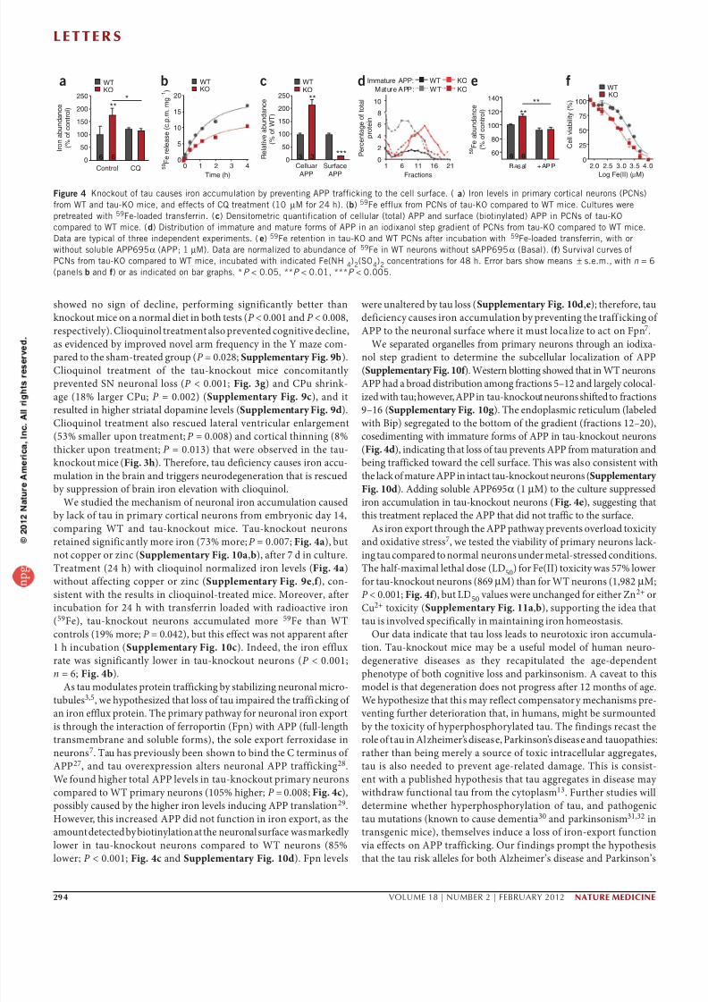
294 VOLUME 18 | NUMBER 2 | FEBRUARY 2012 nature medicine
showed no sign of decine, performing significanty better than
knockout mice on a norma diet in both tests (P < 0.001 and P < 0.008,
respectivey). Cioquino treatment aso prevented cognitive decine,
as evidenced by improved nove arm frequency in the Y maze com-
pared to the sham-treated group (P = 0.028; Supplementary Fig. 9b).
Cioquino treatment of the tau-knockout mice concomitanty
prevented SN neurona oss (P < 0.001; Fig. 3g ) and CPu shrink-
age (18% arger CPu; P = 0.002) (Supplementary Fig. 9c), and it
resuted in higher striata dopamine eves (Supplementary Fig. 9d).
Cioquino treatment aso rescued atera ventricuar enargement
(53% smaer upon treatment; P = 0.008) and cortica thinning (8%
thicker upon treatment; P = 0.013) that were observed in the tau-
knockout mice (Fig. 3h). Therefore, tau deficiency causes iron accu-
muation in the brain and triggers neurodegeneration that is rescued
by suppression of brain iron eevation with cioquino.
We studied the mechanism of neurona iron accumuation caused
by ack of tau in primary cortica neurons from embryonic day 14,
comparing WT and tau-knockout mice. Tau-knockout neuronsretained significanty more iron (73% more; P = 0.007; Fig. 4a), but
not copper or zinc (Supplementary Fig. 10a,b), after 7 d in cuture.
Treatment (24 h) with cioquino normaized iron eves (Fig. 4a)
without affecting copper or zinc (Supplementary Fig. 9e,f ), con-
sistent with the resuts in cioquino-treated mice. Moreover, after
incubation for 24 h with transferrin oaded with radioactive iron
(59Fe), tau-knockout neurons accumuated more 59Fe than WT
contros (19% more; P = 0.042), but this effect was not apparent after
1 h incubation (Supplementary Fig. 10c). Indeed, the iron effux
rate was significanty ower in tau-knockout neurons (P < 0.001;
n = 6; Fig. 4b).
As tau moduates protein trafficking by stabiizing neurona micro-
tubues3,5
, we hypothesized that oss of tau impaired the trafficking of an iron effux protein. The primary pathway for neurona iron export
is through the interaction of ferroportin (Fpn) with APP (fu-ength
transmembrane and soube forms), the soe export ferroxidase in
neurons7. Tau has previousy been shown to bind the C terminus of
APP27, and tau overexpression aters neurona APP trafficking28.
We found higher tota APP eves in tau-knockout primary neurons
compared to WT primary neurons (105% higher; P = 0.008; Fig. 4c),
possiby caused by the higher iron eves inducing APP transation 29.
However, this increased APP did not function in iron export, as the
amount detected by biotinyation at the neurona surface was markedy
ower in tau-knockout neurons compared to WT neurons (85%
ower; P < 0.001; Fig. 4c and Supplementary Fig. 10d). Fpn eves
were unatered by tau oss (Supplementary Fig. 10d,e); therefore, tau
deficiency causes iron accumuation by preventing the traff icking of
APP to the neurona surface where it must ocaize to act on Fpn7.
We separated organees from primary neurons through an iodixa-
no step gradient to determine the subceuar ocaization of APP
(Supplementary Fig. 10f ). Western botting showed that in WT neurons
APP had a broad distribution among fractions 5–12 and argey cooca-
ized with tau; however, APP in tau-knockout neurons shifted to fractions
9–16 (Supplementary Fig. 10g ). The endopasmic reticuum (abeed
with Bip) segregated to the bottom of the gradient (fractions 12–20),
cosedimenting with immature forms of APP in tau-knockout neurons
(Fig. 4d), indicating that oss of tau prevents APP from maturation and
being trafficked toward the ce surface. This was aso consistent with
the ack of mature APP in intact tau-knockout neurons (Supplementary
Fig. 10d). Adding soube APP695α (1 µM) to the cuture suppressed
iron accumuation in tau-knockout neurons (Fig. 4e), suggesting that
this treatment repaced the APP that did not traffic to the surface.
As iron export through the APP pathway prevents overoad toxicity and oxidative stress7, we tested the viabiity of primary neurons ack-
ing tau compared to norma neurons under meta-stressed conditions.
The haf-maxima etha dose (LD50) for Fe(II) toxicity was 57% ower
for tau-knockout neurons (869 µM) than for WT neurons (1,982 µM;
P < 0.001; Fig. 4f ), but LD50 vaues were unchanged for either Zn2+ or
Cu2+ toxicity (Supplementary Fig. 11a,b), supporting the idea that
tau is invoved specificay in maintaining iron homeostasis.
Our data indicate that tau oss eads to neurotoxic iron accumua-
tion. Tau-knockout mice may be a usefu mode of human neuro-
degenerative diseases as they recapituated the age-dependent
phenotype of both cognitive oss and parkinsonism. A caveat to this
mode is that degeneration does not progress after 12 months of age.
We hypothesize that this may refect compensatory mechanisms pre- venting further deterioration that, in humans, might be surmounted
by the toxicity of hyperphosphoryated tau. The findings recast the
roe of tau in Azheimer’s disease, Parkinson’s disease and tauopathies:
rather than being merey a source of toxic intraceuar aggregates,
tau is aso needed to prevent age-reated damage. This is consist-
ent with a pubished hypothesis that tau aggregates in disease may
withdraw functiona tau from the cytopasm13. Further studies wi
determine whether hyperphosphoryation of tau, and pathogenic
tau mutations (known to cause dementia30 and parkinsonism31,32 in
transgenic mice), themseves induce a oss of iron-export function
via effects on APP trafficking. Our f indings prompt the hypothesis
that the tau risk aees for both Azheimer’s disease and Parkinson’s
a b c fd250 20
100250
I r o n a b u n d a n c e
( % o
f c o n t r o l )
P e r c e n t a g e o f t o t a l
p r o t e i n
5 9 F e r e l e a s e ( c . p . m . m g – 1 )
C e l l v i a b i l i t y ( % )
R e l a t i v e a b u n d a n c e
( % o f W T )
200
150
100
50
0Control
WT WTImmature APP:
Mature APP: WT
KO
KOKOWTKO
WTKO WT
KO
6 6 3 3
****
***
*
CQ 0 1
15
10
5
01 2
Time (h)
Celluar
APP
Surface
APP
3 4
200
150
100
50
10
0
8
6
4
2
06 11
Fractions
2.016 21
75
50
25
02.5
Log Fe(II) (µM)
3.0 3.5 4.0
e
140
5 9 F e a b u n d a n c e
( % o f c o n t r o l )
6 6
**
**
Basal +APP
120
100
80
60
Figur 4 Knockout o tau causes iron accumulation by preventing APP traicking to the cell surace. ( a) Iron levels in primary cortical neurons (PCNs)
rom WT and tau-KO mice, and eects o CQ treatment (10 µM or 24 h). (b) 59Fe elux rom PCNs o tau-KO compared to WT mice. Cultures were
pretreated with 59Fe-loaded transerrin. (c) Densitometric quantiication o cellular (total) APP and surace (biotinylated) APP in PCNs o tau-KO
compared to WT mice. () Distribution o immature and mature orms o APP in an iodixanol step gradient o PCNs rom tau-KO compared to WT mice.
Data are typical o three independent experiments. () 59Fe retention in tau-KO and WT PCNs ater incubation with 59Fe-loaded transerrin, with or
without soluble APP695α (APP; 1 µM). Data are normalized to abundance o 59Fe in WT neurons without sAPP695α (Basal). (f) Survival curves o
PCNs rom tau-KO compared to WT mice, incubated with indicated Fe(NH 4)2(SO4)2 concentrations or 48 h. Error bars show means ± s.e.m., with n = 6
(panels b and f) or as indicated on bar graphs. *P < 0.05, **P < 0.01, ***P < 0.005.

8/2/2019 Tau Deficiency Induces ism (1)
http://slidepdf.com/reader/full/tau-deficiency-induces-ism-1 5/6
l e t t e r s
nature medicine VOLUME 18 | NUMBER 2 | FEBRUARY 2012 295
disease prime neurons for age-dependent deterioration by decreas-
ing the eff iciency of neurona iron export. Therefore, strategies that
maintain tau soubiity and abundance may form the basis for new
disease-modifying therapies.
MeTHodS
Methods and any associated references are avaiabe in the onine
version of the paper at http://www.nature.com/naturemedicine/.Note: Supplementary information is available on the Nature Medicine website.
ACKnoWLEDGMEntSThis work was supported by funds f rom the Austraian Research Counci,the Nationa Heath & Medica Research Counci of Austraia (NHMRC) andthe Azheimer’s Association. The Victorian Brain Bank Network is supportedby The University of Mebourne, The Menta Heath Research Institute, TheAfred Hospita and the Victorian Forensic Institute of Medicine, and funded by Neurosciences Austraia and the NHMRC. The authors thank Y.-H. Hung, H. Kim,S.H. Bush and A. Sedjahtera for hepfu discussion and technica assistance,H.N. Dawson (Duke University) for providing the tau-knockout mice, andT.A. Rouaut and D.L. Zhang (US Nationa Institutes of Heath) for MAP23 antibody.
AUtHoR ContRIBUtIonSP.L. and A.I.B. conceived of the study. A.I.B. raised funds for the study. P.L.,
S.A., J.A.D., L.S., D.K.W., P.A.A., D.I.F. and A.I.B. designed and performed theexperiments. G.D.C., B.X.W.W., L.Q.L., B.R.R., I.V. and C.A.M. assisted withthe experiments. G.F.E., P.A.A., R.A.C., R.C., D.I.F. and A.I.B supervised theexperiments. P.L. and A.I.B. anayzed the data and wrote drafts of the manuscript.A authors edited the manuscript.
CoMPEtInG FInAnCIAL IntEREStSThe authors decare competing financia interests: detais accompany the fu-textHTML version of the paper at http://www.nature.com/naturemedicine/ .
Published online at http://www.nature.com/naturemedicine/.
Reprints and permissions information is available online at http://www.nature.com/
reprints/index.html.
1. Laws, S.M. et al. Fine mapping o the MAPT locus using quantitative trait analysis
identies possible causal variants in Alzheimer’s disease. Mol. Psychiatry 12,
510–517 (2007).
2. Laws, S.M. et al. Genetic analysis o MAPT haplotype diversity in rontotemporaldementia. Neurobiol. Aging 29, 1276–1278 (2008).
3. Lei, P. et al. Tau protein: relevance to Parkinson’s disease. Int. J. Biochem.
Cell Biol. 42, 1775–1778 (2010).
4. Höglinger, G.U. et al. Identication o common variants infuencing risk o the
tauopathy progressive supranuclear palsy. Nat. Genet. 43, 699–705 (2011).
5. Lee, V.M.-Y., Goedert, M. & Trojanowski, J.Q. Neurodegenerative tauopathies.
Annu. Rev. Neurosci. 24, 1121–1159 (2001).
6. Smith, M.A., Harris, P.L., Sayre, L.M. & Perry, G. Iron accumulation in Alzheimer
disease is a source o redox-generated ree radicals. Proc. Natl. Acad. Sci. USA 94,
9866–9868 (1997).
7. Duce, J.A. et al. Iron-export erroxidase activity o β-amyloid precursor protein is
inhibited by zinc in Alzheimer’s disease. Cell 142, 857–867 (2010).
8. Bartzokis, G. et al. MRI evaluation o brain iron in earlier- and later-onset Parkinson’s
disease and normal subjects. Magn. Reson. Imaging 17, 213–222 (1999).
9. Zecca, L., Youdim, M.B., Riederer, P., Connor, J.R. & Crichton, R.R. Iron, brain
ageing and neurodegenerative disorders. Nat. Rev. Neurosci. 5, 863–873 (2004).
10. Oakley, A.E. et al. Individual dopaminergic neurons show raised iron levels in
Parkinson disease. Neurology 68, 1820–1825 (2007).
11. Dexter, D.T., Jenner, P., Schapira, A.H. & Marsden, C.D. Alterations in levels o
iron, erritin, and other trace metals in neurodegenerative diseases aecting the
basal ganglia. The Royal Kings and Queens Parkinson’s Disease Research Group.
Ann. Neurol. 32 (suppl.), S94–S100 (1992).
12. Paisán-Ruiz, C. et al. Widespread Lewy body and tau accumulation in childhood
and adult onset dystonia-parkinsonism cases with PLA2G6 mutations. Neurobiol.
Aging (in the press).
13. Khatoon, S., Grundke-Iqbal, I. & Iqbal, K. Levels o normal and abnormally
phosphorylated tau in dierent cellular and regional compartments o Alzheimerdisease and control brains. FEBS Lett. 351, 80–84 (1994).
14. Ksiezak-Reding, H., Binder, L.I. & Yen, S.-H.C. Immunochemical and biochemical
characterization o tau proteins in normal and Alzheimer’s disease brains with Alz
50 and Tau-1. J. Biol. Chem. 263, 7948–7953 (1988).
15. Zhukareva, V. et al. Selective reduction o soluble tau proteins in sporadic and
amilial rontotemporal dementias: an international ollow-up study. Acta
Neuropathol. 105, 469–476 (2003).
16. Mandel, S.A. et al. Multiunctional activities o green tea catechins in neuroprotection.
Modulation o cell survival genes, iron-dependent oxidative stress and PKC signaling
pathway. Neurosignals 14, 46–60 (2005).
17. Kaur, D. et al. Genetic or pharmacological iron chelation prevents MPTP-induced
neurotoxicity in vivo: a novel therapy or Parkinson’s disease. Neuron 37, 899–909
(2003).
18. Dawson, H.N. et al. Inhibition o neuronal maturation in primary
hippocampal neurons rom tau decient mice. J. Cell Sci. 114, 1179–1187
(2001).
19. Harada, A. et al. Altered microtubule organization in small-calibre axons o mice
lacking tau protein. Nature 369, 488–491 (1994).
20. Ikegami, S., Harada, A. & Hirokawa, N. Muscle weakness, hyperactivity, and
impairment in ear conditioning in tau-decient mice. Neurosci. Lett. 279,
129–132 (2000).
21. Roberson, E.D. et al. Reducing endogenous tau ameliorates amyloid beta-induced
decits in an Alzheimer’s disease mouse model. Science 316, 750–754
(2007).
22. Ittner, L.M. et al. Dendritic unction o tau mediates amyloid-beta toxicity in
Alzheimer’s disease mouse models. Cell 142, 387–397 (2010).
23. Cookson, M.R. The biochemistry o Parkinson’s disease. Annu. Rev. Biochem. 74,
29–52 (2005).
24. O’Neill, J. et al. Quantitative 1H magnetic resonance spectroscopy and MRI o
Parkinson’s disease. Mov. Disord. 17, 917–927 (2002).
25. Dauer, W. & Przedborski, S. Parkinson’s disease: mechanisms and models. Neuron 39,
889–909 (2003).
26. Sedelis, M., Schwarting, R.K. & Huston, J.P. Behavioral phenotyping o the
MPTP mouse model o Parkinson’s disease. Behav. Brain Res. 125, 109–125
(2001).
27. Smith, M.A. et al. Tau protein directly interacts with the amyloid beta-
protein precursor: implications or Alzheimer’s disease. Nat. Med. 1, 365–369(1995).
28. Stamer, K., Vogel, R., Thies, E., Mandelkow, E. & Mandelkow, E.-M. Tau blocks
trac o organelles, neurolaments and APP vesicles in neurons and enhances
oxidative stress. J. Cell Biol. 156, 1051–1063 (2002).
29. Rogers, J.T. et al. An iron-responsive element type II in the 5′-untranslated region
o the Alzheimer’s amyloid precursor protein transcript. J. Biol. Chem. 277,
45518–45528 (2002).
30. Ramsden, M. et al. Age-dependent neurobrillary tangle ormation, neuron loss,
and memory impairment in a mouse model o human tauopathy (P301L).
J. Neurosci. 25, 10637–10647 (2005).
31. Lewis, J. et al. Neurobrillary tangles, amyotrophy and progressive motor disturbance
in mice expressing mutant (P301L) tau protein. Nat. Genet. 25, 402–405
(2000).
32. Ittner, L.M. et al. Parkinsonism and impaired axonal transport in a mouse model
o rontotemporal dementia. Proc. Natl. Acad. Sci. USA 105, 15997–16002
(2008).

8/2/2019 Tau Deficiency Induces ism (1)
http://slidepdf.com/reader/full/tau-deficiency-induces-ism-1 6/6
nature medicine doi:10.1038/nm.2613
oNLINe MeTHodSHuman postmortem brain tissues. We obtained postmortem tissues from
the Victorian Brain Bank Network with informed consent from the individua
or the individua’s famiy. A procedures were approved by the Menta Heath
Research Institute Human Ethics Committee and were in accordance with the
Austraian Nationa Heath and Medica Research Counci guideines. Cinica
information is described in Supplementary Table 1. SN from 18 subjects with
cinicay defined Parkinson’s disease (11 mae, 75.3 ± 1.5 years od) and 18 con-
tro individuas without, or with minima, known neuroogica or psychiatricdisorders (13 mae, 77.3 ± 1.2 years od) were seected. In some studies there was
sufficient substantia nigra tissue for comparisons of ony n = 10 in each group
(in heathy contros, six mae, 77.9 ± 2.3 years od; in subjects with Parkinson’s
disease, seven mae, 75.4 ± 1.9 years od). Other brain tissues (fronta cortex
and cerebeum) were taken from a subset of the compete sampe group and
consisted of ten subjects with cinicay defined Parkinson’s disease (six mae,
75.8 ± 2.1 years od) and ten contro individuas (five mae, 76.6 ± 2.1 years od).
There were no significant differences in age or postmortem interva between
these various cohorts. Soube SN tau eves did not correate with postmortem
interva (R2 = 0.004; P = 0.796). We stored tissues at −80 °C unti use.
MPTP time-course study. We intoxicated five-month-od C57BL/6 mae
mice (Monash anima services) with four intraperitonea injections of 10 mg
per kg body weight MPTP (every 2 h in the same day) to give a fina dose of
40 mg per kg body weight33. Mice were kied with an overdose of sodiumpentobarbitone (Lethabarb; 100 mg per kg body weight) on days 3, 10, 21 and
45 after MPTP treatment, and we used untreated ittermate contros (kied
on day 10) for comparison.
Performance and treatment studies of tau-knockout mice. We used tau-
knockout mice18 and background C57BL/6/SV129 contro mice for perform-
ance studies and for biochemica studies. l-DOPA, freshy dissoved in 0.9%
(wt/vo) NaC, 0.5% (wt/vo) sodium carboxymethyceuose, 0.5% (vo/vo)
benzy acoho and 0.4% (vo/vo) Tween-80, was administered oray with the
use of an oroesophagea neede at 10 mg per kg body weight for one dose. We
carried out the performance tests 1 h after the dose. For cioquino treatment,
tau-knockout mice commenced cioquino feeding from 6.5 months of age. We
fed mice a diet of rodent chow mixed with 0.25 g per kg body weight (equivaent
to dosing of ~30 mg per kg body weight per day) of cioquino (Speciaty Feeds)
for 5 months and then kied them for biochemica studies. We administeredperformance tests every 3–4 weeks to monitor the effects of cioquino.
Recombinant APP expression and purification. We expressed soube human
APP695α in the methyotrophic yeast Pichia pastoris strain GS115 and purified
as described7. Briefy, we performed a two-step procedure using an AKTA
FPLC device (GE Heathcare). We purified APP695α from cuture media
using anion exchange on a Q-Sepharose coumn (1.6 cm × 25 cm coumn;
GE Heathcare) foowed by hydrophobic exchange with pheny-Sepharose
(0.5 cm × 5 cm coumn; GE Heathcare).
59Fe-loaded transferrin preparation. We purified human apo-transferrin
and then oaded it with 59Fe (PerkinEmer) as described7. We incubated
neurons with 1.0 × 10⁻⁶ M 59Fe-oaded transferrin for various periodsin serum-free Neurobasa-suppemented medium (Invitrogen). We then
removed medium after 1 h or 24 h, and incubated neurons with or without
soube APP695α (1 µM), washed them thoroughy with PBS and harvested
with trypsin. For the neurona effux assay, we incubated ces at 37 °C and
removed medium at various time points. We performed a comparabe time
course at 4 °C to account for nonspecifi c binding of 59Fe to the outer mem-
brane. We measured a media and ce ysates by γ -ray counter (Wizard 3,
PerkinEmer) in c.p.m.
Metal analysis. Sampes from each experimenta condition were freeze-dried
and then resuspended in 69% nitric acid (utracean grade, Aristar) overnight.
We then heated the sampes for 20 min at 90 °C, and added an equivaent
voume of hydrogen peroxide (30%; Merck) for a further 15 min incubation
at 70 °C. We diuted the sampes in doube-distied water and assayed them
by an AA240 atomic absorption spectrometer (Varian) or inductivey coupedpasma mass spectrometer (Utramass 700, Varian). Each sampe was measured
in tripicate, and the concentrations determined from the standard curve were
normaized to protein concentration or wet tissue weight.
Statistical analyses. Statistica anaysis was carried out in Prism (GraphPad)
or SPSS software. Before t tests or ANOVA post hoc tests were undertaken, a
Leven’s test for homogeneity of error variance was performed. For ANOVA,
Dunnett’s post hoc t tests were used when sampe variances were homoge-
neous. Where variances between groups were found to differ significanty,
Games-Howe post hoc tests were used. A tests were two-taied, with the
eve of significance set at P = 0.05. Detaied tests used in each experiment are
described in the Supplementary Methods.
Additional methods. Detaied methodoogy is described in the Supplementary
Methods.
33. Przedborski, S.et al.The parkinsonian toxin 1-methyl-4-phenyl-1,2,3,6-tetrahydropyridine
(MPTP): a technical review o its utility and saety. J. Neurochem. 76, 1265–1274
(2001).


![08. ism mabni [ism dhomir]](https://img.dokumen.tips/doc/110x75/55a4f0a71a28ab26408b480d/08-ism-mabni-ism-dhomir.jpg)

