Embed Size (px)
Citation preview
This journal is © The Royal Society of Chemistry 2021 Chem. Commun., 2021, 57, 3283–3286 | 3283
Cite this: Chem. Commun., 2021,
57, 3283
Targeting SARS-CoV-2 spike protein by stapledhACE2 peptides†
Marijn N. Maas, Jordi C. J. Hintzen, Philipp M. G. Loffler andJasmin Mecinovic *
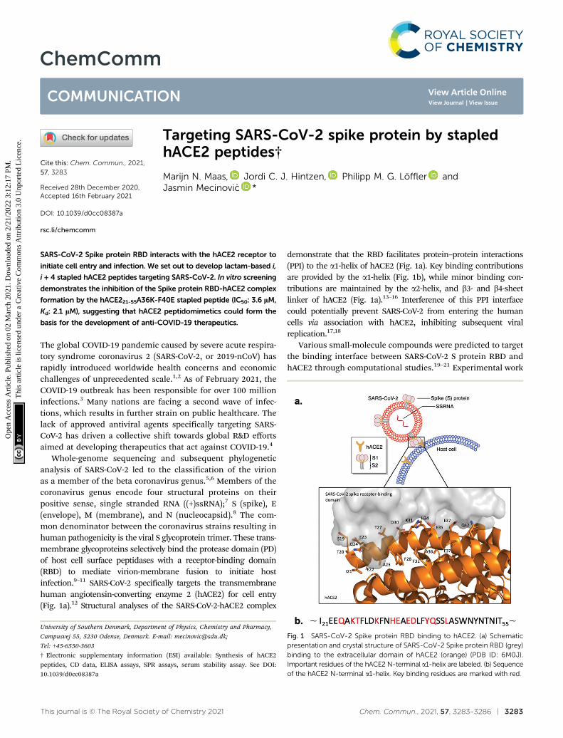
SARS-CoV-2 Spike protein RBD interacts with the hACE2 receptor to
initiate cell entry and infection. We set out to develop lactam-based i,
i + 4 stapled hACE2 peptides targeting SARS-CoV-2. In vitro screening
demonstrates the inhibition of the Spike protein RBD-hACE2 complex
formation by the hACE221-55A36K-F40E stapled peptide (IC50: 3.6 lM,
Kd: 2.1 lM), suggesting that hACE2 peptidomimetics could form the
basis for the development of anti-COVID-19 therapeutics.
The global COVID-19 pandemic caused by severe acute respira-tory syndrome coronavirus 2 (SARS-CoV-2, or 2019-nCoV) hasrapidly introduced worldwide health concerns and economicchallenges of unprecedented scale.1,2 As of February 2021, theCOVID-19 outbreak has been responsible for over 100 millioninfections.3 Many nations are facing a second wave of infec-tions, which results in further strain on public healthcare. Thelack of approved antiviral agents specifically targeting SARS-CoV-2 has driven a collective shift towards global R&D effortsaimed at developing therapeutics that act against COVID-19.4
Whole-genome sequencing and subsequent phylogeneticanalysis of SARS-CoV-2 led to the classification of the virionas a member of the beta coronavirus genus.5,6 Members of thecoronavirus genus encode four structural proteins on theirpositive sense, single stranded RNA ((+)ssRNA);7 S (spike), E(envelope), M (membrane), and N (nucleocapsid).8 The com-mon denominator between the coronavirus strains resulting inhuman pathogenicity is the viral S glycoprotein trimer. These trans-membrane glycoproteins selectively bind the protease domain (PD)of host cell surface peptidases with a receptor-binding domain(RBD) to mediate virion-membrane fusion to initiate hostinfection.9–11 SARS-CoV-2 specifically targets the transmembranehuman angiotensin-converting enzyme 2 (hACE2) for cell entry(Fig. 1a).12 Structural analyses of the SARS-CoV-2-hACE2 complex
demonstrate that the RBD facilitates protein–protein interactions(PPI) to the a1-helix of hACE2 (Fig. 1a). Key binding contributionsare provided by the a1-helix (Fig. 1b), while minor binding con-tributions are maintained by the a2-helix, and b3- and b4-sheetlinker of hACE2 (Fig. 1a).13–16 Interference of this PPI interfacecould potentially prevent SARS-CoV-2 from entering the humancells via association with hACE2, inhibiting subsequent viralreplication.17,18
Various small-molecule compounds were predicted to targetthe binding interface between SARS-CoV-2 S protein RBD andhACE2 through computational studies.19–21 Experimental work
Fig. 1 SARS-CoV-2 Spike protein RBD binding to hACE2. (a) Schematicpresentation and crystal structure of SARS-CoV-2 Spike protein RBD (grey)binding to the extracellular domain of hACE2 (orange) (PDB ID: 6M0J).Important residues of the hACE2 N-terminal a1-helix are labeled. (b) Sequenceof the hACE2 N-terminal a1-helix. Key binding residues are marked with red.
University of Southern Denmark, Department of Physics, Chemistry and Pharmacy,
Campusvej 55, 5230 Odense, Denmark. E-mail: [email protected];
Tel: +45-6550-3603
† Electronic supplementary information (ESI) available: Synthesis of hACE2peptides, CD data, ELISA assays, SPR assays, serum stability assay. See DOI:10.1039/d0cc08387a
Received 28th December 2020,Accepted 16th February 2021
DOI: 10.1039/d0cc08387a
rsc.li/chemcomm
ChemComm
COMMUNICATION
Ope
n A
cces
s A
rtic
le. P
ublis
hed
on 0
2 M
arch
202
1. D
ownl
oade
d on
2/2
1/20
22 3
:12:
17 P
M.
Thi
s ar
ticle
is li
cens
ed u
nder
a C
reat
ive
Com
mon
s A
ttrib
utio
n 3.
0 U
npor
ted
Lic
ence
.
View Article OnlineView Journal | View Issue

3284 | Chem. Commun., 2021, 57, 3283–3286 This journal is © The Royal Society of Chemistry 2021
by Carino et al. demonstrated that small-molecules could leadto PPI inhibition between Spike RBD and the carboxypeptidasedomain of hACE2 with natural and semi synthetic steroidalagents.19 However, small-molecules targeting a large surfacearea of the RBD cannot engage with the entire extended linearridge-like binding region.8
Peptidomimetics are better suited for the inhibition of PPIcompared to small molecules due to efficient engagement withlarge protein surfaces through entropically favored interactionswith protein surfaces, preventing competitive replacement.22,23
hACE2 a1-helix-based peptidomimetics targeting SARS-CoV-2 Sprotein were demonstrated to inhibit RBD-hACE2 complexformation and subsequent host cell infection in both computa-tional and experimental studies.24–26 However, hACE2 a1-helix-based peptides are expected to lose their bio-active conforma-tion in solution, decreasing the RBD binding potential of thepeptidomimetic.17 Furthermore, short and linear peptide-based drugs are generally cell impermeable, not orally avail-able, and subject to rapid proteolysis.27,28 These limitations canbe overcome with i,i + 4 peptide stapling approaches thatstabilise the peptide’s three-dimensional and bio-active struc-ture, increase cellular uptake, decrease enzymatic degradationand improve pharmacokinetic properties through chemicalmodification of the peptide sequence.27,29 However, somewhatconflicting recent reports showed that hydrocarbon-based sta-pling of hACE2 might only partially lead to the disruption of theRBD-hACE2 complex.30,31 In addition, most SARS-CoV-2 repli-cation takes place in the lower and upper airways, whichpotentially allows for targeting of the RBD-hACE2 interactionby inhalation of the aerosolised peptide therapeutics. Thismakes the potential lack of oral availability of peptides a non-issue.32
We hypothesised that the development of a stapled hACE2N-terminal a1-helix might allow for selective and potent inhibi-tion of RBD-hACE2 recognition and subsequent infection. Inthis report we discuss the design and synthesis of lactam-basedstapled 35-mer peptides using the hACE2 N-terminal a1-helixinhibitor 1 sequence to overcome structural instability andincrease peptide binding affinity to SARS-CoV-2 (Fig. 2).
Cryo-EM and X-ray structures, as well as computationalanalysis of the interaction between hACE2 and RBD revealedthat residues 21–55 of the N-terminal a1-helix of hACE2 act asthe major contributors of binding interactions with S proteinRBD (Fig. 1). Kral et al. computationally determined that severalpolar residues of the N-terminal a1-helix are essential media-tors of RBD binding (Fig. 1b).20 We selected peptide i,i + 4stapling sites (F28-F32, F32-A36, A36-F40) based on their posi-tioning in the hACE2 N-terminal a1-helix structure to not inter-fere with the binding interface (Fig. 1b).17,20 Lactam-linkedstapling was shown to effectively induce helicity, uses naturalbuilding blocks, and facilitates straightforward synthesis.33
35-mer native peptide 1 and linear controls 2–4 were synthe-sised by solid-phase peptide synthesis (SPPS) (Fig. 2). Stapledpeptides 5–7 were synthesised using SPPS, followed by staplingof the side-chains with lactam-based i,i + 4 methods, andcleavage of the stapled hACE2 peptides from the resin (Fig. 2).
Mass spectrometry and analytical HPLC confirmed the highpurity (490%) of all RP-HPLC purified synthetic hACE2 peptides(Table S1 and Fig. S1–S7, ESI†).
a-Helical propensity of the linear hACE2 peptides could becomputationally predicted with the use of the PSIPRED secondarystructure prediction server (Fig. S8, ESI†).34 a-Helical content ofthe synthetic hACE2 peptides was confirmed by CD spectroscopyto determine the effect of i,i + 4 lactam stapling on the peptidesecondary structure with the expectation that stapling increasespeptide helicity, using poly-L-lysine as a reference for helicity(Fig. 3 and Fig. S9, ESI†).35,36 Spectra were recorded in 10 mMPBS at pH 7.4 in the absence and the presence of 2,2,2-trifluoroethanol (TFE) at 25 1C (Fig. 3). TFE is known tostimulate the formation of the a-helical secondary structures inpeptides,37 allowing for a comparison of the a-helical content ofhACE2 peptides at maximal helicity (Fractional Helicity, fH),which we observed at 30% TFE (Fig. S9, ESI†). CD spectra weredeconvoluted with the DICHROWEB server to determinehelicities.38 CD spectroscopy demonstrated low helicity for bothlinear and stapled hACE2 peptides in absence of TFE, with
Fig. 2 Design of lactam-based i,i + 4 stapled hACE2-based peptides forthe inhibition of SARS-CoV-2 S protein-hACE2 interactions. The wild-typesequence (1) and linear modified peptides (2–4) were used as controls totheir stapled counterparts (5–7).
Fig. 3 Circular Dichroism spectra of hACE2 peptides in molar ellipticityper residue. 30 mM peptide in 10 mM PBS at pH 7.4 with 30% TFE at 298 K.
Communication ChemComm
Ope
n A
cces
s A
rtic
le. P
ublis
hed
on 0
2 M
arch
202
1. D
ownl
oade
d on
2/2
1/20
22 3
:12:
17 P
M.
Thi
s ar
ticle
is li
cens
ed u
nder
a C
reat
ive
Com
mon
s A
ttrib
utio
n 3.
0 U
npor
ted
Lic
ence
.View Article Online

This journal is © The Royal Society of Chemistry 2021 Chem. Commun., 2021, 57, 3283–3286 | 3285
predictors of helicity averaging to 6-13% helical content (Table S2and Fig. S10, ESI†). A lack of the predominant a-helical secondarystructure of the natural hACE2 peptide sequence in the absence ofTFE is in agreement with findings by Karoyan et al. (Table S2,ESI†).24 In the presence of TFE, however, a-helical structures canbe observed for various synthetic hACE2 peptides with predictorsaveraging from 11 to 52% helical content, with a clear shift innegative ellipticity from 200 to 208 and 222 nm at varyingintensities for all peptides (Fig. 3, Fig. S9, S10, Table S2, ESI†).Stapled peptide 5 displayed the highest helicity across the panel ofhACE2 peptides (average: 52%), with increased helical contentcompared to the wild-type sequence 1 (average: 38%). Staplingcloser to the N-terminus seems to result in a loss of helicitycompared to the native sequence. This observation suggests thati,i + 4 lactam stapling at the A36K-F40E site can positively affectthe helical content, possibly due to the sequence environmenteffects surrounding the peptide staple.
Synthetic hACE2 peptides were evaluated as inhibitors ofSARS-CoV-2 Spike protein-hACE2 complex formation using theELISA based Screening Assay Kit. A concentration–responsecurve of SARS-CoV-2 Spike Protein (RBD) Recombinant HumanMonoclonal Antibody in PBS was used as a positive control forinhibition (0.6 nM–200 mM) and to test the viability of the ELISAkit (Fig. S11, ESI†). A concentration-dependent decrease ofluminescence was observed along with a median inhibitoryconcentration (IC50) for the monoclonal antibody (9.5 nM),similar to the reported IC50 value for the antibody (6.6 nM)targeting SARS-CoV-2 Spike protein by the manufacturer(Fig. S11, ESI†). We evaluated the linear and stapled hACE2peptides at 10 mM and 100 mM to identify potent inhibitors ofthe RBD-hACE2 complex formation (Fig. 4a). Among all peptides,only 5 reduced binding by 480% at 10 mM. Concentration-dependent assays (0.3–300 mM) of the stapled hACE2 peptidesdemonstrated significant inhibition of binding by 5 (IC50: 3.6 mM)compared to the linear wild-type control 1 that displayed onlyminor inhibition at 4100 mM concentrations (Fig. 4b, n = 3replicates). Peptides 6 (IC50: 28.4 mM) and 7 (IC50: 46.8 mM)displayed enhanced inhibitory activity compared to the nativesequence, although 12–18 times less potent when compared to5 (Fig. S11, ESI†). The linear controls 2–4 showed reduced bindingto 50% at 100 mM, suggesting that the linear peptides are poorinhibitors of SARS-CoV-2-hACE2 interactions and that decreasedhACE2 helicity results in less potent inhibitory effects (Fig. 4a).
Surface Plasmon Resonance (SPR) was used to quantify thebinding affinity (Kd) and kinetic association and dissociationparameters (kon and koff) between SARS-CoV-2 Spike proteinand the stapled hACE221-55 peptides (5, 6 and 7) using thepolycarboxylate HC surface SPR sensor chip with pre-immobilizedSARS-CoV-2 Spike protein with a Biacore X100. Full-length hACE2protein and 1 were used as positive and negative controls,respectively (Fig. S12, ESI†). The binding interaction between 5and the immobilized SARS-CoV-2 Spike protein was characterizedby micromolar binding affinity (Kd: 2.1 � 0.2 mM), a fast associa-tion rate (kon: 1.7 � 0.1 � 104 M�1 s�1) and a moderate rate ofdissociation (koff: 0.034 � 0.002 s�1) (Fig. 5 and Table S3, ESI†).The binding affinity of the native sequence 1 and stapled peptides
6 and 7 could not be determined, as the interaction with the Spikeprotein versus the reference surface was too weak for the assess-ment (Fig. S13, ESI†). These results, in combination withthe inhibition assay, indicate direct and potent binding ofhACE221-55A36K-F40E (5) peptide to the RBD domain of Spikeprotein.
Finally, resistance to proteolysis was determined for the35-mer peptides (1, 2 and 5) by treating the peptides with humanserum (1 : 4 v/v, serum:PBS) at 37 1C and recording peptidedegradation over time using analytical HPLC (Fig. 6). Interestingly,the data demonstrated that both 1 and 5 remain highly stable overtime. Peptide 2 was observed to be comparatively less stable than
Fig. 4 SARS-CoV-2 Spike protein-hACE2 inhibitor screening assay.(a) The linear and stapled hACE2 peptides was screened for inhibition at10 mM and 100 mM. Data is mean� SD, n = 2. (b) Concentration-dependent(0.3–300 mM) IC50 determination of 5. Data is mean � SD, n = 3.
Fig. 5 Determination of binding of stapled hACE2 peptide 5 (0–20 mM) toimmobilized SARS-CoV-2 Spike protein with SPR. Conditions: 10 mMsodium phosphate, pH 7.4, 25 1C, n = 2.
ChemComm Communication
Ope
n A
cces
s A
rtic
le. P
ublis
hed
on 0
2 M
arch
202
1. D
ownl
oade
d on
2/2
1/20
22 3
:12:
17 P
M.
Thi
s ar
ticle
is li
cens
ed u
nder
a C
reat
ive
Com
mon
s A
ttrib
utio
n 3.
0 U
npor
ted
Lic
ence
.View Article Online

3286 | Chem. Commun., 2021, 57, 3283–3286 This journal is © The Royal Society of Chemistry 2021
the natural sequence, which is likely due to the introduction ofmore sensitive proteolytic cleavage sites by substitution of A36Kand F40E.
In conclusion, we have developed lactam-based i,i + 4stapled hACE2 a1-helix-based peptidomimetics that targetSARS-CoV-2 S protein and inhibit the RBD-hACE2 complexformation. We envision that hACE2 a1-helix-based peptidomi-metics could potentially prevent SARS-CoV-2 from entering thehuman cells through hACE2 and thus inhibit subsequent viralreplication. Our work highlights that the stapled peptidehACE221-55A36K-F40E (5), which exhibits an increased level ofhelicity, efficiently inhibits SARS-CoV-2 Spike protein-hACE2binding (IC50: 3.6 mM, Kd: 2.1 � 0.2 mM). Related stapledpeptides hACE221-55F32K-A36E (6) and hACE221-55F28K-F32E(7) also resulted in stronger inhibition compared to the naturalsequence 1. Taken together, we believe that continued optimi-sation of the chosen staples and additional sequence modifica-tions might contribute to the design of future anti-coronavirustherapeutics, which can aid with infection prevention.
This work was supported by the ERC Starting Grant to J. M.(ChemEpigen-715691). P. M. G. L. thanks the Villum Fonden(BioNEC-VKR18333). We thank the Danish Molecular BiomedicalImaging Centre (University of Southern Denmark) for the use ofbioimaging facilities.
Conflicts of interest
There are no conflicts to declare.
References1 F. Wu, S. Zhao, B. Yu, Y. M. Chen, W. Wang, Z. G. Song, Y. Hu,
Z. W. Tao, J. H. Tian, Y. Y. Pei, M. L. Yuan, Y. L. Zhang, F. H. Dai,Y. Liu, Q. M. Wang, J. J. Zheng, L. Xu, E. C. Holmes and Y. Z. Zhang,Nature, 2020, 579, 265–269.
2 F. Jiang, L. Deng, L. Zhang, Y. Cai, C. W. Cheung and Z. Xia, J. Gen.Intern. Med., 2020, 35, 1545–1549.
3 Worldometer, Coronavirus Cases, https://www.worldometers.info/coronavirus/coronavirus-cases/#daily-cases, accessed 8 feb 2021.
4 T. Thanh, L. Z. Andreadakis, A. Kumar, R. Gomez Roman,S. Tollefsen, M. Saville and S. Mayhew, Nat. Rev. Drug Discovery,2020, 19, 305–306.
5 P. C. Y. Woo, S. K. P. Lau, C. S. F. Lam, K. K. Y. Lai, Y. Huang, P. Lee,G. S. M. Luk, K. C. Dyrting, K.-H. Chan and K.-Y. Yuen, J. Virol., 2009,83, 908–917.
6 R. Lu, X. Zhao, J. Li, P. Niu, B. Yang, H. Wu, W. Wang, H. Song,B. Huang, N. Zhu, Y. Bi, X. Ma, F. Zhan, L. Wang, T. Hu, H. Zhou,
Z. Hu, W. Zhou, L. Zhao, J. Chen, Y. Meng, J. Wang, Y. Lin, J. Yuan,Z. Xie, J. Ma, W. J. Liu, D. Wang, W. Xu, E. C. Holmes, G. F. Gao,G. Wu, W. Chen, W. Shi and W. Tan, Lancet, 2020, 395, 565–574.
7 G. Lu and D. Liu, Protein Cell, 2012, 803–805.8 C. Wu, Y. Liu, Y. Yang, P. Zhang, W. Zhong, Y. Wang, Q. Wang,
Y. Xu, M. Li, X. Li, M. Zheng, L. Chen and H. Li, Acta Pharm. Sin. B,2020, 10, 766–788.
9 F. Li, W. Li, M. Farzan and S. C. Harrison, Science, 2005, 309,1864–1868.
10 Y. Wan, J. Shang, R. Graham, R. S. Baric and F. Li, J. Virol., 2020,94, e0012720.
11 S. VanPatten, M. He, A. Altiti, K. F. Cheng, M. H. Ghanem andY. Al-Abed, Future Med. Chem., 2020, 12(18), 1647–1656.
12 H. Zhang, J. M. Penninger, Y. Li, N. Zhong and A. S. Slutsky,Intensive Care Med., 2020, 46, 586–590.
13 Q. Wang, Y. Zhang, L. Wu, S. Niu, C. Song, Z. Zhang, G. Lu, C. Qiao,Y. Hu, K. Y. Yuen, Q. Wang, H. Zhou, J. Yan and J. Qi, Cell, 2020,181(4), 894–904.
14 R. Yan, Y. Zhang, Y. Li, L. Xia, Y. Guo and Q. Zhou, Science, 2020,367, 1444–1448.
15 J. Shang, G. Ye, K. Shi, Y. Wan, C. Luo, H. Aihara, Q. Geng,A. Auerbach and F. Li, Nature, 2020, 581, 221–224.
16 D. Wrapp, N. Wang, K. S. Corbett, J. A. Goldsmith, C. L. Hsieh,O. Abiona, B. S. Graham and J. S. McLellan, Science, 2020, 367,1260–1263.
17 C. Lupala, V. Kumar, X. Li, X.-D. Su and H. Liu, bioRxiv, 2020, DOI:10.1101/2020.05.03.075473.
18 S. Xiu, A. Dick, H. Ju, S. Mirzaie, F. Abdi, S. Cocklin, P. Zhan andX. Liu, J. Med. Chem., 2020, 63, 12256–12274.
19 A. Carino, F. Moraca, B. Fiorillo, S. Marchiano, V. Sepe, M. Biagioli,C. Finamore, S. Bozza, D. Francisci, E. Distrutti, B. Catalanotti,A. Zampella and S. Fiorucci, Front. Chem., 2020, 8, 572885.
20 Y. Han, P. Kral and P. Kraı, ACS Nano, 2020, 14, 5143–5147.21 Z. Shang, S. Y. Chan, W. J. Liu, P. Li and W. Huang, ACS Infect. Dis.,
2020, DOI: 10.1021/acsinfecdis.0c00646.22 D. Bojadzic and P. Buchwald, Curr. Top. Med. Chem., 2018, 18,
674–699.23 A. M. Tharappel, S. K. Samrat, Z. Li and H. Li, ACS Infect. Dis., 2020,
6, 2844–2865.24 P. Karoyan, V. Vieillard, E. Odile, A. Denis, A. Guihot, C.-E. Luyt,
L. Gomez-Morales, P. Grondin and O. Lequin, bioRxiv, 2020, DOI:10.1101/2020.08.24.264077.
25 R. C. Larue, E. Xing, A. D. Kenney, Y. Zhang, J. A. Tuazon, J. Li,J. S. Yount, P. K. Li and A. Sharma, Bioconjugate Chem., 2021, 32,215–223.
26 G. Zhang, S. Pomplun, A. R. Loftis, A. Loas and B. L. Pentelute,bioRxiv, 2020, DOI: 10.1101/2020.03.19.999318.
27 L. Nevola and E. Giralt, Chem. Commun., 2015, 51, 3302–3315.28 B. Meibohm, Pharmaceutical Biotechnology: Fundamentals and Appli-
cations, Springer International Publishing, 2019, pp. 99–137.29 T. A. F. F. Cardote and A. Ciulli, ChemMedChem, 2016, 11,
787–794.30 D. C. Morgan, C. Morris, A. Mahindra, C. M. Blair, G. Tejeda,
I. Herbert, M. L. Turnbull, G. Lieber, B. J. Willett, N. Logan,B. Smith, A. B. Tobin, D. Bhella, G. Baillie and A. G. Jamieson,Pept. Sci., 2021, e24217.
31 F. Curreli, S. M. B. Victor, S. Ahmed, A. Drelich, X. Tong,C.-T. K. Tseng, C. D. Hillyer and A. K. Debnath, mBio, 2020,11, e02451.
32 D. Schutz, Y. B. Ruiz-Blanco, J. Munch, F. Kirchhoff,E. Sanchez-Garcia and J. A. Muller, Adv. Drug Delivery Rev., 2020,167, 47–65.
33 A. D. de Araujo, H. N. Hoang, W. M. Kok, F. Diness, P. Gupta,T. A. Hill, R. W. Driver, D. A. Price, S. Liras and D. P. Fairlie,Angew. Chem., Int. Ed., 2014, 53, 6965–6969.
34 D. W. A. Buchan and D. T. Jones, Nucleic Acids Res., 2019, 47,W402–407.
35 N. J. Greenfield, Nat. Protoc., 2006, 1, 2876–2890.36 B. Davidson and G. D. Fasman, Biochemistry, 1967, 6, 1616–1629.37 F. D. Sonnichsen, J. E. Van Eyk, R. S. Hodges and B. D. Sykes,
Biochemistry, 1992, 31, 8790–8798.38 L. Whitmore and B. A. Wallace, Nucleic Acids Res., 2004, 32,
W668–W673.
Fig. 6 The serum stability of hACE2 peptides 1, 2 and 5.
Communication ChemComm
Ope
n A
cces
s A
rtic
le. P
ublis
hed
on 0
2 M
arch
202
1. D
ownl
oade
d on
2/2
1/20
22 3
:12:
17 P
M.
Thi
s ar
ticle
is li
cens
ed u
nder
a C
reat
ive
Com
mon
s A
ttrib
utio
n 3.
0 U
npor
ted
Lic
ence
.View Article Online