Embed Size (px)
Citation preview

DOI: https://doi.org/10.30795/jfootankle.2020.v14.1190
Technical Tips
Copyright © 2020 - Journal of the Foot&Ankle 211J Foot Ankle. 2020;14(2):211-8
Talocalcaneal coalition resection and bone block subtalar joint arthrodesis: a techinical tipCaleb Iehl1 , Elijah Auch1 , Victoria Vivtcharenko1 , Nacime Salomao Barbachan Mansur1,2 , Heather Kowalski1 , Cesar de Cesar Netto1
1. University of Iowa, Carver College of Medicine, Iowa City, USA.2. Universidade Federal de São Paulo, São Paulo, SP, Brazil.
AbstractIn this technical tip, we present the case of an obese 17-year-old female diagnosed with a severe, rigid, and symptomatic flatfoot on a background of exuberant talocalcaneal and residual calcaneonavicular coalition. Through a technical modification of the fusion re-section, both coalitions were quickly and safely removed with two single cuts of an oscillating saw, resecting a medial wedge through a medial approach, without the need for “peel-off” tarsal coalition resection. To protect and guide the resection osteotomy, one Freer elevator ws inserted under direct visualization on the patent posterolateral aspect of the subtalar joint posterior facet and a second elevator was positioned underneath the talar neck. Under fluoroscopic guidance, an osteotomy was performed connecting these two points. The patient also received a bone-block subtalar joint arthrodesis and a Cotton osteotomy. Good short-term alignment correc-tion and functional outcome were achieved.
Level of Evidence V; Therapeutic Studies; Expert Opinion.
Keywords: Tarsal coalition; Talocalcaneal coalition; Calcaneonavicular coalition; Cotton osteotomy; Subtalar fusion.
Study performed at the University of Iowa, Carver College of Medicine, Iowa City, USA.
Corresponding author: Cesar de Cesar Netto. 200 Hawkins Drive, Iowa City, IA, USA, Zip Code: 52240. E-mail: [email protected] Conflicts of interest: none. Source of funding: none. Date received: August 07, 2020. Date accepted: August 08, 2020. Online: August 30, 2020.
How to cite this article: Iehl C, Auch E, Vivtcharenko V, Mansur NSB, Kowalski H, Netto CC. Talocalcaneal coalition resection and bone block subtalar joint arthrodesis: a techinical tip.
J Foot Ankle. 2020;14(2):211-8.
IntroductionTarsal coalitions are a well-known cause of pain and disability
in the foot and ankle, especially in the pediatric population(1,2). These problems often lead to inability to participate in acti-vities of daily living and sports, decrease quality of life, and are often associated with foot deformities, most commonly flatfoot(3). The most common types of tarsal coalitions are cal-caneonavicular (CNC) and talocalcaneal (TCC) coalitions(3-6).
Many of these coalitions are asymptomatic, diagnosed only after trauma or incidentally during investigation of the limb(7). Nevertheless, some cases present with quite exuberant signs and symptoms, as well as pronounced hindfoot deformity, abnormal bone growth, and dysplasia(8). In severe cases with dysplastic talar formation, a ball-and-socket mortise joint may be found(9).
There are osseous, fibrous, and cartilaginous subtypes of coalitions(3-6). Differentiation between subtypes is primarily radiographic, but magnetic resonance imaging (MRI) and
computed tomography (CT) scans can also be used in diag-nosis and treatment planning(10). Most mild and early cases are successfully treated with nonoperative measures, such as physical therapy and insoles, but failure of these modalities usually leads patients to surgical intervention(3-6,11).
A joint-sparing procedure with adequate coalition resection is the cornerstone of surgical treatment and, in several cases, the only required technique for treatment, especially when less than 50% of the mediolateral extension of the subtalar joint is involved(3). Even when deformity is present, removal of the bone bridge is essential to correct foot alignment and relieve pain(12). Different surgical techniques have been des-cribed for this purpose; all share the intention of safely remo-ving the coalition as easily and quickly as possible(13-16).
This article describes the treatment of a 17-year-old fema-le with a symptomatic severe flatfoot deformity and obesity on a background of massive TCC and residual CNC, with his-tory of a prior failed attempt at CNC resection. The patient

Iehl et al. Talocalcaneal coalition resection and bone block subtalar joint arthrodesis: a techinical tip
212 J Foot Ankle. 2020;14(2):211-8
underwent resection of the coalitions through a novel me-dial approach (the subject of this technical tip) consisting of an osteotomy line connecting two fluoroscopic landmarks, followed by a medial wedge resection for hindfoot valgus correction and corrective lateral-wedge bone-block allograft arthrodesis and Cotton osteotomy for residual forefoot su-pination.
Case descriptionA 17-year-old (BMI 36) presented to an outside orthopedic
service with a complaint of chronic right medial foot and ankle pain since childhood. The patient reported a history of rigid symptomatic flatfoot in childhood, which had been treated with physical therapy and insoles. She also had a history of an attempt at surgical CNC resection 18 months earlier, with only partial resolution of her symptoms (Figure 1). Following a re-cent minor injury, she had experienced a flare-up of symp-toms. On physical examination at the time, a severe flatfoot deformity was observed, with significant hindfoot valgus of approximately 20-25° and a supinated forefoot. There was evidence of a healed sinus tarsi approach, with painful, rigid passive hindfoot inversion and eversion. She was also tender to palpation along the medial side of the foot and ankle, whe-re a bony prominence could be palpated at the level of the sustentaculum talus, and had moderate tenderness on the sinus tarsi area. Conventional radiographs showed possible residual CNC, with indirect signs of a TCC (talar beaking and
C-sign) in the right foot, and a mild valgus talar tilt of the talar dome (Figure 2). Initial treatment plans included a walking cast for 6 weeks and brace immobilization; however, there was no significant improvement in the symptoms. MRI and weightbearing CT (WBCT) images were obtained to elucida-te the extent of the coalition, assess the subtalar joint, and, possibly, assist in surgical planning.
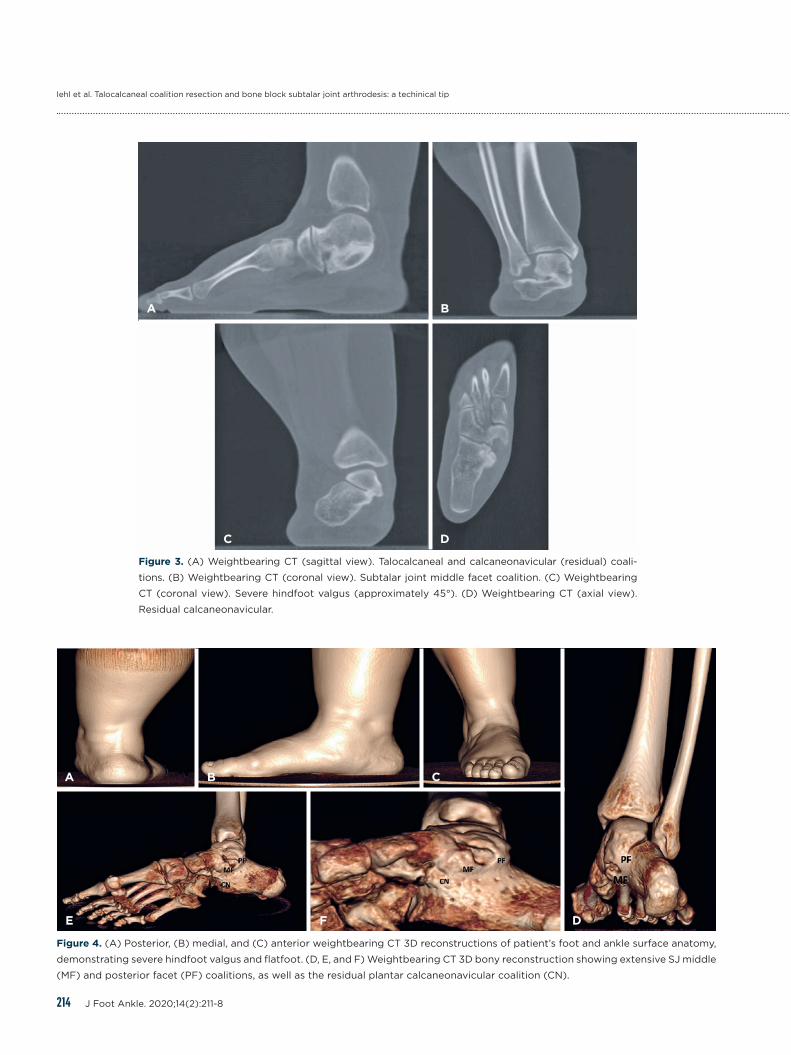
Images demonstrated a recurred/residual CNC, mostly plan-tar, and a massive TCC affecting both the middle and the pos-terior facets of the subtalar joint, compounded by with severe hindfoot valgus (tibiocalcaneal angle of approximately 45°) and a mild valgus tilting of the talar dome (Figures 3 and 4).
The patient and her family decided to proceed with surgi-cal treatment. Coalition resection followed by a bone block arthrodesis and a Cotton osteotomy were proposed to the patient and her family. Once the participants had agreed, sur-gery was formally indicated and performed.
Surgical technique and technical tipThe operation began with aspiration of bone marrow from
the right iliac crest for concentration and later injection into the subtalar joint fusion mass.
An extensive medial foot approach was selected and per-formed along the trajectory of the posterior tibial tendon (PTT), from the medial malleolus down to the navicular tube-rosity. The tendon was exposed and found to be intact pro-
Figure 1. Intraoperative fluoroscopic views of prior surgical treatment of calcaneonavicular coalition, (A) before and (B) after resection.
A B

Iehl et al. Talocalcaneal coalition resection and bone block subtalar joint arthrodesis: a techinical tip
213J Foot Ankle. 2020;14(2):211-8
A
B C
Figure 2. (A) AP bilateral foot radiographs and (B) lateral view of the right foot demonstrating severe flatfoot, forefoot abduction,
dysplastic talar head/neck, and indirect signs of tarsal coalition (C-sign and dorsal talar beak). (C) AP view of the right ankle demons-
trating mild valgus talar tilt.
ximally, but with some insertional tendinopathy. The PTT was partially detached from the navicular tuberosity and retrac-ted plantarly. The flexor digitorum longus (FDL) and flexor hallucis longus (FHL) tendons were identified by periosteal dissection, isolated, and retracted plantarly with the PTT, thus protecting the neurovascular bundle and exposing the medial aspect of the subtalar joint (SJ). A large bony prominence at the level of the sustentaculum tali, middle, and anterome-dial aspect of the SJ posterior facet was identified, consistent with a talocalcaneal bony coalition.
Instead of using a “peel-off” coalition resection technique with resection of multiple slices of bone or a small wedge resection technique, both usually used to identify a line of fibrotic tissue where the articular line was supposed to be positioned, we performed a modified technique that can be
applied in cases where a collation resection or a SJ fusion is plane. The technique modification consists of using two Freer elevators as fluoroscopic markers to guide a single-cut osteo-tomy to separate the talus and the calcaneus. To achieve that, the deep dissection is carried posterolaterally along the pos-terior aspect of the SJ and ankle to identify the posterolateral and patent aspect of the SJ posterior facet. The first Freer elevator is inserted into the joint under direct visualization. The second Freer elevator is positioned under the talar neck, to ensure that no talar neck bone is resected or osteotomized (Figure 5).
Under fluoroscopic guidance, an oscillating saw was used to perform the osteotomy connecting the two markers. In this specific case, as a severe hindfoot valgus existed and a SJ fu-sion was planned, once the talus and calcaneus were separated
Iehl et al. Talocalcaneal coalition resection and bone block subtalar joint arthrodesis: a techinical tip
214 J Foot Ankle. 2020;14(2):211-8
A B
C D
Figure 3. (A) Weightbearing CT (sagittal view). Talocalcaneal and calcaneonavicular (residual) coali-
tions. (B) Weightbearing CT (coronal view). Subtalar joint middle facet coalition. (C) Weightbearing
CT (coronal view). Severe hindfoot valgus (approximately 45°). (D) Weightbearing CT (axial view).
Residual calcaneonavicular.
A B C
DFE
Figure 4. (A) Posterior, (B) medial, and (C) anterior weightbearing CT 3D reconstructions of patient’s foot and ankle surface anatomy,
demonstrating severe hindfoot valgus and flatfoot. (D, E, and F) Weightbearing CT 3D bony reconstruction showing extensive SJ middle
(MF) and posterior facet (PF) coalitions, as well as the residual plantar calcaneonavicular coalition (CN).

Iehl et al. Talocalcaneal coalition resection and bone block subtalar joint arthrodesis: a techinical tip
215J Foot Ankle. 2020;14(2):211-8
A B
Figure 5. Technical tip. (A) Two Freer elevators are inserted under direct visualization to guide the orientation of the osteotomy: one in
the posterolateral patent aspect of the posterior facet of the subtalar joint and one underneath the talar neck. (B) 3D Weightbearing
CT bony reconstruction showing the dots representing the tip of the Freer elevators and a straight line connecting the dots, following
a very similar path when considering a patent posterior facet.
by the first osteotomy, an additional osteotomy line was then performed in the calcaneus to resect a medial wedge of bone aiming to correct the valgus deformity. In cases where the posterior facet would be preserved and an isolated coalition would be performed, this second osteotomy line and wedge resection would not be performed. After removing the wedge and being able to correct the valgus deformity to some ex-tent, the residual cartilage on the talar SJ facets was resected with curettes and osteotomes, and the extra bone on the me-dial surfaces of the talus and calcaneus, composing the bulky aspect of the talocalcaneal coalition, was resected with the oscillating saw and sharp osteotomes.
During a trial for a bone-block arthrodesis with a lateral wedged allograft (inserted from the same medial approach), no adequate correction was achieved secondary to residual stiffness for mobilization of the calcaneus. At that point, the osteotomy line created previously was extended distally in the calcaneus side aiming to separate the residual connection between the calcaneus and the navicular bone, aiming for the plantar aspect of the anterior process of the calcaneus, and avoiding injury to the calcaneocuboid joint. Following this step, adequate mobilization of the calcaneus was possible, and correction of the valgus deformity was achieved with medial displacement of the calcaneus and a lateral-based allograft bone-block trial wedge (12mm lateral base height) (Figure 6).
Clinical evaluation (with the heel centered underneath the leg from a posterior view and the leg elevated) and fluoros-copic assessment demonstrated adequate correction of the deformity and apposition of the trial bone block with the cal-caneal and talar surfaces. The trial was removed and both osseous surfaces were prepared with multiple drill holes to stimulate healing, using a 1.9-mm drill bit. The wedge-shaped allograft was soaked in the bone marrow aspirate for 5 minu-
tes and then inserted into the fusion site. Additional morse-lized cancellous bone allograft with demineralized bone ma-trix and viable cells (with osteoconductive, osteoinductive, and osteogenic potential) was also inserted in between the bone block and calcaneal and talar surfaces (Bonus Triad®, Zimmer-Biomet®). After adequate deformity correction un-der clinical and fluoroscopic guidance, two slightly divergent guidewires for 5.5-mm cannulated screws were inserted. Adequate deformity correction and positioning of the wires was confirmed, followed by insertion of the headed screws, one of them partially threaded (compression) and the second one fully threaded (position). Adequate compression between bone block and bone surfaces was noted under direct visua-lization, as was the stability of the construct. An additional amount of the same morselized cancellous bone graft was inserted in the sinus tarsi and around the bone block.
With the hindfoot corrected into neutral alignment, atten-tion was turned to the forefoot. Palpation of the heads of the first and fifth metatarsals demonstrated residual fixed supi-nation of the forefoot. We then proceeded with a Cotton os-teotomy to reestablish the foot tripod and protect the fusion site and the ankle from a valgus thrust. A 6-mm trial wedge was inserted and was noted to correct the deformity. The trial was removed, and a 6-mm Cotton wedge allograft, soaked in bone marrow aspirate, was inserted into the osteotomy site. Insertion of the allograft provided adequate osteotomy sta-bility and resulted in the correction of forefoot supination, bringing the heads of the first and lesser metatarsals to a more harmonic plantigrade position.
The patient was released with a non-weight–bearing splint. This was replaced with a boot at the 14-day visit, while the non-weight bearing regime gains momentum. During this period, physical therapy was introduced, and ankle range of motion exercises were slowly initiated. By the 7th week, pro-

Iehl et al. Talocalcaneal coalition resection and bone block subtalar joint arthrodesis: a techinical tip
216 J Foot Ankle. 2020;14(2):211-8
Figure 6. (A) The sagittal saw was used to connect the dots marked by the Freer elevators and, later, the osteotomy line in the calca-
neus was extended distally to the plantar aspect of the calcaneocuboid joint to free up the residual calcaneonavicular coalition. (B)
Fluoroscopic view. The bone block (12-mm lateral-based trial wedge) is inserted, and its alignment checked. (C) Fluoroscopic view of
the bone block allograft in place and secured with two guidewires for 5.5-mm cannulated screws. (D) Fluoroscopic view of the 6-mm
trial wedge for Cotton osteotomy correcting the residual forefoot supination. (E) Final lateral fluoroscopic view after insertion of the
Cotton osteotomy wedge allograft. (F and G) Final fluoroscopic calcaneal axial and anterior ankle views demonstrating adequate har-
dware positioning.
A
E F G
B C D
A B
Figure 7. (A and B) Postoperative non-weightbearing radiographs demonstrating the correction achieved on lateral and hindfoot alig-
nment views, with adequate apposition of the bone-block arthrodesis and positioning of the hardware, as well as significant correction
of the hindfoot and forefoot deformities.
gressive bearing was initiated, and the boot was removed by the 12th week. A good short-term alignment correction and functional result were achieved (Figure 7).
During evaluation, this study received approval from the Institutional Review Board and both patient and family provi-ded written informed consent for participation.
DiscussionTarsal coalitions are found in less than 1% of the popula-
tion(17). Though it is not an extremely common condition, its management remains challenging. The TCC in this case was drastic, involving a large fusion between the two bones. Pre-sence of an adjunctive CNC and a severe hindfoot valgus and
Iehl et al. Talocalcaneal coalition resection and bone block subtalar joint arthrodesis: a techinical tip
217J Foot Ankle. 2020;14(2):211-8
residual forefoot supination, although not unprecedented, portended a complex scenario(4,18,19).
Tarsal coalitions are commonly identified in pediatric pa-tients. Although the coalition is already present at birth, ne-gative effects are not normally observed until later growth stages. Pain during physical activity is the main symptom, with recurrent ankle sprain sometimes arising as another. Approximately 90% of tarsal coalitions are either TCC or CNC(1).
Surgery is an expected denouement to these patients, as non-operative treatment usually presents a certain fragility in maintaining a pain-free environment(20). Coalition resection is a well-established technique, often employed alone, espe-cially in patients with small coalitions, focal pain/tenderness, and absence of associated deformities(21,22). Removal of the correct amount of bone without harming good cartilage or neighboring structures is vital to the procedure success. This study presents a technical tip for middle TCC resection using landmarks easily identifiable under direct visualization that can also be used as fluoroscopic landmarks for a safe, rapid, and reliable separation of talus and calcaneus, allowing quicker access to the residual SJ posterior facet.
A subtalar arthrodesis is usually anticipated in severe ca-ses, when more than half of the joint is affected and arthritic changes may be found, as well as in recurrences. It may be also the first line of treatment, as advocated by some authors who believe this is a more durable strategy(23,24). In situ fusions are normally implemented, but vast deformities associated with dysplastic anatomical changes through the bones requi-re hindfoot alignment correction and a bone-block arthrode-sis(25). This is accomplished in order to regain alignment and
talar/hindfoot height, measures that may be lost due to the disease or its withdrawal. Indeed, this was the proposed tactic in this case, which required a medial wedge resection and insertion of an additional lateral-based bone wedge graft for adequate correction of the severe hindfoot valgus. In addi-tion, the forefoot supination and medial column insufficien-cy was addressed by a medial cuneiform plantarflexion os-teotomy (Cotton), reestablishing the foot tripod, correcting the forefoot supination and protecting the ankle joint, that already demonstrated mild valgus talar tilting preoperatively. Although largely performed in the flatfoot environment, the use of bone-block arthrodesis in the tarsal coalition patients remains anecdotal.
In summary, as nonsurgical measures had been exhausted, a prior attempt at surgical calcaneonavicular coalition had been unsuccessful, and pain was persistent, a comprehensi-ve approach to addressing and treating the patient’s severe hindfoot valgus, rigid flatfoot, and exceptional tarsal coali-tions was employed. This approach involved a modification of surgical technique to expedite intraoperative resection time of the talocalcaneal coalition using two Freer elevators as fluoroscopic landmarks, positioned in the posterolateral aspect of the patent posterior facet of the subtalar joint and underneath the talar neck, allowing a single saw cut to se-parate the talus and calcaneus and providing access to the subtalar joint. This specific patient needed additional medial wedge resection and lateral wedge bone-block arthrodesis for adequate correction of severe hindfoot valgus, as well as a Cotton osteotomy to correct forefoot supination and reesta-blish the foot tripod. We would recommend this technical tip/technique modification for resection of TCC in an accelerated fashion intraoperatively.
Authors’ contributions: Each author contributed individually and significantly to the development of this article: CI *(https://orcid.org/0000-0003-1434-2725) survey of the medical records, data collection; EA *(https://orcid.org/0000-0002-7885-7122) survey of the medical records, data collection; VV *(https://orcid.org/0000-0002-1574-3793) bibliographic review, data collection; NSBM *(https://orcid.org/0000-0003-1067-727X) Participated in the review process, Formatting of the article, Approved the final version; HK *(https://orcid.org/0000-0003-1937-0469) bibliographic review, data collection; CCN *(https://orcid.org/0000-0001-6037-0685) conceived and planned the activities that led to the study, participated in the review process, approved the final version, performed the surgeries. *ORCID (Open Researcher and Contributor ID) .
References1. Docquier PL, Maldaque P, Bouchard M. Tarsal coalition in paediatric
patients. Orthop Traumatol Surg Res. 2019;105(1S):S123-31.
2. Myerson M, Kadakia AR. Reconstructive foot and ankle surgery: management of complications. Philadelphia: Elsevier; 2019.
3. Kothari A, Masquijo J. Surgical treatment of tarsal coalitions in children and adolescents. EFORT Open Rev. 2020;5(2):80-9.
4. Kulik Jr SA, Clanton TO. Tarsal coalition. Foot Ankle Int. 1996; 17(5):286-96.
5. Lemley F, Berlet G, Hill K, Philbin T, Isaac B, Lee T. Current concepts review: Tarsal coalition. Foot Ankle Int. 2006;27(12):1163-9.
6. Soni JF, Valenza W, Matsunaga C. Tarsal coalition. Curr Opin Pediatr. 2020;32(1):93-9.
7. Vossen JA, Abbassi M, Qian Y, Hayes CW, Haar PJ, Hoover KB. Correlation between the accessory anterolateral talar facet, bone marrow edema, and tarsal coalitions. Skelet Radiol. 2020; 49(5):699-705.
8. Song W, Liu W, Chen B, Anand A, Cheng X, Yang T. Posteromedial ankle impingement caused by hypertrophy of talocalcaneal coalition: a report of five cases and introduction of a novel index system. J Foot Ankle Surg. 2016;55(6):1312-17.

Iehl et al. Talocalcaneal coalition resection and bone block subtalar joint arthrodesis: a techinical tip
218 J Foot Ankle. 2020;14(2):211-8
9. Steingard M, Percy E. The ball-and-socket ankle: a case history and literature review. Foot Ankle Int. 1995;16(5):302-5.
10. Umul A. MRI findings of talocalcaneal coalition: two case reports. Acta Inform Med. 2015;23(4):248-9.
11. Di Gennaro GL, Stallone S, Olivotto E, Zarantonello P, Magnani M, Tavernini SS. Operative versus nonoperative treatment in children with painful rigid flatfoot and talocalcaneal coalition. BMC Musculoskelet Disord. 2020;21(1):185.
12. Luhmann SJ, Schoenecker PL. Symptomatic talocalcaneal coalition resection: indications and results. J Pediatr Orthop. 1998;18(6):748-54.
13. Aldahshan W, Hamed A, Elsherief F, Abdelaziz AM. Endoscopic resection of different types of talocalcaneal coalition. Foot Ankle Int. 2018;39(9):1082-88.
14. Edmonds WB, Wiley K, Panas K. Technique article: tarsal coalition resection using kirschner wires across the subtalar joint in a two-incision approach. J Foot Ankle Surg. 2019;58(2):337-40.
15. King A, Parsons S. Endoscopic resection of tarsal coalitions. Foot Ankle Clin. 2020;25(3):493-503.
16. Sobron FB, Benjumea A, Alonso MB, Parra G, Perez-Mananes R, Vaquero J. 3D printing surgical guide for talocalcaneal coalition resection: technique tip. Foot Ankle Int. 2019;40(6):727-32.
17. Murphy JS, Mubarak SJ. Talocalcaneal coalitions. Foot Ankle Clin. 2015;20(4):681-91.
18. Alatassi R, Alsiddiky A, Alajlan A, Koaban S. Nonsyndromic
massive tarsal and tarsometatarsal coalitions in a young female. J Foot Ankle Surg. 2019;58(3):581-5.
19. Quinn EA, Peterson KS, Hyer CF. Calcaneonavicular coalition resection with pes planovalgus reconstruction. J Foot Ankle Surg. 2016;55(3):578-82.
20. Birisik F, Demirel M, Bilgili F, Salduz A, Yeldan I, Kilicoglu OI. The natural course of pain in patients with symptomatic tarsal coalitions: A retrospective clinical study. Foot Ankle Surg. 2020;26(2):228-32.
21. Khoshbin A, Law PW, Caspi L, Wright JG. Long-term functional outcomes of resected tarsal coalitions. Foot Ankle Int. 2013;34(10): 1370-5.
22. Yildiz KI, Misir A, Kizkapan TB, Keskin A, Akbulut D. Functional and radiological outcomes after tarsal coalition resections: a minimum 5-year follow-up. J Foot Ankle Surg. 2019;58(6):1223-28.
23. Gougoulias N, O’Flaherty M, Sakellariou A. Taking out the tarsal coalition was easy: but now the foot is even flatter. What now? Foot Ankle Clin. 2014;19(3):555-68.
24. Khoshbin A, Bouchard M, Wasserstein D, Leroux T, Law PW, Kreder HG, et al. Reoperations after tarsal coalition resection: a population-based study. J Foot Ankle Surg. 2015;54(3):306-10.
25. Schwartz JM, Kihm CA, Camasta CA. Subtalar joint distraction arthrodesis to correct calcaneal valgus in pediatric patients with tarsal coalition: a case series. J Foot Ankle Surg. 2015;54(6):1151-7.


![Wilkerson Subtalar Joint[1]](https://img.dokumen.tips/doc/110x75/577c7b1f1a28abe05497586a/wilkerson-subtalar-joint1.jpg)