Embed Size (px)
Citation preview


2 | 4th International Summit on Nail Diseases • Athens, Greece | Book of Abstracts
Table of Contents Welcome Address ...................................................................................................................................... 3
Organisation - Committees ...................................................................................................................... 4
Poster Presentations .......................................................................................................................... 5-42
Authors Index .................................................................................................................................... 43-47
Disclaimer: Abstracts are published in the present Book of Abstracts as submitted by speakers/authors. Missing presentation numbers represent abstracts withdrawn by authors or not submitted by speakers.

3 | 4th International Summit on Nail Diseases • Athens, Greece | Book of Abstracts
Dear Colleagues, We are very happy to welcome you to the 4th International Summit on Nail Diseases, at the Divani Caravel Hotel, in Athens, Greece. Building on the success of the previous International Summits for Nail Diseases, this 4th edition aspires to bring together distinguished experts from around the globe and provide attendees with the opportunity to be informed of the latest scientific data and updated on the diagnosis and treatment of nail diseases. A variety of session types, featuring among them hands-on workshops, will allow a balanced combination of the didactic approach with interactive learning and information exchange, ultimately leading to the enhancement of clinical practice, to the benefit of the patient. Moreover, a significant part of the scientific content has been built on your own contributions, through the submission of your abstracts. We look forward to your active participation in the 4th International Summit on Nail Diseases and we hope you will benefit from this experience!
Dimitrios Rigopoulos President of the Summit
Professor of Dermatology University of Athens Medical School 23-25, 2017 Divani Caravel Hotel ATHENS-GREECE

4 | 4th International Summit on Nail Diseases • Athens, Greece | Book of Abstracts
Organiser
Board of Directors & Organising Committee President: D. Rigopoulos Vice-President: A. Petridis Secretary General: D. Mandrakis Secretary Special: M. Makris Treasurer: S. Grigoriou Members: X. Angelidis - E. Chliva
Under the auspices of
H.S.D.V. Board of Directors President: D. Rigopoulos Vice-President: G. Kontochristopoulos Secretary General: I. Barkis Secretary Special: P. Kostakis Treasurer: D. Ioannidis Members: N. Kalogeropoulos - M. Loukatou - P. G. Stavropoulos - D. Sotiriadis
Faculty J. Andre (Belgium) R. Baran (France) M. Caucanas (France) S. Chiheb (Morocco) C. R. Daniel (USA) N. Di Chiacchio (Brazil) S. Goettmann (France) S. Gregoriou (Greece) C. Grover (India) A. K. Gupta (Canada) E. Haneke (Germany) M. Hinshaw (USA) M. Holzberg (USA) A. Howard (Australia) M. Iorizzo (Switzerland) D.-Y. Lee (Korea) A. Lencastre (Portugal)
M. Pasch (The Netherlands) B.-M. Piraccini (Italy) C. Prevezas (Greece) P. Rich (USA) B. Richert (Belgium) D. Rigopoulos (Greece) B. Ruben (USA) A. Rubin (USA) D. Sgouros (Greece) A. Shemer (Israel) A. Singal (India) M. Starace (Italy) A. Tosti (USA) M.-G. Trakatelli (Belgium) I. Triantafyllopoulou (Greece) M. Zaiac (USA)

5 | 4th International Summit on Nail Diseases • Athens, Greece | Book of Abstracts
Poster Presentations
P01
Glycolic acid in nail disorders Amal Elfiboumi, Soumiya Chiheb HUC IBN ROCHD, Casablanca, Morocco Introduction: Surface abnormalities of the nail may be due to nail diseases or to chemical abuse. So, every nail plate change does not require medical treatment. Several studies reported the role of different aesthetic procedures in the management of nail disorders. But, only single one explored chemical peels. The aim of our study is to support the benefit of glycolic acid (GA) in both pathological conditions and dry, discolored nails due to cosmetics. Materials and Methods: A prospective single open label incontrolled study is conducted in our departement from January until April 2017. We divided our cases into two groups: G1 (consequences of cosmetics) and G2 (pathological conditions). We included dry rough dsicolored nails due to chemical abuse, hyperkeratotic nail plate due to onychomycosis (OM), lichen planus (LP), and nail ridges due to nutritional deficiencies or aging. We added randomly two cases: eczema and chemotherapy side effects (CH). In G1, we use 1-2 coat of GA 50% in 1-2 sitting at weekly intervals but we need multiple sitting (6 - 12 weeks) in G2 with 2-4 coats. Result evaluation is done every 2 weeks. Results: we have collected to date 15 cases: 9 in G1 and 6 in G2 (3 LP, 1 OM, 1 eczema, 1 CH). In G1, 6 patients had good response and 3 still treating. In G2, two LP with no response, 1 LP and 1 OM still treating but beginning of improvement is noted in eczema and CH in the second sitting. Conclusion: good response in G1 was also noted in the Indian report. But non response of LP does not eliminate the benefit of GA cause of the low sample. The improvement in the new indications leads us to widen the sample in order to have more credible results.
•
P02
Contact allergy to acrylates and methacrylates in nail artists in a Greek population sample Anna Tagka, Eleni Hatzidimitriou, Evgeneia Tsagroni, Simeon Kedikoglou, Alexandra Katsarou National and Kapodistrian University of Athens, Athens, Greece Background: Allergic Contact dermatitis (ACD) caused by acrylates, in artificial nail cosmetics is a major problem for nail artists. To analyze the frequency of the problem retrospective studies are necessary. Methods: A retrospective study was performed, including nail artist patients with Contact Dermatitis (CD) submitted to our departmentfrom January 2012 to February 2017. Patients were clinically evaluated by a dermatologist whereas a short medical history was taken as well as the location of lesions was examined.A total of 95 patients, all females with mean age 32.75±8.07 years, have been tested for ACD.Patients’ occupation included
nail artists (n=77, 81.05%), cosmetologists (n=11, 11.58%) and hair dressing professionals (n=7, 7.37 %). Lesions prevalence included upper extremities (98.95%), lower extremities (4.21%), face (3.16%) and trunk (4.21%). Patch testing was performed, according to the European Standard and additional series for acrylates and methacrylates. Results:Top five acrylate and methacrylate allergens included EthyleneglycolDimethacrylate (65.26%), TriethyleneglycolDimethacrylate (26.32%), DiurethaneDimethacrylate (23.16%), Methyl Methacrylate (22.11%) and BIS-GMA (15.79%). At the same time the top five allergens from the Standard Series were found to be Nickel Sulfate (51.58%), Fragrance Mix (18.75%), Thiomersal (15.79%), Cobalt Chloride (13.68%) and Balsam of Peru
(9.47%). The duration of CD has been estimated to be 11.2±11.23 months. Finally, 9.74% (n=9) of patients were
found negative for all allergens included in the patch testing.

6 | 4th International Summit on Nail Diseases • Athens, Greece | Book of Abstracts
Conclusions: The present investigation has estimated the prevalence of ACD in female patients based on their professional profile. Our findings indicated that acrylates and methacrylates, are among the most potent allergens, a finding that agrees with other reports suggesting that acrylates and methacrylates as significant factors in ACD.
•
P03
Combined infections affecting the nail and periungual tissue Hana Zelenkova SANARE spol. s.r.; DOST-Private Clinic of Dermatov, Svidnik, Slovakia Onychomycosis is probably the most frequent condition treated by dermatovenerologists nowadays. In relation to the mass spreading of nail design, we often see rare clinical findings. Differential diagnostics - especially considering the coincidence of a bacterial and a fungal infection – remains a problem. Many times these two conditions affect the location at the same time and neglecting the proper treatment of one or the other pathogen results in prolonged treatment, lengthy healing, and patient discomfort. The question is: what is the priority here? – is it the treatment of the fungal or of the bacterial infection? Is the combination of these two types of infections a rule or rather an exception? In the presentation we shall point out to the cases of great peculiarity from the point of view of differential diagnostics and to the bottlenecks of complex therapy.
•
P04
Toenail onychomycosis by Trichophyon rubrum and concurrent infestation with Tyrophagus putrescentiae Manuela Papini1, Valentina Fabrizi1, Mario Principato2, Silvia Crotti3, Deborah Cruciani3 1University of Perugia - Terni School of Medicine, Terni, Italy; 2 University of Perugia - Department of Veterinary, Perugia, Italy; 3 Istituto Zooprofilattico Sperimentale Umbria e Marche, Perugia, Italy Background: Fungal infections of the nail unit represent up to 50% of all nail problems and 30% of all skin mycoses. Up to one third of people with diabetes are affected by onychomycosis. The present case of a diabetic man is notable from a biologic and a clinical point of view, as it combines a common form of toenail mycosis due to Trichophyton rubrum with an exceptional infestation of the same nails with Tyrophagus putrescentiae, an environmental mite usually harmeless to humans. Case report: A 74-year-old farmer underwent podiatric screening as part of a diabetic foot prevention project. Typical aspects of tinea pedis and distal-lateral onychomycosis of both big toes were present. Mycological tests identified T. rubrum. The surprising observation was the presence in the subungual debris of several small mites in the form of adults, larvae and ova (figure 1), indicating a true colonization. Onychoscopy confirmed their presence (figure 2). The parasitologist identified the mite as Tyrophagus putrescentiae (figure 3). Conclusions: Tyrophagus putrescentiae is a cosmopolitan mite species known to feed on decaying organic material and on products stored under poor conditions. It can colonize different human habitats such as houses, farms, food industry, and laboratory facilities. This species also feeds on different fungi including moulds and dermatophytes, becoming a pest of mycological laboratories. Tyrophagus putrescentiae can cause papular skin eruptions, contact dermatitis, and respiratory allergies among people professionally exposed. Rarely it can also parasitize lung, bowel, and urinary tract. To our knowledge, there is only one previous similar report. We cannot know whether our patient would have developed other problems if the mite infestation was left untreated. Certainly the presence of tinea pedis and onychomycosis in a diabetic can be a potential entrance gate for other pathogens, included mites and their bacterial microbiota.

7 | 4th International Summit on Nail Diseases • Athens, Greece | Book of Abstracts
Figure 1
Figure 2
Figure 3
•
P05 Dermoscopic appearance of onychomycosis: A series of 57 cases Salim Gallouj, Fatima Zahra Mernissi Dermatology Department, Hassan Second University Hospital, Fez, Morocco Onychomycosis is the most prevalent nail disease.amounting to about 50% of all onychopathies. The diagnosis is still a challenge considering that the gold standard complementary test (direct microscopy and culture) may be false negative in up to 35% of the cases. We have performed a dermoscopic study in a serie of 57 cases in our consultation for nail diseases. The Purpose of the study was to Study of the sensitivity ,specificity and positive predictive value of dermoscopic signs in the diagnosis of onychomycosis ;it was a Cross-sectional study conducted in our dermatology department at Hassan second University Hospital Over a period of one year from June 2014 to June 2015 57 cases have been collected .Sex ratio F / M was 1.6 (63,2%F ,36,8% M). The average age was 45 years.Mycological examination found Trichophyton in 44 cases (77%) (88% rubrum) .Candida albicans in 14 cases (22%).Microsporum gypsum 1case (1%) .The dermoscopic signs of onychomycosis with trichophyton were Subungual hyperkeratosis with ruins aspect in 84% Pachyonychia in 75% .Longitudinal white -yellow streaks in 63% .Onycholysis found in 54% .In addition the dermoscopic signs found in candida’s onychomycosis were: Nail hyperkeratosis with ruins aspect found in 85.7% .Pachyonychia in 71.4% . By showing the importance of dermoscopy in the diagnostic investigation .It can be used to follow-up of onychomycosis treatment .We think that this study will contribute to daily dermatological practice, even though more studies are required to confirm our findings.
•
P06
Treatment adherence among patients with onychomycosis Manuela Papini1, Elisa Difonzo2, Valentina Fabrizi1, Massimiliano Galeone2 1University of Perugia, Dermatologic Clinic of Terni, Terni, Italy; 2University of Florence, Dermatologic Clinic, Florence, Italy Background: Treatment of onychomycosis is often complex, long lasting and still unsatisfactory. Several factors may affect therapeutic success. Among others, adherence to treatment is one of the most important and less investigated. Methods: A series of 1052 patients observed in 2012-14 for mycologically confirmed onychomycosis, were invited to a follow-up visit to evaluate their adherence to treatment and therapeutic outcome. Results:477 Patients agreed to participate in the study (264 M and 213 F; mean age 62.5 years). Of these, 299 patients (62.7%) declared they had strictly followed therapeutic prescriptions. In 39 cases (8.2%), treatment was not even initiated, while 139 patients (29.1%) reported that they interrupted it or only partly followed the prescriptions. Among those declaring a good adherence, 56% were clinically and mycologically cured and 32%

8 | 4th International Summit on Nail Diseases • Athens, Greece | Book of Abstracts
greatly improved, while only 12% experienced a treatment failure. In the group of patients who had not followed the prescribed treatment, the reasons given for early termination of therapy and/or failure to initiate were: excessively long therapy (57%), therapy too complex or difficult to execute (8%), feeling of non-effectiveness of the undertaken treatment (32%), occurrence of side effects (6%), therapy too expensive (13%). Treatment adherence was not significantly related with age, gender, education level, type and location of onychomycosis, or type of prescribed treatment (topical, systemic, combined). Conclusions: This survey suggests that poor therapeutic adherence is a major factor in determining treatment failure in cases of onychomycosis. A better communication with patients is likely to improve compliance in these subjects.
•
P07
The onychomycosis/onychodystrophy dermoscopy study Rita Ramos Pinheiro1, Tiago Dias Domingues2, Virginia Coelho de Sousa1, Celia Galhardas1, Margarida Apetato1, André Lencastre1 1Hospital Santo Antonio dos Capuchos, Centro Hospitalar Lisboa Central, Lisboa, Portugal; 2CEAUL, Centro de Estatistica e Aplicacoes da Universidade de Lisboa, Lisboa, Portugal Background: Onychomycosis (OM) and traumatic onychodystrophy (OD) are common toenail abnormalities. Their treatment and prognosis differ, so early diagnosis is essential and questionable without mycology. We aimed to identify and describe onychoscopic patterns associated with OM and OD, proposing an onychoscopy-based algorithm to guide their differential. Methods: We performed an observational prospective study that included 113 patients with toenail abnormalities. All patients underwent physical, onychoscopic, nail clipping and mycological examinations. We evaluated onychoscopy, comparing it with clinical and mycological findings, looking for an association between onychoscopic patterns and the final diagnosis of OM or OD. All the results with a p-value less than 0.05 were considered statistically significant. The software used was SPSS version 23. Results: 113 patients, 62 male and 51 female, with a mean age of 58 years, were included. Mycological examination was positive in 62 patients (52 positive KOH exams and 40 fungal cultures). Nail clipping revealed onychomycosis in 52,7% of the patients. Onychoscopic patterns were classified as follows: regular macular (n=10), irregular macular (n=32), macular with grayish margin (n=4), longitudinal lines (n=8), distal pulverization (n=8), total hazy homogeneous background (HHB) (n=17), partial HHB (n=11), focal macular (n=1), unspecified (n=11) and fine lines pattern (n=11). The diagnoses of OM (n=46), OD (n=51) and OM in a traumatic dystrophic nail (n=16) were confirmed by the mycological and histological results. The irregular macular, the longitudinal lines and the distal pulverization patterns were significantly associated with OM (p<0,05). Additionally, we found a statistically significant association between 3 patterns and an OD diagnosis – the total and partial HHB, and the fine lines pattern (p<0,05). Conclusion: Our results showed distinctive onychoscopic findings of OM and OD. Detection of these patterns is simple and can rapidly guide the diagnosis before mycology results are available.
•
P08
Photodynamic therapy for nail disorders: Our experience Marianna Donnarumma, Maria Carmela Annunziata, Gabriella Fabbrocini Dipartimento di Medicina Clinica e Chirurgia, Federico II University, Naples, Italy PDT is a non-invasive therapy that utilizes light to activate a photosensitizing agent applied topically or systemically, which generates reactive oxygen species (ROS) that initiate the destruction of cells by necrosis or apoptosis. Photosensitizers (PSs) act by absorbing energy from ultraviolet or visible light and transferring it to adjacent molecules.

9 | 4th International Summit on Nail Diseases • Athens, Greece | Book of Abstracts
Onychomycosis is an exceptionally very popular dermatosis, with a prevalence around 14% in the general population. Trichophyton rubrum and Trichophyton mentagrophytes affect from 10 through 30% of theglobal population. Clinically, there are five different modalities: distal and lateral subungual onychomycosis, proximal subungual onychomycosis, superficial onychomycosis, endonyx onychomycosis and mixed onychomycosis. In recent years, PDT has been extensively studied with the aim to be efficacy and suitable treatment modality for onychomycosis. PDT is an easily reproducible, well tolerated, local treatment that does not interact with other drugs and can be combined with any antifungal agent. It can be a treatment option for longstanding onychomycosis that has not responded to the usual antifungal therapies and in patients who having anunderlying disease, received multiple medications.
•
P09
Raman spectroscopy for the rapid ex vivo confirmation of onychomycosis Nikolaos Kourkoumelis1, Georgios Gaitanis1, Aristea Velegraki2, Ioannis Bassukas1
1University of Ioannina, Ioannina, Greece; 2 National and Kapodistrian University of Athens, Athens, Greece Background: Onychomycosis remains one of the most prevalent causes of nail disease worldwide. T. rubrum and Candida species comprise the majority of pathogens. Aim of the present study was to evaluate Raman spectroscopy in the differentiation between healthy and either T. rubrum or Candida infected nails. Methods: Institutional Ethical Review Committee permission was granted and Raman measurements were performed on clippings (N=52) from double (direct microscopy and culture) confirmed onychomycosis. Infecting pathogens included either T. rubrum (N=12) or Candida species: C. parapsilosis (N=12), C. glabrata (N=1), C.albicans (N=2). Clinically and laboratory healthy nails (N=26) were used as controls. In total 208 Raman spectra (4/sample) were acquired with a 785 nm diode laser at ~4.5cm-1 resolution. Signal processing and multivariate statistical analysis (Principal Component Analysis, PCA, with full cross validation) were performed with UnscramblerX (CAMO Software AS, Norway). Results: PCA analysis employing the 22 most informative Raman bands successfully differentiated healthy, T. rubrum and Candida species infected nails. Changes were most evident in the 500-520 cm-1 band, attributed to the disulfide stretching bond of cystine and cysteine residues. In this spectral area Candida infected nails featured an additional shoulder at 519 cm-1, corresponding to a less stable gauche-gauche-trans conformation of the disulphide bond. Two additional bands at 619 and 648 cm-1 corresponding to the C-S stretching vibration were more prominent in the T.rubrum infected nails. Finally, a band attributable to amide II (60% N–H bend and 40% C–N stretch) and tryptophan (Trp) content at 1550 cm-1 was absent from Candida infected nails spectra. Conclusions: Raman spectroscopy is a promising method for the differentiation of healthy vs diseased nails, including efficient separation between onychomycosis caused by T. rubrum and Candida species.
•
P10
The use of 1064nm long-pulsed Nd:YAG Laser in the treatment of onychomycosis Konstantina Mamali, Dimitra Zafeiratou, Theodora Zafeiropoulou, Konstantina Bonatsou, Amalia Tsiatoura Cosmetic Derma Medicine, Athens, Greece Background: Onychomycosis is the most common nail fungal infection of both cosmetic and medical concern. Till nowadays a large variety of treatments has been tested with controversial success. The present case series focusses on the results of the use of 1064nm, long-pulsed Nd:YAG laser for the treatment of nail infections. Methods: Fifty seven unselected patients (79%F; 21%M) were enrolled in the clinical research, conducted in our multicenter private clinic in Athens. Approximately 85% of the patients had already undergone almost all standard forms of treatment without any clinical improvement. All of the patients were treated at 4 weeks intervals, for a total of 6 sessions, using a long-pulsed 1064nm Nd:YAG laser. The laser was adjusted at 6mm spot size, 16J/cm2

10 | 4th International Summit on Nail Diseases • Athens, Greece | Book of Abstracts
fluency, 0.5ms pulse duration and 3Hz pulse rate. In each session all nails, affected and not affected, received the same amount of energy and three passages were performed across each nail. Results: The evaluation of the clinical severity before and after the treatment was based on both fungal culture outcome and the onychomycosis severity index (OSI). One month after the last treatment, approximately 53% of the nail cultures were negative, a result reinforced by a fungal free clinical appearance. None of the patients declared using any other kind of treatment except for topical agents, during our therapeutic plan. Ten patients showed no change in their nail culture results although two of them presented an improved clinical status of their nails. None of the patients experienced any discomfort during the sessions. Conclusions: A subjective clinical improvement of the damaged nails was observed in this case series. The results established Nd:YAG laser as a safe, effective adjuvant treatment option of onychomycosis.
•
P11
Prolonged treatment of the fungal nail infection – Ruling out immunological disease? Lucija Bartolić1, Ivana Seketa2, Iva Kedmenec3, Stefica Kedmenec Bartolic1 1Private specialist Dermatology and Venerology practice Dr. Kedmenec Bartolic, Cakovec, Croatia; 2General hospital “Dr. Ivo Pedicic”, Department of Dermatology and Venereology, Sisak, Croatia; 3The Faculty of Medicine, University of Rijeka, Rijeka, Croatia Background: Fungal nail infections increase with age, rarely are they seen in young children. There are several most common causes of the fungal infection but clinical presentations can mimic other nail conditions such as psoriasis, bacterial infection, and lichen planus that should be ruled out. Methods: We report a case of 36-year-old woman who presented herself with waxing and waning fingernail discolorations for over ten years ago. Results: Ten years ago she presented herself for the first time with visible discolorations of three fingernails. Direct microscopy and the culture were negative. Discolorations appeared on other fingernails so tests were
repeated and Candida was isolated. Topical antifungals were applied locally with visible improvement after a few
weeks. The patient stopped the treatment because of pregnancy. Three years ago she reported with dark
discolorations of the same nails and pain in her fingertips. P. aeruginosa was isolated along with yeasts. Antibiotics and fluconazole were administered with no improvement. Itraconazole was administered in three pulse doses. After the treatment the growth of healthy nail plate was visible and there was no more pain of the fingertips. Two years ago discolorations reappeared but also the edema and pain of the right index finger. Topical antifungal drug was applied and the discolorations vanished but the edema persisted. Considering repeated nail discolorations, edema and the pain the patient was directed to the specialist of the immunology so that the psoriasis, arthritis or other immunological reactions could be ruled out. Conclusion: Fungal nail infections are hard to treat because in most cases the treatment is long and the progress in terms of visible growth of the heathy nail is very slow. Although fungal nail infections can reoccur, even on the same nail, repeated discolorations and negative mycological findings should implicate to exclude other nail diseases.
•
P12
Analysis of distal lateral onychomycosis: Through the dermoscope Thansiha Nargis, Malcolm Pinto, Manjunath Shenoy, Spandana Hegde Yenepoya Medical College, Mangalore, India Background: Onychomycosis is a very common disease and accounts for 50% of the diseases affecting the nail apparatus. Diagnosis of onychomycosis is usually confirmed with the help of a potassium hydroxide mount and fungal culture. Many dermatologists may not have an easy access to a good mycology laboratory. Dermoscopy can be a handy tool in evaluation and can be an additional tool for the diagnosis. Aims and objectives: To determine the dermoscopic findings in distal lateral subungual onychomycosis.

11 | 4th International Summit on Nail Diseases • Athens, Greece | Book of Abstracts
Methods: A prospective study of 60 nails from 49 patients with a clinical diagnosis of distal lateral subungual onychomycosis was conducted. Mycological diagnosis for the presence of funagalelements were carried out by direct microscopic examination with KOH with Chicago sky blue. Those nails that were positive for fungal elements were examined Heine delta 20 plus dermoscope and the features were recorded. Results: Longitudinal striae and jagged proximal edges seen in all 60 (100%) patients. Intermittent spiked pattern was seen in 47 nails (78.3%). Chromonychia and distal irregular termination were noticed in 23 (38.3%) and 7 (11.7%) nails respectively. Out of the 60 nails, 11 nails had associated white superficial onychomycosis and was observed as yellowish white scaly patches over the nail. Conclusions: Dermoscopy may be used as an important diagnostic tool when evaluating nail disease especially in diagnosis of nails with a clinical suspicion of distal lateral onychomycosis.
•
P13
Efficacy and Safety of VT-1161 in a randomized, double-blinded, placebo-controlled phase 2 study of four oral dosing regimens in the treatment of patients with moderate-to-severe Distal-Lateral Subungual Onychomycosis (DLSO) B. Elewski1, S. Kempers2, N. Bhatia3, A. Blauvelt4, S. Curelop5, S. Brand5, T. Degenhardt5, R. Schotzinger5, A. Tavakkol5 1 University of Birmingham, Birmingham, AL; 2Associated Skin Care Specialists, PA, Fridley, MN; 3 Therapeutics Clinical Research, San Diego, CA; 4Oregon Medical Research Center, Portland OR; 5 Viamet Pharmaceuticals, Durham, NC. Background: Current topical and oral therapies for onychomycosis suffer from low efficacy and/or poor safety profiles. VT-1161 is a novel, orally administered, selective inhibitor of fungal CYP51. It is highly potent against dermatophytes in vitro and exhibits favorable pharmacokinetics with sustained target tissue concentrations. Methods: 259 patients with DLSO of the great toenail and 25%-75% nail involvement were randomized to 300 or 600 mg once-weekly VT-1161 for 10 or 22 weeks following a two-week daily loading dose, or a matching placebo regimen. The primary efficacy endpoint was complete cure of the great toenail at Week 48, a composite endpoint of a 100% clear nail and a negative dermatophyte culture and KOH. Results: In the intent-to-treat analysis, complete cure rates were 0% in the placebo arm compared to 32-42% across the VT-1161 arms (all arms p< 0.001 vs. placebo). In the per protocol analyses, which included evaluable patients at Week 48, complete cure rates were as high as 55% with VT-1161. There was an 87% median reduction in the percentage of nail involvement at Week 48 across the VT-1161 arms, as compared to a 9% reduction in placebo (p< 0.005). At Week 48, across all VT-1161 arms, 63% of patients had ≤ 10% nail involvement compared to 8% of patients in the placebo arm. The overall incidence of adverse events (AE) was similar across the VT-1161 arms relative to placebo. AEs leading to study drug discontinuation in the 300 mg 12- or 24-Week arms were similar to placebo (0%, 2%, 2 %, respectively), but higher in the 600-mg group at 12 (4%) and 24 Weeks (6%). No patient discontinued due to laboratory abnormalities and there was no evidence of adverse effects on liver function. Conclusions: VT-1161 exhibited high efficacy and favourable safety, characteristics that are ideal for the treatment of onychomycosis.
•
P14
Candida albicans and non-Candida albicans yeasts isolated from nails over the past 5 years (2012–2016) in Greece. Identification and antifungal drug susceptibilities Michael Arabatzis1, Aristea Velegraki2 1First Department of Dermatology-Venereology, Medical School, Aristotle University, Thessaloniki, Greece; 2Mycology Res Laboratory, Microbiology, Medical Sch, National and Kapodistrian University of Athens, Athens, Greece
12 | 4th International Summit on Nail Diseases • Athens, Greece | Book of Abstracts
Yeasts constitute increasingly recognized pathogens of the nails and the periungual tissues. These comprise,
besides Candida albicans, a number of rare species which are difficult to accurately identify and often present increased resistance to common antifungals. In the present study, we accurately identified a collection of clinical yeast strains, isolated over the past 5 years (2012–2016) in Greece and deposited in the University Of Athens Hellenic Collection Of Pathogenic Fungi (UOA/HCPF). Additionally, we determined their susceptibility to 10 antifungal drugs. The strains were isolated from diseased nails of 15054 patients, and evaluated as clinically relevant if the four following criteria were fulfilled: appropriate clinical findings, positive microscopy, confluent growth in culture and no growth of non-dermatophyte and dermatophyte nail pathogens. They were identified by conventional mycology and, where needed, by sequencing of the D1/D2 domain of the Large Subunit (LSU) rDNA or the Internal transcribed Spacer (ITS). The susceptibility of a representative sample of 70 strains to clotrimazole, fluconazole, flutrimazole, itraconazole, ketoconazole, miconazole, terbinafine, amorolfine, ciclopirox and griseofulvin was determined by the CLSI M44-A method.
In total 2025 strains were identified, 663 from hand nails, 1352 from foot nails and 5 from both, comprising C. albicans (n=1236), C. parapsilosis (314), C. glabrata (266), C. krusei (128), C. tropicalis (76), C. lusitaniae
(3), and C. guilliermondii (2). There were 13 mixed (double) infections. Amorolfine, ciclopirox, flutrimazole, griseofulvin, and ketoconazole exhibited the best in vitro susceptibility, itraconazole and miconazole had intermediate susceptibilities and clotrimazole, fluconazole and terbinafine presented the lower susceptibilities with
22%, 64% and 58% of the strains correspondingly resistant. C. glabrata and C. krusei showed highest resistance to fluconazole.
Yeasts are increasingly isolated from diseased nails and non-C. albicans species now comprise about one third
of these isolates. Itraconazole remains the recommended systemic agent for these infections and amorolfine, ciclopirox, and flutrimazole prove excellent topical choices.
Funded by UOA K.A. 70/3/6915 and AUTH Grant 91391
•
P15
Nail dermoscopy and onychomycosis Hafssa Chehab, Fouzia Hali, Kenza Baline, Soumiya Chiheb Department of Dermatology, Ibn Rochd University Hospital, Casablanca, Morocco Introduction: Onychomycosis is one of the most common nail disorders, accounting for nearly 50% of them. The differential diagnosis of onychomycosis includes inflammatory diseases like psoriasis, lichen planus. Dermoscopy is a non invasive tool that can be useful to make clinical onychomycosis diagnosis. We reported a review of 40 patients presenting chronic pachyonychia. Dermoscopic images were compared with mycological findings. The intent of our study is to determinate if dermoscopy can replace mycological examination followed by culture of the samples since laboratory are not always available? Materials and Methods: We included in our study 40 patients with clinical and mycological diagnosis of onychomycosis that were studied between 1 January 2017 and 17 April 2017 in the Dermatology and Mycology Departments of the university Hospital in Casablanca. Patients with previous treatment for onychomycosis were excluded. Diagnosis of onychomycosis was made by culture in all patients. Macroscopic images of the affected nails were obtained and digital dermoscopic images were obtained with handy scope dermoscopy. Digital dermoscopic images was compared with results of mycology. Results: A total of 40 patients with clinical suspicion of onychomycosis were initially included in the study. 35 had a positive culture for onychomycosis and were finally included. 20 were women and 15 men. Clinically classified as total dystrophic onychomycosis. For 30 patients only T. rubrum was identified as the causative agent ( toenails). Four patients presented T. rubrum on toenails and Candida albicans on fingernails. Only one patient presented Candida albican isolated on fingernails. The spiked pattern was present in 26 patients , the longitudinal

13 | 4th International Summit on Nail Diseases • Athens, Greece | Book of Abstracts
striae in 32, Other findings included chromonychia (discoloration of the nail plate) were also seen with predominance of yellow and wight colour. Discussion and Conclusion: Onychomycosis is a disease that we dermatologist faced frequently, representing approximately 50% of nail affections. The clinical picture is a critical element for establishing the diagnosis, although it may be insufficient. In our study the culture was performed in all patients, being positive in only 35 patients. The intent of our study is to determinate if dermoscopy can replace mycological examination followed by culture of the samples; The “longitudinal striae pattern” was more frequently observed . Our finding is in accordance to what was previously described by Piracinni .The spiked pattern, which was also frequently identified as sharp jagged edge of the proximal end and compared it with traumatic onycholysis. It was observed that traumatic onycholysis had a linear edge without the sharp spiked border. The results of our study show that the diagnosis of onychomycosis need to rely on tools such as dermoscopy and mycological culture. This feature can help in diagnosing onychomycosis but it could definitely not replace the mycological examination and culture of samples.
•
P16
Cumulative arsenic exposure is associated with fungal infections: Two cohort studies based on southwestern and northeastern basins in Taiwan Chih-Hung Lee1, Yu-Wen Cheng1, Ling-I Hsu2, Chien-Jen Chen3 1Kaohsiung Chang Gung Memorial Hospital, Kaohsiung, Taiwan; 2Chang Gung University, Taoyuan, Taiwan; 3Academia Sinica, Taipei, Taiwan Background: Long-term arsenic exposure results in atherosclerosis and cancers, along with aberrant immune responses. Animal-based and epidemiological studies indicate that arsenic exposure increases susceptibility to viral and bacterial infections. This study aimed to assess whether arsenic exposure is associated with the development of fungal infection, which is substantially attributed to as a cause of aberrant immunity. Methods: Based on two well-established cohorts from two basins in southwestern (SW; high arsenic area) and northeastern (NE; low arsenic area) Taiwan (n=297 and 2738, respectively), the arsenic exposure in well water was estimated using HPLC-ICP-MS. Fungal infections were defined via clinical and mycological assessments (PCR of fungal 18S rRNA) of nail samples. Results: Individuals in SW cohort with cumulative arsenic exposure >10000 ug/L*years had a higher risk of developing fungal infections (OR=1.57, 95%CI=1.08–1.92) after adjusting for diabetes and occupation. In NE cohort, female sex, alcohol consumption, and chronic kidney diseases were associated with toenail infections. In contrast, fingernail infections (OR=1.33, 95%CI=1.05–1.68) were highly associated with arsenic exposure in a dose-dependent manner. We are the first to report palmar and plantar hyperkeratosis upon low arsenic exposure in 3.9% and 6.7% individuals, respectively. Conclusions: This is the first large-scale study showing arsenic exposure is associated with fungal infections in a dose-dependent manner.
•
P17
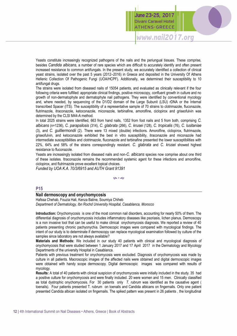
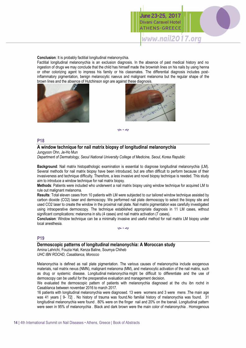
Factitial longitudinal melanonychia or post-inflammatory pigmentation Eleni Klimi Thriasio General Hospital, Athens, Greece Purpose of this study: To show a case of factitial melanonychia. Patient and method: A 10 year old boy of phototype four consulted for longitudinal brown lines on one of the fingers on his right hand that appeared one month prior to consultation. No history of atopy was reported and his past medical history was free of diseases. No ingestion of drugs has been reported. On clinical examination longitudinal brownish lines were observed on his nails and post inflammatory pigmentation on the surrounding the nail skin.
14 | 4th International Summit on Nail Diseases • Athens, Greece | Book of Abstracts
Conclusion: It is probably factitial longitudinal melanonychia. Factitial longitudinal melanonychia is an exclusion diagnosis. In the absence of past medical history and no ingestion of drugs we may conclude that the child has himself made the brownish lines on his nails by using henna or other colorizing agent to impress his family or his classmates. The differential diagnosis includes post-inflammatory pigmentation, benign melanocytic naevus and malignant melanoma but the regular shape of the brown lines and the absence of Hutchinson sign are against these diagnosis.
•
P18
A window technique for nail matrix biopsy of longitudinal melanonychia Jungyoon Ohn, Je-Ho Mun Department of Dermatology, Seoul National University College of Medicine, Seoul, Korea Republic Background: Nail matrix histopathologic examination is essential to diagnose longitudinal melanonychia (LM). Several methods for nail matrix biopsy have been introduced, but are often difficult to perform because of their invasiveness and technique difficulty. Therefore, a less invasive and novel biopsy technique is needed. This study aim to introduce a window technique for nail matrix biopsy. Methods: Patients were included who underwent a nail matrix biopsy using window technique for acquired LM to rule out malignant melanoma. Results: Total eleven cases from 10 patients with LM were subjected to our tailored window technique assisted by carbon dioxide (CO2) laser and dermoscopy. We performed nail plate dermoscopy to select the biopsy site and used CO2 laser to create the window in the proximal nail plate. Nail matrix pigmentation was carefully investigated using intraoperative dermoscopy. The technique established appropriate diagnosis in 11 LM cases, without significant complications: melanoma in situ (4 cases) and nail matrix activation (7 cases). Conclusion: Window technique can be a minimally invasive and useful method for nail matrix LM biopsy under local anesthesia.
•
P19
Dermoscopic patterns of longitudinal melanonychia: A Moroccan study Amina Lahrichi, Fouzia Hali, Kenza Baline, Soumya Chiheb UHC IBN ROCHD, Casablanca, Morocco Melanonychia is defined as nail plate pigmentation. The various causes of melanonychia include exogenous materials, nail matrix nevus (NMN), malignant melanoma (MM), and melanocytic activation of the nail matrix, such as drug or systemic disease. Longitudinal melanonychia might be difficult to differentiate and the use of dermoscopy can be useful for the preoperative evaluation and management decision. We evaluated the dermoscopic pattern of patients with melanonychia diagnosed at the chu ibn rochd in Casablanca between november 2016 to march 2017. 16 patients with longitudinal melanonychia were diagnosed. 13 were womens and 3 were mens .The main age was 41 years [ 9- 72] . No history of trauma was found.No familial history of melanonychia was found. 31 longitudinal melanonychia were found . 80% were on the finger nail and 20% on the toenail. Longitudinal pattern were seen in 95% of melanonychia . Black and dark brown were the main color of melanonychia . Homogenous

15 | 4th International Summit on Nail Diseases • Athens, Greece | Book of Abstracts
pattern was found in 95% of cases. Hutchinson sign were found in 1 case and pseudo hutchinson in 4 cases . The subungueal keratosis was found in 5 cases specially in toenail . No triangular sign or ulceration were found. Dots / globules was found in one case. 3 histological biopsy were made and we found 3 naevus and a malignant melanoma in situ. Melanonychia may have several etiologies, from physiologic lesions to malignant neoplasms, therefore the importance of early etiological diagnosis.Dermoscopy has been widely used by clinicians to improve the accuracy of diagnosing nail pigmentation. Currently, dermatoscopy of the nail bed and of the distal nail fold have also been used.3,4,5 Longitudinal melanonychia in adult must be monitored to avoid the onset of melanoma. In some cases, we can be helped by histology. However melanonychia in children don’t need biopsy despite the dermoscopic pattern of malignancy.But if the longitudinal melanonychia still progressing in adolescence we shoud do a biospy to diagnose a melanoma. Dermoscopic examination of longitudinal melanonychia provides useful information that could help clinicians to improve melanoma recognition.
•
P20
Delayed diagnosis of subungual melanoma misjudged as onychomycosis Thomas Fotas1, Eftychia Platsidaki1, Grigorios Xampsas1, Konstantina Fragia2, Pantelis Panagakis1, Dorothea Polydorou1
1Department of Dermatology and Venereology, Andreas Sygros Skin Hospital, Athens, Greece; 2Private Office, Athens, Greece Introduction: Subungual melanoma (SUM) is a rare variant of melanoma that occurs in the nail unit. Not only early skin biopsy of the nail is difficult but also in its early stage SUM is often misdiagnosed histopathologically. The delay in diagnosis results in progression of the disease, which may be associated with poor prognosis. Case Presentation: A 55-year-old man presented with a 1-year old history of melanonychia of the 1st fingernail which was gradually getting wider. Hutchinson's sign was positive. The nail was neither painful nor symptomatic. There was no personal or family history of melanoma. It was initially diagnosed as onychomycosis (culture positive for Trichophyton rubrum) and being treated with a specific topical and oral treatment for 6 months without any improvement. Biopsy specimens were taken from the nail bed and nail matrix. Histopathology revealed atypical melanocytes and a diagnosis of melanoma arising in a subungual nevus was made. SUM extended to a Breslow depth of 1.77 mm. The atypical melanocytes were positive for HMB-45. Plastic surgeons advised for distal phalanx amputation and sentinel lymph node biopsy. Discussion: SUM often presents clinically as longitudinal melanonychia. Gradually it becomes wider, more irregular in pigmentation, extends to involve the adjacent nail fold (Hutchinson sign), may develop a nodule, ulcerate or bleed and may cause nail dystrophy. The differential diagnosis of longitudinal melanonychia includes drugs, trauma, squamous cell carcinoma and fungal infection. Dermoscopy is helpful. When the pigment is heterogeneous in both the longitudinal and transverse axes, the likelihood of melanoma is greater. The diagnosis is confirmed by biopsy of the nail matrix and nail bed. As in our case delay in diagnosis is common, particularly when total melanonychia affects the toe. Thus dermatologists should be cautious when giving any advice to the patient with melanonychia about potential diagnoses and request biopsy when the diagnosis is not clear. References:
1. Elloumi-Jellouli A, Triki S, Driss M, Derbel F, Zghal M, Mrad K, Rhomdhnane KhB. A misdiagnosed nail bed melanoma. Dermatol Online J. 2010; 16:13.
2. Álvarez-Salafranca M, Hernández-Ostiz S, Salvo Gonzalo S, Ara Martín M. Proximal Subungual Onychomycosis Due to Aspergillus niger: A Simulator of Subungual Malignant Melanoma. Actas Dermosifiliogr. 2016. pii: S0001-7310(16)30428-8.
3. De Giorgi V, Saggini A, Grazzini M, Gori A, Rossari S, Scarfì F, Verdelli A, Chimenti S, Lotti T, Massi D. Specific challenges in the management of subungual melanoma. Expert Rev Anticancer Ther. 2011; 11:749-61.
4. Bae SH, Kim NH, Lee JB, Yun SJ. Total melanonychia caused by Trichophyton rubrum mimicking subungual melanoma. J Dermatol. 2016; 43(:1358-1359.

16 | 4th International Summit on Nail Diseases • Athens, Greece | Book of Abstracts
•
P21
Acquired ungual fibrokeratoma Amina Kissou, Siham Mansouri, Badreddine Hassam CHU IBN SINA, Rabat, Morocco Introduction: Acquired ungual fibrokeratoma is a relatively rare benign lesion commonly found on fingers and toes, but may also occur on palms and sole. We report the case of a 51-year-old patient. Observation: 51 years old male, smoker, who was consulting for a lesion evolving for 8 years, following a minimal trauma. It was a nodular lesion in ungual area of its right major. That was gradually increasing in size. The clinical examination found a flesh-colored mass measuring 1.5 cm in length. The nodule was firm, sessile, verruciform, but painless and not cystic on palpation. Mild hyperkeratosis was noted. The rest of the examination was normal. The rx-ray of the finger was normal. Surgery of the lesion was performed. The histological examination confirmed an Acquired ungual fibrokeratoma. There was no recidive after 4years of fellow-up. Discussion Acquired ungual fibrokeratoma is a relatively rare skin lesion, its incidence is unknown. According to previous reports, it is a more frequent lesion in men, with an average age of 40 years. The lesions were generally sessile, they are solitary lesions with slow growth and without signs of regression over time. Minor trauma plays a role in the etiology of Acquired ungual fibrokeratoma although this has not been proven. Excision is considered curative, and recurrence has been extremely rare. Various surgical techniques can be used to resect these tumors. Recurrences are rare.
•
P22
Toenail onychomycosis in chronic venous insufficiency Mateja Dolenc-Voljc1, Vid Bajuk2, Nada Kecelj Leskovec1, Tanja Planinsek Rucigaj1 1Department of Dermatovenereology, University Medical Centre Ljubljana, Ljubljana, Slovenia; 2Faculty of Medicine, University of Ljubljana, Ljubljana, Slovenia Background: Toenail alterations are very common in patients with chronic venous insufficiency (CVI) and may often mimic onychomycosis. The aim of our study was to assess the frequency and clinical characteristics of toenail onychomycosis in patients with CVI. Methods: In the prospective study 50 patients with CVI were included, 36 women (66.0±16.0 years) and 14 men (71.7±7.3 years). Patients’ history and clinical findings were evaluated. Clinical stage of CVI was determined according to CEAP (Clinical, Etiology, Anatomy, Patophysiology) classification from C1 to C6. Mycological examination of nail scrapings was performed with direct microscopy and cultivation on Sabouraud’s agar. Clinical type of onychomycosis and onychomycosis severity index (OSI) were assessed. Results were evaluated with Pearson’s chi-squared test (χ2), Fisher’s exact test, Student’s t-test and Cramer’s V coefficient (φc). Results: Toenail abnormalities were observed in 44 patients (88.0%). Onychomycosis was confirmed in 19 patients (38.0%). Distolateral subungual onychomycosis was most often observed. Majority of patients (68.0%)
17 | 4th International Summit on Nail Diseases • Athens, Greece | Book of Abstracts
had severe onychomycosis (OSI 16-35). Trichophyton rubrum was isolated in 10 patients, T. mentagrophytes var. interdigitale in 3 and Candida spp. in 2 patients. In 4 patients, onychomycosis was confirmed with microscopic examination only. Tinea pedis was diagnosed clinically in 28 patients (56%). Clinical stage of CVI correlated with OSI (φc=0,621). Severe onychomycosis was more frequent in higher stages of CVI. Diabetes mellitus was present in 12 patients (24.0%) and was more common in higher stages of CVI (p=0.002) and in patients with onychomycosis (p=0.002). Arterial hypertension and hyperlipidaemia were also common comorbidities. Conclusions: Our study showed that toenail onychomycosis is more frequent in CVI compared to estimated prevalence in general population. Patients with CVI often have other co-occurring risk factors for onychomycosis, especially diabetes mellitus. Advanced stage of onychomycosis and comorbidities has an important role in the choice of treatment and its results.
•
P23
New insights on the management of nail toxicities induced by anticancer drugs Nikolaos Makris National and Kapodistrian University of Athens, Faculty of Medicine, Athens, Greece Background: Cytotoxic and targeted anticancer therapies can cause nail tocixities. This study aims to present these nail changes and their management, which includes both preventive and therapeutic measures. Methods: A literature review took place in the electronic database ‘’Pubmed’’ and ‘’Google Scholar’’ covering the period 2008 to 2016 in order to provide the most comprehensive nail toxic reactions of anticancer drugs and their management. Criteria for the exclusion of articles were languages different from English, as well as articles reffered to other type of adverse events . Finally, forty-two (42) articles (qualitative, quantitative researches and systematic reviews) were included. Results: Nail abnormalities induced by both cytotoxic or targeted therapies may result from toxicity to the matrix, the nail bed or the periungual tissues and may involve all or some nails. Toxicity can be asymptomatic and limited to cosmetic concerns (Beau’s lines, onychomadesis, melanonychia, leukonychia), however, more severe effects, involving pain and discomfort can occur (paronychia, pyogenic granuloma-like lesions). Before instituting chemotherapy, patients should be educated regarding potential nail toxicities and prevention strategies should be implemented. Those recommendations vary from avoidance of repeated trauma or manipulation of the nail, restriction of contact with detergents , to the use of topical emollients and protective gloves. Treatment varies from debridement, partial or total avulsion, antiseptic soaks and topical steroids to surgical partial matricectomy under local anaesthesia. Conclusion: Recognizing nail toxic reactions, understanding their mechanisms and finding the appropriate treatment for each case is crucial for ensuring appropriate rapid intervention and thereby abrogating the need to delay or even withhold these essential treatments. Key words: nail, toxicities, anticancer therapies, cutaneous
•
P24
Conventional versus targeted chemotherapy: Pattern of nail affection Daniela Ledic Drvar1, Ivana Manola2 1University Hospital Centre Zagreb, Department of Dermatology and Venereology, School of Medicine, Zagreb, Croatia; 2 Polyclinic Manola, Zagreb, Croatia Background: Anticancer chemotherapy is associated with a variety of nail changes. However, there are differences in pattern of nail affection in patients receiving conventional versus targeted chemotherapy. Targeted therapies that lead to significant cutaneous side effects include primarily the epidermal growth factor receptor inhibitors (EGFRIs).

18 | 4th International Summit on Nail Diseases • Athens, Greece | Book of Abstracts
Common EGFRIs dermatologic adverse events are acneiform rash and xerosis. Paronychia is described in 5-20% of patients and usually develops after 1-2 months. In severe cases ingrown nail, periungual abscess and pyogenic granuloma-like lesions can occur. Conventional chemotherapy, especially taxans, mostly leads to onycholysis, subungual haematoma and abscesses. Management strategies include wearing comfortable shoes, trimming nails but avoiding aggressive manicuring, wearing gloves while cleaning. Topical corticosteroid and anti-inflammatory dose tetracyclines to decrease periungual inflammation and antibiotic soaks are advisable. Electrocautery, silver-nitrate and nail avulsion are recommended to eliminate excessive granulation tissue. Our aim was to present two patients who developed different pattern of nail affection while receiving different classes of antineoplastic drugs. Methods: Patient 1 A 46-year-old female patient with breast cancer presented to us with nail changes. She was receiving conventional neoadjuvant chemotherapy according to AC-T protocol (doxorubicin and cyclophosphamide followed by paclitaxel). Discoloration (chromo – and melano-nychia) and onycholysis were observed. Fungal superinfection was confirmed on her fingernails. Antifungal therapy was prescribed. Patient 2 A 57-year-old female patient with EGFR positive adenocarcinoma of the lungs, on EGFRI inhibitor, erlotinib, was referred to us because of acneiform erruption and nail changes. Nail changes presented as paronychia and granuloma pyogenicum-like lesions affecting fingernails, primarily thumbs. Patient was administered doxicyclne 2x100 mg for 14 days, followed by 1x100 mg. Both patients were given advice on supportive measures. Results: On follow-up visit both patients reported improved physical status and quality of life. Conclusions: Nails affection influences quality of life of oncology patients. It is important to recognise and treat as well as to administer efficaceous therapeutic measures timely.
•
P25
Persistent subungual superficial acral fibromyxoma successfully treated with surgical excision with preservation of nail unit Susan Pei, Molly Hinshaw University of Wisconsin Department of Dermatology, Madison, USA Background: Superficial acral fibromyxoma (SAF) is an uncommon benign soft tissue tumor usually located on digits. Subungual or periungual SAFs have high reported local recurrence rate of up to 24%, at times necessitating distal digital amputation for clearance. We hypothesize conventional excision may treat persistent subungual SAF. A 49 year old male presented for a 2-3 year history of a slowly enlarging, asymptomatic lesion affecting the nail unit of the right hand second finger. Examination showed a firm subungual lesion under a relatively unremarkable nail plate. Methods: This is a longitudinal 17 months follow-up of a single case. Radiographic imaging, biopsy with hematoxylin and eosin and immunohistochemistry, and surgical excision were performed. Results: X-Ray showed irregularity, cortical loss and remodeling of the radial and dorsal aspect of the distal phalanx of the index finger due to long-standing mass. Partial nail avulsion revealed an exophytic 1.2cm by 0.8cm soft firm rubbery nodule involving the radial half of the nail bed and distal matrix, the entire domed portion of which was shave biopsied. Histopathology showed in the onychodermis a neoplasm composed mostly of spindle-shaped cells arranged loosely in a fascicular architecture in myxoid stroma. Immunoperoxidase staining of tumor cells was positive for CD10, CD99 and negative for S100, CD34, EMA, Desmin and MUC4. Alcian blue stain showed abundant stromal mucin. A diagnosis of SAF was made. Within 9 months after biopsy, the tumor regrew to a size

19 | 4th International Summit on Nail Diseases • Athens, Greece | Book of Abstracts
of 2cm and was painful. Partial nail avulsion and excision of tumor with 1mm margins was performed without further recurrence at 6 months and with successful preservation of the nail unit. Conclusions: We present a case of a persistent subungual SAF successfully treated with surgical excision with 1mm margins, with resultant preservation of the nail unit and excellent cosmesis.
•
P26
Nail toxicity of Taxanes O. ELJouari, K. Moustaide, H. Baybay, S. Gallouj, F.Z. Mernissi Department of Dermatology CHU HASSAN II, FEZ, Morocco Background: Taxanes (docetaxel and paclitaxel) are the most commonly pre-scribed anticancer drugs approved for the treatment of different neoplasms. The dermatological adverse effects of these antimicrotubules agents are very frequent in particular nail toxicity who true incidence has not been estimated and tends to vary significantly in the literature. In our study we analyze the different nail changes in the patients who received the taxanes. Material and method: Prospective study for a year included 80 cases referred from the oncology department for nail toxicity Compared to 104 patients received taxanes. Result: Nail toxicity was demonstrated in 77% of cases, the highest incidence 39, 8 % with paclitaxel once-weekly and 37.2% with docetaxel every three-week. The toxicity was noted after the 4th cure, increases with the number of cycles, resulting from a direct toxic effect. The changes affected the nail matrix, the nail bed and the periungual tissue Onycholysis accounted for 90% of the toxicity, the number of nails affected was highly variable, 50% was diffuse, with superiority for the fingernails. Subungual hyperkeratosis was also highly observed 78%, while the subungual haematoma, haemorrhage, Or abscess with purulent discharge were noted in 30% of cases. Paronychia was objective in 28 patients. Management of nail changes depends on the type of nail Affliction and the impact on activities of daily living. All patients benefited from an education on prophylactic measures (emollients cream, protective varnish, cotton gloves, trauma must be avoided, wearing frozen gloves/socks during chemotherapy, use ice packs), 26 patients was treated with an appropriate antibiotic and 16 patients with potent topical steroids. Conclusion: Identification and management of nail adverse effects is critical for maintaining the quality of life in cancer patients and for minimizing dose modifications of their antineoplastic regimen.
•
P27
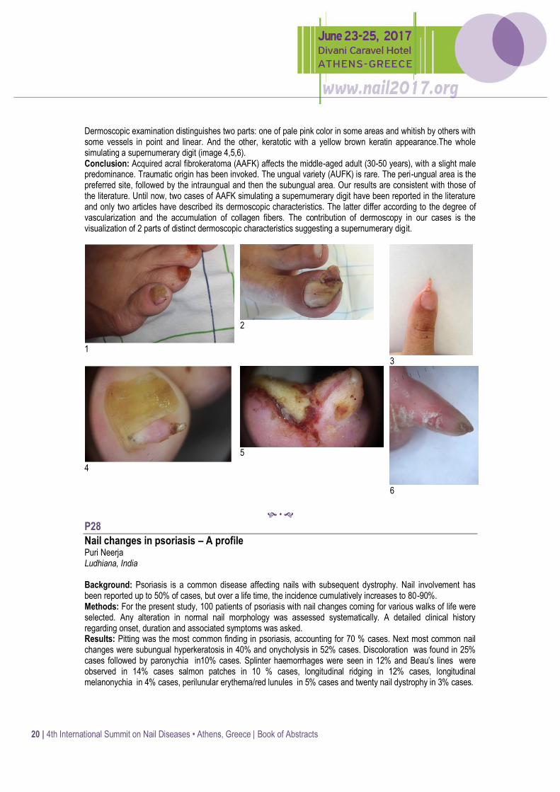
Acquired ungual fibrokeratoma simulating a supernumerary digit: Interest of dermoscopy Mariame Meziane, Salma Salim, Ilhma Meknassi, Badreddine Hassam University Hopsital Ibn Sina, Rabat, Morocco Background: Acquired ungual fibrokeratoma (AUFK) is a benign and rare fibrous tumor of unknown etiology. We report a series of 3 rare cases of AUFK simulating a supernumerary digit with description of clinical aspect and dermoscopic characteristics. Methods: It is a series of three rare cases of AUFK simulating a supernumerary digit, recruited in dermatology department of university hospital of Rabat. Clinical and dermoscopic aspects were noted and anatomo-pathological study carried out after total removal of the lesions. Results: The average age is 34 years; the sex ratio is 2 with a concept of trauma in 2 cases. Two AUFK are localized at toes and the third one on the index. It is based on periungueal area for 2 cases and inside the nail for the third one. The lesions are glove-shaped, infracentimetric, firm in consistency, with a fleshy part and a keratosic one (image 1,2,3).
20 | 4th International Summit on Nail Diseases • Athens, Greece | Book of Abstracts
Dermoscopic examination distinguishes two parts: one of pale pink color in some areas and whitish by others with some vessels in point and linear. And the other, keratotic with a yellow brown keratin appearance.The whole simulating a supernumerary digit (image 4,5,6). Conclusion: Acquired acral fibrokeratoma (AAFK) affects the middle-aged adult (30-50 years), with a slight male predominance. Traumatic origin has been invoked. The ungual variety (AUFK) is rare. The peri-ungual area is the preferred site, followed by the intraungual and then the subungual area. Our results are consistent with those of the literature. Until now, two cases of AAFK simulating a supernumerary digit have been reported in the literature and only two articles have described its dermoscopic characteristics. The latter differ according to the degree of vascularization and the accumulation of collagen fibers. The contribution of dermoscopy in our cases is the visualization of 2 parts of distinct dermoscopic characteristics suggesting a supernumerary digit.
1
2
3
4
5
6
•
P28
Nail changes in psoriasis – A profile Puri Neerja Ludhiana, India Background: Psoriasis is a common disease affecting nails with subsequent dystrophy. Nail involvement has been reported up to 50% of cases, but over a life time, the incidence cumulatively increases to 80-90%.
Methods: For the present study, 100 patients of psoriasis with nail changes coming for various walks of life were selected. Any alteration in normal nail morphology was assessed systematically. A detailed clinical history regarding onset, duration and associated symptoms was asked. Results: Pitting was the most common finding in psoriasis, accounting for 70 % cases. Next most common nail changes were subungual hyperkeratosis in 40% and onycholysis in 52% cases. Discoloration was found in 25% cases followed by paronychia in10% cases. Splinter haemorrhages were seen in 12% and Beau’s lines were observed in 14% cases salmon patches in 10 % cases, longitudinal ridging in 12% cases, longitudinal melanonychia in 4% cases, perilunular erythema/red lunules in 5% cases and twenty nail dystrophy in 3% cases.

21 | 4th International Summit on Nail Diseases • Athens, Greece | Book of Abstracts
Conclusions: Pitting is most common nail abnormality seen in psoriasis and affects finger nail more commonly than toe nails. Pitting in psoriasis is deep, irregularly and randomly placed. In psoriasis, onycholysis commonly starts at the free distal edge of the nail plate. In nail psoriasis, Onychomycosis due to candida, non-dermatophytes and bacterial infections like pseudomonas is seen commonly but dermatophytes are never isolated in psoriatic nails. Splinter haemorrhages are due to trauma together with increased vascularity and fragility of the nail bed dermis.
•
P29
Secukinumab: Efficacy in patients with moderate-to-severe plaque and nail psoriasis; a 32-weeks observational clinical study Ioanna Triantafyllopoulou, Dorothea Polydorou, Anastasios Vogiatzoglou, Pantelis Panagakis Psoriasis Clinic, Athens, Greece Background: Psoriasis is a chronic condition that most often requires lifelong effective and safe treatment. Secukinumab is a recombinant, fully human immunoglobulin monoclonal antibody with a selective binding and neutralization of interleukin-17A. Our purpose was to demonstrate the efficacy of this relatively new agent in plaque and nail psoriasis of selected patients of the Psoriasis Clinic of “A. Syggros” Hospital. Methods: Eighteen patients (12 men ; 6 women) suffering from plaque as well as nail psoriasis were followed –up after starting receiving Secukinumab. Ten patients presented with both fingernails and toenails affected, the remaining eight presented with psoriasis of the toenails. All patients had previously received methotrexate or cyclosporine while 4 of them had also received infliximab which was interrupted due to loss of efficacy (mean time of administration 52 weeks). Mean PASI (Psoriasis Area and Severity Index) score at week 0 was 17 and mean NAPSI (Nail Psoriasis Severity Index) score was 35. Three-hundred mg of Secukinumab were administered by subcutaneous injection at Weeks 0, 1, 2, 3, and 4 followed by 300 mg every 4 weeks. Results: PASI and NAPSI scores were evaluated at weeks 16 and 32. PASI90 was achieved in all patients (100%) as early as week 16. At week 16 all patients (100%) presented with a 50% improvement of their NAPSI score. At week 32, eleven patients (61%) continued to show at least 75% improvement of their NAPSI score (fig1,2). All patients tolerated well Secukinumab and continue receiving it. Conclusions: In our set of patients, administration of Secukinumab was associated with sustained efficacy in terms of both plaque and nail psoriasis, and a good safety profile. These results are in line with the only double-blind randomized 2-year study of Secukinumab (TRANSFIGURE study)
Fig 1. Fingernail involvement of patient 5 at week 0.
Fig 2. Improvement of the NAPSI score of the same patient at week 32.
•

22 | 4th International Summit on Nail Diseases • Athens, Greece | Book of Abstracts
P30
Dermoscopy of nail psoriasis (prospective study of 30 cases and review of the literature) Salma Salim, Badreddine Hassam, Mariame Meziane University Hospital Ibn Sina, Rabat, Morocco Background: Nail involvement is classic in psoriasis. It can be isolated, inaugural or a part of a wider skin involvement.The dermoscope is an effective tool for detection of subclinical signs, early diagnosis and evaluating the severity of nail psoriasis. Methods: This is a prospective study carried out over 6 months in dermatology department of Rabat university hospital. 30 cases of nail psoriasis were collected. Clinical and dermoscopic aspects were noted. Clinical severity and nail involvement were assessed by PASI and NAPSI scores respectively. We included consenting patients, with all types of psoriasis and not receiving topical nor systemic treatment. The data were analyzed using the SPSS 20.0 software. Results: In our study, the mean age is 42 years, the sex ratio 0.6 and the average duration of the disease 12.7 years. The average number of nails affected per patient is 7. The most frequent type of psoriasis is psoriasis in plaques (85%), but other forms may be associated (56.7%).The average PASI and NAPSI scores are 16.4 and 45.4 respectively. By comparing the clinical and dermoscopic data, superiority of dermoscopy is demonstrated in detection of different signs of nail psoriasis; in particular nail bed involvement and vascular abnormalities. Vascular involvement particularly affects vessels of the nail bed and those of the proximal fold, vessels of hyponychium are much less visualizable (image 1,2,3). The relationship between PASI/NAPSI and vascular involvement is positive and statistically significant (image 4). Conclusion: Our study demonstrates the superiority of dermoscopy compared to the clinic in analysis of nail involvement in psoriasis, especially nail bed involvement and vascular abnormalities. Dermoscope is a non-invasive, rapidly applied and inexpensive tool that facilitates the early diagnosis of nail psoriasis. Subclinical lesions can be detected and appropriate treatment early instituted. Vascular analysis is particularly important in assessing the severity of the disease.
•

23 | 4th International Summit on Nail Diseases • Athens, Greece | Book of Abstracts
P31
Pattern of nail involvement in Indian patients with chronic plaque psoriasis Neena Khanna, Tamanna Nazli All India Institute of Medical Science, New Delhi, India Background: The prevalence of nail changes varies from 10% to 50% in patients with chronic plaque psoriasis (CPP) and is thought to be higher (80%) in patients with psoriatic arthritis. The primary objective was to determine prevalence and pattern of nail changes in Indian patients with CPP. The secondary objectives was to correlate severity of nail changes (using Nail Psoriasis Severity Index; NAPSI) with patient demographics (age, gender), disease demographics (age of patient at onset ofdisease, duration of disease), disease severity (body surface area; BSA and psoriasis area severity index; PASI) as also psoriatic arthritis. Methods: Data on nail changes (both due to nail bed and nail matrix involvement) and the NAPSI score was retrospectively collected from 271 consecutive case files of patients with CPP attending the Dermatology OPD at All India Institute of Medical Sciences, New Delhi, India. Patient demographics (age, gender), disease demographics (age of patient at onset of disease, duration of disease), disease severity (using BSA and PASI) as also presence of psoriatic arthritis were also noted from the patient’s case file. Results: Of the 271 psoriasis patients, 102 (37.6% 95% CI: 31.8, 43.7) had nail changes. The mean age of the patients was 36.4±13.0 years, with 76.4% being males and23.6% being females.The most common changes observed included nail pitting (30.7%), followed by onycholysis (24.3%) and dyschromia (20.3%). There was no correlation between age of onset of skin manifestations (r=0.16, p= 0.10), duration of disease (r=0.14, p= 0.15), PASI (r=0.04, p=0.66), BSA (r=0.02, p=0.83) and presence of arthritis (p=0.63) with the NAPSI score. Conclusions: Nail changes are seen in a third of Indian patients with CPP with nail pitting and onycholysis being the commonest. However, there is no correlation between age of onset of skin manifestations, duration of disease, arthritis, PASI and BSA with the NAPSI score.
•
P32
Psoriasis as an independent risk of renal disease: A nationwide retrospective cohort study from 1996 to 2010 Chien-Hui Hong1, Wei-Tai Yu2, Hung-Pin Tu2, Chih-Hung Lee3 1Kaohsiung Veterans General Hospital and National Yang Ming University, Kaohsiung, Taiwan; 2Kaohsiung Medical University, Kaohsiung, Taiwan; 3Kaohsiung Chang Gung University Hospital and Chang Gung University College of Medicine, Kaohsiung, Taiwan Background: Psoriasis is a chronic inflammatory disease that affects the skin, joints, and cardiovascular system. Although a potential association of end-stage renal disease (ESRD) has been demonstrated in patients with psoriasis, whether the use of cyclosporin in these patients would modulate the course of ESRD remains doubted. Further, the association between psoriasis and renal diseases in general remains unclear. This study aimed to investigate the risk of renal disease in psoriatic patients with or without the use of cyclosporin. Methods: We performed a retrospective cohort study using the National Health Insurance Database of Taiwan from 1996 to 2010 to identify patients with psoriasis, renal disease, chronic renal failure, end-stage renal disease, and the use of cyclosporin. Totally, 3502 psoriatic patients and 10,506 matched population comparisons were identified. The hazard ratios (HR) for renal events during the follow-up period were computed. Results: Patients with psoriasis had an increased risk of chronic renal failure (HR = 3.00, 95% confidence interval [CI] 2.30-3.93, P<0.0001) and end-stage renal disease (HR = 2.03, 95% CI 1.04-3.96, P=0.0393). Cyclosporin increased the risk of renal disease in patients without psoriasis (HR=6.34, 95% CI 3.57-11.26, P<0.0001), but not in patients with psoriasis (HR=1.33, 95% CI = 0.66-2.69, P=0.4299). Conclusion: Psoriasis is an independent risk of chronic renal failure and end-stage renal disease. Cyclosporin, a commonly used antipsoriatic agent, does not significantly increase the risk of renal disease in patients with psoriasis.
•

24 | 4th International Summit on Nail Diseases • Athens, Greece | Book of Abstracts
P33
Acrodermatitis Continua of Hallopeau: A case report Kalliopi Tomai, Dimitra Tasioula, Aikaterini Malouhou, Christina Vourlakou, Georgios Anastasiadis Evangelismos Hospital, Athens, Greece Acrodermatitis Continua of Hallopeau (ACE) is a rare inflammatory disease characterized by pustular eruptions first located on the tips of fingers and toes. It often presents at the nail folds of one or two fingers and then extends to others. Sometimes it remains in only one finger and that makes the diagnosis difficult. We present such a case. A 64-year-old woman presented to the dermatological department of our hospital because of the loss of the nail and inflammation of the nail bed of the third finger of her left hand, appeared two years ago. Clinical examination revealed edema of the nail bed, exudation and periungual pustules on the tip of the finger. It was treated with antifungal topical and systematic therapy for two years without improvement. A skin biopsy was performed which revealed epithelial hyperplasia and intra-epidermal spongiform pustules. Diagnosis of Hallopeau was made and she started therapy with acitretin. One month later there was significant clinical improvement. Acrodermatitis continua is an intriguing entity. The clinical diagnosis of Hallopeau's acrodermatitis restricted to nail and digital pulp is difficult. Even dermatologists many times fail to recognize it, so they must always be alert of this condition.
•
P34
A constellation of nail manifestations in autoimmune blistering disorders Vaishnavi Gopal, Manjunath Shenoy, Vishal Bharath Yenepoya Medical College, Mangalore, India Background: Autoimmune blistering disorders are a heterogenous group of disorders associated with autoantibodies directed against desmosomal or basement membrane zone structural proteins. Cutaneous and oral findings are the main manifestations. The incidences of significant nail changes have been reported in very few studies. Material and methods: We report a constellation of nail changes in 30 consecutive cases of autoimmune blistering disorders- pemphigus vulgaris and its variant pemphigus vegtans, pemphigus foliaceous, paraneoplastic pemphigus and bullous pemphigoid. Detailed clinical examination and laboratory work up with immunofluorescence studies were done. Special attention was given to nail changes. Results: Out of 30 cases, 21 cases were pemphigus vulgaris, 3 were paraneoplastic pemphigus, 3 pemphigus foliaceous, 2 bullous pemphigoid and 1 case of pemphigus vegetans. 21 cases had nail changes, most common findings being onychorrhexis and paronychia indicating defective keratinisation of proximal nail matrix and infection of nail folds respectively. The next common findings were onychomaedesis and beaus lines indicating arrest of nail matrix activity. Other nail changes- pterygium & distal lateral subungual onychomycosis. Out of 21 cases in pemphigus vulgaris group 7 had chronic paronychia suggesting lateral nail fold onycholysis. Limitations- Small sample size and nail biopsy could not be done in all cases. Conclusion: These findings implicate that the inflammatory nature of underlying cutaneous disease is reflected conspicuously in nails- conveying that nail mirrors the intemperate nature of autoimmune diseases. Nail examination can also throw a light on its diagnostic significance and the severity of damage to the protein antigens. Certain nail changes such as pterygium are irreversible; however others can revert with treatment.
•

25 | 4th International Summit on Nail Diseases • Athens, Greece | Book of Abstracts
P35 Nail fold dermoscopy in systemic diseases: A prospective study about 70 patients Salim Gallouj, Nisrine Amraoui, Fatima Zahra Mernissi Dermatology Department, Hassan Second University Hospital, Fez, Morocco Nail fold dermoscopy is a non-invasive tool for the qualitative and quantitative study of microcirculation in the proximal nasal fold. It is increasingly used in dermatology in the study of systemic diseases. We describe the nail fold dermoscopic aspect of 70 patients followed for various systemic diseases . The average age of our sample was 39.47 years, including 64 women and 6 men, 38 patients had a Raynaud phenomenon evolving over an average duration of 2.8 years. The average duration of follow-up was 2 years. 15 cases of systemic scleroderma were identified, 26 patients had lupus, 16 had dermatomyositis, 4 had gougerot syndrom, 9 had mixed connective tissue diseases with a sclerodermiform pattern in 10, 8, 8, 2, 4 Patients. The presence of megacapillaris was significantly associated with a progression time of more than 2 years during scleroderma and the presence of avascular zones was associated with the positivity of anti-Scl 70. In the case of lupus, haemorrhages were More common if pulmonary or haematological joint signs. A sclerodermiform pattern was found significantly if associated pulmonary or haematological involvement. Nail fold dermoscopy may also be used to assess the risk of developing digital ulcers and visceral complications in scleroderma patients. Knowledge of the semiology of nail fold dermoscopy is not complicated and allows the dermatologist to comfort his diagnosis if there is a suspicion of a systemic disease, to reassure his patient before a primary raynaud and to have an idea about the prognosis of The pathology of the patient.
•
P36
Beau’s lines – Epidemiological and etiopathogenic data Vassiliki Vosynioti, Themis Sgontzou, Apostolos Bantavanos, Christina Stefanaki, Elina Remountaki, Eirini Potouridou, Georgios Kontochristopoulos, Alexandra Katsarou-Katsari Pediatric Dermatology Clinic, 1st Department of Dermatology & Venereology,”A. Syggros” Hospital, National and Kapodistrian University of Athens, Athens, Greece Background: Nail disorders are not so common in children. However, nail changes may be useful for the clinical identification of systemic diseases. Beau's lines are grooves or depressions that run across nails. Most common causes of Beau's lines (onychomadesis) in children are malnutrition, infections, psoriasis, a toxic event as chemotherapy and late intra-uterine or birth trauma. The aim of the study is to determine the epidemiology of Beau's lines and the possible etiologic factors in childhood. Methods: The study was conducted in the Pediatric Dermatology Clinic of the 1st Department of Dermatology & Venereology University of Athens, "A.Syggros" Hospital,in the period Jan. 2013 to Feb. 2017. A total of13,852 outpatients aged between 0 and 12 years old were initially screened.28 patients were diagnosed with Beau's lines in this period. Patients were classified into 3 age groups (0-4, 5-8 and 9-12 years old). Results: From the 28 patients with Beau's lines, 17(60,7%)were boys and 11(39,3%) were girls. 14, 8, 6 patients were classified into 0-4, 5-8, and 9-12 years old respectively. Acute febrile illness, due to upper respiratory tract virus diseases was the most common cause in 20 patients (71,4%). Hand-foot-and-mouthdisease, according to clinical history and laboratory examinations was the second cause in 5 patients(17,9%) andfinally a nervous habit of nail picking in 3 patients(10,7%). Most of the patients presented bilateral affection on the hand nails and additionally in two children the toe nails were affected simultaneously. Conclusions: In the present study, Beau's lines were more common in boys, with a ratio of 2/3 for boys and girls. Half of the affected children were between 0 to 4 years old. Moreover, Beau's lines mostly appeared after acute illness and secondarily after habitual nail picking.
•

26 | 4th International Summit on Nail Diseases • Athens, Greece | Book of Abstracts
P37
Contribution of nailfold dermoscopy in connective tissue diseases Alaa Aboutaam1, Rojosoa Razanapinaritra1, Wassila Bouissar2, Khadija Echellali2, Amina Moudatir2, Fatima Zahra Alaoui2, Hassan EL Kabli2, Soumya Chiheb1 1Department of dermatology and venereology, UH Ibn Rochd, Casablanca, Morocco; 2Department of Internal Medicine, UH Ibn Rochd, Casablanca, Morocco Background: The study of nail fold capillaries seems to provide elements that can guide the diagnosis of these pathologies, especially with Raynaud's phenomenon. Dermoscopy is a non-invasive and easy-to-use device, when capillaroscopy is not available.The aim of our study was to describe the different nail fold capillaries features of patients with connective tissue diseases or Raynaud's phenomenon. Methods: All patients hospitalized for connective tissue disorders such as systemic scleroderma, systemic lupus erythematosus, Gougerot Sjogren's syndrome, dermatomyositis, or mixed connective tissue disease during the period from June 2016 to February 2017. Cases were collected within the departments of Internal Medicine and Dermatology and Venereology, UH Ibn Rochd of Casablanca. The dermoscopic images were taken using the Handyscope. The statistical studies were carried out using the SPSS 17.0 software. Results: Thirty one female patients with an average age of 38.24 (± 13.71, range 15 years to 68 years) were enrolled. The secondary Raynaud's phenomenon was found in 62% of the cases. The pathologies identified were mainly systemic lupus erythematosus (52.4%), systemic scleroderma (28.6%), Gougerot Sjogren syndrome in 9.5% and dermatomyositis and mixed connective tissue disease in 4.8% each. The mean duration of the pathology was 3 years and a half, with extremes ranging from 2 months to 16 years. The patients were under general corticosteroids at the time of the dermoscopic examination in 76.2% of the cases. According to the scleroderma pattern, capillary distribution was heterogeneous in 47.6%, mega capillaries in 47.6%, punctiform haemorrhages in 68.1%, avascular areas in 28.6%, and hypertrophic cuticles in 47.6%. The analytical study showed that scleroderma is the most correlated pathology to enlarged capillaries (p = 0.002) and to the presence of microhemorrhage (p = 0.007), and also hypertrophy of the cuticle (p = 0.04). Conclusions: The nail fold dermoscopy is great contribution and simple in the diagnosis and prognosis of connective tissue diseases. A study was carried out by the dermatology team of the CHU Hassan II of Fes in 2015, showing the significant association between frequent haemorrhages if associated pulmonary and haematological involvement in lupus, the presence of megacapillars with a duration of evolution more than 2 years during scleroderma, and the presence of avascular zones which was associated with the positivity of anti-Scl 70. In our study, the hypertrophic aspect of the cuticle seems to be characteristic of progressive systemic scleroderma as well.Further studies are needed to support the contribution of dermoscopy in the diagnosis of systemic diseases, or the phenomenon of secondary Raynaud essentially.
•
P38
Nail disorders in patients with chronic renal failure Niema Aqil, Kaoutar Moustaide, Aicha Nassiri, Salim Gallouj, Fatima Zahra Mernissi Department of Dermatology of University Hospital Hassan II, Fez, Morocco Background: Ungual manifestations occurring in patients with end stage renal failure are polymorphic and diverse. The aim of our study was to assess the prevalence and characteristics of different ungual manifestations in patients with end stage renal failure. Patients and methods: We led a transverse investigation of all patients with end stage renal failure followed in the department of nephrology of University Hospital Hassan II, during a period of three months from 01 August 2016 to 01 November 2016. Results: We examined 70 patients (34 men and 36 women). The renal failure history ranged from 6 months to 13 years.

27 | 4th International Summit on Nail Diseases • Athens, Greece | Book of Abstracts
A total of 56/70 (80%) patients had ungual abnormalities. These included absence of lunula (51.7% of patients), half and half nails (33.9%), splinter hemorrhages (58.9%), melanonychia (14.2%), onycholysis (71.4%), Beau’s lines (35.7%), koilonychias and Muehrcke lines (7.1%), onychomycosis (48.2%), pincer-nail deformity, brittle nails and onychogryphosis (10.7%), leukonychia (25%), longitudinal rifging (64.2%), subungual hyperkeratosis (55,3%), paronychia (5.3%), nail clubbing ( 1,7%). Our results find no significant relationship between the ungual disorders and patient age or history of renal failure. Conclusion: Our study found different nail changes in uraemic patients, these abnormalities varied from the most frequent (onycholysis) to rare conditions such as nail clubbing. However, the cause of them remains obscure and could not be traced to a particular abnormality in the renal condition, medication or the procedure itself and it needs further investigations. The treatment is in most cases symptomatic. Finding pathogenesis of these changes would help us prevent them.
•
P39
Nailfold capillaroscopic changes in patients with Type 2 Diabetes Mellitus: An observational, comparative study Deepak Jakhar1, Chander Grover1, Archana Singal1, S.V Madhu2, Gopal Das3 1Department of Dermatology & STD, UCMS & GTBH, New Delhi, India; 2 Department of Internal medicine, UCMS & GTBH, New Delhi, India; 3 Department of Ophthalmology, UCMS & GTBH, New Delhi, India
Background: Type 2 Diabetes Mellitus (T2DM) is a chronic metabolic disease known to affect the microvascular circulation leading retinopathy, nephropathy and neuropathy. This pilot study attempted to describe and possibly quantify Nailfold capillaroscopy(NFC) changes seen in Type 2 diabetics compared to normal controls; and also to assess possible differences in NFC findings in diabetics with or without microvascular complications. Method: After taking institutional clearance, a total of 96 patients with T2DM (ADA Criteria) and 50 healthy controls (Group 1) were included in this observational comparative study done at University College of Medical Sciences & GTB hospital, New Delhi, India. NFC was done for all 10 finger nails in each patient and microvascular architectural changes were recorded. The diabetics were further sub-divided into two groups: those with microvascular complications (Group 2, n=46) and those without (Group 3, n=50). Results: The demographic profile was similar in all the 3 groups. Overall 80.2 % (77/96) patients with T2DM showed NFC changes (p=0.00). Mean capillary density was reduced in diabetics [Group 2 (6.57±1.02 capillary/mm) and group 3 (7.03±1.09 capillary/mm)] as compared to healthy controls (Group 1, 7.63±1.12 capillary/mm). Angulated capillaries and receding capillaries were unique findings recorded in diabetics. 89.13% of Group 2 patients had NFC changes as compared to 72% in Group 3 (p=0.035). Specific changes in capillary morphology, like capillary drop outs (p=0.012), tortuosity (p=0.035), meandering capillaries (p=0.004) and bizarre capillaries (p=0.002) were significantly higher in Group 2. NFC changes didn't significantly correlate with disease duration. Conclusion: Our study suggests that NFC may have a role in detecting and quantifying microvascular changes in T2DM, delineating patients at risk of microvascular complications.
•
28 | 4th International Summit on Nail Diseases • Athens, Greece | Book of Abstracts
P40
Nail involvement during pemphigus Soumiya Chiheb, Bouchra Baghad UHC IBN ROCHD, Casablanca, Morocco Introduction: The frequency and types of nail changes in pemphigus are rare and unclear. Our aim is to determine the frequency and types of nail changes during pemphigus, and their correlation with the severity of the disease. Patients and methods: We conducted a retrospective and prospective study of patients hospitalized in our Department between January 2010 and 2017. Clinical and paraclinical data of patients were collected from medical records. Results: We collected 33 cases with a pemphigus with nail involvement , sex ratio H/F was 1.1. The average age was 46.5 ans (range from 17 to 78). Nail involvement was found in two types of pemphigus, with a predilection during pemphigus vulgaris . Cutaneous and mucous lesions were associated in majority of patients .The attack was inaugural in 38% of patients . The main seat was hand .The nail invasion was mainly a type of onychomadesis ,paronychia, onycholysis, and rarely , periungueal bullous, perionyxis and under ungual hemorrhage Patients were treated with corticosteroids and immunosuppressant with a paralletic evolution with cutaneous lesions. Discussion: Nail manifestations during the pemphigus are polymorphous and frequent but underestimated. Nail invasion during pemphigus should be systematically investigated .It represents a Criteria for the severity of a pemphigus often requiring the use of immunosuppressant.
•
P41
Hand held dermatoscope is useful in early diagnosis of collagen diseases Simona Fratila1, Roxana Rednic2, Mihaela Matei2 1University of Oradea, Faculty of Medicine and Pharmacy, Oradea, Romania; 2Clinical Emergency County Hospital of Oradea, Oradea, Romania Background: The aspect of the capillary network of the nail folds is an important criteria for the diagnosis of collagen diseases and it can be observed by several methods including dermatoscopy. Even if frequently used for the examination of pigmented and acromic skin lesions, dermatoscopy is still not widely used for capillaroscopy. Methods: We present 2 patients who attended dermatological outpatient clinic for skin complaints and were diagnosed with systemic scleroderma and dermatomyositis respectively, by examining the nail fold capillaries with the hand-held dermatoscope. Results: Female patient aged 38 presented pain and tiny ulceration of the fingertips. The dermatoscopical examination revealed subunghial splinter hemorrhage and in the proximal nail fold enlarged capillaries and hemorrhagia, sustaining the clinical suspicion of systemic scleroderma. Subsequent paraclinical investigation confirmed the diagnostic. The evolution of the disease was rapid and severe. Female patient aged 64 with diffuse erythematous violaceous patches periocular, on the chest, upper back and dorsal interfalangeal joints, slightly pruritic and with no myalgia was examined dermatoscopically at the proximal nail fold. Desorganisation of the normal distribution of capillaries, enlarged capillaries and avascular areas were observed, sustaining the clinical suspicion of dermatomyositis that was confirmed paraclinically afterwards. It proved to be a paraneoplastic form, as breast cancer was diagnosed in the same time. Conclusions: Dermatoscopy is a practical, effective and non-time consuming method for detection of nail fold capillary changes in patients with connective tissue disease.
•

29 | 4th International Summit on Nail Diseases • Athens, Greece | Book of Abstracts
P42
Rubinstrein-Taybi syndrome nails Sofia Couto Da Rocha, Joao Larguito Claro Algarve Central Hospital, Faro, Portugal Background: Rubinstein-Taybi Syndrome is described since 1957, when the first case was reported. Only in 1963 Rubinstein and Taybi did the description of a syndrome of facial abnormalities, short stature, beaked nose, anti-mongoloid slant of eyes, heavy arched eyebrows, microcephaly, broad thumbs and hallux which are often angulated, hyper-extensive joints, small tilted pelvis, hypertrichosis, mental retardation, broad fingers and great toes. According to literature reports the condition is extremely rare, with a prevalence of 1:300,000 up to 1:1,000,000. Case Report: We report a case of a 6 years old male diagnosed with RTS at birth that presented painful ingrown hallux nails in both feet. The hallux were broad bilaterally, incurvated with mild hypertrophy until the ungula labia of the nail folds. In the nail bed there were signs of inflammatory reaction and pain when touched. It was performed a partial nail avulsion with phenolisation, using tourniquet, local anesthetic with rupivacain and nitrous oxide via mask. We obtained very good outcomes at one month, 3 months and 6 months time, with no recurrence. Conclusion: In literature there are just a few cases reported of RTS, and even less in number of onychocryptosis, Nevertheless it is presented in 96% of the cases reported. Partial nail avulsion with phenolisation showed good outcomes until 6 months evaluation.
•
P43
Nail changes mimicking lichen planus in chilblains Aashim Singh, Saurabh Bhatia, Somesh Gupta All India Institute of Medical Sciences, New Delhi, India Background: Chilblains or perniosis is characterized by development of pruritic or painful erythematous and violaceous papules, plaques and vesicles over acral areas on exposure to cold conditions. Most of the cases resolve with rewarming and cold protection without any adverse events. Two patients presented to us with chilblains who developed severe nail changes mimicking nail lichen planus. Clinical cases: A young adult male and a female presented with chilblains on cold exposure. Severe brittleness and thinning of nails developed with onset of chilblains in winters followed by spontaneous and painless falling off of the nails. Some regrowth of nails occurred during summers with recurrence in winters. Besides anonychia, pterygium formation and longitudinal striations were also present. There was no history of smoking, drug exposure, Raynaud’s phenomenon or any other features suggestive of connective tissue disease or systemic illness. Anti-nuclear antibodies were negative. Nail lichen planus was considered initially as the clinical differential in both, however, the histopathology findings were not consistent and detailed history and temporal correlation indicated that these nail changes were related to severe chilblains. Both the patients were adviced cold protection

30 | 4th International Summit on Nail Diseases • Athens, Greece | Book of Abstracts
and oral nifedipine 10 mg twice daily which helped in improving the chilblains, however the nail condition persisted and appeared to be permanent. Conclusions: The pathophysiology of chilblains is said to be an abnormal vascular response to cold temperatures. Chronic vasoconstriction leading to nail matrix ischemia can be postulated as a cause of the nail changes in our patients. Our patients developed severe cosmetically bothering nail changes mimicking lichen planus and even anonychia which have not been described previously.
•
P44
Leukonychia totalis in a patient with renal hypoplasia Evgenia Balamoti, Eftychia Platsidaki, Vasiliki Markantoni, Themis Sgontzou, Theofanis Spiliopoulos, Georgios Kontochristopoulos Introduction: Leukonychia describes a white discoloration of the nail. It may be caused by nail plate (true leukonychia) or nail bed abnormalities. True leukonychia has been classified into four types: totalis, partialis, striata and punctata. We present a case of leukonychia totalis of a patient who was incidentally diagnosed with leukonychia and further investigation revealed renal failure and renal hypoplasia. Case Presentation: A 67 year-old woman was referred to our dermatology outpatient clinic for actinic keratoses on her face. On clinical examination, patient had leukonychia of all her fingernails and toenails. She reported that this finding had been ongoing for the last 4 years. She also denied any history of trauma or the use of any new medication. Her medical history included hypertension and dyslipidemia. Patient’s laboratory work up revealed stage I chronic kidney disease and renal hypoplasia. Discussion: Although true leukonychia is a benign isolated finding, it can be rarely associated with other disorders. Therefore, examination of the nails should be an integral part of a complete dermatological examination. References:
1. Das A, Bandyopadhyay D, Podder I. Idiopathic Acquired True Leukonychia Totalis. Indian J Dermatol. 2016; 61:127.
2. Wagner G, Meyer V, Sachse MM. Leukonychia totalis. Hautarzt. 2016; 67:308-10. 3. Canavan T, Tosti A, Mallory H, McKay K, Cantrell W, Elewski B. An Idiopathic Leukonychia Totalis and
Leukonychia Partialis Case Report and Review of the Literature. Skin Appendage Disord. 2015; 1:38-42. 4. Daniel CR, Zaias N. Pigmentary abnormalities of the nails with emphasis on systemic diseases.
Dermatol Clin. 1988; 6:305-13.
•
P46
Phenol versus TCA for the ingrown toenail matricolysis - About 70 cases Kaoutar Moustaide, Salim Gallouj, Fatima Zahra Mernissi Department of Dermatology and Venerology, CHU Hassan II, Fez, Morocco Background: Ingrown toenail nail results from a painful conflict between the nail plate and adjacent soft tissues. Two techniques of matricectolysis have attracted our interest, phenolization (88%) and matricolysis by trichloracetic acid (TCA) (100%). Materials and methods: We performed a randomized prospective study comparing these two techniques. Patients with a big toe incarnation were recruited and signed a consent form validated by the hospital's ethics committee. Surgical management was standardized and similar, for both arms of our study, except at the time of matricectolysis. A clinical evaluation was performed on d1, d3, d15, d30, d60 and after six months. The parameters evaluated were pain, hemorrhage, inflammation, infection, necrosis, sprouting, wound healing, recurrence, aesthetic result.

31 | 4th International Summit on Nail Diseases • Athens, Greece | Book of Abstracts
Results: 35 patients were included in each randomization group. A clear male predominance was noted (Sex ratio: 1.6). A multivariate analysis allowed us to determine the characteristics of each patient: antecedents, obesity, pregnancy, recurrence, type of foot, type of incarnation. A univariate analysis was then carried out and it was found that: inflammation, pain and seepage were significantly related to the use of phenol with p <0.05, while the budding and rate of Healing were significantly related to the use of TCA, as to the rates of infection it was similar in both groups. Moreover, the rate of reccurence was not related to the agent of matricolysis but was associated with the way to cut the nails. And finally, the aesthetic result and the cost of the treatment were similar in both groups. Discussion: The incarnated nail is a frequent and disabling pathology. The choice of surgery is made in the event of failure of conservative treatments. The nail incarnation is the result of a conflict between the nail plate and the peruvial soft tissues. There are three types of incarnation: involving lateral folds, distal fold and proximal fold. Matricectomy has indications in lateral incarnations. Several techniques have been reported to perform this matricolysis. Chemical ablation of the nail matrix is the surgical treatment of choice for most patients with chronic ingrown nails. This chemical destruction can be achieved with phenol which is rapid and easy to manufacture but has the major disadvantage of post-operative pain, seepage and inflammation. The TCA method is as fast, as easy to perform, and accompanied by less postoperative pain with faster healing. Destruction by TCA is therefore considered a technique of choice and allows good aesthetic results with a low recurrence rate as demonstrated in our study. Conclusion: Our study shows that: the two techniques studied allow good results in terms of recidivism and aesthetic result. On the other hand, it is possible to obtain a faster healing and to avoid the patients post-operative pain and the seepage using the TCA. To our knowledge the first study comparing these two methods, other studies are necessary to confirm our results and adopt this technique of matricolysis.
•
P47
Unexpected diagnosis in a nail tumor Patricia Cristodor, Iasmina-Maria Hancu, Oana Pekurar, Valentin Popa, Amassy Airf, Anca Train, Iustin Hancu Timisoara University of Medicine and Pharmacy, Timisoara, Romania We have examined a 58 years old woman with a painful recent deformity of her right halux. We noticed a purpllish discoloration beneath the nailplate, which we assumed was due to a posttraumatic hematoma. We performed a hole in the nailplate in order to realease the pressure but what drained from it was not blood, but a clear mucoid gel. We observed the further evolution of the case. There was a continous drainage from the hole we performed, so we decided to surgically expose the nailbed and try to treat the cause of all these symptoms. We lifted the nailplate and found a little pinky-purplish tumour, which we excized, sutured the remaining defect and then reapplied the nailplate in its initial position. We suspected an onychomatricoma or a mucoid/myxoid cyst. The histology was a surprise: pyogenic granuloma. We have found in the literature many articles about subungual pyogenic granuloma, but they actually referred to a partially subungual pyogenic granuloma, the tumor overriding the nailfold, unlike our case, in which the tumor was complletely burried under the central plart of the nailplate. I still do not have an explanation for the provenience of the mucous material and also for the entry point.
•

32 | 4th International Summit on Nail Diseases • Athens, Greece | Book of Abstracts
P48
Localized longitudinal erythronychia of the right thumb in a multiple sclerosis female patient Ivana Šeketa1, Ana Jović2, Lucija Bartolić3, Elvira Lazić Mosler1,4
1General hospital "Dr. Ivo Pedišić", Department of Dermatology and Venereology, Sisak, Croatia; 2General hospital "Dr. Ivo Pedišić", Department of Surgery, Sisak, Croatia; 3Private Dermatology and Venereology Practice Dr. Kedmenec Bartolić, Čakovec, Croatia; 4Catholic University of Croatia, Zagreb, Croatia Background: Longitudinal erythronychia (LE) is a linear red band visible on the nail plate, originating at the proximal nail fold and extending to the free edge of the nail plate. It is caused by a focal reduction of function in the distal matrix. A sinle or multiple digits can be involved. Localized LE is usually caused by benign or malignant nail tumors, while polydactylous LE is associated with underlying systemic diseases. Biopsy is indicated in either new and evolving, or symptomatic single digit LE, in order to exclude a malignant tumor. Methods: We report a case of a 40-year-old woman with multiple sclerosis with a 4-month history of a fluctuating and painful LE in the right thumbnail. Results: A longitudinal 1.5 mm-wide red streak was found on the nail plate of the right thumb, with splinter hemorrhages and a small yellow subungual keratosis at the distal edge. The lesion was visualized at the level of the distal matrix by a longitudinal partial plate avulsion, and it was excised completely with longitudinal biopsy of the matrix and the nail bed. The histopathology revealed no acanthosis, only areas of distal papillomatosis and the small basophilic cells, so histopathology could was non-diagnostic, but with certainty excluded malignancies, as well as nail lichen planus. The nail plate was replaced after the avulsion in order to protect the nail bed. The growth of the new healthy nail plate was visible 3 weeks later. Conclusion: Close collaboration between the dermatologist and the surgeon is a necessity for management of erythronychia. In case when a dermatologist is unable to perform nail surgery, he/she should at least be able to localize the pathology and direct the appropriate surgical technique. The ideal surgery provides diagnosis and cure in one setting, while preserving the nail plate and leaving minimal scarring.
•
P49
Mohs surgery in Bowen's disease of the nail bed and periungual area about three cases Salim Gallouj, Fatima Zahra Mernissi Department of Dermatology of University Hospital Hassan II, Fez, Morocco Background: We present three cases of Bowen’s disease of nail bed and periungual area. Observations: The patients presented with the clinical features of verruca vulgaris (n=3), nail dystrophy and onycholysis (n=2). Histologically, the lesions demonstrated acanthosis, hyperkeratosis, and anaplasia, involving the full thickness of the epithelium. All lesions were treated by Mohs micrographic surgery. The surveillance of the three patients did not show any recurrence and the evolution was favorable. Conclusions: Bowen's disease of the nail bed and periungual area may present clinically as various inflammatory and neoplastic conditions. Mohs micrographic surgery is recommended for adequate excision and maximal preservation of normal tissue and function.
•
P50
Phenolization of the nail matrix: Our experience of 180 patients with ingrown toenail Eftychia Zouridaki, Iakovoula Hatzimichail, Fotini Liordou Background: Ingrown toenails are common in children and young adults. Selective lateral matrix horn cauterization with liquefied phenol (phenolization) is probably the commonest surgical method to treat them.

33 | 4th International Summit on Nail Diseases • Athens, Greece | Book of Abstracts
Methods: The same doctor treated 180 patients with phenolization in the same way, under local, total finger block anesthesia with a mixture of lidocaine/adrenaline. A strip of the ingrown side of the nail plate was separated from the nail bed and the proximal nail fold was cut and avulsed. Blood was wiped away. Then 3 wisps of cotton dipped into the liquefied phenol were rubbed, one after the other (for 1 min each) into the lateral matrix horn. Any granulation tissue was cauterized with phenol. Patients were evaluated in a week, a month and after a year. Results: Of the 180 patients, 179 were satisfied with the results of phenolization. Sixty-nine patients had at least one kind of unsuccessful surgery in the past. Results are analyzed and discussed in detail. Conclusions: The method, apart from being easy, had minimal, rare side effects that were dealt with successfully.
•
P51
Neutrophils in nail clipping histology – A retrospective review of 112 cases Nélia Cunha, Alexandre João, André Lencastre Dermatology Department, Hospital de Santo António dos Capuchos - Centro Hospitalar de Lisboa Central, Lisbon, Portugal Background: Neutrophils have been described in microscopic examinations of nail clippings in onychomycosis and nail psoriasis, together with subungueal hyperkeratosis and parakeratosis. The presence of neutrophils is regarded as an additional feature demanding the meticulous search for fungi and, in the absence of the latter, as a diagnostic clue to nail psoriasis. Few studies have reported the frequency of neutrophils in nail clippings, and this seems to be lower than in skin biopsies (11-12% in either of the diagnoses). We aimed to study the frequency of the diagnoses associated with neutrophils in nail clippings. Methods: Retrospective review of the histopathologic examination results of nail clippings in the past 2 years in a single centre. Results: Neutrophils were observed in 112 of the 720 nail clippings examined. Within these 112, the most frequent diagnosis was onychomycosis (75.9%), followed by nail psoriasis (18.8%), other non-specific onychodystrophy (4.5%) and pionychia (0.9%). Within the 720 nail clippings examined, neutrophils were present in 22.8%, 77.8% and 2.9% of the cases of onychomycosis, nail psoriasis and other non-specific onychodystrophy, respectively. Specificity for onychomycosis and nail psoriasis was 92.2% and 86.9%, respectively. Conclusions: Neutrophils in nail clippings point out to the diagnosis of onychomycosis or nail psoriasis. In contrast to previous studies, neutrophils were found in high frequency within patients with nail psoriasis.
•
P52
Nail configuration change under no loading circumstance Hitomi Sano, Rei Ogawa Purpose: Our clinical studies have revealed that mechanical forces have a pronounced effect on nail configuration, and thus may be involved in the development of nail deformities. To assess this mechanism, we assessed the nail configuration change under no loading circumstance using mice tail suspension model. Materials and Methods: Twelve normal mice, aged 7 to 9 weeks were involved in this study. Six mice were suspended by their tails to keep their rear legs off the ground (tail suspension group). The other six mice were bred in general environment. Thirty days later, their rear legs nails were harvested and compared by macroscopic, histological, and immunohistological analyses (pERK, Ki67 and involucrin). Results: The nails in the tail suspension model were significantly more curved in both vertical and horizontal directions. The sterile matrix of the epithelium was significantly thinner in the tail suspension mice and was less stained by markers of cell proliferation whose expression is promoted by mechanical strain.

34 | 4th International Summit on Nail Diseases • Athens, Greece | Book of Abstracts
Conclusion: Nails are composed of a layered structure. This study showed that the sterile matrix epithelium was inhibited by the lack of a mechanical force, and that when a mismatch occurs in the growth rates of nail layers, this may lead to nail deformities.
•
P53
Evaluation of fractional CO2 laser-assisted trans-nail drug delivery with optical coherence tomography Chih-Hsun Yang1, Yung-Yi Lee1, Meng-Tsan Tsai2 1Chang Gung Memorial Hospital, Taipei, Taiwan; 2 Chang Gung University, Taipei, Taiwan Background: The nail plate consists compact, hard keratins, limiting topical drug delivery through the nail. Fractional CO2 laser that produces arrays of micro ablation Zones (MAZs) on nail surface may facilitate drug delivery into the nail plate. Methods: In this study, we utilized optical coherence tomography (OCT) for real-time monitoring of the laser–skin tissue interaction. The time-dependent OCT intensity variance was used to observe drug diffusion through the arrays of micro ablation holes created by fractional CO2 laser. Subsequently, nails were treated with cream and liquid topical drugs to investigate the feasibility and diffusion efficacy of laser-assisted drug delivery. Results: The average depths of holes created by fractional Co2 Laser are 372, 321, 290, and 255 um, at the energy of 50, 40, 30, and 20 mJ respectively. The liquid and cream drug preparation was applied to the nail surface and the nail was continuously scanned with OCT for 60 s. The time-dependent speckle variance (SV) signal began to occur around the boundaries of the induced MAZs, and the area of SV distribution then increased with time. After 10 s, the SV signal could be observed in the nail plate. Conclusions: Our results show that fractional CO2 laser improves the effectiveness of topical drug delivery in the nail plate and that OCT could potentially be used for in vivo monitoring of the depth of laser penetration as well as real-time observations of drug delivery.
•
P54
Nail dystrophy in hand eczema and correlation with severity Kataria Vandana, Pandhi Deepika, N.P. Singh, S.N. Bhattacharya University College of Medical Sciences & GTB Hospital, Delhi, India Background: Hand eczema (HE) causes frequent periungual inflammation, affecting functioning of nail matrix unit leading to varied clinical reaction patterns. Objective : The objective of the study was to study nail dystrophic changes among patients of HE and to investigate association between the prevalence of nail dystrophy and HE severity. Method : In present study 100 consecutive patients of HE presenting to our hospital were evaluated. Morphological features of nail dystrophy, involvement of HE in the fingertip area and presence of paronychia were assessed. Where indicated, samples of nail unit were sent for bacterial and fungal cultures. Result : Nail involvement was seen in 76% of the patients; loss of cuticle was commonest (37%), followed by pitting (30%), longitudinal ridging (28%), beaus lines (26%), paronychia (20%), pulpitis (11%), melanonychia (10%), onycholysis (9%), brittle nails (4%), leukonychia (4%) and onychomadesis (2%). A positive correlation (p=0.012) was found between paronychia and nail dystrophic changes and paronychia was higher in patients engaged in manual work versus non manual (p=0.036). HECSI >12 was seen in 28.9% of patients with nail dystrophy as compared to 12.5% without nail dystrophy (p=0.259). Severity index of 3+ was documented more in presence of nail dystrophy as compared to 0 in patients without nail dystrophy (p=0.001). Out of 20 samples sent for bacterial nail culture, 8(40%) grew staphylococcus aureus and two pseudomonas aeruginosa, Acenitobacter baumanii, Seratia species and Klebsiella pneumoniae. Yeast grew in nail KOH in 7 (30.4%) of 23 samples but cultures were negative.

35 | 4th International Summit on Nail Diseases • Athens, Greece | Book of Abstracts
Conclusion : Nail involvement adds up to clinical severity of disease and may affect chronicity, duration of disease and impact disease prognosis.
•
P55
Chronic paronychia (About 130 cases) Alaa Aboutaam, Hind Erraji, Fouzia Hali, Kenza Baline, Soumya Chiheb UHC, Casablanca, Morocco Introduction: Chronic paronychia is an inflammatory disorder involving one or more of the three nail folds and lasting for more than 6 weeks. It’s the result of numerous conditions in which the main factor is the disappearance of the cuticle. We report a series of 130 cases. Methods: A retrospective study conducted in the department of Dermatology of Casablanca from 2006 to 2017. All cases of chronic paronychia diagnosed during this period were included. Details regarding epidemiological, clinical and etiological profiles were recorded. Results: One hundred and thirty cases were collected including 108 women and 22 men. The average age was 42 years. Forty patients were followed up for diabetes mellitus and immunosuppression. Exposure to moist environments or irritants was found in 63 patients (48%). Multiple fingernails were affected in 90 cases (69%). The proximal nail fold was affected in 88 cases (67%). Nail dystrophy were noted in 70 cases (54%). Candida albicans was isolated in 81 cases (62%). Contact dermatitis was responsible of 25 cases (19%). Other etiologies were found such as psoriasis in 9 cases (7%), onychotillomania in 4 cases (3%), medications (Retinoids, chemotherapy, Etanercept) in 3 cases (2%), benign tumors in 2 cases (1,5%), and retronychia in one case (0,7%). Management consisted mainly on avoiding irritants and using topical steroids with systemic antifungals in infected cases. Discussion: Contact dermatitis is the common etiology reported of chronic paronychia. However, in our study, Candida albicans is the most frequent cause found with multiple fingers involeved. It’s a pathogen commonly associated with chronic paronychia, and it’s generally believed not to be a causative pathogen but a secondary fungal colonization occurring after disruption of the barrier formed by the eponychium and nail vest due to chronic irritation which represents a risk factor noted in 48% of our patients.
•
P56
Longitudinal erythronychia Salim Gallouj, Fatima Zahra Mernissi Department of Dermatology of University Hospital Hassan II, Fez, Morocco Longitudinal erythronychia is a frequent nail presentation with a limited differential diagnosis. It’s defined as a linear red band on the nail plate that originates at the proximal nail fold, traverses the lunula, and extends to the free edge of the nail plate. This clinical entity may be divided into cases that involve one (monodactylous) or multiple (polydactylous) nails. Some other morphologic findings may be associated with this sign, such as onycholysis, splinter hemorrhage, subungual keratosis, splitting, V-shaped nick and thinning. The different presentations have distinct differential diagnoses and workups yet often share a common pathogenesis. Localized longitudinal erythronychia most commonly represents onychopapilloma, yet malignancies may present identically. Therefore biopsy may be required. Polydactylous longitudinal erythronychia usually coincides with a regional or systemic cause such as Darier disease, acantholytic dyskeratotic epidermal nevus, acantholytic epidermolysis bullosa, acrokeratosis verruciformis of Hopf, amyloidosis, lichen planus, and pseudobulbar syndrome. Occasionally, it may herald an important underlying disease. A thorough understanding of the pathogenesis, clinical presentations, and possible diagnoses is necessary for successful evaluation and management.
•

36 | 4th International Summit on Nail Diseases • Athens, Greece | Book of Abstracts
P57
Nail Lichen striatus in a child Siham Mansouri, Sara Mai, Nadia Ismaili, Laila Benzekri, Meriem Meziane, Karima Senouci, Badreddine Hassam Rabat, Morocco Background: Lichen striatus is an acquired eruption that develops in a linear pattern along Blaschko’s lines, most commonly on the limbs. It is most often seen in children aged between 3 and 10 years. Nail involvement is uncommon. We report the case of an infant with lichen striatus and nail involvement. Case report: A 3 years old girl was referred for evaluation of asymptomatic eruption on her right inferior member of 2 years evolution. On physical examination, linear strips of bright pink squamous papules were seen on the right inner edge of the foot and the big toe, and the back side of the right leg (Images 1, 2 and 3). Also lateral longitudinal ridging and splitting were seen on the right big toe’s nail (Image 4). The cutaneous biopsy revealed a lymphocyte infiltrate in the superficial dermis with exocytosis and very few dyskeratosic cells, suggesting a lichen striatus. Discussion: Nail involvement in lichen striatus is uncommon and is frequently associated with typical skin lesions. About thirty cases have been reported in the literature since 1941. Nail involvement was more frequent on the fingers, especially the thumb. As in the present case, longitudinal ridging, splitting and thinning are common clinical features of nail involvement. The diagnosis of lichen striatus is based on the presence of skin lesions and can be confirmed by biopsy.
•
P58
An unusual clinical presentation of Pemphigus vulgaris Siham Mansouri, Asmaa Squalli Houssaini, Nadia Ismaili, Laila Benzekri, Karima Senouci, Badreddine Hassam Rabat, Morocco Background: Pemphigus vulgaris is a bullous auto-immune disease affecting the skin and mucosa. It is characterised by acantholysis that results in the formation of intraepithelial bullous lesions. Herein we report a case distinguished by its unusual clinical presentation. Patients and methods: A 61-year-old man, a chronic smoker, consulted for increased salivation and difficult swallowing caused by painful lesions of the mucosa of his oral cavity; and facial skin lesions present for 03 months. Examination revealed multiple diffuse erosions and ulcerations present in the entire oral cavity, two bilateral erosions were present in front of each mandible appearing fiery with bloody crusting; and impaired toe and finger nails. In addition, Nikolsky’s sign was positive in perilesional. Histological examination of two biopsy sample taken from the erosion of skin and the nail bed of one finger, the direct and the indirect immunofluorescence confirmed the diagnosis of pemphigus. Oral corticosteroid therapy was initiated consisting of prednisone 1.5 mg/kg per day. Improvement of the different lesions occurred after three months of treatment. Discussion: Pemphigus vulgaris is a chronic autoimmune intraepithelial blistering disease. The lesions first appear as small asymptomatic blisters and they can located anywhere within the oral cavity, they are most often found in areas subjected to frictional trauma.

37 | 4th International Summit on Nail Diseases • Athens, Greece | Book of Abstracts
The case we report illustrates a special situation in which pemphigus vulgaris was revealed by unusual lesions.
•
P59
Clinical characteristics of nail lichen planus and follow-up: A descriptive study of 28 patients Sophia Capatas, Fouzia Hali, Hind Haim, Kenza Baline, Soumiya Chiheb HUC IBN ROCHD, Casablanca, Morocco Background: Nail lichen planus (NLP) occurs in 10% of patients with disseminated disease but it can also occur in isolation. The outcome of treatment is usually disappointing and data about its long-term prognosis are lacking. Study aim: To study clinical features, response to treatment and follow-up of a series of 28 patients with NLP. Patients and methods: A descriptive study was conducted of 28 patients with histologically confirmed NLP seen at consultations for nail disorders between September 2009 and August 2016. Results: The mean age was 31,5 years (8—65 years) with no gender preponderance. Forty three of patients were children. Stress was an evident triggering factor in 14% of patients. The mean duration was around 32 months (2months to 10 years). Only 18% of patients had extra-ungual lesions. All 20 nails were affected in 50% of patients. Nail matrix involvement was observed in 71 % of cases and 39% had nail-bed involvement. Six patients (21%) were presenting severe involvement such as pterygium or anonychia. Intramuscular corticos-teroids were given to 15 patients. In 43% of patients, the NLP was limited or regressed rapidly from the third injection. The average follow-up was 18 months. Discussion: Our study highlights the frequency of paediatric forms and of often aesthetically unacceptable nail scarring. Early diagnosis of NLP, notably in children, would allow initiation of adequate treatment that could perhaps limit the risk of such sequelae.
•
P60
Bilateral subungual epidermoid inclusions of big toes Piyush Kumar, Archana Singhal, Sushil Savant, Niharika Lal Katihar Medical College, Katihar, India Background: Epidermoid cysts of nail units are infrequent benign tumor. Multiple bilateral subungual epidermoid inclusions (SEI) are still rarer. Methods: A 68-year-old farmer presented with multiple soft masses under the nails of both big toes of 10-year duration. There was intermittent purulent discharge from the lesions associated with pain that used to resolve with systemic antibiotic therapy and rest. However, this was not followed by any diminution in the size of the lesions. Other toenails or fingernails were unaffected. He gave a history of working bare-feet and kneeling often in the fields. Examination revealed multiple bilateral soft, cysts under the distal free edge of big toes’ nails [Figure 1].

38 | 4th International Summit on Nail Diseases • Athens, Greece | Book of Abstracts
Results: X-rays of the feet did not reveal any abnormality. Excision biopsy from one of the lesions revealed bulbous elongation of the rete ridges with cyst formation that contained compact keratin. The granular layer was absent in the cyst wall [Figure 2]. These findings were consistent with clinical diagnosis of SEI. Punch excision of the lesions in multiple sessions was planned. However, after the first session, the patient was lost to follow-up. Conclusion: SEI may have varied presentation such as clubbing, ridging, onycholysis, onychauxis, paronychia, subungual hyperkeratosis, pincer nail deformity and onychodystrophy, and does not affect bone. Nail biopsy may be both diagnostic and therapeutic if the cyst is completely excised. Our case presented with unique "bilateral multiple subepidermal inclusions". We believe recurrent trauma from kneeling down with the hallux facing down toward the soil while working might have contributed to development of lesions.
Figure 1: Multiple subungual inclusion cysts
Figure 2: Histopathology of the cystic lesion showing bulbous elongation of rete ridge and a keratin filled cyst. (H&E x 400)
•
P61
Onycholysis and onychomadesis in Hailey-Hailey disease Hugo Nestor Cabrera, Maria Daniela Hermida, Elba Maria Griffa, Maria Fernanda Carriquiri, Sandra Garcia Centro Prof Dr Hugo Cabrera, Caba, Argentina Nail compromise in Hailey-Hailey disease is poorly communicated, in spite of being relatively frecuent: multiple longitudinal white bands which is the most characteristic finding, striae, irregular lunular border, irregularly spotted lunula, splinter hemorrhages and longitudinal red bands. We describe two cases of onycholysis and onychomadesis secondary to hyperhydrosis and trauma, usually both causes of acantholysis (in this case of the matrix and nail bed). These signs were interpreted as koebnerization fenomena following trauma and led to investigation and diagnosis of the underlying disease.
•
P62
Gold standard “in vitro” procedure to evaluate safety and efficacy of nail care products Marcello Bracchi1, Giorgio Musitelli1, Priscilla Capra1, Mariella Bleve2, Paola Perugini1 1University, Pavia, Italy; 2EticHub, Pavia, Italy Background: It is well known from literature that animal material, like bovine hooves, can be used as a human nail substitute in studying nail permeability and the effect of ingredients on nail. The purpose of this work is to define a methodological approach to standardize samples obtained from animals to improve reliability and reproducibility of the data. Methods: 200 membranes of thickness ranging between 100 and 400 µm were obtained from cow hooves belonging to Chianina race three years old.

39 | 4th International Summit on Nail Diseases • Athens, Greece | Book of Abstracts
The characterization approach used in this work included the study of membrane thickness and their mechanical properties, expressed as structural firmness, flattening and bending properties, by using the Nail StrainStress Meter NM100, a new apparatus based on a recently patented technology. Furthermore water content was detected by using a Corneometer CM 825 and by NIR technology using a MicroNIR™ Pro Spectrometer. Results: A high number of variables in animal samples affecting their homogeneity were identified. In particular mechanical behavior of membranes was mainly dependent from their morphology and the depth inside the hoof at which the membrane was obtained. it was possible to obtain groups of membranes with homogeneous properties and suitable for any kind of study. In particular, membranes with 100 µm of thickness seemed to be very useful for permeability studies instead of 200-400 µm was the best thickness range to simulate in vivo response in studies concerning the efficacy and the safety of ingredients and final products. Conclusions: This work represents an innovative methodological approach to use animal samples as a substrate for studies concerning nail products. Our effort represents a starting point to find a standardized procedure able both to reduce the number of experiments needed to obtain reliable data and to allow any researcher in this field to manage comparable studies.
•
P63
Nail StrainStress Meter NM 100: A novel, unique in-vivo method to characterize the biomechanical properties of the nail Paola Perugini1,2, Giorgio Musitelli1, Marcello Bracchi1, Priscilla Capra1, Mariella Bleve2 1Department of Drug Sciences, University of Pavia, Pavia, Italy; 2EticHub s.r.l., Academic Spin-off, University of Pavia, Pavia, Italy Background: Actually there are a very few “in vivo” methods to study the nail state and the safety and efficacy of nail products. The purpose of this work was to define the procedure to use the Nail StrainStress Meter NM100, a new apparatus based on a recently patented technology, for the “in vivo” evaluation of nails in terms of thickness, structural firmness, flattening and bending properties. Methods: The device has got an high precision force dosimeter. Special heads can be automatically driven in small steps (as small as 0.1 µm) upon the nail surface. As soon as the head touches the nail, the probe begins to press it, at this time the force gradually increases until the maximum is reached and the apparatus detects the applied force. The results are represented as graphs deplacement versus applied force. From these curves interesting mechanical nail parameters are obtained. Results: The assessment of the resistance to compression measures the cohesion of the nail matrix, while the evaluation of the resistance to transverse deformation detects the response of the nail plate to a force imposed. These parameters are normally used together and they are useful for evaluating treatments of nail reconstruction and reinforcing products. The results are expressed as indexes of structural firmness and of flattening property and they are strictly related to nail thickness and curvature. The thickness of the nail is easily and with extreme accuracy determined by the compression analysis. The acquisition of digital images permits the evaluation of the curvature of the nail. Furthermore the apparatus is able to assess the flexibility of free margin of the nail plate in response to an imposed force; the results are expressed as an index of bending property. This parameter is useful to evaluate products whose claims are both the protective action and the increase of nail resistance against external physical and chemical stresses. Conclusions: In this work practical suggestions for standardization of nail mechanical properties by “in vivo” measurements using Nail StrainStress Meter are provided. The apparatus represents an innovative tool for the safety and efficacy evaluation of nail products in several fields: cosmetics, pharmaceuticals and medical devices.
•

40 | 4th International Summit on Nail Diseases • Athens, Greece | Book of Abstracts
P64
Unusual localisation of acquired digital fibrokeratoma Ouiam El Anzi, Majda Askour, Badreddine Hassam CHU IBN SINA, Rabat, Morocco Background: Fibrokeratoma is a benign solitay tumor that develops in adults non acral areas such as toes and fingers. Fibrokeratoma is usually asymptomatic, and is clinically presented as a flesh-colored papule or nodule domeshaped, with a characteristic collarette. We report an interesting case of acquired subungual fibrokeratoma. Methods: A 19-year-old man presented with a subungual painless nodule on his Big left toe . He had been aware of the lesion for 2years. The rest of the examination does not reveal any hypopigmented macules, angiofibromas, shrinking neurological disorders, tuberous sclerosis or neurofibromatosis. Several diagnosis were referred to such as a glomus tumor, mucoid cyst, neurofibroma and acral fibrokeratoma. Radiography of the foot is normal. Skin biopsy shows a finger-fibroepithelial lesion with collagenous stroma containing fibroblasts arranged in parallel to the axis of the lesion and ectatic vessels, which is in favor of an acquired acral fibrokeratoma .The diagnosis of acral fibrokeratoma was selected. Complete excision was achieved without recurrence with a decline of 3 years. Results: The acral fibrokeratoma is a rare benign tumor of fibrous tissue that is isolated as described by Bart in 1968. The most accepted hypothesis is the trauma suggested by the acral localization. Clinically, these are lesions of 3 to 5 mm, a glove finger, which arises at the above nail fold, or at puberty and again later partially overlapping the nail plate .They are rarely large to the extent that they can destroy the nail. Sometimes they can be pedicled and the surface may appear warty. Koenen tumors is a type of digital fibrokeratomas occuring in tuberous sclerosis . The treatment of choice is the complete excision because local recurrence after partial excision or curettage may occur. In order not to damage the matrix, the tumor should be excised carefully, which can cause permanent nail dystrophy . Conclusions: Acral fibrokeratoma is a rare benign fibrous tumor of an unknown etiology, typically found at the distal end of the toes. A detailed clinical examination is necessary in order to investigate signs of tuberous sclerosis. It presents theonly clinically obvious anomaly.
•

41 | 4th International Summit on Nail Diseases • Athens, Greece | Book of Abstracts
P65
Relationship between nail morph and mechanical forces Mariko Shimomoto, Hitomi Sano, Rei Ogawa Department of Plastic, Reconstructive and Aesthetic Surgery, Nippon Medical School, Tokyo, Japan Background: Mechanical force of walking and load likely affects the nail morph and the pathophysiology of nail deformity. This study aims to evaluate the influence of mechanical force on the nail morph. Methods: This study consists of two studies, comparison of the great toe nails of normal walkers and bedridden patients and of the loaded and the non-loaded sides in the cases with hemiloaded gait. Study 1 - This study comprised 179 cases which were classified to the following 3 groups; independently walk cases, bedridden cases for less than 3 months and for more than 3 months. Study 2 - This study comprised 12 cases with hemiloaded gait for more than 3 months. Study 3 - Sixty-three carpenters and 63 office workers were enrolled and the configurations of their thumb nails were assessed by measuring the curve index and pinch strength. Assessments: The morph of the great toe nails of each group in study 1, of the loaded and the non-loaded sides in study 2 and of thumb nails of each group in study 3 were compared with curve index (defined as nail height / nail width) and thickness. Pinch strengths were also compared in study 3. Results: Study 1 - As the bedridden time was getting longer, the curve indexes became significantly higher. The difference of thickness in each group was not significant. Study 2 - The curve index of the non-loaded side was significantly higher than that of the loaded side. The thickness was not significantly different. Study 3 - The carpenters had a significantly lower mean thumb nail curve index and a higher mean pinch strength. Conclusion: Our result suggested that the lack of mechanical force by walking and load may be associated with the overcurvature of the nails.
•
P66
A successful treatment of severe pincer nail with nail braces Yung-Yi Lee, Chih-Hsun Yang Chang Gung Memorial Hostpital, Taipei, Taiwan Background: Pincer nail is a kind of nail deformity with various degree of transverse over-curved nail plate. Pincer nails wound cause pain and cosmetic embarrassment on toenails or fingernails. The deformity can be corrected with surgery. However, patients with diabetes or the elder have wound healing problems. A 53-year-old woman presented with progressive curved nail with severe pain on the bilateral big toenails for 3 years. She looked for the non-invasive treatment for the nail. Methods: Two kinds of nail brace were fixed on the anterior and lateral sides of the nail plate to correct the nail curvature on the bilateral big toenails. We adjust the nail brace with an interval of 1 to 3 months. Results: The patient didn't feel pain on the lateral nail folds of the both big toes right after the first installation of the nail brace.(Fig. 1) The curvature of nail plate of the bilateral big toenails was greatly improved after 1-year correction of nail brace. We also show the other severe pincer nail treated with nail brace. (Fig. 2) The nail curvature was partial reverse immediately after the fixation of the nail brace. Conclusions: Nail brace is a safe, reliable and effective treatment for severe pincer nail. Patients receiving nail brace can relieve the pain immediately after the nail brace without recovery time. Although the nail braces take a long time to correct nails, it is an alternative treatment for patients with severe pincer nail who are not willing to receive operation.

42 | 4th International Summit on Nail Diseases • Athens, Greece | Book of Abstracts
Fig. 1
Fig. 2

43 | 4th International Summit on Nail Diseases • Athens, Greece | Book of Abstracts
Authors Index
A Aboutaam Alaa P37, P55 Airf Amassy P47 Alaoui Fatima Zahra P37 Amraoui Nisrine P35 Anastasiadis Georgios P33
Annunziata Maria Carmela P08
Apetato Margarida P07 Aqil Niema P38 Arabatzis Michael P14 Askour Majda P64
B Baghad Bouchra P40 Bajuk Vid P22 Balamoti Evgenia P44 Baline Kenza P15, P19, P55, P59 Bantavanos Apostolos P36 Bartolic Lucija P11, P48 Bassukas Ioannis P09 Baybay H. P26 Benzekri Laila P57, P58 Bharath Vishal P34 Bhatia N. P13 Bhatia Saurabh P43 Bhattacharya S.N. P54 Blauvelt A. P13 Bleve Mariella P62, P63 Bonatsou Konstantina P10 Bouissar Wassila P37 Bracchi Marcello P62, P63 Brand S. P13
C Cabrera Hugo Nestor P61 Capatas Sophia P59 Capra Priscilla P62, P63 Carriquiri Maria Fernanda P61 Chehab Hafssa P15 Chen Chien-Jen P16 Cheng Yu-Wen P16 Chiheb Soumiya P01, P15, P40, P59 Chiheb Soumya P19, P37, P55 Coelho de Sousa Virginia P07 Couto Da Rocha Sofia P42 Cristodor Patricia P47 Crotti Silvia P04 Cruciani Deborah P04 Cunha Nélia P51 Curelop S. P13
44 | 4th International Summit on Nail Diseases • Athens, Greece | Book of Abstracts
D Das Gopal P39 Deepika Pandhi P54 Degenhardt T. P13 Dias Domingues Tiago P07 Difonzo Elisa P06 Dolenc-Voljc Mateja P22
Donnarumma Marianna P08
E Echellali Khadija P37 El Anzi Ouiam P64 EL Kabli Hassan P37 Elewski B. P13 Elfiboumi Amal P01 ELJouari O. P26 Erraji Hind P55
F Fabbrocini Gabriella P08 Fabrizi Valentina P04, P06 Fotas Thomas P20 Fragia Konstantina P20 Fratila Simona P41
G Gaitanis Georgios P09 Galeone Massimiliano P06 Galhardas Celia P07 Gallouj S. P26 Gallouj Salim P05, P35, P38, P46, P49, P56 Garcia Sandra P61 Gopal Vaishnavi P34 Griffa Elba Maria P61 Grover Chander P39 Gupta Somesh P43
H Haim Hind P59 Hali Fouzia P15, P19, P55, P59 Hancu Iasmina-Maria P47 Hancu Iustin P47 Hassam Badreddine P21, P27, P30, P57, P58, P64 Hatzidimitriou Eleni P02 Hatzimichail Iakovoula P50 Hegde Spandana P12 Hermida Maria Daniela P61 Hinshaw Molly P25 Hong Chien-Hui P32 Hsu Ling-I P16

45 | 4th International Summit on Nail Diseases • Athens, Greece | Book of Abstracts
I Ismaili Nadia P57, P58
J Jakhar Deepak P39 João Alexandre P51 Jović Ana P48
K Katsarou Alexandra P02 Katsarou-Katsari Alexandra P36 Kecelj Leskovec Nada P22 Kedikoglou Simeon P02 Kedmenec Iva P11 Kedmenec Bartolic Stefica P11 Kempers S. P13 Khanna Neena P31 Kissou Amina P21 Klimi Eleni P17 Kontochristopoulos Georgios P36, P44 Kourkoumelis Nikolaos P09 Kumar Piyush P60
L Lahrichi Amina P19 Lal Niharika P60 Larguito Claro Joao P42 Lazić Mosler Elvira P48 Ledic Drvar Daniela P24 Lee Chih-Hung P16, P32 Lee Yung-Yi P53, P66 Lencastre André P07, P51 Liordou Fotini P50
M Madhu S.V P39 Mai Sara P57 Makris Nikolaos P23 Malouhou Aikaterini P33 Mamali Konstantina P10 Manola Ivana P24 Mansouri Siham P21, P57, P58 Markantoni Vasiliki P44 Matei Mihaela P41 Meknassi Ilhma P27 Mernissi F.Z. P26 Mernissi Fatima Zahra P05, P35, P38, P46, P49, P56 Meziane Mariame P27, P30 Meziane Meriem P57 Moudatir Amina P37 Moustaide K. P26 Moustaide Kaoutar P38, P46

46 | 4th International Summit on Nail Diseases • Athens, Greece | Book of Abstracts
Mun Je-Ho P18 Musitelli Giorgio P62, P63
N Nargis Thansiha P12 Nassiri Aicha P38 Nazli Tamanna P31 Neerja Puri P28
O Ogawa Rei P52, P65 Ohn Jungyoon P18
P Panagakis Pantelis P20, P29 Papini Manuela P04, P06 Pei Susan P25 Pekurar Oana P47 Perugini Paola P62, P63 Pinto Malcolm P12 Planinsek Rucigaj Tanja P22 Platsidaki Eftychia P20, P44 Polydorou Dorothea P20, P29 Popa Valentin P47 Potouridou Eirini P36 Principato Mario P04
R Ramos Pinheiro Rita P07 Razanapinaritra Rojosoa P37 Rednic Roxana P41 Remountaki Elina P36
S Salim Salma P27, P30 Sano Hitomi P52, P65 Savant Sushil P60 Schotzinger R. P13 Seketa Ivana P11, P48 Senouci Karima P57, P58 Sgontzou Themis P36 Sgotzou Themis P44 Shenoy Manjunath P12, P34 Shimomoto Mariko P65 Singal Archana P39 Singh Aashim P43 Singh N.P. P54 Singhal Archana P60 Spiliopoulos Theofanis P44 Squalli Houssaini Asmaa P58 Stefanaki Christina P36

47 | 4th International Summit on Nail Diseases • Athens, Greece | Book of Abstracts
T Tagka Anna P02 Tasioula Dimitra P33 Tavakkol A. P13 Tomai Kalliopi P33 Train Anca P47 Triantafyllopoulou Ioanna P29 Tsagroni Evgeneia P02 Tsai Meng-Tsan P53 Tsiatoura Amalia P10 Tu Hung-Pin P32
V Vandana Kataria P54 Velegraki Aristea P09, P14 Vogiatzoglou Anastasios P29 Vosynioti Vassiliki P36 Vourlakou Christina P33
X Xampsas Grigorios P20
Y Yang Chih-Hsun P53, P66 Yu Wei-Tai P32
Z Zafeiratou Dimitra P10 Zafeiropoulou Theodora P10 Zelenkova Hana P03 Zouridaki Eftychia P50

48 | 4th International Summit on Nail Diseases • Athens, Greece | Book of Abstracts

![Onychomycosis Caused by Fusarium spp. in Dakar, Senegal: …downloads.hindawi.com/journals/drp/2017/1268130.pdf · 2019-07-30 · onychomycosis[17].Likewise,anotherstudyinBrazilshowed](https://img.dokumen.tips/doc/110x75/5f3a4ec3793c8e64b61a276f/onychomycosis-caused-by-fusarium-spp-in-dakar-senegal-2019-07-30-onychomycosis17likewiseanotherstudyinbrazilshowed.jpg)