Embed Size (px)
Citation preview

Apidologie 41 (2010) 488–496 Available online at:c© INRA/DIB-AGIB/EDP Sciences, 2010 www.apidologie.orgDOI: 10.1051/apido/2009078
Original article
Synthesis and chemical composition of mucus glandsecretions in Apis cerana indica*
Arun Baburao Sawarkar, Dnyaneshwar Bapuji Tembhare
Department of Zoology, R.T.M. Nagpur University, Nagpur – 440033, India
Received 26 June 2009 – Revised 20 October 2009 – Accepted 21 October 2009
Abstract – The columnar epithelial cells of the mucus gland begin to synthesize secretory material in thelate pupal stage, and this material gradually accumulates in the lumen, beginning soon after emergence ofthe adult drones. Histochemical tests demonstrated secretory activity in the epithelial cells and revealedthe biochemical nature of the secretions as a mixture of proteins, carbohydrates and lipids. Total proteins,lipids and carbohydrates were detected in concentrations of 333.2 ± 13.883, 208.60 ± 11.69 and 44.82 ±2.94 µg/mg, respectively, showing that proteins form the major constituents of the mucus gland secretorymaterial. SDS-PAGE of mucus gland secretory material revealed about 15 proteins of molecular weightranging from 2.5 to 151.2 kDa. Three proteins of 45, 43 and 37 kDa were stained intensely and can beconsidered as the major class of mucus proteins.
mucus gland / protein profile / Apis cerana indica
1. INTRODUCTION
In honeybees, the mucus glands repre-sent the primary male accessory glands. Theglands of adult drones secrete a protein-richviscous fluid soon after emergence. Duringmating, the secretion has multiple functions,such as aiding in sperm transfer, providinga glue that keeps the drone’s copulatory or-gans attached to the queen, and forming themajor part of the mating sign (Snodgrass,1956; Woyke, 1956; Woyke and Ruttner, 1958;Blum et al., 1962, 1967; Koeniger et al.,1989, 1996; Wyatt and Davey, 1996; Colonelloand Hartfelder, 2003, 2005; Cruz-Landim andDallacqua, 2005; Tozetto et al., 2007).
Most of the information on the structure,development and functions of mucus glandsin honey bees is confined to Apis mellifera.In India, Apis cerana indica F. is widely do-mesticated and it is a dominant hive-bee of
Corresponding author: D.B. Tembhare,[email protected]* Manuscript editor: Klaus Hartfelder
the apiculture industry. To our knowledge,however, only meager information on the re-productive physiology of A. cerana indica isavailable. The present histological, histochem-ical and biochemical study was, therefore, un-dertaken to investigate the structure of the mu-cus glands and to obtain information on thesynthesis and chemical composition of the se-cretory material (mucus) in this species of thehoney bee.
2. MATERIAL AND METHODS
Bees were collected from a hive established onthe premises of the Department of Zoology, RTMNagpur University, Nagpur (India).
2.1. Histological and histochemicalmethods
The mucus glands of the drone honeybees weredissected in insect Ringer solution and immediatelyfixed in Bouin’s or Carnoy’s fixative for 18–24 h,
Article published by EDP Sciences

Mucus gland secretion in Apis cerana indica 489
dehydrated in ethanol, cleared in xylene and embed-ded in paraffin wax at 58–60 ◦C. Sections were cutat 4–6 µm thickness. The Bouin fixed sections werestained with either Ehrlich’s haematoxylin eosin(HE) or Heidenhain’s iron haematoxylin-orange G(Fe-H) histological techniques. Carnoy fixed sec-tions were stained with the Feulgen reaction (FR),toluidine blue (TB), Hg – bromophenol blue (Hg-BPB) and periodic acid Schiff’s reagent (PAS) fordemonstration of DNA, RNA, proteins, and mu-copolysaccharides, respectively. Baker’s calciumformal fixed (12 h) material was frozen immedi-ately and 10 µm thick sections were cut on a cryo-stat at –20 ◦C. These were stained with Sudan blackB (SBB) for lipids (Tembhare, 2008).
2.2. Biochemical methods
Mucus glands were dissected from newlyemerged, 6- and 12 day-old drones in ice-coldRinger solution and the fat body, trachea and mus-cles were carefully removed. The glands werewashed in ice-cold Ringer, weighed to 0.001 mgaccuracy and homogenized for 5 min at 0 ◦C in ice-cold phosphate buffered saline (pH 7.0) using a pes-tle mortar. The supernatant obtained after centrifu-gation at 12 000 g was used for estimation of totalproteins, lipids and carbohydrates with the methodsof Lowry et al. (1951), Frings and Dunn (1970) andDubois et al. (1956), respectively.
2.3. SDS-PAGE
Proteins were separated electrophoretically inSDS polyacrylamide gels (Laemmli, 1970) con-sisting of a 3% stacking gel (pH 6.8) and a 10%separating gel (pH 8.8) containing 1% SDS. Mu-cus glands of newly emerged (0 day old) and 6day-old adult drones were dissected, homogenizedand centrifuged as mentioned above and the su-pernatant was used as the sample. 50 µL of clearsupernatant were mixed with 50 µL (1:1) of sam-ple buffer (Laemmli, 1970). The samples were heattreated for 5 min in a water bath (60 ◦C). The mix-ture was cooled on ice and 20–40 µL were appliedto the gel. A wide-range molecular weight (massweight) marker protein mix (Sigma, USA) was usedto estimate molecular mass. The gel was stainedwith Coomassie brilliant blue for 2 h and destainedwith a mixture of methanol-acetic acid-distilled wa-ter until the bands on the gel became clear.
2.4. Cell measurements
The diameter of cells and their nuclei were mea-sured using a lanometer (PZO< Poland). 25 read-ings were taken for each cell and nucleus from 8–10sections to calculate means and standard errors.
3. RESULTS
3.1. Histology
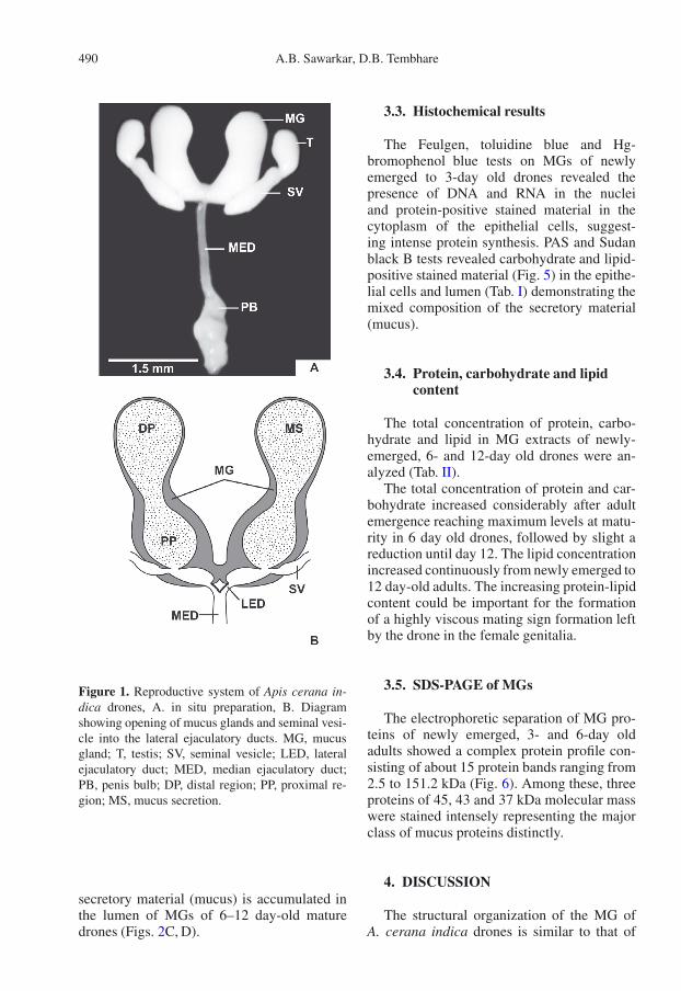
The mucus glands (MG) of A. cerana indicadrones are milky white, large bi-lobed, peanut-shaped, sac-like structures with a wide lumen.Each gland is divided by a well-defined narrowconstriction into a narrow distal and a largeproximal region. The proximal regions of theglands are fused forming a common sac open-ing into the lateral ejaculatory ducts. The lat-eral ejaculatory ducts are rather short and openinto the median ejaculatory duct (Fig. 1).
The wall of the MG consists of a thin in-ner epithelial layer and a thick outer mus-cle coat. It is externally covered by a peri-toneal sheath. The epithelial cells are large andcolumnar in shape and are arranged in a sin-gle tier. They contain spherical centrally lo-cated nuclei and granular cytoplasmic inclu-sion in their perikarya. In the distal region ofthe MGs, the muscle coat is composed of aninner layer of circular muscles and outer layerof longitudinal muscles, while in the proximalregion, the muscle coat is composed of innerand outer layers of longitudinal muscles and amiddle layer of circular muscle (Fig. 2).
3.2. Histomorphological changes
The MGs show a gradual increasein weight, length and diameter duringpupal–adult development (Fig. 3). The nucleiof the epithelial cells gradually increase indiameter from 6.98 ± 0.35 µm in late pupato 10.50 ± 0.48 µm in adult drones (Fig. 4).In the late pupal stage, the epithelial cells arepacked with dense cytoplasmic inclusions.In the newly emerged drones, release ofsecretory material from the epithelial cellsinto the lumen is evident. A large amount of
490 A.B. Sawarkar, D.B. Tembhare
Figure 1. Reproductive system of Apis cerana in-dica drones, A. in situ preparation, B. Diagramshowing opening of mucus glands and seminal vesi-cle into the lateral ejaculatory ducts. MG, mucusgland; T, testis; SV, seminal vesicle; LED, lateralejaculatory duct; MED, median ejaculatory duct;PB, penis bulb; DP, distal region; PP, proximal re-gion; MS, mucus secretion.
secretory material (mucus) is accumulated inthe lumen of MGs of 6–12 day-old maturedrones (Figs. 2C, D).
3.3. Histochemical results
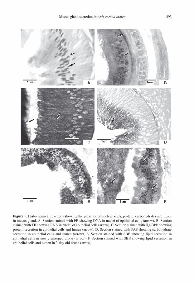
The Feulgen, toluidine blue and Hg-bromophenol blue tests on MGs of newlyemerged to 3-day old drones revealed thepresence of DNA and RNA in the nucleiand protein-positive stained material in thecytoplasm of the epithelial cells, suggest-ing intense protein synthesis. PAS and Sudanblack B tests revealed carbohydrate and lipid-positive stained material (Fig. 5) in the epithe-lial cells and lumen (Tab. I) demonstrating themixed composition of the secretory material(mucus).
3.4. Protein, carbohydrate and lipidcontent
The total concentration of protein, carbo-hydrate and lipid in MG extracts of newly-emerged, 6- and 12-day old drones were an-alyzed (Tab. II).
The total concentration of protein and car-bohydrate increased considerably after adultemergence reaching maximum levels at matu-rity in 6 day old drones, followed by slight areduction until day 12. The lipid concentrationincreased continuously from newly emerged to12 day-old adults. The increasing protein-lipidcontent could be important for the formationof a highly viscous mating sign formation leftby the drone in the female genitalia.
3.5. SDS-PAGE of MGs
The electrophoretic separation of MG pro-teins of newly emerged, 3- and 6-day oldadults showed a complex protein profile con-sisting of about 15 protein bands ranging from2.5 to 151.2 kDa (Fig. 6). Among these, threeproteins of 45, 43 and 37 kDa molecular masswere stained intensely representing the majorclass of mucus proteins distinctly.
4. DISCUSSION
The structural organization of the MG ofA. cerana indica drones is similar to that of

Mucus gland secretion in Apis cerana indica 491
Figure 2. Histology of the mucus gland, A. Cross section of the proximal region of MGs showing a thickwall (W) and a large lumen (L), B. The wall of the MG consists of outer and inner longitudinal musclelayers (LML), a middle circular muscle layer (CML), and an epithelial layer (EL) with a brush border (BB),C. Release of mucus secretion (MS) into the lumen (L) of the MG (→) in late pupae, D. Accumulation ofmucus secretion (MS) in the lumen of the MG of a 6-day old drone. EL, epithelial layer; ML, muscle layer.
A. mellifera, representing the typical mesoder-mal male accessory gland (Snodgrass, 1956;Woyke, 1958; Simpson, 1960; Kapil, 1962;Moors et al., 2005). Full development of theMG by the end of the pupal stage in A. cer-ana indica is also a characteristic feature of A.mellifera (Tozetto et al., 2007). Similarly, thetime course of secretory activity in the epithe-lial cells and gradual accumulation of secre-tory material (mucus) in the lumen of the MGsresembles that seen in A. mellifera (Mindt,1962; Colonello and Hartfelder, 2003; Moorset al., 2005). Bishop (1920) noticed that thehoney bee drones only become capable of mat-ing at 8–10 days after emergence, which is alsothe time that the mucus gland takes to becomefully filled with secretions.
Histochemical reactions demonstrated Hg-BPB positive proteins, PAS positive car-bohydrates (muco-polysaccharides) and su-danophilic lipids in the epithelial cells of MGsof A. cerana indica, similar to results ob-tained for A. mellifera (Blum et al., 1962,1967; Ivanova, 2000; Ivanova et al., 2000;Colonello and Hartfelder, 2003; Cruz-Landimand Dallacqua, 2005) and Bombus terrestris(Baer et al. 2000, 2001). The biochemicalanalysis also showed that proteins are themajor constituents of the secretions, whilecarbohydrates and lipids make smaller con-tributions to the MG secretory material ofA. cerana indica and thus supporting the ob-servations of Colonello and Hartfelder (2003)for A. mellifera. It is now well established that

492 A.B. Sawarkar, D.B. Tembhare
Figure 3. Changes in weight, length and diame-ter of the MGs during pupal-adult development, A.Weight, B. Length, C. Diameter. EP, early pupa;MP, mid pupa; LP, late pupa; NEA, newly emergedadult; 6DA, 6-day old adult; 12DA, 12-day oldadult.
the male accessory glands of various insectspecies secrete predominantly proteins, alongwith some muco-polysaccharides, glycogenand lipids (Chen, 1984; Happ, 1984; Gillott,1988, 2003; Leather and Hardie, 1995).
Figure 4. Nuclear diameter of epithelial cells ofMG during pupal-adult development.
SDS-PAGE revealed about 16–20 pro-teins in A. mellifera mucus (Ivanova, 2000;Colonello and Hartfelder, 2003; Cruz-Landimand Dallacqua, 2005) while the present studyshowed the presence of about 15 protein bandsin the gland extracts of newly emerged and6-day old drones of A. cerana indica. InA. mellifera the molecular mass range of theseproteins is 174 to 25 kDa (Colonello andHartfelder, 2003) , whereas we found proteinsranging from 151.2 to 2.5 kDa molecular massin A. cerana indica. Similarly, Colonello andHartfelder (2003) also report a group of threeproteins of 43–47.5 kDa appearing persistentlyin the mucus of mature drones of A. melliferaand considered them as the major mucus pro-teins. In A. cerana indica, a group of three pro-teins ranging from 37–45 kDa molecular masscan similarly be considered as a class of majormucus proteins.
The presence of a large number of pro-teins suggests a multifunctional role of themucus (Gillott, 1988, 1996, 2003) such asa mating plug in the female genitalia aftercopulation to avoid polyandry (Baer et al.,2000, 2001; Sauter et al., 2001; Strassmann,2001; Moors et al., 2005), as contributingto the mating sign (Koeniger, 1984, 1986a,b, 1991; Koeniger et al., 1996) which couldhave adhesive function fixing the drone tothe queen while copulating freely in air andalso to firmly retain the detached part of thedrone’s endophallus i.e. the cervix filled withsperm in the queen’s vagina (Koeniger, 1984).This may represent a stimulant for oocyte
Mucus gland secretion in Apis cerana indica 493
Figure 5. Histochemical reactions showing the presence of nucleic acids, protein, carbohydrates and lipidsin mucus gland, A. Section stained with FR showing DNA in nuclei of epithelial cells (arrow), B. Sectionstained with TB showing RNA in nuclei of epithelial cells (arrow), C. Section stained with Hg-BPB showingprotein secretion in epithelial cells and lumen (arrow), D. Section stained with PAS showing carbohydratesecretion in epithelial cells and lumen (arrow), E. Section stained with SBB showing lipid secretion inepithelial cells in newly emerged drone (arrow), F. Section stained with SBB showing lipid secretion inepithelial cells and lumen in 3-day old drone (arrow).

494 A.B. Sawarkar, D.B. Tembhare
Table I. Histochemical results on MG.
Presence in mucus glandTest Substance Presence in mucus gland
Nucleus Cell Lumen1. Feulgen reaction (FR) DNA ++ __ __2. Toluidine blue (TB) RNA ++ ++ __3. Mercuric bromophenol blue (Hg-BPB) Protein __ +++ +++
4. Periodic acid-Schiff (PAS) Carbohydrate __ ++ ++
5. Sudan black-B (SBB) Lipid __ + ++
+ (positive), ++ (moderate), +++ (intense), — (no reaction).
Table II. Major components of mucus glands extracts.
Age of Total concentration ofdrones Protein Carbohydrate LipidDay (µg/mg) (µg/mg) (µg/mg)
0 (NEA) 89.68 ± 7.91 26.46 ± 0.54 85.57 ± 3.516 333.2 ± 13.88 44.82 ± 2.94 135.8 ± 6.83
12 273.7 ± 9.29 32.25 ± 1.90 208.6 ± 11.69
Abbr.: MG- mucus gland, NEA –newly emerged adult, ± standard error.
Figure 6. SDS-PAGE of MG extracts of newlyemerged (NEA) and 6-day old drones (6DA).
maturation (Melo et al., 2001; Patricio andCruz-Landim, 2002; Cruz-Landim and Dal-lacqua, 2005), an energy source (Colonelloand Hartfelder, 2003) or be of importance forsperm capacitation and storage, similar to pro-cesses shown in other insects (Chen, 1984;Gillott, 1996).
Synthèse et composition chimique des sécrétionsde la glande à mucus chez Apis cerana indica.
KW : glande à mucus / profil protéinique / mâle/ Apis cerana indica
Zusammenfassung – Synthese und chemischeZusammensetzung des Mucusdrüsensektets vonApis cerana indica F. Obwohl Apis cerana indicain Indien die weitverbreitetste und am häufigstenin der Bienenhaltung anzutreffende Biene ist, sindunsere Kenntnisse über ihre Fortpflanzungsphysio-logie nur beschränkt. Ziel der vorliegenden Studiewar es demzufolge, die Struktur der Mucusdrüseund die Synthese und Zusammensetzung ihres Sek-tets mittels histologischer, histochemischer und bio-chemischer Methoden zu untersuchen.Die Mucusdrüsen (MD) von A. cerana indicasind milchigweiße, sackartige Strukturen von erd-nussartiger zweiteiliger Form und einem großenLumen. Jede Drüse wird durch eine Verengung inein schmales distales und ein weites proximales

Mucus gland secretion in Apis cerana indica 495
Teilstück unterteilt. Die proximalen Teilstücke derbeiden Drüsen bilden einen sackförmigen Ausgangin den jeweiligen medianen Abschnitt der lateralenSamenleiter. Die beiden lateralen Samenleiter sindkurz und laufen in einem medianen gemeinsamenSamenleiter zusammen (Abb. 1).Die Wand der MD besteht aus einem inneren Epi-thel und einer äußeren Muskellage, die von ei-nem Mesenthelium umkleidet ist. Die Epithelzellensind große, Drüsenzellen von säulenartigem For-mat, die eine einzige Zellage bilden. Die rundenZellkerne liegen zentral und die Perikaryen weisengranuläre Zytoplasmaeinschlüsse auf. Im distalenAbschnitt der MD besteht die Muskellage aus ei-ner inneren Ringmuskel- und einer äußeren Längs-muskelschicht, während die Muskulatur im proxi-malen Abschnitt von einer inneren und äusserenLängsmuskel- und einer mittleren Ringmuskellagegebildet wird (Abb. 2).Während der Entwicklung von der Puppe zur adul-ten Bienen nimmt die MD an Gewicht, Länge undDurchmesser zu (Abb. 3). Eine Größenzunahmewar auch für die Kerne der Epithelzellen zu sehen,von 6,98 ± 0,35 µm bei späten Puppen auf 10,50 ±0,48 µm bei adulten Drohnen (Abb. 4).In den Epithelzellen der MD war ab dem spä-ten Puppenstadium die Synthese von sekretori-schem Material zu sehen, das bereits kurz nach demSchlüpfen der adulten Drohnen im Lumen der Drü-sen akkumulierte. Histochemische Tests gaben Auf-schluss über die sekretorische Aktivität und die bio-chemische Zusammensetzung des Sekrets als seineMischung aus Proteinen, Kohlenhydraten und Li-piden (Abb. 5 und Tab. I). Bei einem Gesamtpro-teingehalt von 333,2 ± 13,8 µg/mg stellen Protei-ne die Hauptkomponente des Sekrets dar, das zu-dem 208,6 ± 11,7 µg/mg Lipide und 44,8 ± 2,9µg/mg Kohlenhydrate enthält (Tab. II). In SDS-Polyacrylamidgelen ließen sich elektrophoretisch15 Proteinbanden auftrennen, mit Molekülmassenvon 2,5 bis 151,2 kDa, wobei drei Proteinbandenvon 45, 43 und 37 kDa besonders hervortraten unddementsprechend die Hauptproteine des Mucusdrü-sensekrets darstellen (Abb. 6).Das komplexe Proteinmuster lässt auf eine Multi-funktionalität des Drüsensekrets schließen, das eineRolle spielen könnte sowohl im Spermatransfer, derSpermienkapazitierung, der Spermienlagerung undEnergieproduktion, wie auch in der Oocytenreifungund anderen bei Bienen beschriebenen postkopula-torischen Aktivitäten.
Mucusdrüse / Proteinprofil / Apis cerana indica
REFERENCES
Baer B., Maile R., Schmid-Hempel P., Morgan E.D.,Jones G.R. (2000) Chemistry of a mating plug inBumblebees, J. Chem. Ecol. 26, 1869–1875.
Baer B., Morgan E.D., Schmid-Hempel P. (2001)A nonspecific fatty acid within the bumblebeemating plug prevents females from remating, Proc.Natl. Acad. Sci. USA 98, 3926–3928.
Bishop G.H. (1920) Fertilization in the honeybee. I.The male sexual organs: their histological struc-ture and physiological functioning, J. Exp. Zool.31, 225–265.
Blum M.S., Bumgarner J.E., Taber S. (1967)Composition and possible significance of fattyacids in the lipid classes in honey bee semen, J.Insect Physiol. 13, 1301–1308.
Blum M.S., Glowska Z., Taber S. (1962) Chemistryof the drone honey bee reproductive system. II.Carbohydrates in the reproductive organs and se-men, Ann. Entomol. Soc. Am. 55, 135–139.
Chen P.S. (1984) The functional morphology and bio-chemistry of insect male accessory glands andtheir secretions, Annu. Rev. Entomol. 29, 233–255.
Colonello N.A., Hartfelder K. (2003) Protein con-tent and pattern during mucus gland maturationand its ecdysteroid control in honeybee drones,Apidologie 34, 257–267.
Colonello N.A., Hartfelder K. (2005) She’s my girl-male accessory gland products and their func-tion in the reproductive biology of social bees,Apidologie 36, 231–244.
Cruz-Landim C., Dallacqua R.P. (2005) Morphologyand protein pattern of honeybee drone accessoryglands, Genet. Mol. Res. 4, 473–481.
Dubois M., Gilles K.A., Hamilton J.K., Rebers P.A.,Smith F. (1956) Colorimetric method for determi-nation of sugars and related substances, Analyt.Chim. 28, 350–356.
Frings C.S., Dunn R.T. (1970) A colorimetric methodfor determination of total serum lipids based onsulfo-vanilin reaction, Am. J. Clin. Pathol. 53, 89–91.
Gillott C. (1988) Arthropoda-Insecta, in: AdiyodiK.G., Adiyodi R.G. (Eds.), Reproductive Biologyof Invertebrates, Vol. III, Wiley, New York,pp. 319–471.
Gillott C. (1996) Male insect accessory glands: func-tions and control of secretory activity, Invertebr.Reprod. Dev. 30, 199–205.
Gillott C. (2003) Male accessory gland secretion: mod-ulators of female reproductive physiology and be-havior, Annu. Rev. Entomol. 48, 163–184.
Happ G.M. (1984) Structure and development of maleaccessory glands in insects, in: King R.C., Akai H.(Eds.), Insect Ultrastructure, Plenum, New York,pp. 365–396.
Ivanova E. (2000) Organ specificity of water-solubleproteins during drone (Apis mellifera L.) ontogen-esis, Apidologie 31, 671–677.
Ivanova E., Popov P., Dobrovolov I. (2000)Electrophoretic study of water-soluble proteins

496 A.B. Sawarkar, D.B. Tembhare
during the honeybee (Apis mellifera L.) ontogene-sis, Apidologie 31, 679–687.
Kapil R.P. (1962) Anatomy and histology of themale reproductive system of Apis indica (Apidae,Hymenoptera), Insect. Soc. 9, 73–90.
Koeniger G. (1984) FunktionsmorphologischeBefunde bei der Kopulation der Honigbiene (Apismellifera L.), Apidologie 15, 189–204.
Koeniger G. (1986a) Reproduction and mating be-haviour, in: Rinderer T.E. (Ed.), Bee Genetics andBreeding, Academic Press, San Diego, pp. 255–280.
Koeniger G. (1986b) Mating sign and multiple matingin the honey bee, Bee World 67, 141–150.
Koeniger G. (1991) Diversity in Apis mating systems,Westview Press, pp. 199–211.
Koeniger G., Hänel H., Wissel M., Herth W. (1996)Cornual gland in the honeybee drone (Apis mel-lifera L.): structure and secretion, Apidologie 27,145–156.
Koeniger N., Koeniger G., Wongsiri S. (1989) Matingand sperm transfer in Apis florae, Apidologie 21,413–418.
Laemmli U.K. (1970) Cleavage of structural proteinsduring assembly on the head of bacteriophage T4,Nature 227, 680–685.
Leather S.R., Hardie J. (1995) Insect Reproduction,CRC Press, Boca Raton, Florida, USA.
Lowry O.L., Rosebrough N.J., Farr A.L., Randall R.J.(1951) Protein measurement with the folin phenolreagent, J. Biol. Chem. 193, 265–275.
Melo G.A.R., Buschini M.L.T., Campos L.A.O. (2001)Ovarian activation in Melipona quadrifasciataqueens triggered by mating plug stimulation(Hymenoptera, Apidae), Apidologie 32, 355–361.
Mindt B. (1962) Untersuchungen über das Lebender Drohnen, insbesondere Ernährung undGeschlechtsreife, Z. Bienenforsch. 6, 9–33.
Moors L., Spaas O., Koeniger G., Billen J. (2005)Morphological and ultrastructural changes in
the mucus glands of Apis mellifera dronesduring pupal development and sexual maturation,Apidologie 36, 245–254.
Patricio K., Cruz-Landim C. (2002) Mating influencein the ovary differentiation in adult queens of Apismellifera L. (Hymenoptera, Apidae), Braz. J. Biol.62, 641–649.
Sauter A., Brown M.J.F., Baer B., Schmid-Hempel P.(2001) Males of social insects can prevent queensfrom multiple mating, Proc. R. Soc. London B268, 1449–1454.
Simpson J. (1960) Male genitalia of Apis species,Nature 185, 56.
Snodgrass R.E. (1956) Anatomy of the honeybee,Comstock Publ. Ass. Cornell Univ. Press, Ithaca,N.Y., p. 343.
Strassmann J. (2001) The rarity of multiple mating byfemales in the social Hymenoptera, Insect. Soc.48, 1–13.
Tembhare D.B. (2008) Techniques in Life Sciences,Himalaya Publ. House, Mumbai, India.
Tozetto S.D.O., Bitondi M.M.G., Dallacqua R.P.,Simões Z.L.P. (2007) Protein profiles of testes,seminal vesicles and accessory gland of honey beepupae and their relation to the ecdysteroid titer,Apidologie 38, 1–11.
Woyke J. (1956) Anatomo-physiological changes inqueen-bees returning from mating flights, and theprocess of multiple mating, Bull. Acad. Polon. Sci.4, 81–87.
Woyke J. (1958) The histological structure of thereproductive organs of the drone, Poznan Soc.Friends of Sci., Publ. Sect. Agric. Sylvic. 19, 38–50.
Woyke J., Ruttner F. (1958) An anatomical study of themating process in the honeybee, Bee World 39, 3–18.
Wyatt G.R., Davey K.G. (1996) An anatomical studyof the mating process in the honeybee, Bee World39, 3–18.