Embed Size (px)
Citation preview
Synthesis and Characterisation of Helicate and Mesocate
Forms of a Double-Stranded Diruthenium(II) Complex
of a Di(terpyridine) Ligand
Kate L. Flint,a J. Grant Collins,b Siobhan J. Bradley,c Trevor A. Smith,c Christopher J. Sumby,a and F. Richard Keene*a
SUPPLEMENTARY MATERIAL
Table of Contents NMR Data .................................................................................................................................................. 2
Crude Reaction Mixture ......................................................................................................................... 3Mesocate, 2 ............................................................................................................................................. 3
Assignment of -CH2CH2- bridge ........................................................................................................ 6Helicate, 3 ............................................................................................................................................... 7
X-ray Crystallography ................................................................................................................................ 9Mesocate, 2 ............................................................................................................................................. 9
Packing ............................................................................................................................................. 10Helicate, 3 ............................................................................................................................................. 11
Packing ............................................................................................................................................. 12HR-ESMS Spectra .................................................................................................................................... 13
Mesocate (2) ......................................................................................................................................... 13Helicate (3) ........................................................................................................................................... 14
UV-Vis Spectra ........................................................................................................................................ 15
aDepartment of Chemistry, School of Physical Sciences, The University of Adelaide, Adelaide, South Australia 5005.bSchool of Physical, Environmental & Mathematical Sciences, UNSW Canberra, Australian Defence Force Academy, Canberra, ACT 2600.cARC Centre of Excellence in Exciton Science, School of Chemistry, The University of Melbourne, Victoria 3010.*Email: [email protected].
10.1071/CH19220_AC©CSIRO 2019 Australian Journal of Chemistry 2019, 72(10), 762-768

- 2 -
NMR Data
Table S1. Assigned 1H NMR peaks for the diruthenium(II) mesocate (2), and helicate (3)
complexes (ND = not defined). (Note structure below shows 3D representation of helicate)
1H NMR – Mesocate 2 1H NMR – Helicate 3
Assigned Shift (ppm) #H Multiplicity J (Hz)
H 8.65 4 d 8.14
H 8.58 4 d 8.11
H, H 8.35 8 m 3.94, 3.94, 8.11
H 8.28 4 d 8.25
H 8.00 4 dd 1.50, 8.40
H 7.67 4 dd 0.64, 8.24
H 7.19 4 d 1.46
H 6.67 4 d 0.65
CH2 2.90 4 m ND
CH2 2.50 4 m ND CH3 2.00 12 s -
Assigned Shift (ppm) #H Multiplicity J (Hz)
H 8.83 4 d 8.08
H 8.71 4 d 8.11
H 8.50 4 t 8.13, 8.13
H 8.38 4 d 8.29
H 8.34 4 d 8.24
H 7.74 4 d 7.30
H 7.40 4 d 10.36
H 7.15 4 s
H 6.38 4 s
CH2, CH2 2.63 8 s
CH3 2.03 12 s
- 3 -
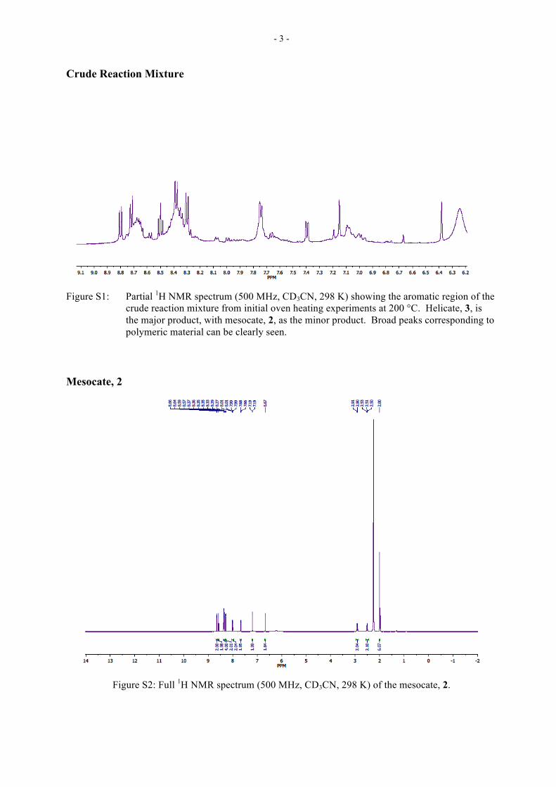
Crude Reaction Mixture
Figure S1: Partial 1H NMR spectrum (500 MHz, CD3CN, 298 K) showing the aromatic region of the
crude reaction mixture from initial oven heating experiments at 200 °C. Helicate, 3, is the major product, with mesocate, 2, as the minor product. Broad peaks corresponding to polymeric material can be clearly seen.
Mesocate, 2
Figure S2: Full 1H NMR spectrum (500 MHz, CD3CN, 298 K) of the mesocate, 2.

- 4 -
Figure S3: Full 13C NMR spectrum (126 MHz, CD3CN, 298 K) of the mesocate, 2.
Figure S4: Partial 1H-1H ROESY NMR spectrum showing 1 mesocate, 2.
- 5 -
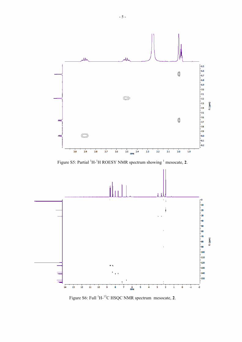
Figure S5: Partial 1H-1H ROESY NMR spectrum showing 1 mesocate, 2.
Figure S6: Full 1H-13C HSQC NMR spectrum mesocate, 2.

- 6 -
Figure S7: Partial 1H-13C HSQC NMR spectrum showing mesocate, 2.
Assignment of -CH2CH2- bridge
Distance between atoms (Å) Chemical Shift
H42 H44 H12 H14 H1A 2.557 3.391 4.674 3.577 H1A/B 2.93 ppm H1B 3.543 2.354 4.561 2.26 H2A/B 2.50 ppm H2A 3.853 4.359 2.858 3.165 H42/12 7.19 ppm H2B 2.614 4.297 2.537 3.524 H44/14 8.00 ppm
Figure S8: Assignment of -CH2CH2- bridge hydrogens of the mesocate, 2 using interatomic
distances from crystal structure.

- 7 -
Helicate, 3
Figure S9: Full 1H NMR spectrum (500 MHz, CD3CN, 298 K) of the helicate, 3.
Figure S10: Full 13C NMR spectrum (126 MHz, CD3CN, 298 K) of the helicate, 3.

- 8 -
Figure S11: Partial 1H-1H ROESY NMR spectrum showing 1cate, 3.
Figure S12: Partial 1H-1H ROESY NMR spectrum showing 1 helicate, 3.

- 9 -
X-ray Crystallography
Mesocate, 2
Figure S13: Crystal structure of the diruthenium mesocate, 2, including van der Waals
surface, illustrating accessible central cavity.
- 10 -
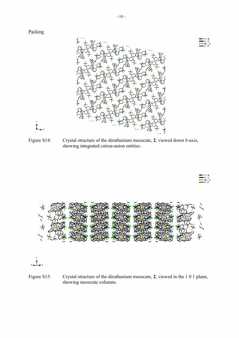
Packing
Figure S14: Crystal structure of the diruthenium mesocate, 2, viewed down b-axis,
showing integrated cation-anion entities.
Figure S15: Crystal structure of the diruthenium mesocate, 2, viewed in the 1 0 1 plane,
showing mesocate columns.

- 11 -
Helicate, 3
Figure S16: Crystal structure of the diruthenium helicate, 3, including van der Waals
surface, illustrating inaccessible central cavity.

- 12 -
Packing
Figure S17: Crystal structure of the diruthenium helicate, 3, viewed down a-axis,
showing alternating cation/anion layers.
Figure S18: Crystal structure of the diruthenium helicate, 3, viewed in the ab plane, with a “loose” herringbone arrangement incorporating anions.

- 13 -
HR-ESMS Spectra
Mesocate (2)
Figure S19: Full HR-ESI MS spectrum (CH3CN) of the mesocate, 2.
Figure S20: Partial HR-ESI MS (CH3CN) and calculated isotopic patten of mesocate 2,
peak at m/z 311.0708 due to [2 – 4(PF6-)]4+.

- 14 -
Helicate (3)
Figure S21: Full HR-ESI MS spectrum (CH3CN) of the helicate, 3.
Figure S22: Partial HR-ESI MS (CH3CN) and calculated isotopic pattern of helicate 3, peak
at m/z 311.0708 due to [3 – 4(PF6-)]4+.

- 15 -
UV-Vis Spectra
Figure S23: Absorbance spectra of diruthenium mesocate, 2, and helicate, 3.
0
10000
20000
30000
40000
50000
60000
70000
80000
200 250 300 350 400 450 500 550 600
Mol
ar A
bsor
btiv
ity (M
-1 c
m-1
)
Wavelength (nm)
MesocateHelicate