Embed Size (px)
Citation preview
European Journal of Neuroscience, Vo!. 2, pp. 414-429 © European Neuroscience Association 0953-816xI90 $3.00
Synaptic and Nonsynaptic Localization of Benzodiazepine/GABAA Receptor/CI- Channel Complex Using Monoclonal Antibodies in the Dorsal Lateral Geniculate Nucleus of the Cat
lvan Soltesz \ J. David B. Roberts \ Hiroshi Takagi1,3, J. Grayson Richards2, Hans Mohler2 and Peter Somogyi1
1 MRC Anatomical Neuropharmacology Unit, South Parks Road, Oxford OX1 UK 2Pharmaceutical Research Department, F. Hoffmann-La Roche & Co. Ltd., CH-4002 Basle, Switzerland
address: 1 st Department of Osaka City University Medical School, 1-4-54 Asahimachi, Abeno-ku, Osaka 545,
Key words: GABA, receptors, synapse, immunocytochemistry, inhibition, thalamus
Abstract
The two monoclonal antibodies, bd-17 and bd-24, are specific for {3- and a-subunits of the GABAA/benzodiazepine receptor/chloride channel complex respectively. An abundance of both subunits has been revealed in the visual thalamus of the cat by light microscopic immunocytochemistry using these antibodies. The a-subunit specific antibody and electron microscopy were used to determine the subcellular distribution of immunoreactivity with respect to specific cell classes in the dorsal lateral geniculate nucleus. Immunoreactivity was always associated with membranes and the degree of immunoreactivity varied greatly between different types of cell as defined by: (i) immunoreactivity for GABA; (ii) soma area; (iii) presence or absence of cytoplasmic laminated bodies (CLB). GABA negative neurons with the smallest soma area showed the strongest immunoreactivity, mainly in the endoplasmic reticulum and also on the somatic plasma membrane. Cytoplasmic laminated bodies could be found in the majority of these neurons. Large GABA negative cells without CLBs were strongly immunoreactive on the plasma membrane of the soma and dendrites, but showed scant if any intracellular immunoreactivity. GABA-positive cells showed weak intracellular immunoreactivity but negligible if any immunoreactivity at the somatic and proximal dendritic plasma membrane. A similar reaction pattern was found in GABA negative cells which contained no CLBs and which constituted a medium sized cell population. It is suggested that the degree of intracellular receptor immunoreactivity is positively correlated with receptor turnover. The dendrites of projection cells, particularly outside the glomeruli, showed strong immunoreactivity on the plasma membrane. The synaptic junctions formed by many boutons (F terminals) establishing symmetrical synapses with dendrites of relay cells were immunopositive, but no immunoreactivity could be detected at the synapses established by the presynaptic dendrites of the local interneurons. Many axo-somatic F1 junctions were also immunoreactiye. However, immunoreactivity for the receptor/channel complex was also widely distribution on nonsynaptic plasma membranes of somata and dendrites. Thus GABA may act at both synaptic and non-synaptic sites. Furthermore, the correlation of immunoreactivity for the GABAA receptor complex with previously published properties of physiologically identified cells suggests that the strongly immunoreactive, small, GABA negative cells with CLBs might correspond to the 'lagged' X-type cells, and the large GABA negative receptor outlined cells without CLBs might correspond to some of the V-type neurons.
Introduction
Gamma-aminobutyric acid (GABA) mediated inhibition has been shown to play a major role in gating the transmission of visual information from the retina to the visual cortex in the dorsal lateral geniculate
Correspondence to: Dr P. Somogyi, as above
Received 11 July 1989, revised 1 December 1989, accepted 20 December 1989
nucleus (LGN) of the cat (Singer and Bedworth, 1973; Dubin and Cleland, 1977; Lindstrom, 1982; Sillito and Kemp, 1983; Berardi and Morrone, 1984; Sherman and Koch, 1985; Ahlsen et al., 1985; Pape
Immunocytochemistry of benzodiazepine/GABAA receptors in the LGN 415
GABA-RB
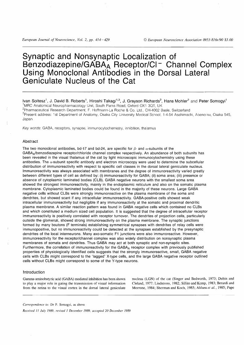
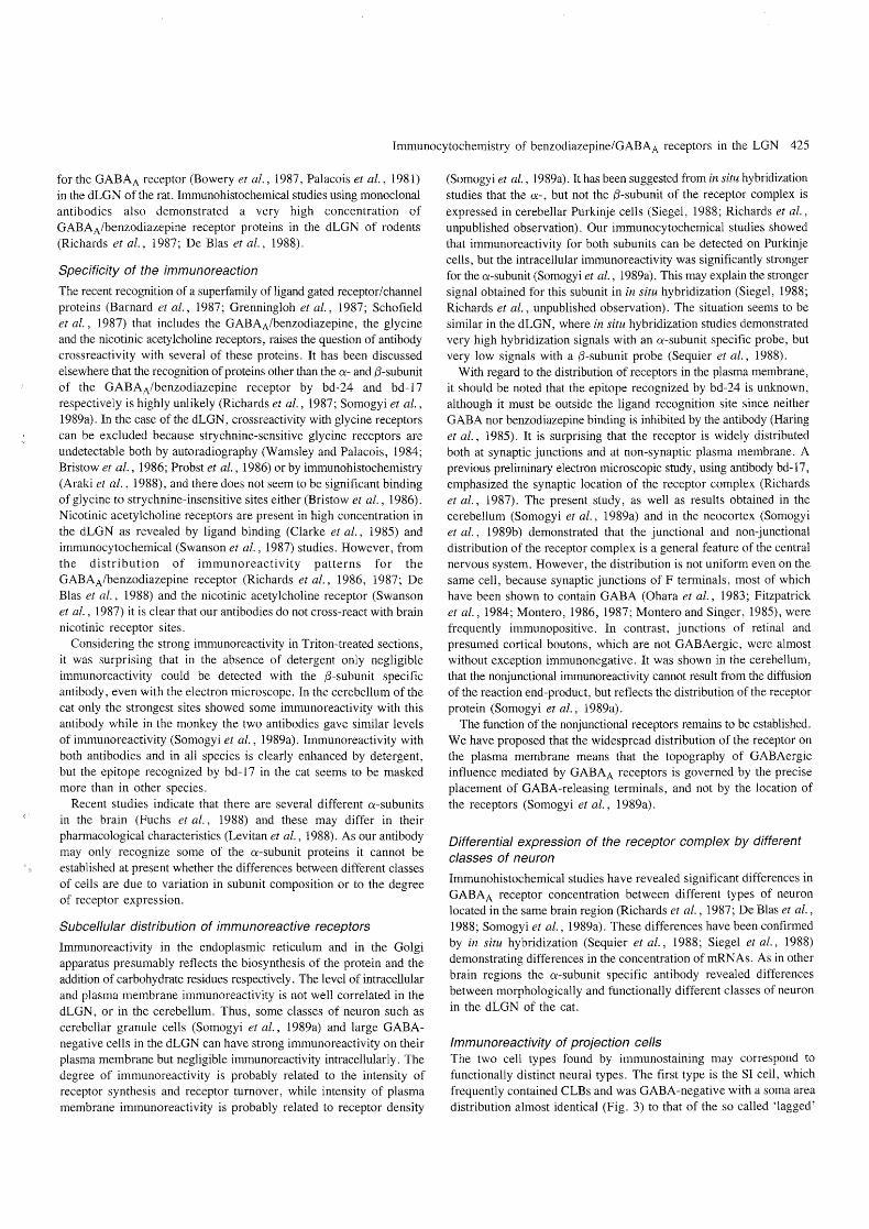
FIG.!' Distribution of GABAA-R immunoreactivity in the visual thalamus of cat, as shown by monoclonal antibody bd-24 (A), specific for the a-subunit of the receptor, and monoclonal antibody bd-17 (B), specific for the ,B-subunit of the receptor complex . Sections (coronal plane) were treated with Triton X-lOO, therefore both antibodies gave strong reaction throughout the neuropil of the thalamus , but not in fibre tracts. The stronger reaction at the periphery of the sections was not observed consistently. A, AI' C , laminae of the dorsal lateral geniculate nucleus ; LGNv , ventral LGN; MIN, medial interlaminar nucleus ; NL, nucleus lateralis; OT, optic tract ; Pul, pulvinar. Scale : 500 fim.
and Eysel , 1986). Neurons in the LGN receive GABAergic input from axons of the perigeniculate nucleus, from axons of intrinsic neurons and from presynaptic dendrites of these same intrinsic neurons
(Guillery, 1969; Hamori et al., 1974; Lieberman and Webster, 1974; Sherrnan and Spear, 1982; Fitzpatrick et al., 1984; Mason et al., 1984; Montero and Singer, 1985; Montero, 1986). In vivo microelectrode
416 Immunocytochemistry of benzodiazepine/GABAA receptors in the LGN
studies have revealed the existence of optic tract activated inhibition, and combined electrophysiological and pharmacological studies using the GABAA antagonist bicuculline have demonstrated that at least a substantial part of this inhibition is mediated by GAB AA receptors (Sillito and Kemp, 1983; Vidyasagar, 1984; Berardi and Morrone, 1984; Pape and Eysel, 1986; Eysel et aI., 1987). It is apparent that different types of cells can be defined on the basis of the spatial and temporal properties of the GABA mediated inhibitory influences.
The LGN of the cat offers great advantages not only for physiological studies on inhibition, but also for the localization of the contributing molecular machinery. The LGN contains at least one local circuit cell population (Guillery, 1966; Szentagothai, 1973; Hamori etal., 1974; Hamos et al., 1985), which probably uses GABA as a transmitter (Montero and Singer, 1985; Montero and Zempel, 1985; Gabbott et al., 1985; Madarasz et aI., 1985; McCormick and Pape, 1988). Physiologically distinct cell populations can be delineated also by size distribution of their somata (Friedlander et al., 1981; Stanford et al. , 1983; Humphrey and Weller, 1988a,b). Furthermore, almost all constituents of the neuropil can be identified by electron microscopy (Peters and 1966; Guillery, 1969; Famiglietti and Peters, 1972; Szentagothai, 1973; Robson and Mason, 1979; Mason et al., 1984; Wilson et aI., 1984; Hamos et al., 1985). The regional distribution of one of the main components of the inhibitory mechanism, the GABAA/benzodiazepine receptor/chloride channel complex has been mapped in the thalamus using radioligand binding (Palacois et al., 1981; Schoch et al., 1985; Bowery et al., 1987) and immunohistochemistry (Schoch et al., 1985; Richards et al., 1987; De BIas et al., 1988; Vitorica et al., 1988). Although these studies show high levels of binding sites and immunoreactivity in the LGN of rodents, they have not provided information on localization of the receptor with regard to particular populations of cells. This study was undertaken to identify the cellular and subcellular distribution of the GABAA/benzodiazepine receptor/chloride channel complex.
We used two monoclonal antibodies specific for the a- and {3-subunit of the receptor complex respectively (Schoch et aI., 1984, 1985; Haring et al., 1985), and an immuno-electronmicroscopic method (Somogyi et al., 1989a) in order to relate the receptor distribution to different classes of cells. The distribution of GABA-containing neurons was also studied in relation to receptor distribution. Some of the results have been presented in preliminary form (Somogyi, 1989).
Materials and methods
Preparation of animals and tissue
Three adult male cats (2 - 3 kg) were used. They were deeply anaesthetized with chloral hydrate (400 i.p. initially, supplemented as required), and perfused transcardially with saline for 1 min followed by fixative for about 30 min at room temperature. In the first 10 min a fixative containing paraformaldehyde (4 % ), glutaraldehyde (0.025%) and picric acid (approx 0.2%), made up in 0.1 M phosphate buffer (PB, pH 7.2-7.4) was used (Somogyi and Takagi, 1982), then perfusion was continued with a similar fixative but without glutaraldehyde.
A tissue block containing the LGN was dissected from the fixed brain and 50pm thick sections were cut on a vibratome. The sections were washed in 0.1 M PB and then placed in solutions of the same buffer containing 10% and 20% sucrose for about an hour each. To facilitate the penetration of reagents most of the sections were frozen in liquid N2 then thawed in 0.1 M PB as described earlier (Somogyi and
Takagi, 1982). Thereafter sections were treated with 1 % sodium borohydride (Willingham, 1983) dissolved in phosphate buffered saline (PBS, pH 7.4). This treatment enhanced immunoreactivity.
Antibodies
The purification of GABAA/benzodiazepine receptors and the preparation of mouse monoclonal antibodies have been described earlier (Haring et al., 1985; Schoch et al., 1984, 1985). As shown by immunoblotting bd-24 recognizes one major protein (Mr 50000-53 000), an a-subunit of the receptor containing the benzodiazepine binding site (Schoch et al., 1984, 1985). The other antibody bd-17 recognizes one major protein band (M r 55000-56000; Fuchs et al., 1988), the {3-subunits of the receptor containing the GAB A binding site. Both antibodies precipitate the GABA, the benzodiazepine and the t-butylbicyclophosphorothionate (TPBS) binding sites in solubilized brain preparations, indicating that the receptor channel complex contains both subunits (Schoch et al., 1985; Mohler et al., 1986). The antibodies are gamma-isotype IgGs. In the present experiment tissue culture supernatant, containing 10% calf serum, was used for immunocytochemistry.
Immunocytochemistry
For the localization of the receptor complex the same methods were used as reported earlier (Somogyi et aI., 1989a). Briefly, free floating sections were incubated either overnight or for 2 days at 4°C, first with serum to block nonspecific binding of the antibodies, then with hybridoma supernatant containing either bd-24 or bd-I7. Supernatants were undiluted or diluted 2 -4 times.
In the ABC method 10% normal sheep serum was used for blocking and then, following the primary antibody, biotinylated sheep IgG (dil. 1 :50, Vector) was applied for 1 h. The sections were then washed in PBS containing 1 % normal sheep serum, followed by incubation in avidin-biotin-HRP complex (dil. 1:100, Vector) for 1 h. In the indirect antibody method 10 % normal rabbit serum was used for blocking, then following the primary antibody the sections were incubated in HRP-conjugated rabbit antimouse IgG (dU. 1:100, Dako) for 3 h. All steps but the primary antibody incubations were done at room temperature with agitation on a shaker. In some cases Triton X-lOO (0.05 %) was added to the primary antibody solution to increase penetration of the antibodies and to reveal possible masked immunoreactive sites.
For specimens used in light microscopy only (Fig. 1) Triton X-lOO (0.2 %) was included in all solutions. These sections received contrast enhancement with 0.1 % OS04 for a few minutes.
We have tried several methods using particulate markers such as colloidal gold, which gives higher resolution localization of antigens than immunoperoxidase. It has not been possible to achieve penetration of these markers into the narrow membrane delineated spaces in tissue sections using pre-embedding incubation. So far we have also failed to achieve immunoreaction in postembedding procedures.
As controls for method specificity, some sections were incubated in tissue culture medium, and other sections were incubated with omission of the primary antibody step from the sequence.
The peroxidase enzyme reaction was carried out in the dark by preincubating the sections for 30 min in 0.05% diaminobenzidine tetrahydrochloride (Sigma), dissolved in 50 mM Tris buffer (pH 7.4), followed by incubation in the same solution containing 0.01 % H20 2 for 3 10 min. After washing in PB the sections for combined light and electron microscopy were treated with OS04 (1 % in PB) for 30 min, dehydrated in ethanol and embedded flat on glass slides in
Immunocytochemistry of benzodiazepine/GABAA receptors in the LGN 417
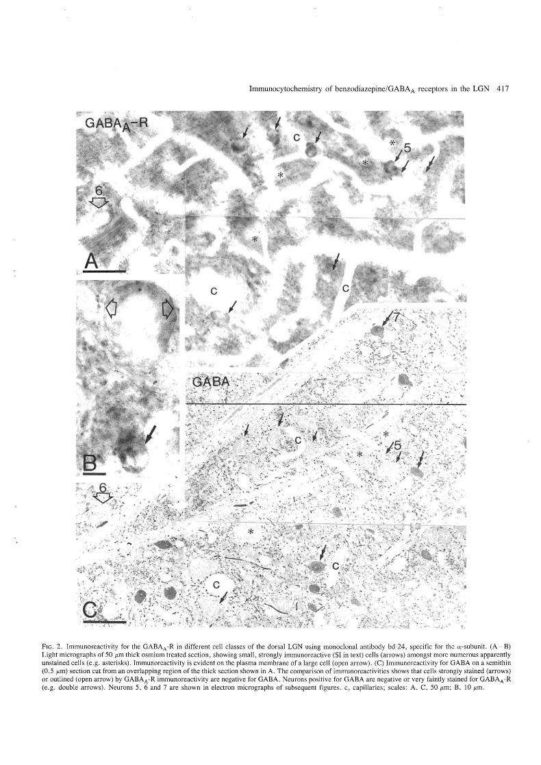
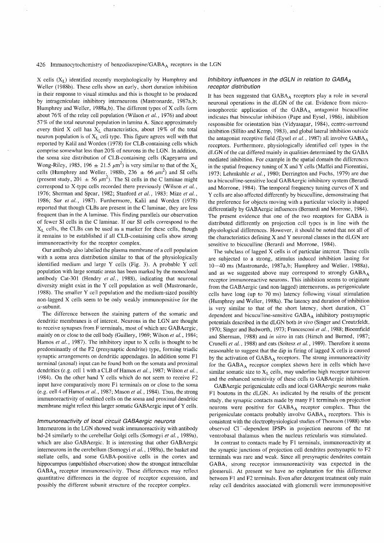
FIG . 2. Immunoreactivity for the GAB AA -R in different cell classes of the dorsal LGN using monoclonal antibody bd-24, specific for the a-subunit. (A - B) Light micrographs of 50 flm thick osmium treated section , showing small, strongly immunoreactive (SI in text) cells (arrows) amongst more numerous apparently unstained cells (e.g. asterisks). Immunoreactivity is evident on the plasma membrane of a large cell (open arrow). (C) Immunoreactivity for GABA on a semithin (0.5 flm) section cut from an overlapping region of the thick section shown in A. The comparison of immunoreactivities shows that cells strongly stained (arrows) or outlined (open arrow) by GABAA -R immunoreactivity are negative for GABA. Neurons positive for GABA are negative or very faintly stained for GABAA-R (e.g. double arrows) . Neurons 5, 6 and 7 are shown in electron micrographs of subsequent figures. c, capillaries; scales : A, C, 50 flm; B, 10 flm .
418 Immunocytochemistry of benzodiazepine/GABAA receptors in the LGN
epoxy resin (DURCUPAN ACM, Fluka). To increase contrast for electron microscopy the sections were treated with 1 % uranyl acetate in 70% ethanol for 40 min during dehydration. Lead staining was not used. Ultrathin sections were cut from the surface layers of the thick vibratome sections because the immunoreactivity was usually limited to the superficial 10-15 j-tm of the sections. At least two areas from each animal were cut for electron microscopy.
Postembedding immunocytochemistry for GABA was carried out on semithin (0.5 j-tm thick) sections cut from the resin-embedded, vibratome sections (50 j-tm thick), which had been immunoreacted for the receptor complex before embedding. Rabbit antiserum to GAB A (Code No. 9) and previously described procedures were used (Hodgson et aI., 1985; Somogyi et al., 1985). Alternate semi thin sections were either stained with toluidine blue/azure II for the identification of cytoplasmic laminated bodies, or immunoreacted for GABA.
Morphometric analysis
The outlines of neuronal somata were drawn along their largest extent, as projected by a drawing tube in the plane of the section, from the osmium-treated vibratome sections immunoreacted for the GABAA receptor complex. Only neurons with immunoreaction recognizable using a 50x oil immersion objective were drawn. The areas of these two dimensional projections were measured with the aid of a bitpad attached to a computer and evaluated using the package Bioquant IV (R. & M. Biometrics Inc.). Statistical analysis was carried out using the two-tailed Student's Hest. Numerical values are expressed in the text as mean ± SD.
Results
Controls
Immunoreactivity could not be detected when the tissue culture supernatant containing the monoc1onal antibodies was either omitted from the incubation or replaced by fresh tissue culture medium.
Distribution of immunoreactivity as detected by light microscopy
There were substantial differences between sections incubated with and without Triton X-WO. When the detergent was included very strong immunoreactivity was observed throughout the neuropil in all areas of the visual thalamus including the dorsal and ventral lateral geniculate nucleus, the medial interlaminar nucleus, the pulvinar and the lateral posterior complex (Fig. 1). The immunoreactivity was similar with both the (X- and the f3-subunit specific antibodies. The strong immunoreactivity in the neuropil made the evaluation of individual neurons difficult in detergent treated sections.
The distribution was different in sections which were not treated with detergent. Immunoreactivity could not be detected light microscopically in and around the LGN with undiluted hybridoma supernatant containing antibody bd-17, specific for the f3-subunit of the receptor complex. With antibody bd-24 the immunoreactivity was easily detectable at both light and electron microscopic level, but immunoreactivity in the neuropil was substantially weaker than in detergent treated sections. This differential decrease in immunoreactivity allowed better visualization of neuronal somata (Fig. 2A). We therefore used antibody bd-24 for detailed analysis. The most striking feature of the pattern of immunoreactivity as seen in the light microscope was the presence of small, strongly immunoreactive (SI) cells amongst the more numerous nonimmunoreactive somata. These cells often seemed to
A
(/I
Cl.) 0 -0
... Q) .0 E ::J c:
B
(/I
Qi 0
'0 to.. Cl.) .0 E ::J c:
C
.!!! Qi 0 -0
to.. Cl.) .0 E ::J c:
200
180
160
140
120
100
80
60
40
20
0
120
100
80
60
40
20
0
6
5
4
3
2
lamina A-At
• Scalls o O.L. cells
~ tOOO soma size (in 1Jm1
lamina C
• S cells o O.L. cells
1000 500 2 soma size (In IJm )
Humphrey and Wailer (for Xl cells) Frledlander et si (for Y cells)
• XL cells
0 Y calls
O~~~~~~~~~~~~-r~~~ SOO 2' 1000
soma size (In IJm )
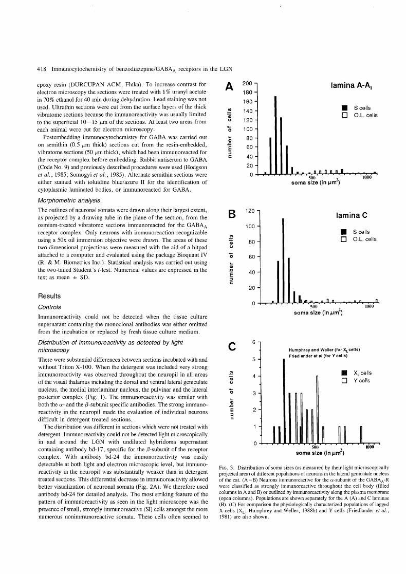
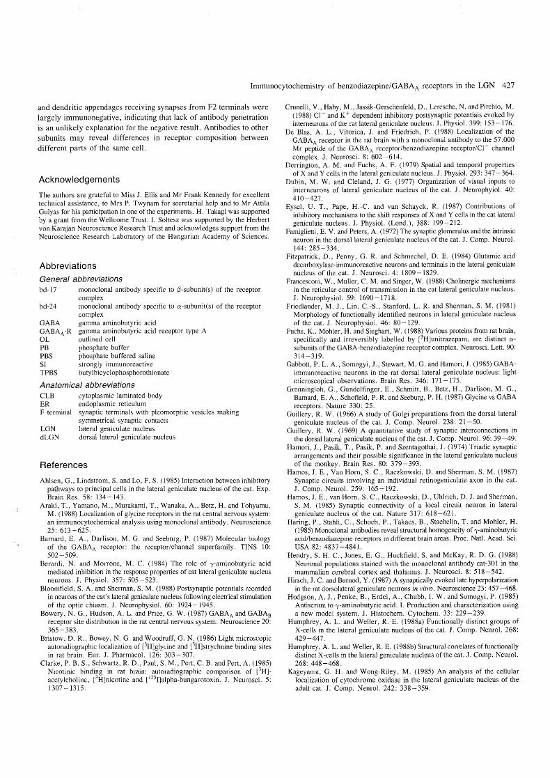
FIG. 3. Distribution of soma sizes (as measured by their light microscopically projected area) of different populations of neurons in the lateral geniculate nucleus of the cat. (A - B) Neurons immunoreactive for the a-subunit of the GABAA-R were classified as strongly immunoreactive throughout the cell body (filled columns in A and B) or outlined by immunoreactivity along the plasma membrane (open columns). Populations are shown separately for the A (A) and C laminae (B). (C) For comparison the physiologically characterized populations of lagged X cells (XL' Humphrey and Weller, 1988b) and Y cells (Friedlander et al., 1981) are also shown.
Immunocytochemistry of benzodiazepine/GABAA receptors in the LGN 419
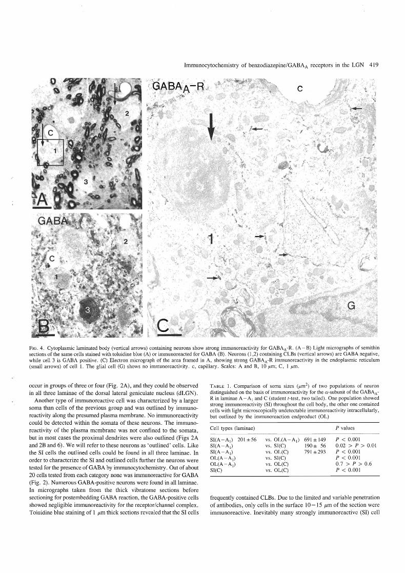
FIG. 4. Cytoplasmic laminated body (vertical arrows) containing neurons show strong immunoreactivity for GABAA -R. (A - B) Light micrographs of semithin sections of the same cells stained with toluidine blue (A) or immunoreacted for GABA (B). Neurons (l ,2) containing CLBs (vertical arrows) are GABA negative , while cell 3 is GABA positive. (C) Electron micrograph of the area framed in A, showing strong GABAA -R immunoreactivity in the endoplasmic reticulum (small arrows) of cell 1. The glial cell (G) shows no immunoreactivity . c, capillary. Scales: A and B, 10 /Lm; C, 1 /Lm.
occur in groups of three or four (Fig. 2A), and they could be observed in all three laminae of the dorsal lateral geniculate nucleus (dLGN).
Another type of immunoreactive cell was characterized by a larger soma than cells of the previous group and was outlined by immunoreactivity along the presumed plasma membrane. No immunoreactivity could be detected within the somata of these neurons . The immunoreactivity of the plasma membrane was not confined to the somata, but in most cases the proximal dendrites were also outlined (Figs 2A and 2B and 6). We will refer to these neurons as 'outlined' cells. Like the SI cells the outlined cells could be found in all three laminae. In order to characterize the SI and outlined cells further the neurons were tested for the presence of GABA by immunocytochemistry. Out of about 20 cells tested from each category none was immunoreactive for GABA (Fig. 2). Numerous GABA-positive neurons were found in all laminae. In micrographs taken from the thick vibratome sections before sectioning for postembedding GAB A reaction, the GABA-positive cells showed negligible immunoreactivity for the receptor/channel complex. Toluidine blue staining of 1 /Lm thick sections revealed that the SI cells
TABLE 1. Comparison of soma sizes (/Lm2) of two populations of neuron distinguished on the basis of immunoreactivity for the a-subunit of the GABAA-R in laminae A - A I and C (student (-test , two tailed). One population showed strong immunoreactivity (SI) throughout the cell body , the other one contained cells with light microscopically undetectable immunoreactivity intracellularly, but outlined by the immunoreaction endproduct (OL)
Cell types (laminae)
SI(A - AI) 201 ± 56 SI(A -AI) SI(A-A I) OL(A-A I) OL(A-A I) SI(C)
vs . OL(A-A I) vs. SI(C) vs. OL(C) vs. SI(C) vs. OL(C) vs. OL(C)
691 ± 149 190± 56 791 ±293
P values
P < 0 .001 0 .02 > P > 0.01 P < 0.001 P < 0.001 0.7 > P > 0.6 P < 0 .001
frequently contained CLBs. Due to the limited and variable penetration of antibodies, only cells in the surface 10 -15 /Lm of the section were immunoreactive. Inevitably many strongly immunoreactive (SI) cell
420 Immunocytochemistry of benzodiazepine/GABAA receptors in the LGN
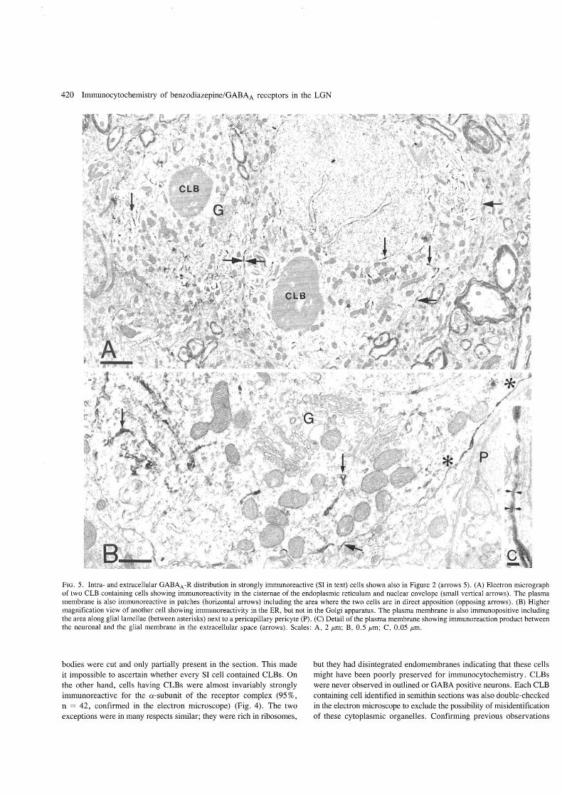
FIG. 5. Intra- and extracellular GABAA-R distribution in strongly immunoreactive (SI in text) cells shown also in Figure 2 (arrows 5). (A) Electron micrograph of two CLB containing cells showing immunoreactivity in the cisternae of the endoplasmic reticulum and nuclear envelope (small vertical arrows). The plasma membrane is also immunoreactive in patches (horizontal arrows) including the area where the two cells are in direct apposition (opposing arrows). (B) Higher magnification view of another cell showing immunoreactivity in the ER, but not in the Golgi apparatus. The plasma membrane is also immunopositive including the area along glial lamellae (between asterisks) next to a pericapillary pericyte (P). (C) Detail of the plasma membrane showing immunoreaction product between the neuronal and the glial membrane in the extracellular space (arrows). Scales: A, 2 /Lm ; B, 0.5 /Lm; C, 0 .05 /Lm.
bodies were cut and only partially present in the section. This made it impossible to ascertain whether every SI cell contained CLBs. On the other hand, cells having CLBs were almost invariably strongly immunoreactive for the a-subunit of the receptor complex (95 %, n = 42, confirmed in the electron microscope) (Fig. 4). The two exceptions were in many respects similar; they were rich in ribosomes,
but they had disintegrated endomembranes indicating that these cells might have been poorly preserved for immunocytochemistry. CLBs were never observed in outlined or GAB A positive neurons. Each CLB containing cell identified in semi thin sections was also double-checked in the electron microscope to exclude the possibility of misidentification of these cytoplasmic organelles. Confirming previous observations
Immunocytochemistry of benzodiazepine/GABAA receptors in the LGN 421
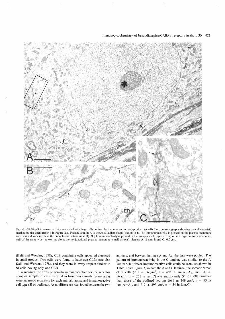
FIG. 6. GABAA -R immunoreactivity associated with large cells outlined by immunoreaction end-product. (A - B) Electron micrographs showing the cell (asterisk) marked by the open arrow 6 in Figure 2A . Framed area in A is shown at higher magnification in B. (B) Immunoreactivity is present on the plasma membrane (arrows) and very rarely in the endoplasmic reticulum (ER). (C) Immunoreactivity is present in the synaptic cleft (open arrow) of an F type bouton and another cell of the same type, as well as along the nonjunctional plasma membrane (small arrows). Scales: A, 2 !tm; Band C, 0 .5 !tm.
(Kalil and Worden, 1978), CLB containing cells appeared clustered in small groups. Two cells were found to have two CLBs (see also Kalil and Worden, 1978), and they were in every respect similar to SI cells having only one CLB.
To measure the sizes of somata immunoreactive for the receptor complex samples of cells were taken from two animals. Soma areas were measured separately for each animal, lamina and immunoreactive cell type (SI or outlined). As no difference was found between the two
animals, and between laminae A and A I, the data were pooled. The pattern of immunoreactivity in the C laminae was similar to the A laminae, but fewer immunoreactive cells could be seen. As shown in Table 1 and Figure 3, in both the A and C laminae, the somatic 'area' of SI cells (201 ± 56 ILm2, n = 462 in lam.A-A 1, and 190 ±
56 J.tm2 , n = 251 in lam.C) was significantly (P < 0.001) smaller than those of the outlined neurons (691 ± 149 J.tm2
, n = 53 in lam.A-A 1, and 712 ± 293 J.tm2 , n = 34 in lam.C) .
422 Immunocytochemistry of benzodiazepine/GABAA receptors in the LGN
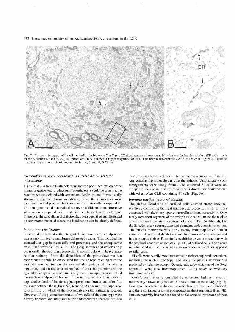
FIG. 7. Electron micrograph of the cell marked by double arrow 7 in Figure 2C showing sparse immunoreactivity in the endoplasmic reticulum (ER and arrows) for the a-subunit of the GABAA -R . Framed area in A is shown at higher magnification in B. This neuron also contains GABA as shown in Figure 2C therefore it is very likely a local circuit neuron. Scales: A, 2 JLm; B, 0.25 JLm.
Distribution of immunoreactivity as detected by electron microscopy
Tissue that was treated with detergent showed poor localization of the immunoreaction end-production. Nevertheless it could be seen that the reaction was associated with somata and dendrites, and it was usually stronger along the plasma membrane. Since the membranes were disrupted the end-product also spread onto all intracellular organelles. The detergent treated material did not reveal additional immunoreactive sites when compared with material not treated with detergent. Therefore, the subcellular distribution has been described and illustrated on nontreated material where the localization can be clearly defined.
Membrane localization In material not treated with detergent the immunoreaction endproduct was mainly limited to membrane delineated spaces. This included the extracellular gap between cells and processes, and the endoplasmic reticulum cisternae (Figs. 4-8) . The Golgi saccules and vesicles only occasionally showed immunoreactivity, even in cells with heavy intracellular staining. From the deposition of the peroxidase reaction endproduct it could be established that the epitope reacting with the antibody was located on the extracellular surface of the plasma membrane and on the internal surface of both the granular and the agranular endoplasmic reticulum. Using the imrnunoperoxidase method the reaction endproduct formed in the narrow extracellular space is deposited on both of the closely juxtaposed membranes and often fills the space between them (Figs. SC , 6 and 9). As a result, it is impossible to determine on which of the two membranes the antigen is located. However, if the plasma membranes of two cells of the same type were directly apposed and imrnunoreaction endproduct was present between
them, this was taken as direct evidence that the membrane of that cell type contains the molecule carrying the epitope. Unfortunately such arrangements were rarely found. The clustered SI cells were an exception; their somata were frequently in direct membrane contact with other, often CLB containing SI cells (Fig. SA).
Immunoreactive neuronal classes The plasma membrane of outlined cells showed strong immunoreactivity confirming the light microscopic prediction (Fig. 6). This contrasted with their very sparse intracellular immunoreactivity. Only rarely were short segments of the endoplasmic reticulum and the nuclear envelope found to contain reaction endproduct (Fig. 6) although, like the SI cells, these neurons also had abundant endoplasmic reticulum. The plasma membrane was fairly evenly immunopositive both at somatic and proximal dendritic sites. Immunoreactivity was present in the synaptic cleft of F terminals establishing synaptic junctions with the proximal dendrites or somata (Fig. 6C) of outlined cells. The plasma membrane of outlined cells was also immunoreactive when apposed to glial cells.
SI cells were heavily immunoreactive in their endoplasmic reticulum, including the nuclear envelope, and along the plasma membrane as predicted by light microscopy. Occasionally a few cisternae of the Golgi apparatus were also immunopositive. CLBs never showed any immunoreactivity .
GABA positive cells identified by correlated light and electron microscopy showed only moderate levels of immunoreactivity (Fig. 7). Few immunoreactive endoplasmic reticulum profiles were observed, and these contained reaction endproduct in short segments (Fig. 7B). Immunoreactivity has not been found on the somatic membrane of these cells.
"
Immunocytochemistry of benzodiazepine/GABAA receptors in the LGN 423
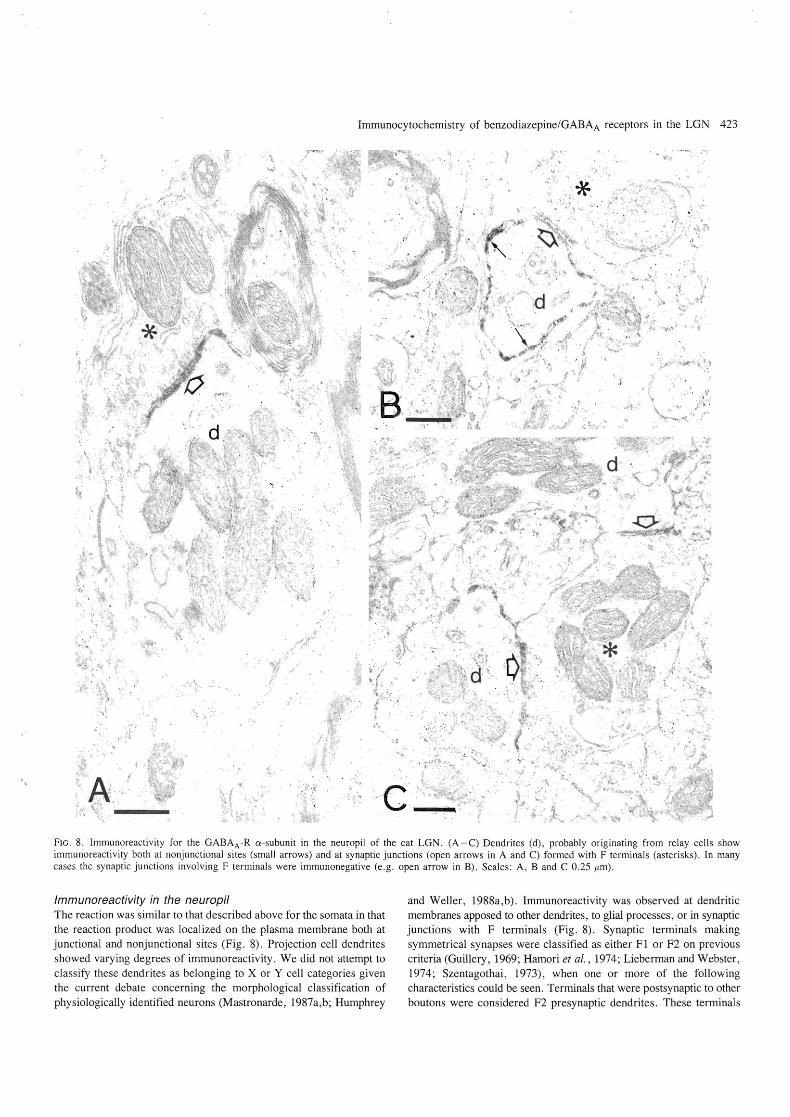
FIG. 8. Immunoreactivity for the GABAA -R a-subunit in the neuropil of the cat LGN. (A -C) Dendrites (d), probably originating from relay cells show immunoreactivity both at nonjunctional sites (small arrows) and at synaptic junctions (open arrows in A and C) formed with F terminals (asterisks). In many cases the synaptic junctions involving F terminals were immunonegative (e.g. open arrow in B). Scales: A, Band C 0.25 jlm) .
Immunoreactivity in the neuropil The reaction was similar to that described above for the somata in that the reaction product was localized on the plasma membrane both at junctional and nonjunctional sites (Fig. 8). Projection cell dendrites showed varying degrees of immunoreactivity. We did not attempt to classify these dendrites as belonging to X or Y cell categories given the current debate concerning the morphological classification of physiologically identified neurons (Mastronarde, 1987a,b; Humphrey
and Weller , 1988a,b). Immunoreactivity was observed at dendritic membranes apposed to other dendrites, to glial processes , or in synaptic junctions with F terminals (Fig. 8). Synaptic terminals making symmetrical synapses were classified as either F 1 or F2 on previous criteria (Guillery, 1969; Hamori et al., 1974; Lieberman and Webster, 1974; Szentagothai, 1973), when one or more of the following characteristics could be seen. Terminals that were postsynaptic to other boutons were considered F2 presynaptic dendrites. These terminals
424 Immunocytochemistry of benzodiazepine/GABAA receptors in the LGN
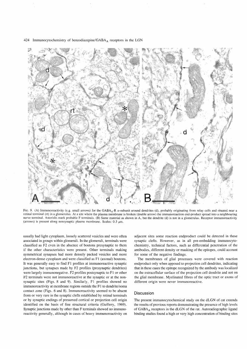
FIG. 9. (A) Immunoreactivity (e .g . small arrows) for the GABAA-R a-subunit around dendrites (d), probably originating from relay cells and situated near a retinal terminal (rt) in a glomerulus . At a site where the plasma membrane is broken (double arrow) the immunoreaction end-product spread into a neighbouring nerve terminal. Asterisks mark probable F terminals . (B) Same material as shown in A, but the dendrite (d) is not in a glomerulus. Receptor immunoreactivity (arrows) is present along nonsynaptic plasma membrane. Scales: 0.5 /Lm.
usually had light cytoplasm, loosely scattered vesicles and were often associated in groups within glomeruli . In the glomeruli, terminals were classified as F2 even in the absence of boutons presynaptic to them if the other characteristics were present. Other terminals making symmetrical synapses had more densely packed vesicles and more electron-dense cytoplasm and were classified as Fl (axonal) boutons. It was generally easy to find Fl profiles at immunoreactive synaptic junctions, but synapses made by F2 profiles (presynaptic dendrites) were largely immunonegative. F2 profiles postsynaptic to Fl or other F2 terminals were not immunoreactive at the synaptic or at the nonsynaptic sites (Figs. 8 and 9). Similarly, F 1 profiles showed no immunoreactivity at membrane regions outside the F 1 to dendrite/soma contact zone (Figs . 6 and 8). Immunoreactivity seemed to be absent from or very rare in the synaptic clefts established by retinal terminals or by synaptic endings of presumed cortical or projection cell origin identified on the basis of fine structural criteria (Guillery, 1969). Synaptic junctions made by other than F terminals showed no immunoreactivity generally, although in cases of heavy immunoreactivity on
adjacent sites some reaction endproduct could be detected in these synaptic clefts. However, as in all pre-embedding immunocytochemistry, technical factors, such as differential penetration of the antibodies, different density or masking of the epitopes, could account for some of the negative findings.
The membranes of glial processes were covered with reaction endproduct only when apposed to projection cell dendrites, indicating that in these cases the epitope recognized by the antibody was localized on the extracellular surface of the projection cell dendrite and not on the glial membrane. Myelinated fibres of the optic tract or axons of different origin were never immunoreactive.
Discussion
The present immunocytochemical study on the dLGN of cat extends the results of previous reports demonstrating the presence of high levels of GABAA receptors in the dLGN of the rat. Autoradiographic ligand binding studies found a high or very high concentration of binding sites
Immunocytochemistry of benzodiazepine/GABAA receptors in the LGN 425
for the GABAA receptor (Bowery et aI., 1987, Palacois et al., 1981) in the dLGN of the rat. Immunohistochemical studies using monoclonal antibodies also demonstrated a very high concentration of GABAA/benzodiazepine receptor proteins in the dLGN of rodents (Richards et aI., 1987; De BIas et al., 1988).
Specificity of the immunoreaction
The recent recognition of a superfamily of ligand gated receptor/channel proteins (Barnard et at., 1987; Grenningloh et aI., 1987; Schofield et al., 1987) that includes the GABAA/benzodiazepine, the glycine and the nicotinic acetylcholine receptors, raises the question of antibody crossreactivity with several of these proteins. It has been discussed elsewhere that the recognition of proteins other than the a- and {3-subunit of the GABAA/benzodiazepine receptor by bd-24 and bd-17 respectively is highly unlikely (Richards et al., 1987; Somogyi et al., 1989a). In the case of the dLGN, crossreactivity with glycine receptors can be excluded because strychnine-sensitive glycine receptors are undetectable both by autoradiography (Wamsley and Palacois, 1984; Bristow et aI., 1986; Probst et al., 1986) or by immunohistochemistry (Araki et al., 1988), and there does not seem to be significant binding of glycine to strychnine-insensitive sites either (Bristow et aI., 1986). Nicotinic acetylcholine receptors are present in high concentration in the dLGN as revealed by ligand binding (Clarke et aI., 1985) and immunocytochemical (Swanson et al., 1987) studies. However, from the distribution of immunoreactivity patterns for the GABAA/benzodiazepine receptor (Richards et al., 1986, 1987; De BIas et al., 1988) and the nicotinic acetylcholine receptor (Swanson et al., 1987) it is clear that our antibodies do not cross-react with brain nicotinic receptor sites.
Considering the strong immunoreactivity in Triton-treated sections, it was surprising that in the absence of detergent only negligible immunoreactivity could be detected with the {3-subunit specific antibody, even with the electron microscope. In the cerebellum of the cat only the strongest sites showed some immunoreactivity with this antibody while in the monkey the two antibodies gave similar levels of immunoreactivity (Somogyi et al., 1989a). Immunoreactivity with both antibodies and in all species is clearly enhanced by detergent, but the epitope recognized by bd-17 in the cat seems to be masked more than in other species.
Recent studies indicate that there are several different a-subunits in the brain (Fuchs et aI., 1988) and these may differ in their pharmacological characteristics (Levitan et al., 1988). As our antibody may only recognize some of the a-subunit proteins it cannot be established at present whether the differences between different classes of cells are due to variation in subunit composition or to the degree of receptor expression.
Subcellular distribution of immunoreactive receptors
Immunoreactivity in the endoplasmic reticulum and in the Golgi apparatus presumably reflects the biosynthesis of the protein and the addition of carbohydrate residues respectively. The level of intracellular and plasma membrane immunoreactivity is not well correlated in the dLGN, or in the cerebellum. Thus, some classes of neuron such as cerebellar granule cells (Somogyi et al., 1989a) and large GABAnegative cells in the dLGN can have strong immunoreactivity on their plasma membrane but negligible immunoreactivity intracellularly. The degree of immunoreactivity is probably related to the intensity of receptor synthesis and receptor turnover, while intensity of plasma membrane immunoreactivity is probably related to receptor density
(Somogyi et al., 1989a). It has been suggested from in situ hybridization studies that the a-, but not the {3-subunit of the receptor complex is expressed in cerebellar Purkinje cells (Siegel, 1988; Richards et al., unpublished observation). Our immunocytochemical studies showed that immunoreactivity for both subunits can be detected on Purkinje cells, but the intracellular immunoreactivity was significantly stronger for the a-subunit (Somogyi et al., 1989a). This may explain the stronger signal obtained for this subunit in in situ hybridization (Siege1, 1988; Richards et al., unpublished observation). The situation seems to be similar in the dLGN, where in situ hybridization studies demonstrated very high hybridization signals with an a-subunit specific probe, but very low signals with a {3-subunit probe (Sequier et al., 1988).
With regard to the distribution of receptors in the plasma membrane, it should be noted that the epitope recognized by bd-24 is unknown, although it must be outside the ligand recognition site since neither GABA nor benzodiazepine binding is inhibited by the antibody (Haring et aI., 1985). It is surprising that the receptor is widely distributed both at synaptic junctions and at non-synaptic plasma membrane. A previous preliminary electron microscopic study, using antibody bd-I7 , emphasized the synaptic location of the receptor complex (Richards et al., 1987). The present study, as well as results obtained in the cerebellum (Somogyi et al., 1989a) and in the neocortex (Somogyi et al., 1989b) demonstrated that the junctional and non-junctional distribution of the receptor complex is a general feature of the central nervous system. However, the distribution is not uniform even on the same cell, because synaptic junctions of F terminals, most of which have been shown to contain GABA (Ohara et al., 1983; Fitzpatrick et al., 1984; Montero, 1986, 1987; Montero and Singer, 1985), were frequently immunopositive. In contrast, junctions of retinal and presumed cortical boutons, which are not GABAergic, were almost without exception immunonegative. It was shown in the cerebellum, that the nonjunctional immunoreactivity cannot result from the diffusion of the reaction end-product, but reflects the distribution of the receptor protein (Somogyi et al., 1989a).
The function of the nonjunctional receptors remains to be established. We have proposed that the widespread distribution of the receptor on the plasma membrane means that the topography of GABAergic influence mediated by GABAA receptors is governed by the precise placement of GABA-releasing terminals, and not by the location of the receptors (Somogyi et al., 1989a).
Differential expression of the receptor complex by different classes of neuron
Immunohistochemical studies have revealed significant differences in GABAA receptor concentration between different types of neuron located in the same brain region (Richards et al., 1987; De BIas et al., 1988; Somogyi et al., 1989a). These differences have been confirmed by in situ hybridization (Sequier et aI., 1988; Siegel et aI., 1988) demonstrating differences in the concentration of mRNAs. As in other brain regions the a-subunit specific antibody revealed differences between morphologically and functionally different classes of neuron in the dLGN of the cat.
Immunoreactivity of projection cells The two cell types found by immunostaining may correspond to functionally distinct neural types. The first type is the SI cell, which frequently contained CLBs and was GABA-negative with a soma area distribution almost identical (Fig. 3) to that of the so called 'lagged'
426 Immunocytochemistry of benzodiazepine/GABAA receptors in the LGN
X cells (Xd identified recently morphologically by Humphrey and Weller (1988b). These cells show an early, short duration inhibition in their response to visual stimulus and this is thought to be produced by intrageniculate inhibitory interneurons (Mastronarde, 1987a,b; Humphrey and Well er, 1988a, b). The different types of X cells form about 76 % of the relay cell population (Wilson et aI., 1976) and about 57 % of the total neuronal population in lamina A. Since approximately every third X cell has characteristics, about 19 % of the total neuron population is of XL cell type. This figure agrees well with that reported by Kalil and Worden (1978) for CLB-containing cells which comprise somewhat less than 20% of neurons in the LGN. In addition, the soma size distribution of CLB-containing cells (Kageyama and Wong-Riley, 1985, 196 ± 21.5 /1-m2) is very similar to that of the XL cells (Humphrey and Weller, 1988b, 236 ± 66 /1-m2) and SI cells (present study, 201 ± 56 /1-m2
). The SI cells in the C laminae might correspond to X -type cells recorded there previously (Wilson et al. , 1976; Sherman and Spear, 1982; Stanford et al., 1983; Mize et aI., 1986; Sur et al., 1987). Furthermore, Kalil and Worden (1978) reported that though CLBs are present in the C laminae, they are less frequent than in the A laminae. This find~ng parallels our observation of fewer SI cells in the C laminae. If our SI cells correspond to the XL cells, the CLBs can be used as a marker for these cells, though it remains to be established if all CLB-containing cells show strong immunoreactivity for the receptor complex.
Our antibody also labelled the plasma membrane of a cell population with a soma area distribution similar to that of the physiologically identified medium and large Y cells (Fig. 3). A probable Y cell population with large somatic areas has been marked by the monoc1onal antibody Cat-301 (Hendry et al., 1988), indicating that neuronal diversity might exist in the Y cell population as well (Mastronarde, 1988). The smaller Y cell population and the medium-sized possibly non-lagged X cells seem to be only weakly immunopositive for the a-subunit.
The difference between the staining pattern of the somatic and dendritic membranes is of interest. Neurons in the LGN are thought to receive synapses from F terminals, most of which are GABAergic, mainly on or close to the cell body (GuiIlery, 1969; Wilson et al., 1984; Hamos et al., 1987). The inhibitory input to X cells is thought to be predominantly of the F2 (presynaptic dendrite) type, forming triadic synaptic arrangements on dendritic appendages. In addition some Fl terminal (axonal) input can be found both on the somata and proximal dendrities cell 1 with a CLB of Hamos et al., 1987; Wilson et al. , 1984). On the other hand Y cells which do not seem to receive F2 input have comparatively more Fl terminals on or close to the soma
cell 4 of Hamos et aI., 1987; Mason et aI., 1984). Thus, the strong immunoreactivity of outlined cells on the soma and proximal dendritic membrane might reflect this larger somatic GABAergic input ofY cells.
Immunoreactivity of local circuit GABAergic neurons Interneurons in the LGN showed weak immunoreactivity with antibody bd-24 similarly to the cerebellar Golgi cells (Somogyi et al., 1989a), which are also GABAergic. It is interesting that other GABAergic interneurons in the cerebellum (Somogyi et aI., 1989a), the basket and stellate cells, and some GABA-positive cells in the cortex and hippocampus (unpublished observation) show the strongest intracellular GABAA receptor immunoreactivity. These differences may reflect quantitative differences in the degree of receptor expression, and possibly the different subunit structure of the receptor complex.
Inhibitory influences in the dGLN in relation to GABAA receptor distribution
It has been suggested that GABAA receptors play a role in several neuronal operations in the dLGN of the cat. Evidence from microionophoretic application of the GABAA antagonist bicuculline indicates that binocular inhibition (Pape and Eysel, 1986), inhibition responsible for orientation bias (Vidyasagar, 1984), centre-surround inhibition (Sillito and Kemp, 1983), and global lateral inhibition outside the antagonist receptive field et al., 1987) all involve GABAA receptors. Furthermore, physiologically identified cell types in the dLGN ofthe cat differed mainly in qualities determined by the GABA mediated inhibition. For example in the spatial domain the differences in the spatial frequency tuning of X and Y cells (Maffei and Fiorentini, 1973; Lehmkuhle et al., 1980; Derrington and Fuchs, 1979) are due to a bicuculline-sensitive local GABAergic inhibitory system (Berardi and Morrone, 1984). The temporal frequency tuning curves of X and Y cells are also affected differently by bicuculline, demonstrating that the for objects moving with a particular velocity is shaped differentially by GABAergic influences (Bernardi and Morrone, 1984). The present evidence that one of the two receptors for GABA is distributed differently on projection cell types is in line with the physiological differences. However, it should be noted that not all of the characteristics defining X and Y neuronal classes in the dLGN are sensitive to bicuculline (Berardi and Morrone, 1984).
The subclass of lagged X cells is of particular interest. These cells are subjected to a strong, stimulus induced inhibition lasting for 10-40 ms (Mastronarde, 1987a,b; Humphrey and WeBer, 1988a), and as we suggested above may correspond to strongly GABAA receptor immunoreactive neurons. This inhibition seems to originate from the GABAergic (and non-lagged) interneurons, as perigeniculate cells have long (up to 70 ms) latency following visual stimulation (Humphrey and W eH er , 1988a). The latency and duration of inhibition is very similar to that of the short latency, short duration, CIdependent and bicuculline-sensitive GABAA inhibitory postsynaptic potentials described in the dLGN both in vivo (Singer and Creutzfeldt, 1970; Singer and Bedworth, 1973; Francesconi et al., 1988; Bloomfield and Sherman, 1988) and in vitro in rats (Hirsch and Burnod, 1987; Cmnelli et aI., 1988) and cats (Soltesz et al., 1989). Therefore it seems reasonable to suggest that the dip in firing of lagged X cells is caused by the activation of GABAA receptors. The strong immunoreactivity for the GABAA receptor complex shown here in cells which have similar somatic size to XL cells, may underline high receptor turnover and the enhanced sensitivity of these cells to GABAergic inhibition.
GABAergic perigeniculate cells and local GABAergic neurons make Fl boutons in the dLGN. As indicated by the results of the present study, the synaptic contacts made by many F 1 terminals on projection neurons were positive for GABAA receptor complex. Thus the perigeniculate contacts probably involve GABAA receptors. This is consistent with the electrophysiological studies of Thomson (1988) who observed CI--dependent IPSPs in projection neurons of the rat ventrobasal thalamus when the nucleus reticularis was stimulated.
In contrast to contacts made by Fl terminals, immunoreactivity at the synaptic junctions of projection cell dendrites postsynaptic to F2 terminals was rare and weak. Since all presynaptic dendrites contain GABA, strong receptor immunoreactivity was expected in the glomeruli. At present we have no explanation for this difference between Fl and F2 terminals. Even after detergent treatment only main relay cell dendrites associated with glomeruli were immunopositive
Immunocytochemistry of benzodiazepine/GABAA receptors in the LGN 427
and dendritic appendages receiving synapses from F2 terminals were largely immunonegative, indicating that lack of antibody penetration is an unlikely explanation for the negative result. Antibodies to other subunits may reveal differences in receptor composition between different parts of the same cell.
Acknowledgements
The authors are grateful to Miss 1. Ellis and Mr Frank Kennedy for excellent technical assistance, to Mrs P. Twynam for secretarial help and to Mr Attila Gulyas for his participation in one of the experiments. H. Takagi was supported by a grant from the Wellcome Trust. I. Soltesz was supported by the Herbert von Karajan Neuroscience Research Trust and acknowledges support from the Neuroscience Research Laboratory of the Hungarian Academy of Sciences.
Abbreviations General abbreviations bd-17 monoclonal antibody specific to {3-subunit(s) of the receptor
complex bd-24 monoclonal antibody specific to a-subunit(s) of the receptor
complex GABA gamma aminobutyric acid GABAA-R gamma aminobutyric acid receptor type A OL outlined cell PB phosphate buffer PBS phosphate buffered saline SI strongly immunoreactive TPBS butylbicyclophosphorothionate
Anatomical abbreviations CLB ER F terminal
LGN dLGN
cytoplasmic laminated body endoplasmic reticulum synaptic terminals with pleomorphic vesicles making symmetrical synaptic contacts lateral geniculate nucleus dorsal lateral geniculate nucleus
References
Ahlsen, G., Lindstrom, S. and Lo, F. S. (1985) Interaction between inhibitory pathways to principal cells in the lateral geniculate nucleus of the cat. Exp. Brain Res. 58: 134-143.
Araki, T., Yamano, M., Murakami, T., Wanaka, A., Betz, H. and Tohyama, M. (1988) Localization of glycine receptors in the rat central nervous system: an immunocytochemical analysis using monoclonal antibody. Neuroscience 25: 613 - 625.
Barnard, E. A., Darlison, M. G. and Seeburg, P. (1987) Molecular biology of the GABAA receptor: the receptor/channel superfamily. TINS 10: 502-509.
Berardi, N. and Morrone, M. C. (l984) The role of ),-aminobutyric acid mediated inhibition in the response properties of cat lateral geniculate nucleus neurons. 1. Physiol. 357: 505-523.
Bloomfield, S. A. and Sherman, S. M. (1988) Postsynaptic potentials recorded in neurons of the cat's lateral geniculate nucleus following electrical stimulation of the optic chiasm. 1. Neurophysiol. 60: 1924-1945.
Bowery, N. G., Hudson, A. L. and Price, G. W. (1987) GABAA and GABAB receptor site distribution in the rat central nervous system. Neuroscience 20: 365-383.
Bristow, D. R., Bowey, N. G. and Woodruff, G. N. (l986) Light microscopic autoradiographic localization of eHlglycine and eHJstrychnine binding sites in rat brain. Eur. J. Pharmacol. 126: 303-307.
Clarke, P. B. S., Schwartz, R. D., Paul, S. M., Pert, C. B. and Pert, A. (1985) Nicotinic binding in rat brain: autoradiographic comparison of eHJacetylcholine, [3H]nicotine and [125I]alpha-bungarotoxin. 1. Neurosci. 5: 1307 -1315.
Crunelli, V., Haby, M., lassik-Gerschenfeld, D., Leresche, N. andPirchio, M. (1988) Cl ~ and K + dependent inhibitory postsynaptic potentials evoked by interneurons of the rat lateral geniculate nucleus. 1. Physiol. 399: 153-176.
De Bias, A. L., Vitorica, J. and Friedrich, P. (1988) Localization of the GABAA in the rat brain with a monoclonal antibody to the 57,000 Mr peptide the GABAA receptor/benzodiazepine receptor/Cl ~ channel complex. J. Neurosci. 8: 602-614.
Derrington, A. M. and Fuchs, A. F. (1979) Spatial and temporal properties of X and Y cells in the lateral geniculate nucleus. 1. Physiol. 293: 347-364.
Dubin, M. W. and Cleland, 1. G. (1977) Organization of visual inputs to interneurons of lateral geniculate nucleus of the cat. 1. Neurophyiol. 40: 410-427.
Eysel, U. T., Pape, H.-C. and van Schayck, R. (l987) Contributions of inhibitory mechanisms to the shift responses of X and Y cells in the cat lateral geniculate nucleus. 1. Physiol. (Lond.), 388: 199-212.
Famiglietti, E. V. and Peters, A. (1972) The synaptic glomerulus and the intrinsic neuron in the dorsal lateral geniculate nucleus of the cat. 1. Comp. Neurol. 144: 285-334.
Fitzpatrick, D., Penny, G. R. and Schmechel, D. E. (l984) Glutamic acid decarboxylase-immunoreactive neurons and terminals in the lateral geniculate nucleus of the cat. 1. Neurosci. 4: 1809-1829.
Francesconi, W., Muller, C. M. and Singer, W. (1988) Cholinergic mechanisms in the reticular control of transmission in the cat lateral geniculate nucleus. 1. Neurophysiol. 59: 1690-1718.
Friedlander, M. 1., Lin, c.-S., Stanford, L. R. and Sherman, S. M. (1981) Morphology of functionally identified neurons in lateral geniculate nucleus of the cat. 1. Neurophysiol. 46: 80-129.
Fuchs, K., Mohler, H. and Sieghart, W. (1988) Various proteins from rat brain, specifically and irreversibly labelled by [3Hlunitrazepam, are distinct asubunits of the GABA-benzodiazepine receptor complex. Neurosci. Lett. 90: 314-319.
Gabbott, P. L. A., Somogyi, 1., Stewart, M. G. and Hamori, 1. (1985) GABAimmunoreactive neurons in the rat dorsal lateral geniculate nucleus: light microscopical observations. Brain Res. 346: 171 175.
Grenningloh, G., Gundelfinger, E., Schmitt, B., Betz, H., Dariison, M. G., Bamard, E. A., Schofield, P. R. and Seeburg, P. H. (1987) Glycine vs GABA receptors. Nature 330: 25.
Guillery, R. W. (1966) A study of Golgi preparations from the dorsal lateral geniculate nucleus of the cat. 1. Comp. Neurol. 238: 21-50.
Guillery, R. W. (1969) A quantitative study of synaptic interconnections in the dorsal lateral geniculate nucleus of the cat. 1. Comp. Neurol. 96: 39 -49.
Hamori, 1., Pasik, T., Pasik, P. and Szentagothai, 1. (1974) Triadic synaptic arrangements and their possible significance in the lateral geniculate nucleus of the monkey. Brain Res. 80: 379-393.
Hamos, 1. E., Van Horn, S. C., Raczkowski, D. and Sherman, S. M. (1987) Synaptic circuits involving an individual retinogeniculate axon in the cat. 1. Camp. Neurol. 259: 165-192.
Hamos, J. E., van Horn, S. C., Raczkowski, D., Uhlrich, D. 1. and Sherman, S. M. (1985) Synaptic connectivity of a local circuit neuron in lateral geniculate nucleus of the cat. Nature 317: 618 - 62l.
Haring, P., Stahli, C., Schoch, P., Takacs, B., Staehelin, T. and Mohler, H. (1985) Monoclonal antibodies reveal structural homogeneity of ),-arninobutyric acid/benzodiazepine receptors in different brain areas. Proc. Natl. Acad. Sci. USA 82: 4837 -4841.
Hendry, S. H. C., lones, E. G., Hockfield, S. and McKay, R. D. G. (1988) Neuronal populations stained with the monoclonal antibody cat-301 in the mammalian cerebral cortex and thalamus. 1. Neurosci. 8: 518-542.
Hirsch, J. C. and Burnod, Y. (1987) A synaptically evoked late hyperpolarization in the rat dorsolateral geniculate neurons in vitro. Neuroscience 23: 457 -468.
Hodgson, A. 1., Penke, B., Erdei, A., Chubb, 1. W. and Somogyi, P. (1985) Antiserum to ),-aminobutyric acid. 1. Production and characterization using a new model system. 1. Histochem. Cytochem. 33: 229-239.
Humphrey, A. L. and Weller, R. E. (l988a) Functionally distinct groups of X-cells in the lateral geniculate nucleus of the cat. 1. Comp. Neurol. 268: 429-447.
Humphrey, A. L. and Weller, R. E. (l988b) Structural correlates of functionally distinct X-cells in the lateral geniculate nucleus of the cat. 1. Comp. Neurol. 268: 448 -468.
Kageyama, G. H. and Wong-Riley, M. (1985) An analysis of the cellular localization of cytochrome oxidase in the lateral geniculate nucleus of the adult cat. 1. Camp. Neurol. 242: 338-359.
428 Immunocytochemistry of benzodiazepine/GABAA receptors in the LGN
Kalil, R. and Worden, 1. (1978) Cytoplasmic laminated bodies in the lateral geniculate nucleus of normal and dark reared cats. J. Comp. Neurol. 178: 469-486.
Lehmkuhle, S., Kenneth, E. K., Mangel, S. C. and Sherman, S. M. (1980) Spatial and temporal sensitivity of X-and Y -cells in dorsal lateral geniculate nucleus of the cat. J. Neurophysiol. 43: 520 - 541.
Levitan, E. S., Schofield, P. R., Burt, D. R., Rhee, L. M., Wisden, W., Kohler, M., Fujita, N., Rodriguez, H. F., Stephenson, A., Darlison, M. G., Barnard, E. A. and Seeburg, P. H. (1988) Structural and functional basis for receptor heterogeneity. Nature 335: 76 - 79.
Lieberman, R. and Webster, K. E. (1974) of synaptic organization of intrinsic neurons in the dorsal lateral nucleus. An ultrastructural study of the normal and of the experimentally deafferented nucleus in the rat. J. Neurocyto!. 3: 677 - 710.
Lindstrom, S. (1982) Synaptic organization of inhibitory pathways to principal cells in the lateral geniculate nucleus of the cat. Brain Res. 234: 447 -453.
Madarasz, M., Somogyi, Gy., Somogyi, J. and Hamori, J. (1985) Numerical estimation of ),-aminobutyric acid (GABA)-containing neurons in three thalamic nuclei of the cat: direct GABA immunocytochemistry. Neurosci. Lett. 61: 73-78.
Maffei, L. and Fiorentini, A. (1972) convergence and analysis of contrast. J. Neurophysiol. 35:
Mason, C. A., GuiIIery, R. W. and Rosner, M. C. (1984) Patterns ofsynaptic contact individually labeled cells of the dorsal lateral geniculate nucleus the cat. Neuroscience 1 319 ~ 329.
Mastronarde, D. M. (1987) Two classes of single-input X-cells in cat lateral geniculate nucleus. n. Retinal inputs and the generation of receptive-field properties. J. Neurophysiol. 57: 381-413.
Mastronarde, D. M. (1987) Two classes of single-input X-cells in catlateral geniculate nucleus. I. Receptive-field properties and classification of cells. J. Neurophysiol. 57: 357-380.
Mastronarde, D. N. (1988) Branching of X and Y functional pathways in cat lateral geniculate nucleus. Soc. Neurosci. Abstr. 14: 309
McCormick, D. A. and Pape, H.-C. (1988) Acetylcholine inhibits identified interneurons in the cat lateral nucleus. Nature 334: 246-248.
Mize, R. R., Spencer, R. F. and L. H. (1986) Quantitative comparison of retinal synapses in the dorsal and ventral (parvicellular) C lam,inae of the cat dorsal lateral geniculate nucleus. 1. Comp. Neurol. 248: 57 -73.
Mohler, H., Schoch, P., Richards, J. G., Haring, P., Takacs, B. and Stahli, C. (1986) Monoclonal antibodies: probes for structure and location of the GABA receptor/benzodiazepine receptor/chloride channel complex. In: Olsen, R. W. and Venter, J. C. (eds), Benzodiazepine/GABA Receptors and Chloride Channels: Structural and Functional Properties, pp. 258-297. Alan R. Liss, New York.
Montero, V. M. (1986) Localization of ),-aminobutyric acid (GABA) in type 3 cells and demonstration of their source to F2 terminals in the cat lateral geniculate nucleus: a Golgi-electron-microscopic GABA-immunocytochemical study. J. Comp. Neurol. 254: 228-245.
Montero, V. M. (1987) Ultrastructural identification of synaptic terminals from the axon of type 3 interneurons in the cat lateral geniculate nucleus. J. Comp. Neurol. 264: 268-283.
Montero, V. M. and Singer, W. (1985) Ultrastructural identification of somata and neural processes immunoreactive to antibodies against glutamic acid decarboxylase (GAD) in the dorsal lateral geniculate nucleus of the cat. Exp. Brain Res. 59: 151-165.
Montero, V. M. and Zempel, J. (1985) Evidence for two types of GABAcontaining intemeurons in the A-laminae of the cat lateral geniculate nucleus: a double-label HRP and GABA-immunocytochemical study. Exp. Brain Res. 60: 603 -609.
Ohara, P. T., Lieberman, A. R., Hunt, S. P. and Wu, 1. Y. (1983) Neural elements containing glutamic acid decarboxylase (GAD) in the dorsal lateral gel1icllllate nucleus of the rat. Immunohistochemical studies by light and
microscopy. Neuroscience 8: 189 - 211. Palacois, J. M., Wamsley, J. K. and Kuhar, M. J. (1981) t1Hm-~nIllrmv
receptors: autoradiographic localization. Brain Res. Pape, H.-C. and Eysel, U. T. (1986) Binocular interactions in the lateral
geniculate nucleus of the cat: GABAergic inhibition reduced by dominant afferent activity. Exp. Brain Res. 61: 265-271.
Peters, A. and Palay, S. L. (1966) The morphology of lamina A and Al of the dorsal nucleus of the lateral geniculate body of the cat. 1. Anat. 100: 451-486.
Probst, A., Cortes, R. and Palacois, J. M. (1986) The distribution of receptors in the human brain. A light microscopic study using [3Hlstrychnine. Neuroscience, 17: 11-35.
Richards, G., Mohler, H. and Haefely, W. (1986) Mapping bertzo,jla:lepme receptors in the CNS by radiohistochemistry and immunohistochemistry. Panula, P., Paivarinta, H. and Soinila, S. (eds) Neurohistochemistry: Modern Methods and Applications, pp. 629-677. Alan R. Liss Inc., New York.
Richards, J. G., Schoch, P., Haring, P., Takacs, B. and Mohler, H. (1987) Resolving GABAA/benzodiazepine receptors: cellular and subcellular localization in the CNS with monoclonal antibodies. J. Neurosci. 7: 1866-1886.
Robson, J. A. and Mason, C. A. (1979) The synaptic organization of terminals traced from individual retino-geniculate axons in the cat. Neuroscience 4: 99-112.
Schoch, P., Haring, P., Takacs, B., Stahli, C. and Mohler, H. (1984) A GABA/benzodiazepine receptor complex from bovine brain: nlllrifir::ltifln
reconstitution and immunological characterization. J. Rec. Res. Schoch, P., Richards, J. G., Haring, P., Takacs, B., Stahli, c., Staehelin, T.,
Haefely, W. and Mohler, H. (1985) Co-localization of GABAA receptors and benzodiazepine receptors in the brain shown by monoclonal antibodies. Nature 314: 168-171.
Schofield, P. R., Darlison, M. G., Fujita, N., Burt, D. R., Stephenson, F. A., Rodriguez, H., Rhee, L. M., Ramachandran, J., Reale, V., Glencorse, T. A., Seeburg, P. H. and Bamard, E. A. (1987) Sequence and functional expression of the GABAA receptor shows a ligand-gated receptor super-family. Nature 328: 221-227.
Sequier, J. M., Richards, J. G., Malherbe, P., Price, G. W., Mathews, S. and Mohler, H. (1988) of brain areas containing RNA homologous to cDNAs encoding the 0;- of the rat GABAA ),-aminobutyrate receptor. Proc. Natl. Acad. Sci. USA 85: 7815-7819.
Sherman, S. M. and Koch, C. (1986) The control of retinogeniculate transmission in the mammalian lateral geniculate nucleus. Exp. Brain Res. 63: 1-20.
Sherman, S. M. and Spear, P. D. (1982) Organization of visual pathways in normal and visually deprived cats. Physiol. Rev. 62: 738-855.
Siegel, R. E. (1988) The mRNAs encoding GABAA/benzodiazepine receptor subunits are localized in different cell populations of the bovine cerebellum. Neuron 1: 579-584.
Sillito, A. M. and Kemp, J. A. (1983) The influence of GABAergic inhibitory processes on the field structure of X and Y cells in cat dorsal lateral geniculate nucleus Brain Res. 277: 63-78.
Singer, W. and Bedworth, N. (1973) Inhibitory interaction between X and Y units in the cat lateral geniculate nucleus. Brain Res. 49: 291-307.
Singer, W. and Creutzfeldt, O. D. (1970) Reciprocal lateral inhibition of Onand Off-center neurons in the lateral geniculate body of the cat. Exp. Brain Res. 10: 311-330.
SoJtesz, I., Lightowler, S., Leresche, N. and Crunelli, V. (1989) On the properties and origin of the GABAB inhibitory postsynaptic potential recorded in morphologically identified projection cells of the cat dorsal lateral geniculate nucleus. Neuroscience 33: 23-33.
Somogyi, P. (1989) Synaptic organization of neurons and GAB AA receptors in the lateral geniculate nucleus and cortex. In: Lam, D. K.-T., Gilbert, C. D. (eds) Neural Mechanisms of Visual Perception. Proc. Ret. Res. Found. Symp. Vol. 2, pp. 35-62. Portfolio Pub!. Co., Houston.
Somogyi, P. and Hodgson, A. J. (1985) Antiserum to acid. Ill. Demonstration of GABA in neurons and in conventional electron microscopic sections cortex. J. Histochem. Cytochem. 33: 249-257.
Somogyi, P. and Takagi, H. (1982) A note on the use of picric acid-paraformaldehyde-glutaraldehyde fixative for correlated light and electron microscopic immunocytochemistry. Neuroscience 7: 1779 -1783.
Somogyi, P., Takagi, H., Richards, J. G. and Mohler, H. (1989a) Subcellular localization of benzodiazepine/GABAA receptors using monoclonal antibodies in the cerebellum of rat, cat and monkey. 1. Neurosci. 9: 2197-2209.
Somogyi, P., Roberts, J. D. B., Gulyas, A., Richards, J. G. and De Bias, A. L. (1989b) GABA and the synaptic or localization of benzo-diazepine/GABAA receptor/Cl- channel in visual cortex of cats. Soc. Neurosci. Abstr. 15: 1397.
Stanford, L. R., Friedlander, M. 1. and Sherman, S. M. (1983) Morphological and physiological properties of geniculate W-celJs of the cat: a comparison with X- and Y-cells. J. Neurophysiol. 50: 582-608.
Immunocytochemistry of benzodiazepine/GABAA receptors in the LGN 429
Sur, M., Esguerra, M., Garraghty, P. E., Kritzer, M. F. andSherman, S. M. (1987) Morphology of physiologically identified retinogeniculate X-and Yaxons in the cat. J. Neurophysiol. 58: 1-32.
Swanson, L. W., Simmons, D. M., Whiting, P. J. and Lindstrom, J. (1987) Immunohistochemical localization of neuronal nicotinic receptors in the rodent central nervous system. J. Neurosci. 7: 3334-3342.
Szentagothai, J. (1973) Neuronal and synaptic architecture of the lateral geniculate nucleus. In Jung, R. (ed.) Handbook of Sensory Physiology, Volume VIII3, Central Processing of Visual Information, Part B, Visual Centers in the Brain, pp. 141 176. Springer-Verlag, Berlin.
Thomson, A. M. (1988) Inhibitory postsynaptic potentials evoked in thalamic neurons by stimulation of the reticularis nucleus evoke slow spikes in isolated rat brain slices-I. Neuroscience 25: 491-502.
Vidyasagar, T. R. (1984) Contribution of inhibitory mechanisms to the orientation sensitivity ofcatdLGN neurons. Exp. Brain Res. 55: 192-195.
Vitorica, J., Park, D., Chin, G. and de Bias, A. L. (1988) Monoclonal antibodies
and conventional antisera to the GABAA receptor/benzodiazepine receptor/CI- channel complex. J. Neurosci. 8: 615-622.
Wamsley, J. K. and Palacois, J. M. (1984) Amino acid and benzodiazepine receptors. In: Bjorklund, A., Hokfelt, T. and Kuhar, M. J. Handbook of Chemical Neuroanatomy, Vol. 3: Classical Transmitters and Transmitter Receptors in the CNS, pp. 352-385. Elsevier, Amsterdam.
Willingham, M. C. (1983) An alternative fixation-processing method for preembedding ultrastructural immunocytochemistry of cytoplasmic antigens: the GBS (glutaraldehyde-borohydride-saponin) procedure. J. Histochem. Cytochem. 31: 791 - 798.
Wilson, J. R., Friedlander, M. J. and Sherman, S. M. (1984) Fine structural morphology of identified X- and Y -cells in the cat's lateral geniculate nucleus. Proc. R. Soc. Lond. B. 221: 411-436.
Wilson, P. D., Rowe, M. H. and Stone, J. (1976) Properties of relay cells in the cat's lateral geniculate nucleus: a comparison of W-cells with X- and Y -cells. J. Neurophysiol. 39: 1193 -1209.


![benzodiazepine 2016.ppt [modalità compatibilità]](https://img.dokumen.tips/doc/110x75/61b1192fb6856845036e1b69/benzodiazepine-2016ppt-modalit-compatibilit.jpg)