Embed Size (px)
Citation preview

Page 12
Patient Information
Superficial (Non-invasive)
Bladder Cancer Urology Department

Page 2
Page 11
Further Information We endeavour to provide an excellent service at all times, but should you have any concerns please, in the first instance, raise these with the Matron, Senior Nurse or Manager on duty. If they cannot resolve your concern, please contact our Patient Advice and Liaison Service (PALS) on 01932 723553 or email [email protected]. If you remain concerned, PALS can also advise upon how to make a formal complaint.
Author: Alison B. Gidlow & Brian W. Ellis Department: Urology
Version: 1 Published: Sept 2014 Review: Sept 2016

Page 10
Are the tumours likely to go further into the wall of the bladder?
In a very few cases the tumours change into an invasive type and start to grow into the muscle wall of the bladder. In this case the treatment may be surgery to remove the bladder (cystectomy) or radiotherapy.
Remember that most people with superficial bladder cancer are cured completely by cystoscopy (and resection using the resectoscope) with or without drug treatment to the bladder. Regular check cystoscopies are then all that is required to make sure the bladder remains clear of tumour. In some cases the tumour/s do persist in coming back despite surgical removal or drug treatments, but always remain superficial (non-invasive). This is a nuisance, but does not cause serious harm and is not a threat to life. We hope this information has been of help to you. If you have any further worries or queries do not hesitate to contact Terri Hess, the Specialist Urology Nurse who is available 9am-5pm, Monday-Friday. Telephone No: 01932 872000 and ask for bleep 8944.
Updated October 2014, Terri Hess
Page 3
Superficial (Non-invasive) Bladder Cancer If you have been told that you have cancer of the bladder you are probably feeling shocked and anxious about how this might affect you. This leaflet answers questions many people ask about this common condition.
What is cancer?
The organs and tissues of the body are made up of tiny building blocks called cells. Normally these cells repair and reproduce themselves in an orderly and controlled manner. However, if for some reason the process becomes out of control, the cells continue to divide, developing into a lump called a tumour. Tumours can be either benign or malignant. Benign tumours do not spread and although they may grow they do not usually cause very much trouble. On the other hand, malignant or cancerous tumours tend to grow, invade and destroy surrounding tissues. They may also spread to other parts of the body; they are more serious. You will read below that in the case of bladder cancer there are two types of malignant tumours which we call invasive and non-invasive. The type you have is the non-invasive or superficial (surface) type. The behaviour of these non-invasive tumours is more like a benign than a malignant tumour.
Page 4
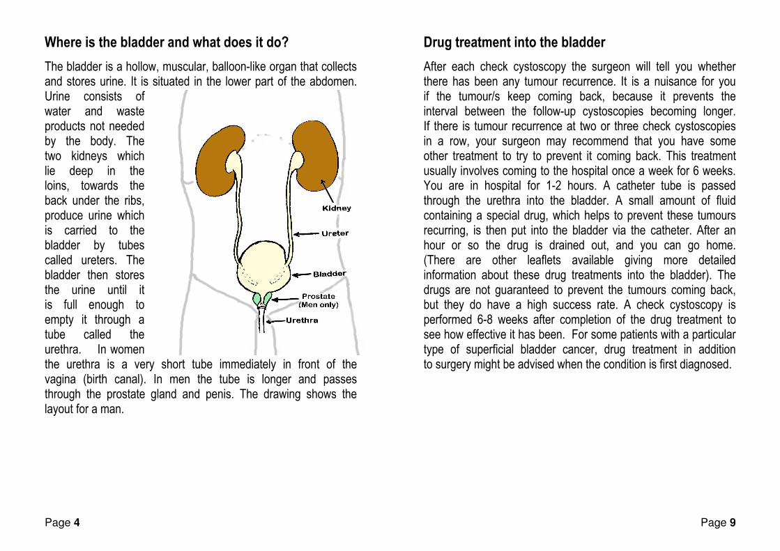
Where is the bladder and what does it do?
The bladder is a hollow, muscular, balloon-like organ that collects and stores urine. It is situated in the lower part of the abdomen. Urine consists of water and waste products not needed by the body. The two kidneys which lie deep in the loins, towards the back under the ribs, produce urine which is carried to the bladder by tubes called ureters. The bladder then stores the urine until it is full enough to empty it through a tube called the urethra. In women the urethra is a very short tube immediately in front of the vagina (birth canal). In men the tube is longer and passes through the prostate gland and penis. The drawing shows the layout for a man.
Page 9
Drug treatment into the bladder
After each check cystoscopy the surgeon will tell you whether there has been any tumour recurrence. It is a nuisance for you if the tumour/s keep coming back, because it prevents the interval between the follow-up cystoscopies becoming longer. If there is tumour recurrence at two or three check cystoscopies in a row, your surgeon may recommend that you have some other treatment to try to prevent it coming back. This treatment usually involves coming to the hospital once a week for 6 weeks. You are in hospital for 1-2 hours. A catheter tube is passed through the urethra into the bladder. A small amount of fluid containing a special drug, which helps to prevent these tumours recurring, is then put into the bladder via the catheter. After an hour or so the drug is drained out, and you can go home. (There are other leaflets available giving more detailed information about these drug treatments into the bladder). The drugs are not guaranteed to prevent the tumours coming back, but they do have a high success rate. A check cystoscopy is performed 6-8 weeks after completion of the drug treatment to see how effective it has been. For some patients with a particular type of superficial bladder cancer, drug treatment in addition to surgery might be advised when the condition is first diagnosed.

Page 8
What is the treatment for superficial bladder cancer?
Surgery
Superficial bladder cancer only affects the lining of the bladder, and the tumours can usually be removed very easily using a resectoscope. The tumour is shaved off the bladder wall and the area is cauterised using a mild electrical current to prevent excessive bleeding. In this way several tumours can be treated at the same time. Sometimes tiny tumours are treated simply with cautery through the telescope. After this type of treatment, follow-up (or check) cystoscopies are needed at regular intervals (usually every 3 months at first) because the tumours can come back. If there is any recurrence of the tumours they can again be removed during the examination. Often this is the only treatment needed for superficial bladder cancer. You will have regular check cystoscopies, with any tumours dealt with as they recur. If there is no sign of recurrence the interval between the check cystoscopies becomes longer, until they are just once a year. Many of these follow-up cystoscopies are now carried out as day cases, using the flexible cystoscope without any need for a general anaesthetic.
Page 5
What is superficial cancer of the bladder? The tumours in Superficial bladder cancer look like tiny frilly mushrooms, with their stems attached to the inner lining of the bladder. They are called superficial (surface) bladder cancers because they are confined to the inner lining of the bladder and do not extend into the muscle wall, as is the case with invasive tumours which do penetrate into the bladder wall. We also use the term "non-invasive" bladder cancer. They may develop as a single tumour, or there may be many; they may be small (only a few millimetres across) or large (up to several centimetres).

Page 6
What are the causes of cancer of the bladder?
The causes of cancer of the bladder are largely unknown, but research is going on all the time into possible causes of the disease. However, cigarette smoking is known to increase the risk of developing bladder cancer. It is also known that exposure to some industrial dyes and solvents can put people at risk, especially those who used to work in rubber and leather manufacturing. If you think that you may have been exposed in this way, do discuss it with us. We have a list of those chemicals for which exposure leading to a bladder cancer might enable you to receive financial compensation. 7000 new cases are diagnosed in England and Wales each year. It is twice as common in men as in women. The condition most commonly occurs between 50 and 70 years of age, but may occur in people in their 30s and 40s.
How is it diagnosed?
The commonest symptom of cancer of the bladder is blood in the urine (haematuria). This usually occurs suddenly and is generally not painful. Initial tests usually include scans or X-rays to check both the bladder and the kidneys. The presence of bladder tumours may be confirmed when a surgeon looks inside the bladder with an instrument called a cystoscope. An initial inspection can be undertaken using a ‘flexible cystoscope’ that is passed through the urethra and into the bladder under local anaesthetic. Using this instrument the surgeon can look at the bladder. In order to remove tumours it is usual to be given a general anaesthetic. The surgeon then uses a resectoscope,
Page 7
which is a thin tube fitted with a telescope through which removal of tissue is possible. The surgeon can often tell by looking at the tumours whether they are invasive or non-invasive, but samples are always sent to the laboratory for examination under the microscope. Results are usually available about 10 days later. This gives the surgeon further information about the type of tumour, and whether it extends into the bladder wall.


![Muscle-invasive and Metastatic Bladder Cancer · muscle-invasive bladder cancer (Ta,T1 and carcinoma in situ) [2], and primary urethral carcinomas [3]. 1.2 Panel Composition The EAU](https://img.dokumen.tips/doc/110x75/5e558374ee435e2e4f1b6d29/muscle-invasive-and-metastatic-bladder-cancer-muscle-invasive-bladder-cancer-tat1.jpg)


![Muscle invasive bladder Cancer [Dr.Edmond Wong]](https://img.dokumen.tips/doc/110x75/554af03bb4c90559058b477d/muscle-invasive-bladder-cancer-dredmond-wong.jpg)


